histology of the cartilage
TRANSCRIPT

HISTOLOGY OF THE CARTILAGE

INTRODUCTION
➢It is a specialized connective tissue designed to givesupport, bear weight andwithstand tension, torsion andbending.

• Avascular tissue
• Non nervous structure
• Poor regeneration capacity
• Usually surrounded by perichondrium (exceptarticular cartilage and fibro cartilage)
General Features

PERICHONDRIUM
Cartilage is covered by dense irregular connective tissue
known as Perichondrium except articular cartilage and fibro
cartilage.
•Has two layers:
➢ Outer fibrous layer (vascular)
➢ Inner chondrogenic layer (cellular)
•Has cells which can regrow cartilage to some extent if the
cartilage is damaged.


• Cells:➢Chondroblasts➢Chondrocytes
• Extra cellular matrix
• Fibres-collagen and elastic
• Ground substance- mucopolysaccharides(chondroitin sulphate, Keratan sulphateand hyaluronic acid)
COMPONENTS
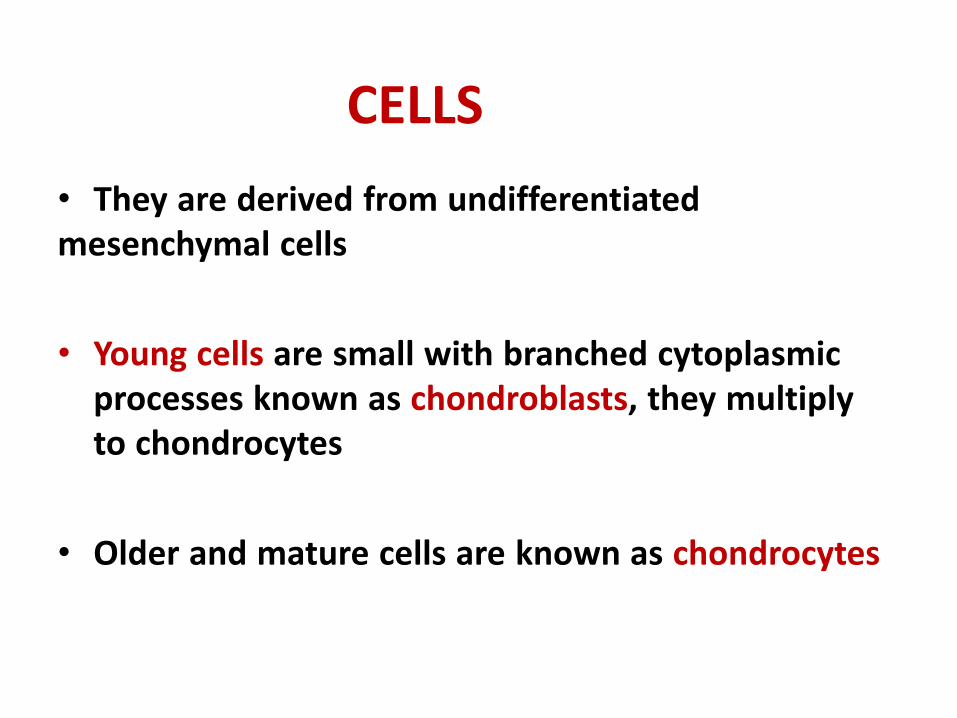
• They are derived from undifferentiatedmesenchymal cells
• Young cells are small with branched cytoplasmicprocesses known as chondroblasts, they multiplyto chondrocytes
• Older and mature cells are known as chondrocytes
CELLS

• Chondrocytes are bigger in size and are found inspaces called as lacunae
• They are found either groups of 2-4 cells togetherknown as cell nest or individual cells
• They are responsible for production of fibres andground substance of the cartilage
• Old mature cells are incapable of multiplication

TYPES
• HYALINE CARTILAGE
• ELASTIC CARTILAGE
• FIBRO CARTILAGE

HYALINE CARTILAGE
Hyaline cartilage is the most common type, found in several places.

Hyaline Cartilage
The hyaline cartilage is covered by perichondrium except for articular cartilage of synovial joints.

• Cells encapsulated in groups of 2-4 cells
• Matrix around the cells is brighter and deepin color than other areas, this matrix is knownterritorial matrix.
• Two groups of cells are separated by a lightlycolored matrix known inter-territorial matrix
TYPES OF HYALINE CARTILAGE
It has two types1. Articular
2. Costal
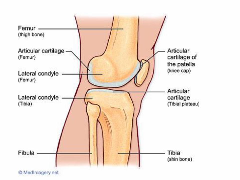

ARTICULAR CARTILAGE
Covers articular areas of bones forming synovial joints.


CARTILAGE GROWTH
AppositionalIncreasing in WIDTH; chondroblasts deposit matrix on surface of pre-existing cartilage
InterstitialIncreasing in LENGTH; chondrocytes divide and secrete matrix from w/in lacunae

• Costal cartilages
• Articular cartilages (devoid of perichondrium)
• Nasal cartilage
• Cartilage of trachea and bronchi
EXAMPLES


Hyaline cartilage- magnified
Territorial matrix
Interterritorial matrix
Chondrocytes in
lacuna
Cell nest

ELASTIC CARTILAGE
• Perichondrium present
• Characterized by the presence of elasticfibers in abundance.
• Chondrocytes are larger than those of hyalinecartilage
• Chondrocytes are found in singles or in twosin lacuna.


EXAMPLES
• Auricle or pinna
• Epiglottis
• External auditory meatus

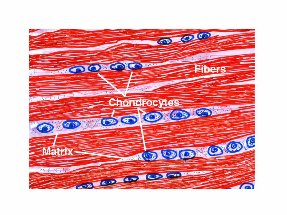
• Perichondrium is characteristically absent
• Has thick bundles of collagen fibers
• Chondrocytes are seen between these fibersin single or in narrow rows
FIBROCARTILAGE


EXAMPLES
• Pubic Symphysis
• Manubriosternal joint
• Intervertebral discs
MCQ
A section of Hyaline cartilage can be identified
by the presence of
1.Homogenous matrix
2.Elastic fibres
3.Collagen fibres
4.Chondrocytes arranged in row
MCQ
Hyaline cartilage is present in
1.Tracheal ring
2.Epiglottis
3.Intervertebral disc
4.Glenoidal Labrum

MCQ
Perichondrium is absent in
1.Elastic and Hyaline Cartilage
2.Hyaline Cartilage
3.Fibrocartilage and articular cartilage
4.Costal Cartilage and Ear Pinna

MCQ
Which of the following features is NOT TRUE
about cartilage?
1.Firm and flexible
2.Highly vascular
3.Insensitive
4.Poor in regeneration