histology lab blood and lymphatics. blood laboratory this laboratory is concerned with the...
TRANSCRIPT

Histology Lab
Blood and Lymphatics

BLOOD LABORATORY
This laboratory is concerned with the identification of the cellular components of peripheral blood and the bone marrow.
Be able to distinguish each of the cells of this system and recognize their salient features as described in the text associated with each of the images.

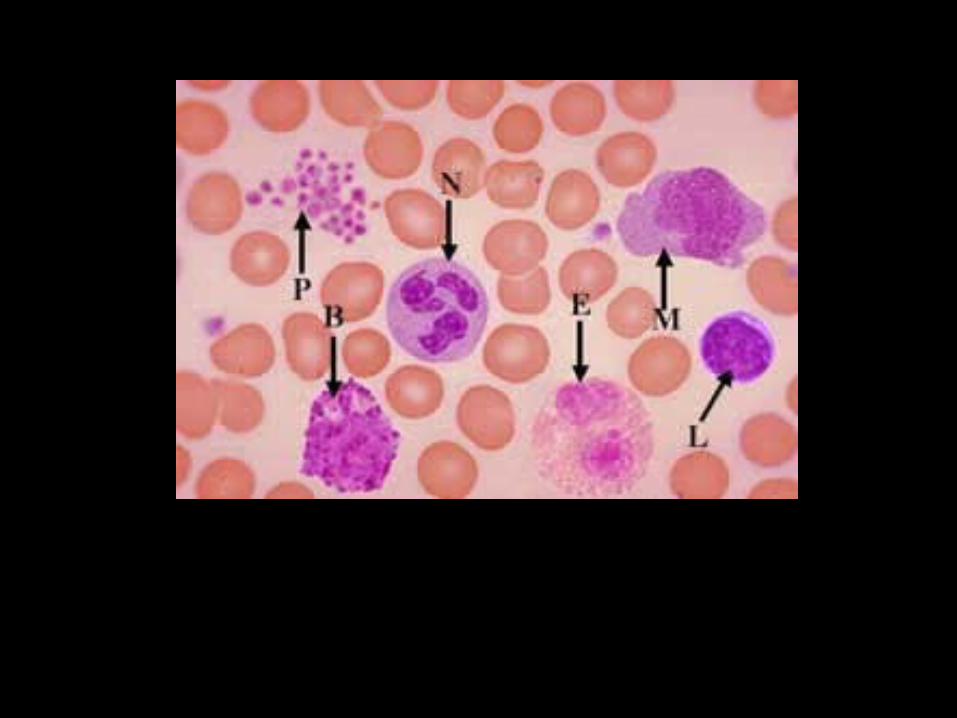
• Blood smear-Peripheral. Wright’s stain. This is a smear of peripheral blood illustrating erythrocytes, leukocytes (white blood cells), and platelets. Erythrocytes or red blood cells comprise the largest cell population or peripheral blood. These anucleated cells are approximately 7.5u in diameter and number about 5 million/ul. They are biconcave in shape and thus the central part of the cell appears lighter than the periphery. Leukocytes number approximately 7-10,000/ul. They utilize the vascular system for transport; however, their main functions are carried out in connective tissue. Platelets number about 250,000/ul.
N: Neutrophils contain lots of membrane-bound granules in their cytoplasm, however, with this stain, they stain very pale lavender. They tend to have two to five nuclear lobes each connected by thin strands of chromatin. Neutrophils comprise about 70% of the leukocytes.E: Eosinophils have bi-lobed nuclei. The cytoplasm of these cells is filled with membrane-bound granules that stain pink. The granules tend to be about the same in size and shape. Eosinophils comprise about 1-3% of the leukocytes.B: Basophils have granules scattered about the cytoplasm. The granules are somewhat irregular in size and shape and tend to mask out the nucleus. Basophils comprise about 0.5-1% of the leukocytes.L: Lymphocytes are about the same size as the erythrocytes (red blood cells). The darkly staining nucleus almost fills the entire cell. There is a small rim of basophilic cytoplasm. Lymphocytes comprise about 20-30% of the leukocytes.M: Monocytes tend to be 2-3 times the size of erythrocytes. They have light-staining nuclei that are somewhat irregular in shape. Monocytes comprise about 3-8% of the leukocytes.P: Platelets are small fragments of cytoplasm surrounded by plasmalemma. They bud off from megakaryocytes located in the bone marrow.


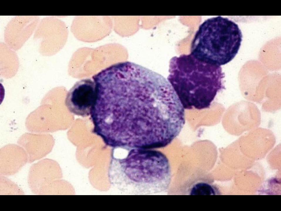
• Proerythroblast. The central cell in this image is a Proerythroblast (red arrow), one of the first identifiable stages of red cell development. The cytoplasm stains basophilic due to a number of ribosomes that will be involved in the production of hemoglobin. The nucleus, which occupies about 80% of the cell volume, contains fine chromatin and 1-2 nucleoli (yellow arrows). Note that with Wright’s stain, the nucleoli do not stain dark as with H & E. Wright’s stain colors DNA but not RNA. As a result, the nucleoli appears light with a thin rim of dark-staining material around it (this represents the nucleolar-associated DNA of the acrocentric chromosomes. The black arrow is indicating a polychromatophilic erythroblast (a later stage of development).


• Proerythroblast. The central cell is another example of a proerythroblast (red arrow). The yellow arrows are pointing to nucleoli. Note the text of the previous image for a description of this developmental stage. The green arrow is indicating a polychromtophilic erythroblast and the black arrow is pointing to an eosinophilic myelocyte, a developmental stage of one of the leukocytes.


• Basophilic Erythroblast. Proerythroblasts undergo several mitoses; some of the daughter cells differentiate into the next stage of development, Basophilic Erythroblasts (red arrows). The cytoplasm is usually more basophilic than that of proerythroblasts due to larger numbers of ribosomes. Nucleoli are absent and the chromatin appears darker than the previous stage. Note that the nucleus still occupies are large portion of the cell volume. The black arrow is indicating a polychromatophilic erythroblast.


• Polychromatophilic Erythroblast. The black arrows are pointing to basophilic erythroblasts. These cells undergo several mitoses; some of the daughter cells differentiate into the next stage of development, Polychromatophilic Erythroblast (red arrow). Note that the cytoplasm appears somewhat gray. This is due the presence of hemoglobin that is being produced by the ribosomes. There are fewer ribosomes at this stage than previous stages, thus less blue and more pink = gray color. Note also that the nucleus is denser than previous stages.


• Polychromatophilic Erythroblast. Another image of a polychromatophilic erythroblast. Note the dense nucleus and gray cytoplasm. These cells undergo mitoses; some of the daughter cells differentiate into the next type, the orthochromatophilic erythroblast.


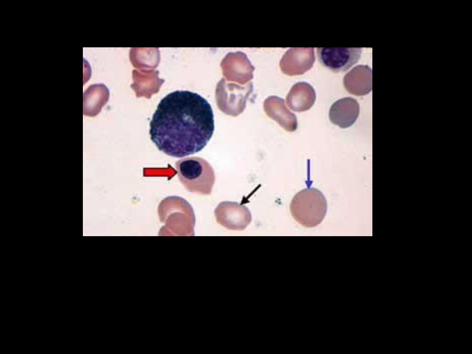
• Orthochromatophilic Erythroblast/ Reticulocyte. Orthochromatophilic Erythroblasts (red arrow) have a cytoplasmic color that approximates that of an erythrocyte. Thus nucleus initially is somewhat centrally located and very dense. It then moves to an eccentric position, becomes pyncnotic and then is extruded. Once the nucleus is lost, the cell is termed a Reticulocyte (blue arrow). It is not yet biconcave and thus lacks a central halo. It still contains a scant amount of RNA and thus stains positive with an RNA stain. After about 24 hours, all RNA is gone and the cell assumes a biconcave shape and is then termed an erythrocyte (black arrow). About 1% of the circulating red blood cells are reticulocytes.


• Myeloblast. The Myeloblast (red arrow) is one of the first recognizable stages in the development of granulocytes. The nucleus occupies about 50-60% of the cell volume. It has a fine chromatin pattern and 1 or 2 nucleoli (yellow arrows). No granules are present at this stage thus one cannot determine whether this cell will develop into a neutrophil, eosinophil, or basophil. The cytoplasm is basophilic due to the content of ribosomes. Present in this image are two orthochromatophilic erythroblasts (black arrows) and a reticulocyte (blue arrow). These cells undergo mitoses; some of the daughter cells differentiate into the next type, the promyelocyte.


• Promyelocyte. The characteristic feature of this stage is the presence of primary or azurophilic granules in the basophilic cytoplasm. One is still unable to determine whether this cell will develop into a neutrophil, eosinophil, or basophil. Like the myeloblast, the nucleus has a fine chromatin pattern and 1 or 2 nucleoli (yellow arrows). The cytoplasm is basophilic. Can you name the cell indicated by the black pointer??


• Promyelocyte. This is another image of a promyelocyte. The green arrows are pointing to the azurophilic granules; the yellow arrow to a nucleolus. This cell undergoes mitoses; some of the daughter cells will differentiate into the next stage, a myelocyte.


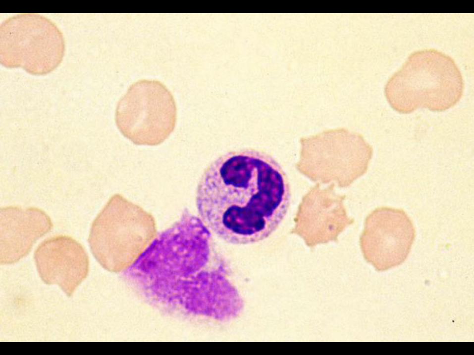
• Neutrophilic Myelocyte. An early stage of Myelocyte development is the production of secondary granules. The staining characteristic of these granules allows the identification of the cell as a neutrophilic myelocyte, eosinophilic myelocyte, or basophilic myelocyte. The central cell indicated by the green arrow is a neutrophilic myelocyte. The yellow arrow is pointing a light-staining area of the cell representing the Golgi apparatus producing neutrophilic granules. As the myelocyte becomes more differentiated, the secondary granules become more numerous, masking the azurophilic granules. The cell becomes somewhat smaller and the chromatin denser. Nucleoli are absent at this stage. The red arrows are pointing to three neutrophilic myelocytes.


• Neutrophilic Myelocyte. Two neutrophilic myelocytes are illustrated in this image. A few azurophilic granules can be visualized in the cytoplasm of the left cell. Note that the nucleus of the cell indicated by the black pointer is just beginning to segment.


• Neutrophilic Myelocyte. A neutrophilic myelocyte is illustrated in this image. Note the azurophilic granules in the cytoplasm and the dense chromatin of the nucleus. The black arrow indicates a neutrophilic band cell. Myelocytes undergo mitoses and give rise to more myelocytes some of which will differentiate into metamyelocytes. Myelocytes are the last stage to undergo cell division.


• Neutrophilic Metamyelocyte. The identification of the granulocytes beyond the myelocyte stage is based on nuclear morphology. The nucleus indents on one side and eventually becomes “band” shaped. Illustrated in this image is a neutrophilic metamyelocyte. This cell stage is identified as having nuclear indentation (black arrow) less than 50%.


• Neutrophilic Bands. As the nuclear indentation progresses, the nucleus becomes horseshoe or band-shaped (red arrows). Band cells are occasionally found in the peripheral blood.


• Early Neutrophil. The early sign of maturation is nuclear segmentation (black arrow). This is a process whereby the nucleus segments into two or more lobes, each connected by strands of chromatin. Neutrophils may contain 2-5 lobes whereas eosinophils are normally bi-lobed.


• Neutrophils. These are two images of neutrophils, each having three nuclear lobes. The left neutrophil has a small amount of chromatin projecting off one of the lobes (black arrow). This known as a Barr body (which are common in females) and represents an inactive X-chromosome.


• Neutrophils. This is an image of two neutrophils, each having four nuclear lobes.


• Megakaryoblasts. Megakaryoblasts are located in the bone marrow near the sinusoids. They develop into megakaryocytes that function in the production of platelets. This process involves the budding of small portions of cytoplasm surrounded by a plasmalemma. Megakaryoblasts are large cells, measuring about 30 um in diameter. During the period of differentiation of a megakaryocyte, the cytoplasm increases in amount and the nuclear DNA undergoes several replications. The end result is a large cell, measuring about 70-100 um, with a multi-lobulated nucleus.


• Megakaryocytes


LYMPHATIC SYSTEM LABORATORY
This laboratory is concerned with the identification of the structural components of lymphatic tissue. The main cellular components of lymphatic tissue include lymphocytes, plasma cells, macrophages, and phagocytic reticular cells.
Be able to distinguish each of the tissues/organs of this system and recognize their salient features as described in the text associated with each of the images.

• Diffuse Lymphatic Tissue. This is a section of the ileum illustrating diffuse lymphatic tissue. Diffuse lymphatic tissue may be classified as either loose or dense. Loose diffuse lymphatic tissue consists of lymphocytes that are scattered about within connective tissue (red arrows). When large numbers of lymphocytes are very closely associated with each other, the tissue is classified as dense diffuse lymphatic tissue (yellow arrow).


• Diffuse Lymphatic Tissue. This is a section of the jejunum illustrating a dense diffuse lymphatic tissue consisting primarily of large numbers of closely associated lymphocytes giving it a dark appearance (red arrows).


• Nodular lymphatic tissue. This is a section of appendix, which typically contains large numbers of lymphatic nodules (red arrows). These nodules are classified as secondary due to the presence of “germinal centers” (yellow arrows). The green arrow is indicating a patch of dense diffuse lymphatic tissue.


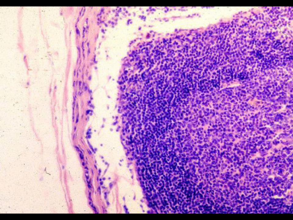
• Palatine Tonsil. This is a section of palatine tonsil containing several secondary nodules (yellow arrows) embedded in dense diffuse lymphatic tissue (orange arrow). The surface of the tonsil is lined by a non-keratinized stratified squamous epithelium (red arrow). A small amount of connective tissue lies just deep to the epithelium.


• Palatine Tonsil. This is a higher magnification of the previous image illustrating the epithelial lining (red arrow). A small amount of connective tissue lies just deep to the epithelium (green arrow). Nodular lymphatic tissue is located deep to the connective tissue.


• Palatine Tonsil. This image illustrates one of several crypts typically found associated with palatine tonsil (red arrow). Debris can collect in these crypts, as there is no effective flushing mechanism.


• Palatine Tonsil - Capsule. Palatine tonsils are classified (along with pharyngeal and lingual tonsils) as partially encapsulated lymphatic organs. Dense connective tissue forms a band (partial capsule) around the deep portion of the tonsil (green line).


• Lymph Node. Lymph nodes are surrounded by a dense connective tissue capsule (black arrows). Deep to the capsule is the cortex of the lymph node (C). This area contains lymphatic nodules (red arrows) embedded in a dense diffuse lymphatic tissue. The central portion of a lymph node is called the medulla (M) and contains cords of lymphatic tissue, but no nodules. The two areas are separated by the red line in this illustration.


• Lymph Node-Medulla. This is a high magnification of the medullary portion of a lymph node. Red arrows are indicating “medullary sinuses” containing lymph coming from the cortical region on its way to the hilus of the node where it will enter efferent lymphatic vessels.


• Lymph Node-Cortex. This is a high magnification of the outer portion of a lymph node. The green arrow is indicating the dense connective capsule. Projecting off of the capsule are bands of connective tissue, termed trabeculae (blue arrow) that penetrate the cortex. Just deep to the capsule is the subcapsular sinus (red arrow) containing lymph that entered this region via afferent lymphatic vessels. The lymph may percolate through the nodular lymphatic tissue to the medulla or pass to the medulla via trabecular sinuses (yellow arrow) surrounding the trabeculae.


• Lymph Node-Cortex. This is a higher magnification of the outer portion of a lymph node illustrating the capsule (green arrow), subcapsular sinus (red arrow), and trabecular sinus (yellow arrow). The cortical region of a secondary nodule, containing small lymphocytes, is separated from the germinal center (GC) by the red line. The germinal center contains small and large lymphocytes, macrophages, and plasma cells.


• Lymph Node-Afferent Vessels. Lymph enters the subcapsular sinuses via afferent lymphatic vessels (red arrow). The lymph then passes to the medullary sinuses via the trabecular sinuses (fast route) or by percolating through the cortical lymphatic tissue (slow route).


• Spleen. The image on the left is a low magnification of a spleen. A capsule of dense connective tissue and smooth muscle surrounds this organ. The bulk of the spleen contains red pulp (black arrows), which consists of cords of lymphatic tissue among blood vascular sinuses. The white pulp (encircled in red) consists of dense collections of lymphocytes surrounding central arteries (yellow arrows). The image on the right is a high magnification of the red pulp illustrating several splenic cords or cords of Billroth (red arrows).


• Spleen. This is high magnification of the splenic capsule (green arrow). This structure consists of dense connective tissue and smooth muscle. Numerous trabeculae (black arrow) project off of the capsule and penetrate the substance of the organ. White pulp is separated from the red pulp by red lines. Yellow arrows point to the central arteries.


• Spleen. This is a high magnification of the parenchyma of the spleen. Numerous areas of white pulp are separated from the red pulp by red lines.


• Spleen-White Pulp. This is a high magnification of white pulp (encircled in red). A central artery is indicated by a black arrow. The red pulp contains numerous blood vascular sinuses.


• Thymus. The thymus is surrounded by a capsule of connective tissue (not shown). Projections from the capsule separate the organ into lobes and lobules. The central portion of a lobule, the medulla (M), is light staining and contains small and large lymphocytes. The outer part of the lobule is dark staining and termed the cortex and contains small lymphocytes. The thymus lacks lymphatic nodules. Stroma is composed of epithelial reticular cells.

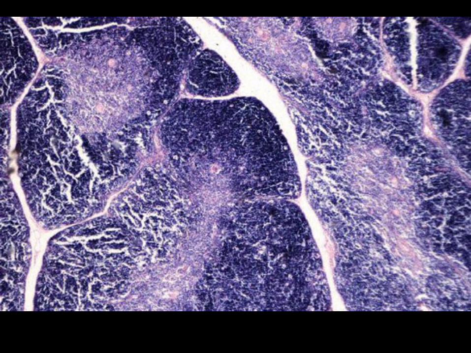

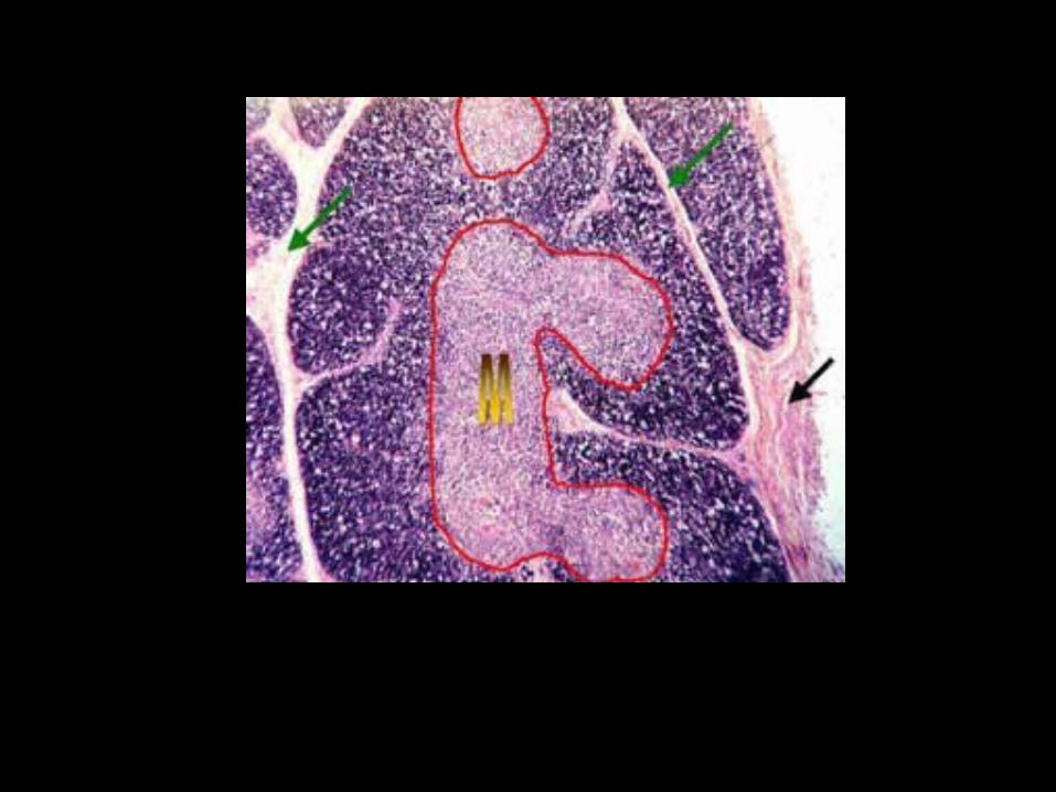
• Thymus. The thymus is surrounded by a capsule of connective tissue (black arrow). Projections from the capsule separate the organ into lobes and lobules (green arrows). The central portion of a lobule, the medulla (M), is light staining and contains small and large lymphocytes. The outer part of the lobule is dark staining and termed the cortex and contains small lymphocytes. The thymus lacks lymphatic nodules. The stroma is composed of epithelial reticular cells.


• Thymus. This image is a high magnification of the approximate separation of the medulla and the cortex (red line). The medulla contains unique structures termed Hassall’s (thymic) corpuscles (red arrows). These whorl-shaped structures, which increase with age, consist of concentric layers of epithelial reticular cells.


• Thymus-Medulla. The image on the left is a high magnification of a thymic medulla illustrating Hassall’s corpuscles (red arrows). The image on the right is a high magnification of a Hassall’s corpuscle (yellow arrows) demonstrating the concentric arrangement of the epithelial reticular cells. The central portion of the corpuscle may contain keratin and occasionally, calcification.


Welcome back!
I hope you had a great summer break and that your
mind will allow you to get back into studying 26 hours a
day.
Enjoy Histo and watch your summer
break fade away