histology and histochemistry of the long bones in …
TRANSCRIPT
Arch. Dis. Childh., 1963, 38, 295.
HISTOLOGY AND HISTOCHEMISTRYOF THE LONG BONES IN MICROMELIA
BY
A. T. SPENCERFrom the Department ofPathology, University of Birmingham
(RECEIVED FOR PUBLICATION OCTOBER 11, 1962)
There are many recorded cases of micromelia butno report of the histochemistry of the bones in thiscondition in papers published recently.The object of this paper is to describe a case of
micromelia in a newborn infant, and to recordcertain histochemical changes in the epiphyses.
Case Report
Clinical Findings. The infant was the fifth child bornto parents of normal build and stature. The first childwas a male stillborn; the second was a female dwarfnow aged 12 years; the third and fourth children wereboth male stillborns, and so far as can be ascertainedthe stillborn males showed no evidence of dwarfism.On this occasion pregnancy had been normal and the
mother, a patient of Prof. H. C. McLaren, went intolabour nine days before the expected date of delivery.The child was delivered by a high forceps techniquebecause of foetal distress as shown by irregularity of thefoetal heart. The baby breathed immediately at birth,but its condition soon deteriorated and it becamecyanosed and died after 25 hours.
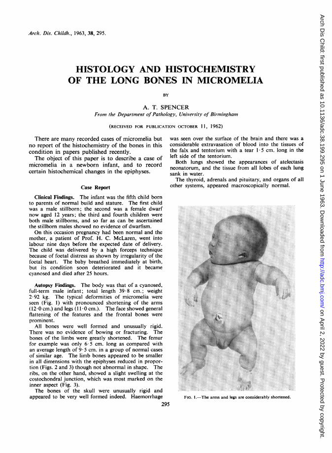
Autopsy Findings. The body was that of a cyanosed,full-term male infant; total length 39-8 cm.; weight2 92 kg. The typical deformities of micromelia wereseen (Fig. 1) with pronounced shortening of the arms(12-0 cm.) and legs (11-0cm.). The face showed generalflattening of the features and the frontal bones wereprominent.
All bones were well formed and unusually rigid.There was no evidence of bowing or fracturing. Thebones of the limbs were greatly shortened. The femurfor example was only 6 5 cm. long as compared withan average length of 9 5 cm. in a group of normal casesof similar age. The limb bones appeared to be smallerin all dimensions with the epiphyses reduced in propor-tion (Figs. 2 and 3) though not abnormal in shape. Theribs, on the other hand, showed a slight swelling at thecostochondral junction, which was most marked on theinner aspect (Fig. 3).The bones of the skull were unusually rigid and
appeared to be very well formed indeed. Haemorrhage
was seen over the surface of the brain and there was aconsiderable extravasation of blood into the tissues ofthe falx and tentorium with a tear 1 5 cm. long in theleft side of the tentorium.Both lungs showed the appearances of atelectasis
neonatorum, and the tissue from all lobes of each lungsank in water.The thyroid, adrenals and pituitary, and organs of all
other systems, appeared macroscopically normal.
FIG. I.-The arms and legs are considerably shortened.
295
on April 2, 2022 by guest. P
rotected by copyright.http://adc.bm
j.com/
Arch D
is Child: first published as 10.1136/adc.38.199.295 on 1 June 1963. D
ownloaded from

ARCHIVES OF DISEASE IN CHILDHOOD
FIG. 2.-The tibia and femur are shortened and the epiphyses are ofproportionately smaller size.
FIG. 3.-The humerus is shortened. A swelling at the costochondraljunction is present.
FIG. 4.-Lower end of femur; the cartilage cells are much smallerthan normal. The cartilaginous matrix of the epiphysis stains deep
pink. (Iron haematoxylin and van Gieson's stain x 100.)
FIG. 5.-Similar field to Fig. 4 from lower end of femur of a control.Iron haematoxylin and van Gieson's stain x 100.)
TABLE
RESULTS OF NITROGEN AND HYDROXYPROLINE ASSAYS
Mg. HydroxyprolineSample of % Nitrogen Hydroxyproline per mg. NitrogenEpiphysial __Cartilage Original Defatted and Original Defatted and Original Defatted and
Dehydrated Dehydrated Dehydrated
Lower end femur (Case) .. 242 13*4 0*85 5*9 0 351 0-440
Lower end femur (control) .1. -07 14-5 0-434 5-72 0-406 0- 394
Upper end femur (Case) 2-53 13-2 0.99 7-6 0-391 0-576
Upper end femur (control).. 1-50 13-3 0-21 3-19 0*140 0*240
296
on April 2, 2022 by guest. P
rotected by copyright.http://adc.bm
j.com/
Arch D
is Child: first published as 10.1136/adc.38.199.295 on 1 June 1963. D
ownloaded from
HISTOLOGY AND HISTOCHEMISTRY OF LONG BONES IN MICROMELIA 297
Materials and MethodsA femur, tibia, humerus and several ribs were fixed
in 4% formaldehyde saline. Control material wasobtained at autopsy from newborn babies showing noevidence of any bony abnormality. All bones weredecalcified in 25% formic acid, double embedded incelloidin and paraffin wax, and sections were preparedof their entire length wherever practicable.
Staining Methods. Sections from the case andcontrol material were stained at one and the same timewitb a variety of stains including Ehrlich's haematoxylinand eosin; iron haematoxylin and van Gieson's stain;cresyl fast violet; alcian blue (Steedman, 1950); dialysediron (Hale, 1946); the Laidlaw's method for reticulin;the periodic acid Schiff reaction (P.A.S.) (McManus,1946); and the bi-col method of Wolman (1956).Substances staining with alcian blue, dialysed iron,
and blue with the bi-col stain are accepted as acid muco-polysaccharides.
Sections were also examined with a polarizing micro-scope to confirm the presence of lamellar bone by itsbirefringent properties.
Chemical Examination. Samples of epiphysial car-tilage from the case and from controls were chemicallyanalysed for hydroxyproline so that a quantitativeestimation of collagen content could be made. AKjeldahl nitrogen assay was also done and the investiga-tion was carried out by Dr. D. L. Woodhouse using themethod of Neuman and Logan (1950). The resultsof the analysis are recorded in the Table. The percentagenitrogen and hydroxyproline is given in respect of theoriginal and dehydrated and defatted material. Hydroxy-proline is also given in terms of mg. hydroxyprolineper mg. nitrogen.
ResultsExamination of the last column of the Table shows
a higher concentration of hydroxyproline in the casematerial. The percentage of nitrogen in the casematerial is also higher than in the control basedon the original sample weights, but the percentagenitrogen in all specimens after dehydration anddefatting is approximately the same.
This indicates that there is an absolute increase inthe hydroxyproline content of the case material inboth instances and that the relatively greater increaseindicated in the third column of the results is dueto a greater water and fat content in the control.
Histology and HistochemistryEpiphysis. The most outstanding feature in the
cartilaginous epiphysis is the great increase in thenumber of cartilage cells with a considerable reduc-tion in the ratio of intercellular matrix to cell. Thecartilage cells are very much smaller in size thannormal and the zones of resting, proliferating andmaturing cells can easily be seen. The zone of
proliferating cells is the same depth in the longaxis of the bone as normal but the maturing zone isabout two-thirds as deep. The individual cells ofthe maturing zone do increase in size and becomevacuolated towards the epiphysial line as the normal,but they never attain the dimensions of the normal(Figs. 4 and 5).Consequent upon the tight packing of the smaller
cartilage cells there is an increase in the number ofossification rows in the width of the epiphysis.Associated with this, the projecting spicules ofcartilaginous matrix on which bone is laid are muchnarrower and more numerous and there also appearsto be an increase in the number of osteoclasts in thisarea.Another interesting feature is the pronounced
deep pink staining of the cartilaginous matrix ofthe epiphysis with the van Gieson's stain, although,except near the epiphysial line, a fibrous structurecannot be seen with an ordinary light microscop eUnder the polarizing microscope, however, birefrin-gent material is prominent in this region. Also incontrast to the normal, this cartilaginous matrix isP.A.S. positive (Figs. 6 and 7), although with thealcian blue, dialysed iron, and bi-col preparationsthere is no apparent increase in the acid mucopoly-saccharide content. This taken into considerationwith the results of the chemical analysis indicatesan increased collagen content.
Diaphysis. Numerous plump osteoblasts arepresent around the spicules of cartilaginous matrixmade available at the epiphysial line. Bone is laiddown in the normal manner though possibly at agreater rate, for at corresponding points from theepiphysial line in this Case and in the control a muchthicker layer of bone is present on the spicules ofcartilaginous matrix in the Case. An importantdifference from normal is that the spicules ofcartilage are smaller and are replaced by bone at apoint nearer the epiphysial line, though they stillretain their P.A.S. positive character. In spite ofthe increased number of ossification rows of theepiphysis and the consequently increased number ofcartilaginous spicules available on which bone maybe laid there is not an increased number of bonytrabeculae in the diaphysis. In fact the bonytrabeculae are reduced in number, and several,although they are thicker than normal, are notaligned in the axis of the bony shaft (Figs. 8 and 9).The bone which constitutes the trabeculae
appears to be of normal lamellar type and isbirefringent and shows a normal acid mucopoly-saccharide content. P.A.S. positive woven boneformed next to the periosteum appears to be pro-
on April 2, 2022 by guest. P
rotected by copyright.http://adc.bm
j.com/
Arch D
is Child: first published as 10.1136/adc.38.199.295 on 1 June 1963. D
ownloaded from

298 ARCHIVES OF DISEASE IN CHILDHOOD
FIG. 6.-Lower end of femur; the cartilaginous matrix of the epiphysis(to the left) is P.A.S. positive. (P.A.S. x 100.)
FIG. 7.-Similar field of control. The cartilaginous matrix is P.A.S.negative. (P.A.S. x 100.)
FiG. 8.-Costochondral junction; the spicules of cartilage are replaced by bone nearer to the costalcartilage than normal (the cartilage appears black). (Cresyl fast violet x 24.)
on April 2, 2022 by guest. P
rotected by copyright.http://adc.bm
j.com/
Arch D
is Child: first published as 10.1136/adc.38.199.295 on 1 June 1963. D
ownloaded from

HISTOLOGY AND HISTOCHEMISTR Y OF LONG BONES IN MICROMELIA 299duced normally and is replaced by lamellar bonefurther from the epiphysis as in the normal.
DiscussionRecently, much attention has been focused on
micromelia and other skeletal abnormalities asso-ciated with the administration of the thalidomidedrugs (Morgan, 1962; Leck and Millar, 1962; Lenz,1962; Pfeiffer and Kosenow, 1962; Somers, 1962;Speirs, 1962; Taussig, 1962). Many photographsof the infants are included in the published reports,but there is no account of the histology or histo-chemistry.The administration of several of the alkylating
agents has also been shown to produce similarabnormalities in rats (Murphy, 1959). Exposure toX rays has been shown to cause micromelia (Murphy,1959; Murphy and Goldstein, 1930), and riboflavinedeficiency has been cited as a cause (Warkany, 1947;Nelson, Baird, Wright and Evans, 1956; Kalter,1959). An association between hypoglycaemia andinsulin micromelia in chicks has also been reportedby several authors (Landauer, 1947; Zwilling, 1948;Anderson, Crane and Harper, 1959). However,none of these causes can be incriminated in thepresent case, and the fact that there is a seconddwarfed child in an otherwise normal family suggeststhat an inherited recessive factor is probably respon-sible.
It is not known whether the histology and histo-chemistry of micromelia is identical whatever thecause, but it will be seen that the chief abnormalitiesin this case are (1) the increase in the collagencontent of the epiphysial cartilage, (2) the increasedrate of ossification and (3) the more numeroussmaller cartilage cells. In other respects ossificationappears to proceed normally. The cartilage cellsmature and align themselves normally so thiscondition is easily distinguished from achondro-plasia where one of the outstanding abnormalities isthe failure of the cartilage cells of the epiphysis toalign in normal palisades (Knaggs, 1927; Harris andRussell, 1933; Pearce and Brown, 1945; Weinmannand Sicher, 1947; Potter, 1952).The acid mucopolysaccharide content of the
epiphysial cartilage is not apparently disturbed.There is evidence that an increase in acid mucopoly-saccharide may inhibit the crystal nucleation processof ossification (Neuman, 1960; Spencer, 1962).
Osteoblasts are present and normal lamellar boneis laid down.The increased collagen content of the epiphysial
cartilage may be important in explaining the abnor-mally short bones, for it is established that collagencan produce an epitactic growth of hydroxy-apatitecrystals (Neuman and Neuman, 1958). Thereforewith an increased amount of collagen in the epi-physial cartilage it may be postulated that many more
FIG. 9.-Costochondral junction of control. Compare with Fig. 8.(Cresyl fast violet x 24.)
8A
on April 2, 2022 by guest. P
rotected by copyright.http://adc.bm
j.com/
Arch D
is Child: first published as 10.1136/adc.38.199.295 on 1 June 1963. D
ownloaded from

300 ARCHIVES OF DISEASE IN CHILDHOODcrystal-seeding points than normal may be available.Hence the laying down of bone mineral may beestablished at a greater rate than normal with theresult that the area of growth at the line of ossi-fication would become prematurely fixed, resultingin an inhibition in the growth in length of the bone.
Summary
A case of micromelia in a full-term newborninfant is described. Histochemical examinationand chemical analysis confirm an increase in thecollagen content of the epiphysial cartilage. It issuggested that this may account for the apparentincrease in the rate of ossification in the long bonesleading to inhibition of growth in their length.
I am grateful to Professor J. W. Orr for helpfulcriticism and advice, and to Professor H. C. McLaren forpermission to use the clinical records. I am alsoindebted to Dr. D. L. Woodhouse and Mr. G. Fare forthe chemical estimations and for help with the inter-pretation of the results.
REFERENCES
Anderson, C. E., Crane, J. T. and Harper H. A. (1959). Alterationsin growth-cartilage in experimental dwarfism. I. Studies oninsulin-dwarfed chicks. J. Bone lt Surg., 41A, 1094.
Hale, C. W. (1946). Histochemical demonstration of acid poly-saccharides in animal tissues. Nature (Lond.), 157, 802.
Harris, H. A. and Russell, A. E. (1933). The atypical growth incartilage as the fundamental factor in dwarfism and achondro-plasia. Proc. roy. Soc. Med., 26, 779.
Kalter, H. (1959). Congenital malformations induced by riboflavindeficiency in strains of inbred mice. Pediatrics, 23, 222.
Knaggs, R. L. (1927). Achondroplasia (chondrodystrophia foetalis).Brit. J. Surg., 15, 10.
Landauer, W. (1947). Potentiating effects of adrenal cortical extracton insulin-induced abnormalities of chick development.Endocrinology, 41, 489.
Leck, I. M. and Millar, E. L. M. (1962). Incidence of malformationssince the introduction of thalidomide. Brit. med. J., 2, 16.
Lenz, W. (1962). Thalidomide and congenital abnormalities.Lancet, 1, 45.
McManus, J. F. A. (1946). Histological demonstration of Mucinafter periodic acid. Nature (Lond.), 158, 202.
Morgan, B. C. (1962). Thalidomide ("Distaval") and foetal abnor-malities. Brit. med. J., 1. 792.
Murphy, D. P. and Goldstein, L. (1930). Micromelia in a childirradiated in utero. Surg. Gynec. Obstet., 50, 79.
Murphy, M. L. (1959). A comparison of the teratogenic effectsof five polyfunctional alkylating agents on the rat fetus.Pediatrics, 23, 231.
Nelson, M. M., Baird, C. D. C., Wright, H. V. and Evans, H. M.(1956). Multiple congenital abnormalities in the rat resultingfrom riboflavin deficiency induced by the antimetabolite galacto-flavin. J. Nutr., 58, 125.
Neuman, R. E. and Logan, M. A. (1950). The determination ofhydroxyproline. J. biol. Chem., 184, 299.
Neuman, W. F. (1960). In Bone as a Tissue, ed. K. Rodahl, J. T.Nicholson and E. M. Brown, Jr., p. 103. McGraw-Hill, NewYork.and Neuman, M. W. (1958). The Chemical Dynamics of BoneMineral, p. 176. University Chicago Press, Chicago.
Pearce, L. and Brown, W. H. (1945). Hereditary achondroplasiain the rabbit. II. Pathologic aspects. J. exp. Med., 82, 261.
Pfeiffer, R. A. and Kosenow, W. (1962). Thalidomide and con-genital abnormalities. Lancet, 1, 45.
Potter, E. L. (1952). Pathology of the Fetus and the Newborn, p. 485.Year Book Publishers, Chicago.
Somers, G. F. (1962). Thalidomide and congenital abnormalities.Lancet, 1, 912.
Speirs, A. L. (1962). Thalidomide and congenital abnormalities.ibid., 1, 303.
Spencer, A. T. (1962). A histochemical study of long bones inosteogenesis imperfecta congenita. J. Path. Bact., 83, 423.
Steedman, H. F. (1950). Alcian blue 8GS: A new stain for mucin.Quart. J. micr. Sci., 91, 477.
Taussig, H. B. (1962). A study of the German outbreak of phoco-melia. The thalidomide syndrome. J. Amer. med. Ass.,180, 1106.
Warkany, J. (1947). Etiology of congenital malformations. Advanc.Pediat., 2, 1.
Weinmann, J. P. and Sicher, H. (1947). Bone and Bones. Funda-mentals of Bone Biology, p. 147. Kimpton, London.
Wolman, M. (1956). A histochemical method for the differentialstaining of acidic tissue components (particularly ground-substance polysaccharides). Bull. Res. Coun. Israel E, 6E, 27.
Zwilling, E. (1948). Association of hypoglycemia with insulinmicromelia in chick embryos. J. exp. Zool., 109, 197.
on April 2, 2022 by guest. P
rotected by copyright.http://adc.bm
j.com/
Arch D
is Child: first published as 10.1136/adc.38.199.295 on 1 June 1963. D
ownloaded from