histological seasonal changes in ovaries of spotted · pdf filedeveloping follicles with signs...
TRANSCRIPT

353
Int. J. Morphol.,26(2):353-361, 2008.
Histological Seasonal Changes in Ovaries ofSpotted Tinamous (Nothura maculosa Tinamidae,
Temminck, 1815) Related to Gonadotrope Population
Cambios Histológicos Estacionales en Ovarios de Perdiz Común (Nothura maculosa Tinamidae,Temminck, 1815) ) y su Relación con la Población de Gonadotropas
Juan Claver; Juan M. Rosa; Daniel M. Lombardo & María C. Soñez
CLAVER, J.; ROSA, J. M.; LOMBARDO, D. M. & SOÑEZ, M. C. Histological seasonal changes in ovaries of spotted tinamous(Nothura maculosa Tinamidae, Temminck, 1815) related to gonadotrope population. Int. J. Morphol., 26(2):353-361, 2008.
SUMMARY: Nothura maculosa is a South American Tinamidae with a marked seasonal reproductive pattern. This work descri-bes ovarian seasonal changes in this species related to gonadotrope (GTHs) population. Ovary and pituitary samples were collectedmonthly from adult birds during four annual periods. They were fixed in Bouin’s solution and processed for light microscopy. The data ofpost-fixation gonadal weight were analysed using STATISTIX 4.0. Histological sections of the ovaries were stained with H/E, PAS andGoldner-Masson trichrome. Single and double immunostaining were applied on pituitary sections with anti-chicken-FSH and anti-chicken-LH antibodies. The samples were analysed in quarterly periods of the year, P1: March-May (resting stage); P2: June-August (developingstage); P3: September-November (reproductive stage); P4: December-February (involutive stage). Ovary weight (ow) significantly variedamong periods (p<0.001). During P1, only primordial and previtellogenic follicles were observed, ow 0.09± 0.01 g (n=25); during P2,developing follicles with signs of vitellogenesis were detected, ow 0.13± 0.01 g (n=14); during P3, maximum follicular development wasfound, ow 0.9 ± 0.15 g (n=39); P4 exhibited great variability in follicular stages, ow 0.18± 0.18 g (n=19). Involutive atresia was observedin all the periods, while bursting atresia and post-ovulatory follicles were only characterized at P3 and P4. The GTHs containing few LHand FSH immunoreactive (ir) granules were predominant during P1-P2. The GTHs with LH ir granules were abundant in intermediatezone and caudal lobe in P3 and P4 while few cells contained both types of granules. The number of FSH cells was increased during P3and P4. The histological ovarian changes were narrowly correlated with the variations in the gonadotrope population.
KEY WORDS: Birds; Gonadotropes; Nothura maculosa; Ovary; Pituitary; Reproduction; Tinamidae.
INTRODUCTION
The genus Nothura includes small Tinamidae speciesliving in open areas of South America. Three species live inArgentina: Nothura maculosa (Temminck), Nothura darwini(Gray) and Nothura chacoensis (Conover) (Dabbene, 1972;del Hoyo & Elliott, 1992). The spotted tinamou (Nothuramaculosa) is the most widespread and abundant tinamou inArgentina with 6 sub-species described (Bump & Bump,1969; Del Hoyo & Elliot) where is popular as a game birdenjoyed by hunters and appreciated for its delicious meat.Bump & Bump have reported habitat characteristics,distribution and behaviour of this species in Argentina. Asin all members of the family, incubation and chick care arecarried out by the males (Handford & Mares, 1985). With
few exceptions, mating systems in tinamous are describedas simultaneous polygyny for males and sequential polyandryfor females (Handford & Mares; Del Hoyo & Elliot;Garitano-Zavala, 2003). In Nothura maculosa, matingsystems are poorly known but it was suggested that may bemonogamous at first but polygamous when older (Del Hoyo& Elliot). Seasonal reproduction have been described inNothura maculosa, with maximal (October) and minimal(April-May) periods of activity (Menegheti, 1981, 1984;Fleming Batalha da Silveira et al., 1981). Attempts weremade to characterize stages of the annual cycle of thesespecies based on the morphological changes in ovaries(Arriaga et al., 1983; Burger, 1991) and testes (Burger, 1992).
Área Histología y Embriología. Facultad de Ciencias Veterinarias, Universidad de Buenos Aires. Chorroarin 280. C1427CWO Buenos Aires, Argentina.Patrocinio: UBACYT 074-040

354
Seasonal reproduction in birds is marked by activation ofthe hypothalamo-pituitary-gonadal axis (Leska & Dusza.,2007). Adenohypophysis activity under hypothalamic con-trol regulates annual cyclic changes in gonads (Benoit, 1962;Follett et al., 1966). Changes in cell pituitary populations ofNothura maculosa relative to the annual cycle have beenpreviously determined (Soñez et al., 1995, 1997) but theywere not correlated with ovarian histology. With the aim ofbetter understanding reproductive parameters of this species,in this work we describe seasonal histological changes inovaries correlated with immunocytochemical detection ofgonadotropes during the annual cycle in a wild population ofNothura maculosa from Buenos Aires Province, Argentina.
MATERIAL AND METHOD
One hundred and twenty mature female Spottedtinamous were captured during four years, 5-20 animalsmonthly. The birds were maintained in cages 24 hours withfood and water ad libitum. The population studied proceededfrom San Miguel del Monte (35º 27’ S-Lat.- 58º 48’ W-Length) and Florencio Varela (30º 50’ S-Lat.- 58º 06’ W-Length) districts in Buenos Aires province. These zones arecharacterized by a temperate climate with mean temperatures8-23 ºC (winter-summer), annual rainy regime of 80 mmaverage, with 15L/9D daily photoperiod in summer and 9L/15D during winter.
The samples were analysed in four quarterly periodsof the year: P1 (March-April-May), P2 (June-July-August),P3 (September-October-November) and P4 (December-January-February).
Because age determination is not easy in this species,only females exceeding a weight of 200 g were consideredmature according to published data (Bump & Bump).
The animals were sacrificed by decapitation afterchloroform inhalation. After macroscopic observations ofthe ovaries and follicles (number, colour and size),hypophysis, ovaries and oviducts were removed immediately,fixed in Bouin’s solution for 3 h (hypophysis) and 24 h(ovaries and oviducts), dehydrated and embedded inParaplast (Monoject Scientific, U.S.A.) for light microscopy.Gonads and oviducts were measured in fresh and ovarieswere weighted after fixation. The oviducts length wasconsidered as an additional parameter of the gonadaldevelopment. Ovary sections of 4-5 µm were stained withhaematoxylin-eosin (H&E), Goldner–Masson trichrome andperiodic acid Schiff - haematoxylin (PAS/H). Weight datawere statistically analysed by descriptive statistics, ANOVA,
post-ANOVA (Bonferroni test) and median test (nonparametric) with significance of 0.05 %, using Statistics 4.0.
Immunocytochemical method (ICC). Pituitary serialsections were deparafinised, gradually hydrated in ethanoland washed in 0.5 M Tris saline buffer (TS), pH 7.6. Anindirect ABC system (Vectastain Universal Elite ABC kit,PK-6200, Vector Labs.) was applied with polyclonalantibodies, anti-chicken FSH 1:500 and anti-chicken LH1:2000, anti-turkey GH 1:1000 (kindly provided by J.A.Proudman, USDA– ARS Animal Hormone Program, USA)and monoclonal anti-chicken PRL 1:2000 (a gift from L.R.Berghman, Neuroendocrinology Lab., Zoological Institute,Leuven, Belgium). The endogenous peroxidase activity wasinhibited using hydrogen peroxide 3%. The sectionspreviously treated with normal rabbit serum were incubatedin a moist chamber at room temperature, for 30 min with thefirst specific antibody and the biotinilated second antibody.The immunocomplex was revealed by 3-3’diaminobenzidine(DAB, SIGMA), lightly counterstained with Mayer’shaematoxylin, dehydrated, cleared and cover-slipped. Doubleimmunostaining was carried out in some of the serialsections, washing with TS, pH 7.6 after the DAB reaction.The ICC procedure was repeated with the second specificantibody and biotinilated IgG. Incubation with alkalinephosphatase conjugated streptavidine (Label, BiogenexLabs.) was performed for 20 min at room temperature andrevealed by BCIP/NBT substrate solution (5-bromo, 4-chloro, 3-indolyl phosphate nitroblue tetrazolium, BiogenexLabs.). Finally, the sections were counterstained, dehydrated,cleared and mounted.
In the adjacent serial sections of each slide, the firstincubation with the specific antibody was omitted or replacedby pre-adsorbed antibody (working dilution of antibody with7 mg/ml hormone, 24 h, 4º C) performing the subsequenttechnique steps and substrate reactions described above forcontrol staining.
RESULTS
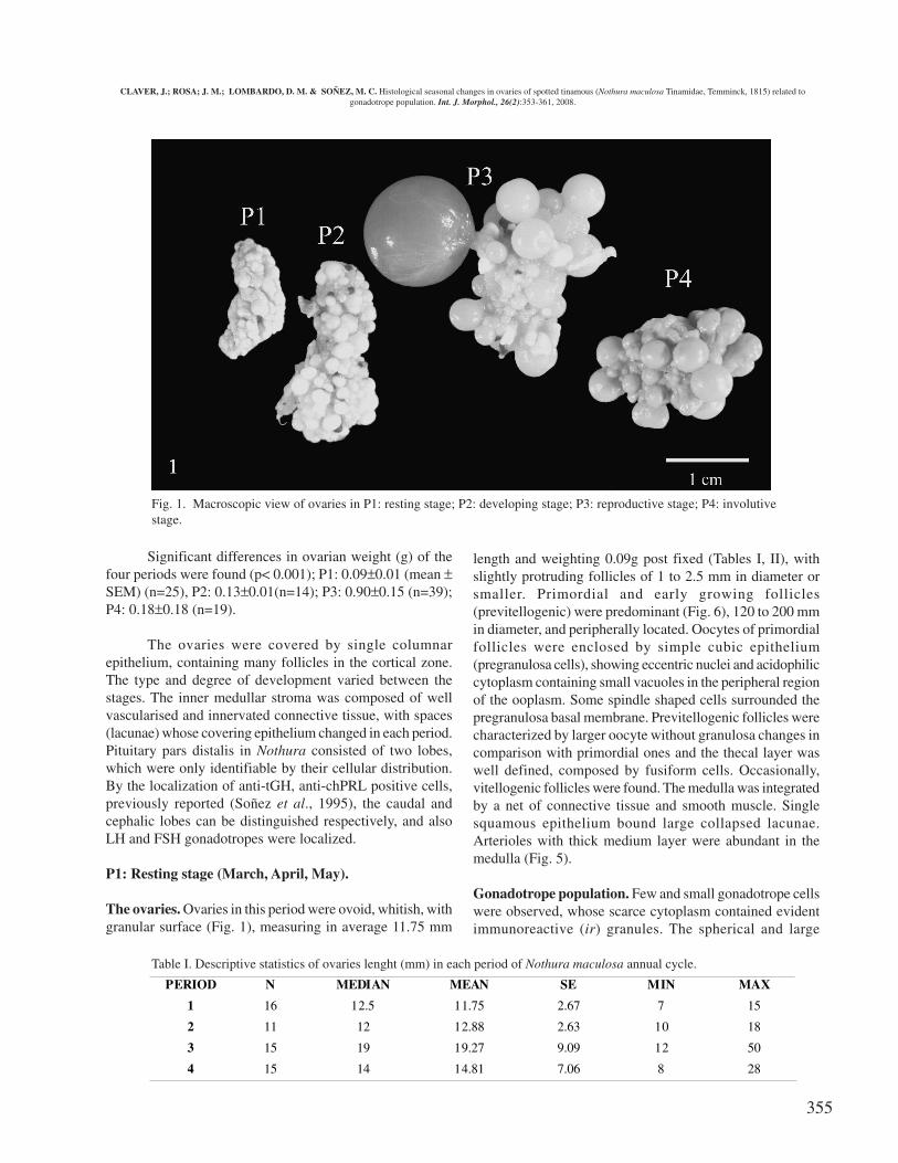
Marked macroscopic differences in size and featureswere observed in gonads of each period (Fig 1). Also theoviduct length changed remarkably. During P1 and P2, theoviducts were scarcely developed and very thin; varying inlength from 60 to 80 mm. Their maximum development wasobserved in P3 with 170.28 mm average of length duringOctober and November, while in September their features weresimilar to those in P2. They gradually decreased in lengthduring P4 with 136.33 mm average in December, 35.65 mmaverage in January and similar to those in P1 during February.
CLAVER, J.; ROSA; J. M.; LOMBARDO, D. M. & SOÑEZ, M. C. Histological seasonal changes in ovaries of spotted tinamous (Nothura maculosa Tinamidae, Temminck, 1815) related togonadotrope population. Int. J. Morphol., 26(2):353-361, 2008.
355
Significant differences in ovarian weight (g) of thefour periods were found (p< 0.001); P1: 0.09± 0.01 (mean ±SEM) (n=25), P2: 0.13 ± 0.01(n=14); P3: 0.90 ± 0.15 (n=39);P4: 0.18 ± 0.18 (n=19).
The ovaries were covered by single columnarepithelium, containing many follicles in the cortical zone.The type and degree of development varied between thestages. The inner medullar stroma was composed of wellvascularised and innervated connective tissue, with spaces(lacunae) whose covering epithelium changed in each period.Pituitary pars distalis in Nothura consisted of two lobes,which were only identifiable by their cellular distribution.By the localization of anti-tGH, anti-chPRL positive cells,previously reported (Soñez et al., 1995), the caudal andcephalic lobes can be distinguished respectively, and alsoLH and FSH gonadotropes were localized.
P1: Resting stage (March, April, May).
The ovaries. Ovaries in this period were ovoid, whitish, withgranular surface (Fig. 1), measuring in average 11.75 mm
length and weighting 0.09g post fixed (Tables I, II), withslightly protruding follicles of 1 to 2.5 mm in diameter orsmaller. Primordial and early growing follicles(previtellogenic) were predominant (Fig. 6), 120 to 200 mmin diameter, and peripherally located. Oocytes of primordialfollicles were enclosed by simple cubic epithelium(pregranulosa cells), showing eccentric nuclei and acidophiliccytoplasm containing small vacuoles in the peripheral regionof the ooplasm. Some spindle shaped cells surrounded thepregranulosa basal membrane. Previtellogenic follicles werecharacterized by larger oocyte without granulosa changes incomparison with primordial ones and the thecal layer waswell defined, composed by fusiform cells. Occasionally,vitellogenic follicles were found. The medulla was integratedby a net of connective tissue and smooth muscle. Singlesquamous epithelium bound large collapsed lacunae.Arterioles with thick medium layer were abundant in themedulla (Fig. 5).
Gonadotrope population. Few and small gonadotrope cellswere observed, whose scarce cytoplasm contained evidentimmunoreactive (ir) granules. The spherical and large
PERIOD N MEDIAN MEAN SE MIN MAX
1 16 12.5 11.75 2.67 7 15
2 11 12 12.88 2.63 10 18
3 15 19 19.27 9.09 12 50
4 15 14 14.81 7.06 8 28
Fig. 1. Macroscopic view of ovaries in P1: resting stage; P2: developing stage; P3: reproductive stage; P4: involutivestage.
Table I. Descriptive statistics of ovaries lenght (mm) in each period of Nothura maculosa annual cycle.
CLAVER, J.; ROSA; J. M.; LOMBARDO, D. M. & SOÑEZ, M. C. Histological seasonal changes in ovaries of spotted tinamous (Nothura maculosa Tinamidae, Temminck, 1815) related togonadotrope population. Int. J. Morphol., 26(2):353-361, 2008.
356
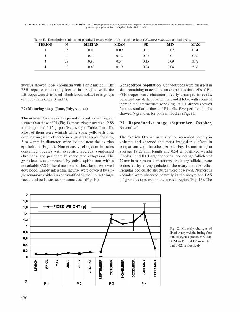
PERIOD N MEDIAN MEAN SE MIN MAX
1 25 0.09 0.09 0.01 0.02 0.31
2 14 0.14 0.12 0.02 0.07 0.32
3 39 0.90 0.54 0.15 0.09 3.72
4 19 0.69 0.19 0.28 0.04 5.33
nucleus showed loose chromatin with 1 or 2 nucleoli. TheFSH-tropes were centrally located in the gland while theLH-tropes were distributed in both lobes, isolated or in groupsof two ir cells (Figs. 3 and 4).
P2: Maturing stage (June, July, August)
The ovaries. Ovaries in this period showed more irregularsurface than those of P1 (Fig. 1), measuring in average 12.88mm length and 0.12 g. postfixed weight (Tables I and II).Most of them were whitish while some yellowish ones(vitellogenic) were observed in August. The largest follicles,2 to 4 mm in diameter, were located near the ovarianepithelium (Fig. 9). Numerous vitellogenic folliclescontained oocytes with eccentric nucleus, condensedchromatin and peripherally vacuolated cytoplasm. Thegranulosa was composed by cubic epithelium with aremarkable PAS (+) basal membrane. Theca layers were welldeveloped. Empty interstitial lacunae were covered by sin-gle squamous epithelium but stratified epithelium with largevacuolated cells was seen in some cases (Fig. 10).
Gonadotrope population. Gonadotropes were enlarged insize, containing more abundant ir granules than cells of P1.FSH-tropes were characteristically arranged in cords,polarized and distributed in the caudal lobe, with some ofthem in the intermediate zone (Fig. 7). LH-tropes showedfeatures similar to those of P1 cells. Few peripheral cellsshowed ir granules for both antibodies (Fig. 8).
P3: Reproductive stage (September, October,November)
The ovaries. Ovaries in this period increased notably involume and showed the most irregular surface incomparison with the other periods (Fig. 1), measuring inaverage 19.27 mm length and 0.54 g. postfixed weight(Tables I and II). Larger spherical and orange follicles of22 mm in maximum diameter (pre-ovulatory follicles) wereconnected by a long pedicle to the ovary and also otherirregular pediculate structures were observed. Numerousvacuoles were observed centrally in the oocyte and PAS(+) granules appeared in the cortical region (Fig. 13). The
Table II. Descriptive statistics of postfixed ovary weight (g) in each period of Nothura maculosa annual cycle.
Fig. 2. Monthly changes offixed ovary weight during fourannual cycles (mean ± SEM).SEM in P1 and P2 were 0.01and 0.02, respectively.
CLAVER, J.; ROSA; J. M.; LOMBARDO, D. M. & SOÑEZ, M. C. Histological seasonal changes in ovaries of spotted tinamous (Nothura maculosa Tinamidae, Temminck, 1815) related togonadotrope population. Int. J. Morphol., 26(2):353-361, 2008.

357
zona radiata, a peripheral region of the oocyte with finecytoplasmic processes, and the perivitelline layer were welldefined (Fig. 13). The granulosa was composed of apseudostratified columnar epithelium with strong PAS (+)basal membrane. The theca was a vascular sheath of cellularconnective tissue with some isolate theca gland cells,permeated with large and irregular capillaries. The first signof non-bursting atresia, in primordial and growing follicles,was shrinkage, while the lipid bodies coalesced to formacidophilic masses in the ooplasm. Few large folliclesshowed features of bursting atresia, characterized byfollicular wall break and vitellium escaping into theneighbouring lacunae (Fig. 14), and inside lacunae epithelialcells. Many of these atretic follicles presented follicularwall projections crossing the center, showing a central axisof dense connective tissue and covered by thecal andhypertrophied granulosa cells. Post ovulatory follicles werefrequently found in P3 and P4 (Fig. 18) and were locatednear the gonad surface. Their hypertrophied and vacuolatedcells filled the space occupied by the ovulated oocyte andsurrounded by thick layer of small, acidophilic cells.Condensed connective tissue bounded this structure.
Gonadotrope population. The ir granules storage wasincreased in gonadotropes which were more voluminous thanP1 and P2, and were observed in both lobes of the gland.FSH tropes were arranged in the characteristic cords in thecaudal lobe and in groups of 3 to 4 ir cells or isolated in thecephalic lobe (Fig. 11), while LH tropes were also seen asisolated or in small groups of 3 to 4 cells (Fig.12).
P4: Involutive stage (December, January, February)
The ovaries. The ovaries measured in average 14.81 mmlength and 0.19 g post fixed weight (Tables 1, 2). Some ofthem showed similar active signs as in P3 with predominantlarge orange follicles while others were in clear regressionwith whitish follicles. Atretic and post ovulatory follicleswere frequently observed. Interstitial lacunae were filledwith vitelline and surrounded by phagocytic epithelial cells(Fig. 17). The late stages of regression of postovulatoryfollicle and atresia were difficult to distinguish due to thefibrosis of the theca.
Gonadotrope population. Gonadotropes showed similarfeatures to those in P3. They were numerous and distributedin the entire gland. FSH tropes were arranged in cords andLH tropes were isolated or formed groups of scarce cells(Fig 15). Double immunostaining allowed distinguishingcells containing only FSH (+) granules at the periphery andLH (+) cells in the middle of the gland. Cells containingpositive granules for both antibodies were located amongthem (Fig. 16).
DISCUSSION
Although most histological features observed inNothura maculosa ovaries were similar to those of domesticand wild avian species (Hodges 1974; Johnson, 2000;Jones, 1978; Kern, 1972) they showed some specificcharacteristics. The two types of atresia, non-burstingatresia (Type 1) and bursting atresia (Type 2), described inthe ovary of the domestic hen and white-crowned sparrow(Gupta et al., 1988; Kern), were seen in small and largerfollicles of Nothura maculosa, respectively. Changes innon-bursting atresia of this species were similar to thosereported in Columba livia and Gallus domesticus by Guraya(1976). Bursting atresia was described previously inNothura maculosa (Arriaga et al.) in october ovaries.Internal follicular wall projections that we observed inbursting atresia of Nothura have not been described in otheravian species. Bursting atresia in hens occurs at the end ofthe egg-laying period (Nili & Kelly, 1996). Bump & Bumpreported the possibility of more than one egg-laying periodin Nothura maculosa, but we have not seen the remarkableregression among periods reported by the authors. Probablythe frequency of bursting atresia that we have observed inNothura maculosa during P3 and P4 may indicate morethan one egg-laying periods.
Atretic vitellogenic follicles finally disappearedfrom the ovary of Nothura maculosa without making avisible contribution to the stroma. So during P1 the ovarianstroma did not show any lipidic cells such as interstitialgland-cells which have been reported in domestic species(Gupta et al.; Guraya; Hodges and Kern). The large stromalspaces (lacunae) boundered by a phagocytic epitheliumand containing erythrocytes, yolk and the ooplasm ofruptured follicles initially observed in hens by Callebaut(1979) was described by Arriaga et al. in Nothura maculosaas stromal spaces. Similar structures were described inWhite crowned Sparrow’s ovary during spring and thebreeding period, disappearing in July (Kern). We also foundlacunar spaces in Nothura maculosa ovaries of P3 and P4.During P1, these spaces did not disappear but they wereemptied. Transformation of lacuna-lining cells intophagocytic cells occurred in the ovary of laying hens afterbursting atresia and exposure of these cells to released yolk(Nili & Kelly). In contrast, we observed that thistransformation occurred in Nothura maculosa beforebursting atresia. So in this species the presence of yolk inthe lacunae spaces seems not to be the cause oftransformation of this lining epithelium. Yolk vacuoles inthe cytoplasm of these cells may indicate that they werephagocytically active when the yolk was released to thestroma. Largest follicles presented a perivitelline
CLAVER, J.; ROSA; J. M.; LOMBARDO, D. M. & SOÑEZ, M. C. Histological seasonal changes in ovaries of spotted tinamous (Nothura maculosa Tinamidae, Temminck, 1815) related togonadotrope population. Int. J. Morphol., 26(2):353-361, 2008.

358

359
membrane, similar to that described in laying hens byWaclawek et al. (1998).
The distribution of gonadotropes in the pituitary glandand their morphological changes would be correlated togonadotrophin secretion and seasonal ovarian changes.Separate LH and FSH positives cells were almost exclusivelyfound in accordance with the findings in chicken pituitary(Proudman et al., 1999). However, the dual-label stainingrevealed cells containing both types of granules near theperiphery of the gland and their distribution was different.LH-tropes were distributed throughout the anterior pituitaryduring all the annual cycle but FSH-tropes were not foundin the cephalic lobe of P1 and P2, while they were localizedthroughout the gland in P3 and P4. Both cell types changedin number and in the storage in granules which increased inP3 and P4 in agreement with previous ultrastructuralobservations. (Soñez et al., 1997).
The annual reproductive cycle reported in Nothuramaculosa of South Eastern Brazil (Burger) shows similarpatterns in ovarian and oviductal development and involutioncompared with our results.
In conclusion, Nothura maculosa ovaries showedclear seasonal changes of growth and regression correlatedwith changes in gonadotrope population. During P1 theovaries were clearly in the resting stage and there were fewand small gonadotropes. Histological changes graduallyappeared in ovaries and the gonadotrope population of P2.Follicular growth began at the end of P2 in relation withFSH-trope increase. Maximum ovary and gonadotropedevelopment was reached during P3. P4 was the period whenmost ovarian variability appeared without changes ingonadotrope population.
The present study provides a basis for a thoroughinvestigation on the reproduction of this species.
ACKNOWLEDGMENTS
We thank Dr. J. A. Proudman at USDA – ARSMaryland for the gift of anti-chicken FSH-LH sera. We arevery gratefully to Dr. Irene Von Lawzewitch for her supportto complete this work.
Fig. 3. Anti-chicken FSH antibody positive cells in cephalic lobe of pituitary gland during P1 (arrow head) isolated and scarcely distributed.Counterstained with haematoxylin (H). X330.Fig. 4. Anti-chicken LH antibody positive cells in caudal lobe of pituitary gland during P1 (arrow head), isolated and more abundant incomparison with anti-chicken FSH positive cells. Counterstained with haematoxylin (H). X132.Fig. 5. Ovary stroma during P1 showing empty interstitial lacunae (arrow head) and abundant vascularisation (V). H/E. X33.Fig. 6. Ovary cortex during P1 showing abundant primordial follicles (P), some secondary follicles (S) and few tertiary follicles (T). H/E. X33.Fig. 7. Anti-chicken FSH positive cells only in caudal lobe of pituitary gland during P2. The FSH-tropes were arranged in cords (arrowhead). Counterstained with haematoxylin (H). X132.Fig. 8. Double immunocytochemical staining using anti-chicken FSH and LH serum. Blue cells were FSH positive peripherally located(a) while brown LH-positive cells (b) were found in all the gland and few cells containing both positive granules (arrow head) wereobserved. Counterstained with haematoxylin (H). X132.Fig. 9. Ovary cortex during P2 showing secondary follicles (S) near the ovary epithelium (E). Thecal layer is strongly PAS positive(arrow head). PAS / H. X132.Fig. 10. Interstitial stroma with dilated lacunae (L) covered by numerous vacuolated cells (*). Connective tissue (arrow head). PAS / H. X132.Fig. 11. Sagittal section of pituitary gland during P3 showing anti-ch FSH positive cells found in both lobes. Counterstained withhaematoxylin (H). X13.Fig. 12. Anti-chicken LH positive cells among the lobes of pituitary gland during P3 (≠ ). They were isolated. Counterstained withhaematoxylin (H). X132.Fig. 13. Vitellogenic follicle in ovary of P3. There are PAS positive thecal layers (T). The granulosa ephytelium (E) is seudostratified. Inoocyte: the radiata zone (arrow ahead), yolk granules (G) and the vacuolated cytoplasm (V). PAS / H. X132.Fig. 14. Bursting atretia in ovary of P3. Follicular wall is broken (arrow head), interstitial lacunae contain vitellium granules (L). Goldner-Masson trichrome. X33.Fig. 15. FSH-tropes in caudal lobe of pituitary gland during P4. They contain abundant granules (arrow head). Counterstained withhaematoxylin (H). X330.Fig. 16. Double immunocytochemical staining using anti-chicken FSH and LH serum in pituitary gland during P4. Some cells containboth types of granules (Blue: anti-cFSH, brown: anti-cLH) (arrow head). Counterstained with haematoxylin (H). X330.Fig. 17. Interstitial lacunae containing PAS positive vitellium granules (L) in ovary during P4. PAS / H. X132.Fig. 18. Post-ovulatory follicle in ovary during P4. The granulosa layer is hypertrophic (G) with central cells vacuolated ( ≠ ). PAS / H. X33.
CLAVER, J.; ROSA; J. M.; LOMBARDO, D. M. & SOÑEZ, M. C. Histological seasonal changes in ovaries of spotted tinamous (Nothura maculosa Tinamidae, Temminck, 1815) related togonadotrope population. Int. J. Morphol., 26(2):353-361, 2008.

360
REFERENCES
Arriaga, A.; Nicora, O. & Ibañez, N. Variacionesestacionales en ovario de inambu chico comun (Nothuramaculosa). El Hornero Nº extr.: 14-27, 1983.
Benoit, J. Hypothalamo-hypophyseal control of the sexualactivity in birds. Gen Comp Endocrinol 1:254-74, 1962.
Bump, G.; Bump, J.W. A study of the spotted tinamous andthe pale spotted tinamous of Argentina. Spec. Sci. Rep.US Fish Wildl Serv., 120:1-160, 1969.
Burger, M. I. Ciclo reproductivo de fêmeas de umapopulação de Nothura maculosa TEMMINK, 1815(Aves,Tinamidae) no Rio Grande de Sul, Brasil.Iheringia, Sér Zool. 71:161-74, 1991.
Burger, M.I. Ciclo reprodutivo de machos de uma populaçâode Nothura maculosa TEMMINCK, 1815 (Aves,Tinamidae) no Rio Grande Do Sul, Brasil. Iheringia,Sér. Zool., 73:77-90, 1992.
Callebaut, M. The avian ovary is an open organ. Anat.Embryol., 158:103-19, 1979.
Dabbene, R. Los tinamidos o perdices de la Argentina. InAves de caza de la República Argentina. Ed. Albatros,Buenos Aires, 1972. pp 85-113.
Del Hoyo, J. & Elliott, A. eds Order Tinamiformes. In“Handbook of the birds of the world” Ed by Lynx,
Barcelona, Spain, 1992. V. I. pp 111-38.
Fleming Batalha da Silveira, C. & Menegheti, J.O. Estudosobre a relaçâo peso e sexo em Nothura maculosa(TEMMINCK,1815) (Aves, Tinamiformes, Tinamidae).Iheringia, Sér Zool., 58:7-16, 1981.
Follett, B. K. & Farner, D.S. Pituitary gonadotropins in theJapanese Quail (Coturnix japonica) duringphotoperiodically induced gonadal growth. Gen. Comp.Endocrinol., 7:125-31, 1966.
Garitano-Zavala, A: Proyectos demostrativos de crianzade pisacca (Nothoprocta ornata) en Bolivia. Informedel Instituto de Ecología, Universidad Mayor de SanAndrés. Fundación para el Desarrollo de la Ecología.La Paz, Bolivia, 2003.
Gupta, S. K.; Gilbert, A. B. & Walker, M.A. Histologicalstudy of follicular atresia in the ovary of the domestichen (Gallus domesticus). J. Reprod. Fert., 82:219-25,1988.
Guraya, S.S. Morphological and histochemical observationson follicular atresia and interstitial gland tissue in thecolumbid ovary. Gen. Comp. Endocrinol., 30:534-8, 1976.
Handford, P. & Mares, M. A. The mating systems of ratitesand tinamous. An evolutionary perspective. Biol. J.Linn. Soc., 25:77-104, 1985.
CLAVER, J.; ROSA, J. M.; LOMBARDO, D. M. & SOÑEZ, M. C. Cambios histológicos estacionales en ovarios de perdiz (Nothuramaculosa Tinamidae, Temminck, 1815) y su relación con la población de gonadotropas. Int. J. Morphol., 26(2):353-361, 2008.
RESUMEN: Nothura maculosa es un tinámido sudamericano que presenta marcada estacionalidad reproductiva. Este trabajodescribe los cambios estacionales del ovario de esta especie, en relación con la población de gonadotropas (GTHs). Muestras de ovariosy pituitarias de ejemplares adultos fueron colectadas mensualmente durante cuatro años; se fijaron en solución de Bouin y procesadaspara M.O. Los datos del peso gonadal posfijación fueron analizados usando STATISTIX 4.0. Los cortes de ovarios fueron coloreados conH/E, P.A.S. y Tricrómico de Goldner-Masson. En cortes de adenohipófisis se aplicó inmunocitoquímica simple y doble (sistema ABC,Vector Lab.), empleando anticuerpos anti-pollo FSH y anti-pollo LH. Las muestras se analizaron en períodos trimestrales de cada año(P): P1: Marzo-Abril-Mayo (etapa de reposo), P2: Junio-Julio-Agosto (etapa de desarrollo), P3: Septiembre-Octubre-Noviembre (etapareproductiva), P4: Diciembre-Enero-Febrero (etapa involutiva). El peso de los ovarios (PO) varió significativamente entre los periodos(p< 0.001). Durante P1, sólo se observaron folículos primordiales y pre-vitelogénicos, PO 0.09± 0.01 g (n=25); durante P2, se detectaronfolículos en desarrollo con signos de vitelogénesis, PO 0.13± 0.01 g (n=14); durante P3, se encontró máximo desarrollo folicular, PO0.90 ± 0.15 g (n=39); P4 exhibió gran variabilidad folicular, PO 0.18± 0.18 g (n=19). La atresia involutiva se observó en todos los perío-dos, mientras que la atresia explosiva y los folículos postovulatorios caracterizaron a P3 y P4. Las GTHs conteniendo escasos gránulosLH y FSH inmunoreactivos (ir) predominaron durante P1 y P2. Las GTHs con gránulos LHir eran abundantes en la zona intermedia y enel lóbulo caudal en P3 y P4 mientras que escasas células contenían ambos tipos de gránulos. El número de células FSHir se incrementódurante P3 y P4. Los cambios histológicos del ovario se correlacionaron estrechamente con las variaciones en la población de gonadotropas.
PALABRAS CLAVE: Aves; Gonadotropas; Nothura maculosa; Ovario; Pituitaria; Reproducción; Tinamidae.
CLAVER, J.; ROSA; J. M.; LOMBARDO, D. M. & SOÑEZ, M. C. Histological seasonal changes in ovaries of spotted tinamous (Nothura maculosa Tinamidae, Temminck, 1815) related togonadotrope population. Int. J. Morphol., 26(2):353-361, 2008.
361
Hodges, R. D. The histology of the Fowl. Academic Press.Inc., London, 1974.
Johnson, A. L. Reproduction in female. In “Sturkie’s AvianPhysiology” Ed by G. Causey Whiton. Acadeemic Press,2000. pp. 569-96.
Jones, R. E. The vertebrate ovary. Comparative biology andevolution. Plenum press, New York and London, 1978.
Kern, M. D. Seasonal changes in the reproductive system ofthe female White-crowned sparrow, Zonotrichiagambelii, in captivity and in the field. Z. Zellforsch.,126:297-319, 1972.
Leska, A. & Dusza, L. Seasonal changes in the hypothalamo-pituitary-gonadal axis in birds. Reproductive Biology.,7(2): 99-126, 2007.
Menegheti, J. O. Acasalamento em Nothura maculosa(Temminck,1815) (Tinamidae) duraçâo, período,magnitude e sua variaçâo. Iheringia Sér. Zool., 64:3-14,1984.
Menegheti, J.O. Observações preliminares sobre oacasalamento e recrutamento em Nothura maculosa(Temminck, 1815) (Aves Tinamidae) no Rio Grande doSul, Brasil. Iheringia Sér. Zool., 59:65-75, 1981.
Nili, H. & Kelly, W.R. Form and function of lacunae in theovary of the laying hen. Anat. Rec., 244:165-74, 1996.
Proudman, J.A.; Vandesande, F. & Berghman, L.R.Immunochemical evidence that follicle-stimulatinghormone and luteinizing hormone reside in separate cellsin the chicken pituitary. Biol. Reprod., 60:1324-8, 1999.
Soñez, M.C.; Stancato, M.R. & Von Lawzewitsch, I.Immunocytochemical identification of prolactin andsomatotropin-containing cells in adenohypophysis ofNothura maculosa (Tinamidae. Temminck, 1815) duringrecess and active reproduction periods using humanhormone antisera. Com. Biol., 13:85-100, 1995.
Soñez, M.C. & Von Lawzewitsch, I. Ultrastructuralidentification of pituitary cells in Nothura maculosa(Tinamidae. Temminck, 1815). Biocell, 21:103-14, 1997.
Waclawek, M,; Foisner, R.; Nimpf, J. & Schneider, W. J.The chicken homologue of zona pellucida protein-3 issynthesized by granulose cells. Biol. Reprod., 59:1230-9, 1998.
Correspondence to:
Dr. Juan Claver
Histología y Embriología
Facultad de Ciencias Veterinarias
Universidad de Buenos Aires
Chorroarin 280
C1427CWO
Buenos Aires
ARGENTINA
Tel.: 54-011-4524-8458
Fax.: 54-011-4524-8480
Email: [email protected]
Received: 14-02-2008
Accepted: 22-03-2008
CLAVER, J.; ROSA; J. M.; LOMBARDO, D. M. & SOÑEZ, M. C. Histological seasonal changes in ovaries of spotted tinamous (Nothura maculosa Tinamidae, Temminck, 1815) related togonadotrope population. Int. J. Morphol., 26(2):353-361, 2008.

362