histological evidence of carcinoma in hepatic tumour
TRANSCRIPT
496 BRITISH MEDICAL JOURNAL 29 NOVEMBER 1975
There is circumstantial evidence to suggest that a repeatedsuccession of menstrual cycles before the first pregnancy mayeven be harmful to the breast'; recent epidemiological evidenceshows that the risk of breast cancer increases with time elapsedfrom menarche to first pregnancy.'5 16 Such considerationshighlight the importance of a fuller understanding of the changestaking place in the breast during the normal menstrual cycle andafter the use of oral contraceptives.
We thank those women who generously volunteered to take part inthis study and are grateful to Dr C S Corker and Mr R Sharpe forstatistical advice.
References
McCance, R A, Luff, M C, and Widdowson, E E, J7ournal of Hygiene,1937, 37, 571.
2Royal College of General Practitioners, Report on Oral Contraceptives andHealth. London, Pitman Medical, 1974.
3Ingleby, H, Bzulletint of the International Associationi of Medical Museumns,1949, 29, 87.
4Reimann, S P, and Seabold, P S, Amterican Journal of Cancer, 1933, 17, 34.5Geschickter, C F, Diseases of the Breast. Philadelphia, Lippincott, 1945.Masters, W H, and Johnson, V E, Humzan Sexual Response. London,
Churchill, 1966.Hytten, F E, British Medical Journal, 1954, 1, 912.Dawson, E K, Edinburgh Medical)Journal, 1934, 41, 653.
9 Marshall, W A, and Tanner, J M, Archives of Disease in Childhood, 1969,44, 291.
1'1 Haagensen, C D, Diseases of the Breast. London, Saunders, 1956.1' Ozzello, L, and Speer, F D, American 7ourrnal of Pathology, 1958, 34 (5),
993.12 Engel, S, Proceedings of the Royal Society of Medicine, 1947, 40, 899.13 Speert, H, Contributions to Enmbryology, 1948, 32, 9.14 Short, R V, in Physiology and Genetics of Reproduiction, part A, ed E M
Coutinho and F Fuchs. New York, Plenum, 1974.1 MacMahon, B, Cole, P, and Brown, J, Journal of the National Cancer
Instituite, 1973, 50, 21.16 Shapiro, S, et al, in Host Environment Interactions in the Etiology of Cancer
in Man, ed R Doll and I Vodopija, p 169. Lyon, International Agencyfor Research on Cancer, 1973.
Histological evidence of carcinoma in a hepatic tumourassociated with oral contraceptives
M DAVIS, B PORTMANN, M SEARLE, RALPH WRIGHT, ROGER WILLIAMS
British Medical3Journal, 1975, 4, 496-498
Summary
A primary hepatic tumour occurred in a 21-year-oldwoman who had been taking oral contraceptives for twoyears; she was treated by partial hepatectomy. Part ofthe neoplasm showed features suggestive of focal nodularhyperplasia, while the remainder had the histologicalcharacteristics of a well-differentiated hepatocellularcarcinoma. This is the first report of malignant trans-formation of a tumour in a patient taking oral contra-ceptives.
Introduction
Since 1972 25 cases of benign primary tumour of the liver havebeen described in women taking oral contraceptives. -12 Aparticular feature of these neoplasms has been their extremevascularity, and two-thirds of the patients have presented withhaemoperitoneum from spontaneous rupture, which carries amortality of 60"0. We describe here for the first time a furthercomplication of this condition: malignant transformation ofthe tumour.
Liver Unit, King's College Hospital and Medical School, LondonSE5 8RX
M DAVIS, MB, MRCP, senior lecturer and honorary consultant physicianB PORTMANN, MD, research histopathologistROGER WILLIAMS, MD, FRCP, consultant physician and director of unit
University Department of Medicine, Royal South Hants Hospital,Southampton
M SEARLE, BSC, medical studentRALPH WRIGHT, MD, FRCP, professor of medicine
Case report
A 21-year-old woman was found on a routine blood check to beanaemic with a haemoglobin of 9 9 g/dl and an erythrocyte sedimenta-tion rate of 100 mm in one hour (Westergren). Her history wasnegative except for an attack of acute hepatitis some two years earlier,from which she had made a complete recovery, with return to normalliver function. Hepatitis B surface antigen (HBsAg) had never beenfound in her serum. She had been taking Volidan (megestrol acetate4 mg, ethinyloestradiol 50 tlg) for contraception continuously for twoyears, since her recovery from hepatitis.
Physical examination showed, in addition to the anaemia, firmenlargement of the left lobe of the liver. Liver function tests gaveabnormal results, with a serum bilirubin of 20 ,imol/l (1 2 mg/100 ml) and alkaline phosphatase of 244 IU/1. Other investigations,including estimations of immunoglobulins, a-fetoprotein, andHBsAg, showed no abnormalities. Coagulation test results werenormal except for a slightly prolonged partial thromboplastin time.A technetium liver scan showed a filling defect in the left lobe of theliver, and a selenium scan showed uptake in this area. Hepaticarteriography confirmed the presence of a highly vascular tumour inthe left lobe, supplied by the middle and left hepatic arteries. Atlaparoscopy the left lobe of the liver was diffusely enlarged and some-what purple.The patient underwent left hepatectomy on 6 November 1974,
and left hospital three weeks later after an uneventful postoperativecourse. She has since remained well, with no evidence of tumour onserial liver scans.
PATHOLOGICAL APPEARANCES OF RESECTED TUMOUR
The left hepatectomy specimen weighed 820 g and contained awell-circumscribed multilobular mass 14 cm in diameter. Some partsof the lobules were light brown, but most of the tumour was greyand more friable. The central part of the mass was occupied by partiallycalcified fibrous tissue, which radiated between the lobules. Patchyhaemorrhages were scattered throughout the mass, but no majorhaemorrhage was seen.
Microscopic examination showed the whole tumour to be welldemarcated from the surrounding liver tissue, which was histologicallynormal except for mild portal tract inflammation, although a truecapsule was not present. The light brown areas of the tumour werecomposed of liver cells that appeared fairly normal, although rather
on 10 Decem
ber 2021 by guest. Protected by copyright.
http://ww
w.bm
j.com/
Br M
ed J: first published as 10.1136/bmj.4.5995.496 on 29 N
ovember 1975. D
ownloaded from
BRITISH MEDICAL JOURNAL 29 NOVEMBER 1975
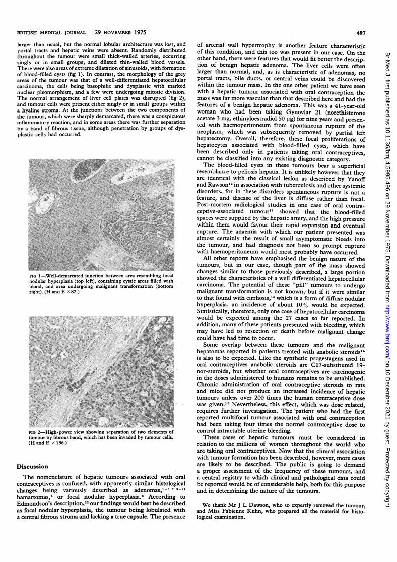
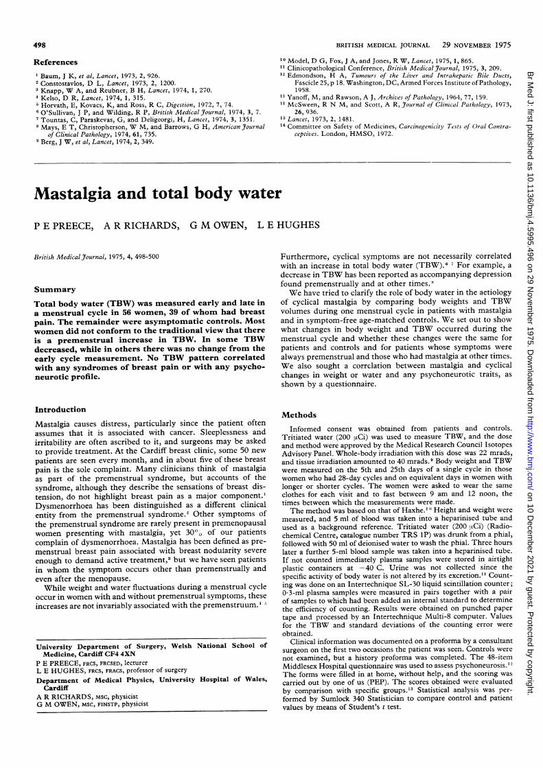
larger than usual, but the normal lobular architecture was lost, andportal tracts and hepatic veins were absent. Randomly distributedthroughout the tumour were small thick-walled arteries, occurringsingly or in small groups, and dilated thin-walled blood vessels.There were also areas of extreme dilatation of sinusoids, with formationof blood-filled cysts (fig 1). In contrast, the morphology of the greyareas of the tumour was that of a well-differentiated hepatocellularcarcinoma, the cells being basophilic and dysplastic with markednuclear pleomorphism, and a few were undergoing mitotic division.The normal arrangement of liver cell plates was disrupted (fig 2),and tumour cells were present either singly or in small groups withina hyaline stroma. At the junctions between the two components ofthe tumour, which were sharply demarcated, there was a conspicuousinflammatory reaction, and in some areas there was further separationby a band of fibrous tissue, although penetration by groups of dys-plastic cells had occurred.
FIG 1-Well-demarcated junction between area resembling focalnodular hyperplasia (top left), containing cystic areas filled withblood, and area undergoing malignant transformation (bottomright). (H and E x 82.)
FIG 2-High-power view showing separation of two elements oftumour by fibrous band, which has been invaded by tumour cells.(HandE x136.)
Discussion
The nomenclature of hepatic tumours associated with oralcontraceptives is confused, with apparently similar histologicalchanges being variously described as adenomas,' -5 7 9-11hamartomas,6 or focal nodular hyperplasia.8 According toEdmondson's description,22 our findings would best be described
as focal nodular hyperplasia, the tumour being lobulated witha central fibrous stroma and lacking a true capsule. The presence
497
of arterial wall hypertrophy is another feature characteristicof this condition, and this too was present in our case. On theother hand, there were features that would fit better the descrip-tion of benign hepatic adenoma. The liver cells were oftenlarger than normal, and, as is characteristic of adenomas, noportal tracts, bile ducts, or central veins could be discoveredwithin the tumour mass. In the one other patient we have seenwith a hepatic tumour associated with oral contraception themass was far more vascular than that described here and had thefeatures of a benign hepatic adenoma. This was a 41-year-oldwoman who had been taking Gynovlar 21 (norethisteroneacetate 3 mg, ethinyloestradiol 50 tLg) for nine years and presen-ted with haemoperitoneum from spontaneous rupture of theneoplasm, which was subsequently removed by partial lefthepatectomy. Overall, therefore, these focal proliferations ofhepatocytes associated with blood-filled cysts, which havebeen described only in patients taking oral contraceptives,cannot be classified into any existing diagnostic category.The blood-filled cysts in these tumours bear a superficial
resemblance to peliosis hepatis. It is unlikely however that theyare identical with the classical lesion as described by Yanoffand Rawson" in association with tuberculosis and other systemicdisorders, for in these disorders spontaneous rupture is not afeature, and disease of the liver is diffuse rather than focal.Post-mortem radiological studies in one case of oral contra-ceptive-associated tumour'1 showed that the blood-filledspaces were supplied by the hepatic artery, and the high pressurewithin them would favour their rapid expansion and eventualrupture. The anaemia with which our patient presented wasalmost certainly the result of small asymptomatic bleeds intothe tumour, and had diagnosis not been so prompt rupturewith haemoperitoneum would most probably have occurred.
All other reports have emphasised the benign nature of thetumours, but in our case, though part of ihe mass showedchanges similar to those previously described, a large portionshowed the characteristics of a well differentiated hepatocellularcarcinoma. The potential of these "pill" tumours to undergomalignant transformation is not known,-but if it were similarto that found with cirrhosis,"1 which is a form of diffuse nodularhyperplasia, an incidence of about 10/0 would be expected.Statistically, therefore, only one case of hepatocellular carcinomawould be expected among the 27 cases so far reported. Inaddition, many of these patients presented with bleeding, whichmay have led to resection or death before malignant changecould have had time to occur.Some overlap between these tumours and the malignant
hepatomas reported in patients treated with anabolic steroids'is also to be expected. Like the synthetic progestagens used inoral contraceptives anabolic steroids are C17-substituted 19-nor-steroids, but whether oral contraceptives are'carcinogenicin the doses administered to humans remains to be established.Chronic administration of oral contraceptive steroids to ratsand mice did not produce an increased incidence of hepatictumours unless over 200 times the human contraceptive dosewas given.'6 Nevertheless, this effect, which was dose related,requires further investigation. The patient who had the firstreported multifocal- tumour associated with oral contraceptionhad been taking four times the normal contrgceptive dose tocontrol intractable uterine bleeding.These cases of hepatic tumours must be considered in
relation to the millions of women throughout the world whoare taking oral contraceptives. Now that the clinical associationwith tumour formation has been described, however, more casesare likely to be described. The public is going to demand
a proper assessment of the frequency of these tumours, anda central registry to which clinical and pathological data couldbe reported would be of considerable help, both for this purposeand in determnining the nature of the tumours.
We thank Mr J L Dawson, who so expertly removed the tumour,and Miss Fabienne Kuhn, who prepared all the material for histo-logical examination.
on 10 Decem
ber 2021 by guest. Protected by copyright.
http://ww
w.bm
j.com/
Br M
ed J: first published as 10.1136/bmj.4.5995.496 on 29 N
ovember 1975. D
ownloaded from
498 BRITISH MEDICAL JOURNAL 29 NOVEMBER 1975
References
Baum, J K, et al, Lancet, 1973, 2, 926.2 Constostavlos, D L, Lancet, 1973, 2, 1200.3 Knapp, W A, and Reubner, B H, Lancet, 1974, 1, 270.4 Kelso, D R, Lancet, 1974, 1, 315.Horvath, E, Kovacs, K, and Ross, R C, Digestion, 1972, 7, 74.
6 O'Sullivan, J P, and Wilding, R P, British Medical Journal, 1974, 3, 7.7 Tountas, C, Paraskevas, G, and Deligeorgi, H, Lancet, 1974, 3, 1351.8 Mays, E T, Christopherson, W M, and Barrows, G H, American Journal
of Clinical Pathology, 1974, 61, 735.9 Berg, J W, et al, Lancet, 1974, 2, 349.
10 Model, D G, Fox, J A, and Jones, R W, Lancet, 1975, 1, 865.11 Clinicopathological Conference, British Medical Journal, 1975, 3, 209.12 Edmondson, H A, Tuniours of the Liver and Intrahepatic Bile Ducts,
Fascicle 25, p 18. Washington, DC, Armed Forces Institute of Pathology,1958.
13 Yanoff, M, and Rawson, A J, Archives of Pathology, 1964, 77, 159.14 McSween, R N M, and Scott, A R, J7ournal of Clinical Pathology, 1973,
26, 936.15 Lancet, 1973, 2, 1481.16 Committee on Safety of Medicines, Carcinogenicity Tests of Oral Contra-
ceptives. London, HMSO, 1972.
Mastalgia and total body water
P E PREECE, A R RICHARDS, G M OWEN, L E HUGHES
British Medical3Journal, 1975, 4, 498-500
Summary
Total body water (TBW) was measured early and late ina menstrual cycle in 56 women, 39 of whom had breastpain. The remainder were asymptomatic controls. Mostwomen did not conform to the traditional view that thereis a premenstrual increase in TBW. In some TBWdecreased, while in others there was no change from theearly cycle measurement. No TBW pattern correlatedwith any syndromes of breast pain or with any psycho-neurotic profile.
Introduction
Mastalgia causes distress, particularly since the patient oftenassumes that it is associated with cancer. Sleeplessness andirritability are often ascribed to it, and surgeons may be askedto provide treatment. At the Cardiff breast clinic, some 50 newpatients are seen every month, and in about five of these breastpain is the sole complaint. Many clinicians think of mastalgiaas part of the premenstrual syndrome, but accounts of thesyndrome, although they describe the sensations of breast dis-tension, do not highlight breast pain as a major component.'Dysmenorrhoea has been distinguished as a different clinicalentity from the premenstrual syndrome.2 Other symptoms ofthe premenstrual syndrome are rarely present in premenopausalwomen presenting with mastalgia, yet 300o of our patientscomplain of dysmenorrhoea. Mastalgia has been defined as pre-menstrual breast pain associated with breast nodularity severeenough to demand active treatment,' but we have seen patientsin whom the symptom occurs other than premenstrually andeven after the menopause.
While weight and water fluctuations during a menstrual cycleoccur in women with and without premenstrual symptoms, theseincreases are not invariably associated with the premenstruum.4
University Department of Surgery, Welsh National School ofMedicine, Cardiff CF4 4XN
P E PREECE, FRCS, FRCSED, lecturerL E HUGHES, FRCS, FRACS, professor of surgery
Department of Medical Physics, University Hospital of Wales,Cardiff
A R RICHARDS, MSC, physicistG M OWEN, MSC, FINSTP, physicist
Furthermore, cyclical symptoms are not necessarily correlatedwith an increase in total body water (TBW).6 7 For example, adecrease in TBW has been reported as accompanying depressionfound premenstrually and at other times.8We have tried to clarify the role of body water in the aetiology
of cyclical mastalgia by comparing body weights and TBWvolumes during one menstrual cycle in patients with mastalgiaand in symptom-free age-matched controls. We set out to showwhat changes in body weight and TBW occurred during themenstrual cycle and whether these changes were the same forpatients and controls and for patients whose symptoms werealways premenstrual and those who had mastalgia at other times.We also sought a correlation between mastalgia and cyclicalchanges in weight or water and any psychoneurotic traits, asshown by a questionnaire.
Methods
Informed consent was obtained from patients and controls.Tritiated water (200 uiCi) was used to measure TBW, and the doseand method were approved by the Medical Research Council IsotopesAdvisory Panel. Whole-body irradiation with this dose was 22 mrads,and tissue irradiation amounted to 40 mrads. 9 Body weight and TBWwere measured on the 5th and 25th days of a single cycle in thosewomen who had 28-day cycles and on equivalent days in women withlonger or shorter cycles. The women were asked to wear the sameclothes for each visit and to fast between 9 am and 12 noon, thetimes between which the measurements were made.The method was based on that of Haxhe." Height and weight were
measured, and 5 ml of blood was taken into a heparinised tube andused as a background reference. Tritiated water (200 1LCi) (Radio-chemical Centre, catalogue number TRS 1P) was drunk from a phial,followed with 50 ml of deionised water to wash the phial. Three hourslater a further 5-ml blood sample was taken into a heparinised tube.If not counted immediately plasma samples were stored in airtightplastic containers at -40 C. Urine was not collected since thespecific activity of body water is not altered by its excretion." Count-ing was done on an Intertechnique SL-30 liquid scintillation counter;0 3-ml plasma samples were measured in pairs together with a pairof samples to which had been added an internal standard to determinethe efficiency of counting. Results were obtained on punched papertape and processed by an Intertechnique Multi-8 computer. Valuesfor the TBW and standard deviations of the counting error wereobtained.
Clinical information was documented on a proforma by a consultantsurgeon on the first two occasions the patient was seen. Controls werenot examined, but a history proforma was completed. The 48-itemMiddlesex Hospital questionnaire was used to assess psychoneurosis.'2The forms were filled in at home, without help, and the scoring wascarried out by one of us (PEP). The scores obtained were evaluatedby comparison with specific groups.'3 Statistical analysis was per-formed by Sumlock 340 Statistician to compare control and patientvalues by means of Student's t test.
on 10 Decem
ber 2021 by guest. Protected by copyright.
http://ww
w.bm
j.com/
Br M
ed J: first published as 10.1136/bmj.4.5995.496 on 29 N
ovember 1975. D
ownloaded from