hip test-complete1
TRANSCRIPT

Hip Examination

Applied anatomy
• The hip joint is multiaxial ball-and-socket joint.• comprised of – femoral head– articulates – acetabulum

Applied anatomy

Applied anatomy

Inspection

Gait
ดูการเดิน มเีดินกะเผลก (anthalgic gait) หรือไม่ และการยืนของผู้ป่วย ลักษณะการเดินมกีารเคลื่อนไหว
มากนอ้ยเพยีงใด เปรียบเทยีบกันทัง้สองด้าน• พยาธิสภาพที่ข้อสะโพก การเดินของผู้ป่วยมกั
เคลื่อนไหวของสะโพกน้อย มกัลงนำ้าหนกัข้างทีดี่ ในทา่ นอนจะพบว่าขายาวไม่เทา่กัน (Apparent
shortening) • ตรวจ Trendelenburg test โดยให้ผู้ป่วยยนืลงนำ้า
หนักบนขาข้างหนึง่ และยกเข่าของข้างตรงข้าม ตาม ปกติระดับของกระดูก Pelvis จะยกข้ึนในด้านที่ไมไ่ด้ลง
นำ้าหนกั แต่ถ้ามพียาธิสภาพในข้อสะโพก เช่นCongenital dislocation หรือ การอ่อนแรงของกล้าม
เนือ้สะโพก ระดับกระดูก Pelvis จะลดตำ่ากว่าอีกข้างหนึง่

Trendelenburg test
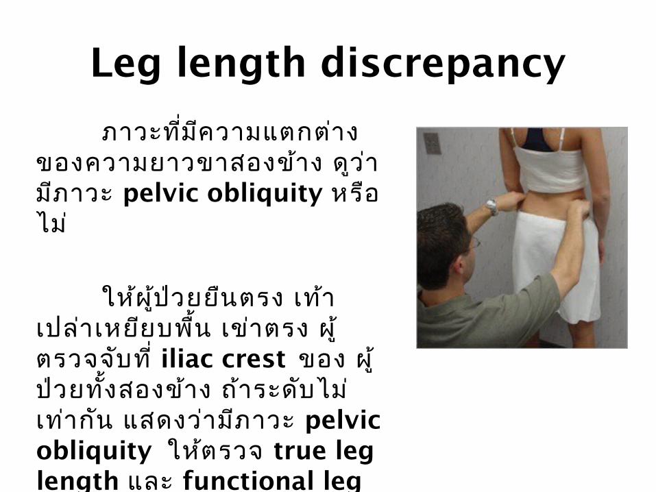
Leg length discrepancy
ภาวะทีม่คีวามแตกต่าง ของความยาวขาสองข้าง ด ูว ่า
มภีาวะ pelvic obliquity หรือ ไม่
ใหผ้ ู้ป ่วยยนืตรง เทา้ เปล ่าเหยยีบพื้น เข ่าตรง ผู้
ตรวจจบัท ี่ iliac crest ของ ผู้ ป่วยทัง้สองข้าง ถ ้าระดบัไม ่
เทา่ก ัน แสดงว ่ามภีาวะ pelvic obliquity ใหต้รวจ true leg length และ functional leg length ต่อไป

True leg length discrepancy
1. ว ัดจาก ASIS ไปถึง medial malleolus
2. ว ัดจาก ASIS ไปถึง lateral malleolus
ถา้ความยาวแตกต่างก ันมากกว่า 5 cm ถือว ่า แตกต่างอยา่งมนียัส ำาค ัญ แสดงถึงความยาวของ
ค่าท ีไ่มเ่ท ่าก ัน จากนั้นดตู ่อว ่าเป ็นท ีค่วามยาว ของ femur หรือ tibia ที่ยาวไมเ่ทา่ก ัน

Test for Measuring femoral lengths
• ใหผ้ ู้ป ่วยนอนหงาย งอเข ่าและสะโพก 90°• ถา้มกีระดกู femur ทีย่าวกว ่าอ ีกข ้างหนึง่ ขาข้างน ัน้จะอย ูส่ ูงกว ่า

Test for Measuring tibia lengths
(Prone knee flexion test)
• ใหผ้ ู้ป ่วยนอนควำ่า งอเข ่า 90° เปร ียบเทยีบความสูงของส ้นเทา้
• ถา้มกีระด ูก tibia ทีส่ ัน้กว ่าอ ีกข ้างหนึง่ ขาข้างน ัน้จะอย ูต่ ำ่ากว ่า

Functional leg length discrepancy
• ว ัดจาก umbilicus ไป ถึง medial malleolus
• ถ้าว ัด true leg length discrepancy ไมแ่ตก
ต่าง แต่ functional leg length discrepancy มคีวามแตกต่างอาจเกดิจาก
ภาวะ scoliosis, ความผิดร ูปของกระดกู
เชงิกราน หรือม ีabduction/adduction contructure ของข้อสะโพก

Hip dislocation
• Posterior hip dislocation : สะโพกจะอยูใ่น ทา่ flexion, adduction และ internal
rotation ไมส่ามารถขยับเหยยีดได้ท ัง้ active และ passive
• Anterior hip dislocation : สะโพกจะอยูใ่นทา่abduction และ external rotation

การตรวจข้อตะโพกที่สงส ัยว ่าม ีการอ ักเสบ
• ข้อสะโพกอาจอย ูใ่นท่า flexion และ adduction เน ื่องจากปวด เก ิดการหดเกร ็งของ adductor
muscle• Fabere test : ใหผ้ ู้ป ่วยงอเข ่าขางหนึ่ง โดยวาง
เท ้าอย ูท่ ี่บร ิเวณกระด ูก patella ของเข ่าอกีข ้าง หนึ่ง ใชม้ ือด ันเข ่าข ้างน ั้นลงต ิดพ ืน้เพ ือ่ใหข้าหมุน ออก หมุนข ้อสะโพกเข ้าด ้านใน โดยจับเข ่าหม ุน
เข ้าข ้างในและเท ้าหม ุนออกข้างนอก ถ้าม ีการ อกัเสบที่ข ้อสะโพก จะด ันเข ่าข ้างน ั้นไดน้อยและ
จะปวดที่สะโพก

Palpation

การคลำา• คลำาต ำาแหนง่ landmarks ทีส่ ำาค ัญ ได้แก ่ ASIS,
iliac crest, lesser trochanter, greater trochanter, pubis ดูว ่ามอีาการเจ ็บหร ือไม ่
อาจเกดิ avultion fracture ทีบ่ร ิเวณดังกล ่าว ได้
• การคลำา hip joint ใหค้ล ำา lateral ตอ่ femoral pulse มา 2 cm และลงมาอีก 2 cm ถ้ามภีาวะ
อักเสบหรือต ิดเช ื้อของข้อ หร ือมภีาวะกระดกูข ้อ สะโพกหกั จะทำาใหก้ดเจ ็บบร ิเวณนีไ้ด ้

Manipulation

การขยับ ใหต้รวจการทำางานของกล้ามเน ื้อต ่าง ๆ ดงัน ี้


Flexion

Extension

Abduction

Adduction
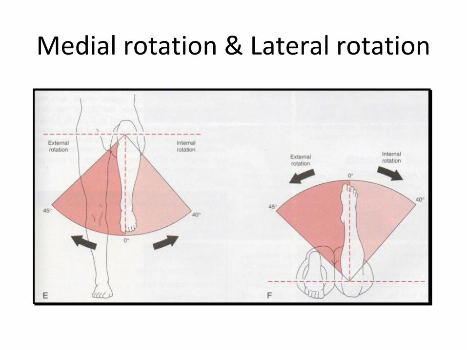
Medial rotation & Lateral rotation

Resisted isometric movement

Resisted isometric movements
Resisted isometric movements around the hip. (A) Flexion. (B) Extension. (C) Adduction. (D) Abduction. (E) Medial rotation. (F) Lateral rotation. (G) Knee flex-on. (H) Knee extension

Functional assessment

Special TestsPatrick's Test (Faber or Figure-
Four Test) ท่าเตร ียม : supine และ flexion, abduction,
and external rotation ของ hip (คล้ายเลข4)
ว ิธ ีตรวจ : foot of the test leg is on top of the knee of the opposite leg
: examiner then slowly lowers the knee of the test leg toward the examining table

Special Tests
Patrick's Test (Faber or Figure-Four Test) (Cont.)
• Negative : leg's knee falling to the table or at least being parallel with the opposite leg.
• Positive : leg's knee remaining above the opposite straight leg.
– hip joint ,ilio-psoas spasm, or the sacroiliac joint

Special TestsTrendelenburg's Signประเม ิน : Assess stability of the hip and ability of the hip
abductors to stabilize the pelvis on the femur
ว ิธ ีตรวจ : stand on one lower limb• Negative : • Positive : pelvis on the opposite side (nonstance side) drops
when the patient stands on the affected leg– weak gluteus medius– an unstable hip on the affected or stance side

Special TestsAnterior Labral Tear Testท่าเตร ียม : supine position ,Take hip full flexion, lateral
rotation, abduction
ว ิธ ีตรวจ : extends hip, medial rotation, adduction• Positive : pain, reproduction patient’s symptom

Special TestsPosterior Labral Tear Testท่าเตร ียม : Supine position, Take hip full flexion, adduction,
medial rotation
ว ิธ ีตรวจ : extends hip, abduction and lateral rotation• Positive : pain, reproduction patient’s symptom
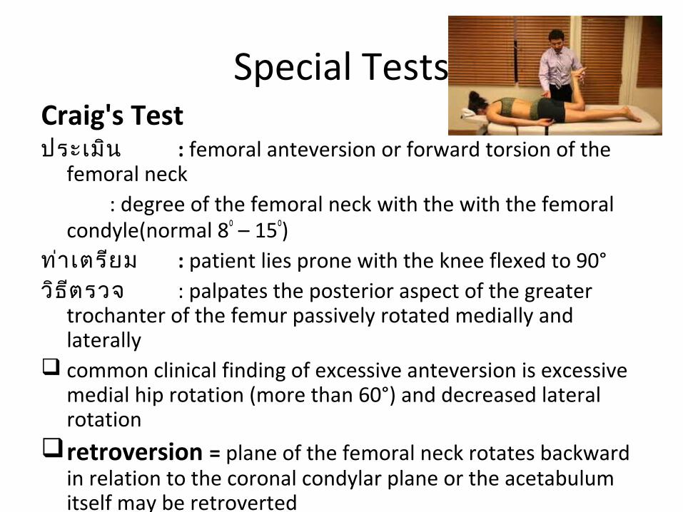
Special TestsCraig's Testประเม ิน : femoral anteversion or forward torsion of the
femoral neck: degree of the femoral neck with the with the femoral
condyle(normal 8o – 15o)ท่าเตร ียม : patient lies prone with the knee flexed to 90°ว ิธ ีตรวจ : palpates the posterior aspect of the greater
trochanter of the femur passively rotated medially and laterally
common clinical finding of excessive anteversion is excessive medial hip rotation (more than 60°) and decreased lateral rotation
retroversion = plane of the femoral neck rotates backward in relation to the coronal condylar plane or the acetabulum itself may be retroverted

Special TestsTorque Testประเม ิน : Supine position
ท่าเตร ียม : supine close to the edge of the examining table with the femur of the test
leg extended over the edge of the table
ว ิธ ีตรวจ : one hand to medially rotate the femur to the end of range and the other hand to apply a slow posterolateral pressure along the line of the neck of the femur for 20 seconds to stress the capsular ligaments and test the stability of the hip joint

Special TestsStinchfield Testว ิธ ีตรวจ : supine and flex hip with knee straight to
30° of hip flexion against resistance• Positive : Hip or groin pain hip pathology
: Posterior hip pain or back pain lumbar or sacroiliac pathology

Special TestsNelaton's Line • Imaginary line drawn from the ischial tuberosity of
the pelvis to theASIS of the pelvis on the same side • Positive : greater trochanter of the femur is
palpated well above the line– dislocated hip– coxa vara

Special TestsBryant's Triangle• imaginary perpendicular Iine : ASIS of t pelvis
examining table (A)• Second imaginary line : ASIS of t pelvis tip of
greater trochanter (B)• Third imaginary line : B A• Positive : – two sides are compared = Difference• coxa vara • Congeni dislocation of the hip
AB
C

Special TestsRotational Deformitiesท่าเตร ียม : supine with the lower limbs straight
ว ิธ ีตรวจ : examiner looks at the patellae• Patellae face in ; medial rotation of femur or
tibia.• Patellae face up, out, away ; lateral rotation of
femur or tibia

Pediatric TestsOrtolani's Signประเม ิน : congenital dislocation of the hip
Negative : highly suggestive that the problem (i.e., congenital dislocation of the hip)
Positive : does not necessarily rule out the problem

Pediatric TestsBarlow’sประเม ิน : developmental dysplasia of hip
: used for infants up to 6 month• Positive : hip dislocation

Pediatric TestsGaleazzi Sign (Allis or Galeazzi Test)ประเม ิน : unilateral congenital dislocation of the
hip
unilateral development dysplasia of the hip
3 to 18 months of age
ว ิธ ีตรวจ : supine ,knees flexed ,hips flexed to 90°. :
• Positive : one knee is higher than the other

Pediatric TestsTelescoping Sign (Piston or Dupuytren's Test).ประเม ิน : child with a dislocated hip
ท่าเตร ียม : supine position
ว ิธตีรวจ : flexes the knee and hip to 90° , The femur is push down and lift up
• Negative : normal hip, little movement occcurs with this action
• Positive : excessive movement is called telescoping, or pistoning

Pediatric TestsAbduction Test (Harts' Sign)ประเม ิน : congenital dislocation, developmental dysplasia
ท่าเตร ียม : supine with the hips and knees flexed to 90°
ว ิธ ีตรวจ : passively abducts both legs, noting any asymmetry or limitation of movement
• If one hip is dislocated, that shows asymmetry of fat folds in the gluteal and upper leg area

The Weber-Barstow maneuver for leg length asymmetry
• Patient lifts hips off bed then comparing height of medial malleolus with the legs extended (Leg length discrepancy)
Tests for Muscle Tightness
or Pathology

Sign of the Buttock
• Passively straight leg raised. If there is limitation, the examiner flexes the patient's knee to see whether further hip flexion can be obtained.
• If hip flexion does not increase, the lesion is in the buttock or the hip, not the sciatic nerve or hamstring muscles.
• There may also some limited trunk flexion. Causes of a positive include ischial bursitis, a neoplasm, an abscess in the buttock, or hip pathology.

Thomas Test
• Test : assess a hip flexion contracture of the hip
• positive test. : the patient's straight leg rises off the table and a muscle stretch end feel will be felt .
(A) Negative test
(B) Positive test

Rectus Femoris Contracture Test (Kendall Test)
• The movement leg is brought to the chest• Negative test : (A). The test leg remains
bent over the end of the examining table
• Positive test : (B) The test leg have knee extends

Ely's Test (Tight Rectus Femoris)
• The patient lies prone, passively flexes the patient's knee.
• Positive test : On flexion of the knee, the patient's hip on the same side spontaneously flexes, indicating that the rectus femoris muscle is tight on that side.
• The two sides should be tested and compared

Ober's Test
• Test : assess the tensor fasciae latae (iliotibial band) for contracture
• Lying position with the lower leg flexed at the hip and knee for stability. Passively abducts and extends upper leg with knee straight or flexed to 90°.
• positive test : if a contracture is present, the leg remains abducted and does not fall to the table. The leg remains abducted while the patient's muscles are relaxed.

• Determind : iliotibial band friction syndrome
• Positive test : severe pain over the lateral femoral condyle
Noble Compression Test

Piriformis Test
• Test : piriformis syndrome• Positive : If the piriformis muscle is tight,
pain is elicited in the muscle. If the piriformis muscle is pinching the sciatic nerve, pain results in the buttock
The patient is in the side lying position
flexes the test hip to 60° with the knee flexed. The examiner stabilizes the hip with one hand and applies a downward pressure to the knee

Test for Hamstrings Contracture

1) 90-90 Straight Leg Raising Test
• Normal flexibility in the hamstrings : knee extensior should be within 20° of full extension
• Positive : if the hamstrings are tight, the end feel will be muscle stretch

1) 90-90 Straight Leg Raising Test
• modify to test the length of gluteus maximus.
• If the thigh flexes 110° to 120° before the ASIS moves up, gluteus maximus length is normal.
• If the ASIS moves up before the thigh reaches the trunk, gluteus maximus is tight. Both sides should be compared.
Flex hip flex knee

2) Hamstrings Contracture Test
• Normally, the patient should be able to at least touch the toes while keeping the knee extended.
• If he is unable to do so, it is an indication of tight hamstrings on the straight leg.
Pt sit with one knee flexed against the chest to stabilize the pelvis and the other knee extended.then flex the trunk and touch the toes of the extended lower limb with the fingers. Repeated on the other side.

3) Tripod Sign
• Passively extends one knee. If the hamstring muscles on that side are tight, the patient extends the trunk to relieve the tension in the hamstring.
• Extension of the spine is indicative of a positive test.
patient is seated with both knees flexed to 90° over the table The examiner then passively extends one knee. If the hamstring muscles on that side are tight, the patientextends the trunk to relieve the tension in the hamstringThe leg is returned to its starting position, and the other leg is tested and compared with the first side.
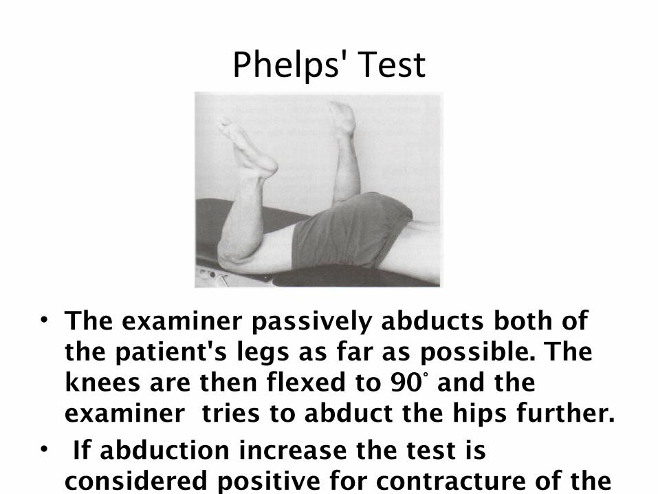
Phelps' Test
• The examiner passively abducts both of the patient's legs as far as possible. The knees are then flexed to 90° and the examiner tries to abduct the hips further.
• If abduction increase the test is considered positive for contracture of the gracilis muscle.

Tightness of Hip Rotators
•Tightness of the lateral rotators :
medial rotate hip by rotating the leg outward. •If the lateral rotators are tight :
medial rotation will be less than 30° to 40° and the end feel will be muscle stretch rather than tissue (capsular) stretch.
Pt lie supine with the hip and knee flexed to 90

Lateral Step Down Manoeuver (Pelvis Drop Test)
• Stand up straight on the step one foot. slowly lowers the nonweight - bearing leg to the floor.
(A) Negative test - normal
(B) Positive test - pelvis drop

Fulcrum Test of the Hip
• Assess for possible stress fracture of the femoral shaft
• Places arm under femur and carefully applies a downward force at the knee.
• The fulcrum arm is move from distal to proximal along the thigh as gentle pressure. If a stress fracture is present.
• Patient complains of a sharp pain and express

Cutaneous Distribution

Dermatomes around the hip

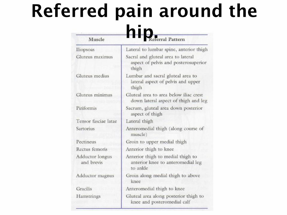
Referred pain around the hip.
•Right side demonstrates referral to the hip. •Left side shows referral from hip
True hip pain is usually referred to the groin, but it may also be referred to the ankle, knee, lumbar spine, and sacroiliac jointsSimilarly, the knee, sacroiliac joints, and lumbar spine may refer pain to the hip
Referred pain around the hip.

Peripheral Nerve Injuries About the Hip

Sciatic Nerve (L4 through S3)• Injured in the pelvis
or upper femur area (e.g., posterior hip dislocation)
• Hamstrings and all muscles below the knee can be affected.
• Result : high steppage gait with an inability to stand on the heel or toes
• compressed by the piriformis : pain and weakness on abduction and lateral rotation of the hip

• Weakness of Gluteus medius, Gluteus minimus, Tensor fasciae latae
• Hip : medial rotated, and weakness of the hip abductors resulting in a Trendelenburg's gait.
Superior Gluteal Nerve (L4 through S1)

Femoral Nerve (L2 through L4)
• compressed during childbirth, ant. dislocation of femur or traumatic surgery.
• Not able to : flex the thigh on the trunk or extend the knee.
• Reflex : lost deep tendon knee reflex • Sensory loss : medial side of thigh (ant.
femoral cutaneous nerve) and the leg and foot (saphenous nerve).

Obturator Nerve (L2 through L4)
• Caused by pelvic or hip surgery, pregnancy(obstetric palsy), fractures or tumors
• Controls primarily the adductors, hip adduction is affected, as are knee flexion and hip lateral rotation
• Sensory deficit is small ; medial part of thigh
• Pain from the symphysis pubis to the medial aspect of the knee

Joint Play Movements
• Patient in the supine position.• The examiner should attempt to compare
the amounts of available movement on the two sides.

1. Caudal Glide of the femur
(long leg traction or long-axis extension
The examiner places both hanaround the patient's leg, slightly above the ankle. Thexaminerthen leans back, applying a long-axis extension(traction) to the entire lower limb. Part of thmovement occurs in the knee. If one suspects sompathology in the knee or the knee is stiff, both hanshould be placed around the thigh just proximal to thknee, and traction force should again be applied ( eFig. 11-54A). The first method enables the examineto apply a greater force. During the movement, antelescoping or excessive movement occurring in thhip should be

2. Compression
The examiner places the patient’s knee in the resting position and then applies a compressiveforce to the hip through the longitudinal of the femur by pushing through the femoral condyle

3. Lateral Distraction
hand ; placing a wide strap around the leg as high up in the groin as possible. The strap is then wrapped around the examiner's buttockThe examiner leans back, using the buttocks to apply the distraction force to the hip. The proximal palpate the hip or greater trochanter movement, distal hand prevents abduction of the leg, and, hence, torque to the hip

4. Quadrant (Scouring) Tests
The examiner flex and adducts the patient's hip so that the hip faces the patient's opposite shoulder and resistance to the movement is felt. As slight resistance is maintained, thepatient's hip is taken into abduction while maintainingflexion in an arc of movement. As the movement isperformed, the examiner should look for any irregularityin the movement (e.g., "bumps"), pain, or patientapprehension, which may give an indication of wherethe pathology is occurring in the hip