hip functional motion assessment bilateral...
TRANSCRIPT
Virginia Orthopedic Manual Physical Therapy Institute
Hip Technique Manual
Hip Functional Motion Assessment
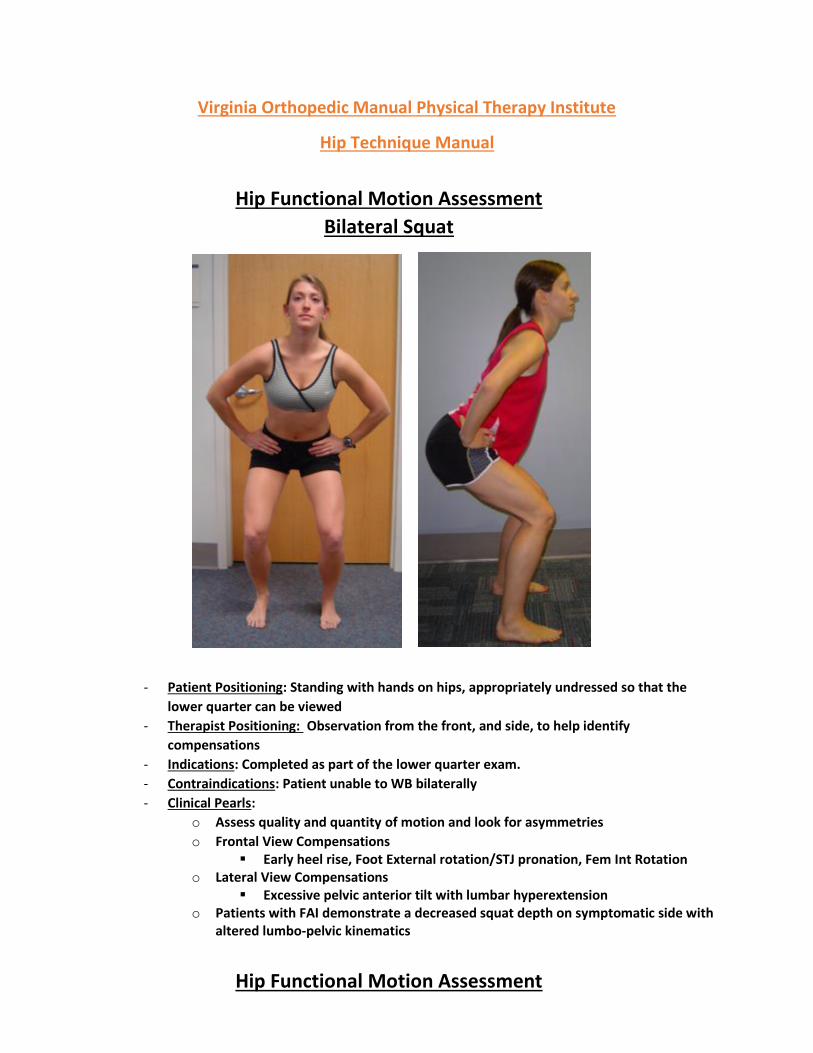
Bilateral Squat
- Patient Positioning: Standing with hands on hips, appropriately undressed so that the
lower quarter can be viewed
- Therapist Positioning: Observation from the front, and side, to help identify
compensations
- Indications: Completed as part of the lower quarter exam.
- Contraindications: Patient unable to WB bilaterally
- Clinical Pearls:
o Assess quality and quantity of motion and look for asymmetries
o Frontal View Compensations Early heel rise, Foot External rotation/STJ pronation, Fem Int Rotation
o Lateral View Compensations Excessive pelvic anterior tilt with lumbar hyperextension
o Patients with FAI demonstrate a decreased squat depth on symptomatic side with altered lumbo-pelvic kinematics
Hip Functional Motion Assessment
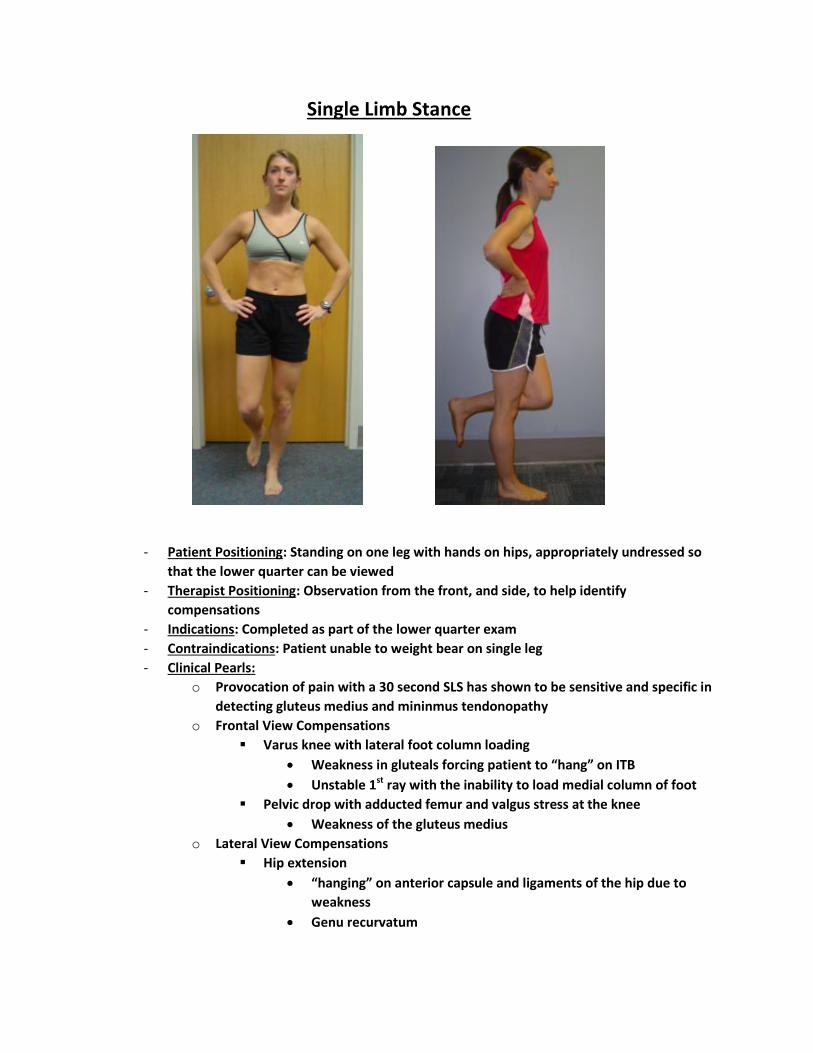
Single Limb Stance
- Patient Positioning: Standing on one leg with hands on hips, appropriately undressed so
that the lower quarter can be viewed
- Therapist Positioning: Observation from the front, and side, to help identify
compensations
- Indications: Completed as part of the lower quarter exam
- Contraindications: Patient unable to weight bear on single leg
- Clinical Pearls:
o Provocation of pain with a 30 second SLS has shown to be sensitive and specific in
detecting gluteus medius and mininmus tendonopathy
o Frontal View Compensations
Varus knee with lateral foot column loading
Weakness in gluteals forcing patient to “hang” on ITB
Unstable 1st ray with the inability to load medial column of foot
Pelvic drop with adducted femur and valgus stress at the knee
Weakness of the gluteus medius
o Lateral View Compensations
Hip extension
“hanging” on anterior capsule and ligaments of the hip due to
weakness
Genu recurvatum

Hip Functional Motion Assessment
Single Limb Squat
- Patient Positioning: Standing on one leg with hands on hips, appropriately undressed so
that the lower quarter can be viewed
- Therapist Positioning: Observation from the front, and side, to help identify
compensations
- Indications: Completed as part of the lower quarter exam
- Contraindications: Patient unable to single weight bear
- Clinical Pearls:
o Assess quality and quantity of motion and look for asymmetries
o Ask the patient to perform 5 times and assessing…
Ability to maintain balance
Perturbations
Depth of squat
Speed of squat
o Frontal View Compensations
Compensated trendelenburg
Weakness in gluteals
Excessive medial torsion of the femur with increased dynamic valgus at
the knee
Weakness of the hip in the transverse plane
o Lateral View Compensations
Forward trunk lean
Quadriceps weakness or avoidance of using the quad

Hip Functional Motion Assessment
Step Down Test
- Patient Positioning: Standing with one foot on the edge of a step with hands on hips
appropriately undressed so that the lower quarter can be viewed
o Utilize a step that is roughly half the height of the patient’s tibia
o Patient asked to touch the floor with the heel and raise back up
- Therapist Positioning: Observation from the front, and side, to help identify
compensations
- Indications: Completed as part of the lower quarter exam
- Contraindications: Patient unable to single weight bear
- Clinical Pearls:
o Assess quality and quantity of motion and look for asymmetries
o Graded on a 5 point scale
<1 good, >4 poor
Arm strategy to recover balance (+1)
Trunk mvt – lean either direction (+1)
Pelvic mvt – rotated/elevated either side (+1)
Knee position
Deviated medial to 2nd toe (+1)
Deviated past medial border foot (+2)
Difficulty with balance (+1)

Hip Functional Motion Assessment
Star Excursion or Y Test
- Patient Positioning: Standing on one foot appropriately undressed so that the lower
quarter can be viewed
o Patient asked to reach posteromedical and posterolateral at a target
- Therapist Positioning: Observation from the front, and side, to help identify
compensations and distance reached
- Indications: Completed as part of the lower quarter exam
- Contraindications: Patient unable to single weight bear
- Clinical Pearls:
o Assess quality and quantity of motion and look for asymmetries
o Posterio-medial/Posterio- lateral reach distances o – Correlated with Hip ABD/EXT strength
Hip Functional Motion Assessment
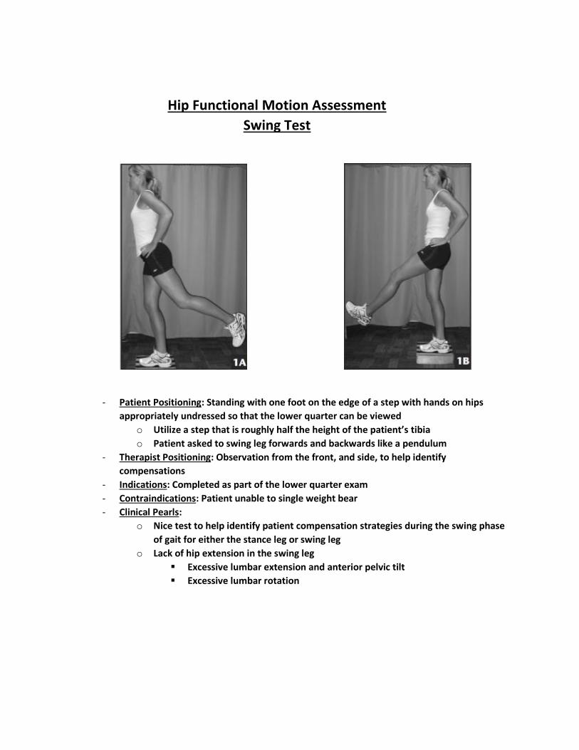
Swing Test
- Patient Positioning: Standing with one foot on the edge of a step with hands on hips
appropriately undressed so that the lower quarter can be viewed
o Utilize a step that is roughly half the height of the patient’s tibia
o Patient asked to swing leg forwards and backwards like a pendulum
- Therapist Positioning: Observation from the front, and side, to help identify
compensations
- Indications: Completed as part of the lower quarter exam
- Contraindications: Patient unable to single weight bear
- Clinical Pearls:
o Nice test to help identify patient compensation strategies during the swing phase
of gait for either the stance leg or swing leg
o Lack of hip extension in the swing leg
Excessive lumbar extension and anterior pelvic tilt
Excessive lumbar rotation

Hip Functional Motion Assessment
Closed Chain Rotation Test
- Patient Positioning: Standing on one foot with knee slightly flexed, holding lightly to the
therapists hands, appropriately undressed so that the lower quarter can be viewed
o The patient is asked to turn their body and follow the therapist
- Therapist Positioning: Standing in front of the patient holding their hands lightly.
Therapist walks slowly to the right and then to the left forcing the patient to rotate on the
stance lower extremity.
- Indications: Completed as part of the lower quarter exam
- Contraindications: Patient unable to single weight bear
- Clinical Pearls:
o Hip screen for potential labral pathology
Looking for mechanical signs and symptom reproduction
o Knee meniscal screen as well (Thessaly/Ochiai Test)
Hip Functional Motion Assessment
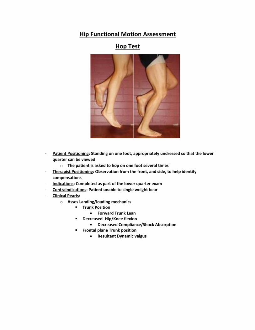
Hop Test
- Patient Positioning: Standing on one foot, appropriately undressed so that the lower
quarter can be viewed
o The patient is asked to hop on one foot several times
- Therapist Positioning: Observation from the front, and side, to help identify
compensations
- Indications: Completed as part of the lower quarter exam
- Contraindications: Patient unable to single weight bear
- Clinical Pearls:
o Asses Landing/loading mechanics Trunk Position
Forward Trunk Lean Decreased Hip/Knee flexion
Decreased Compliance/Shock Absorption Frontal plane Trunk position
Resultant Dynamic valgus
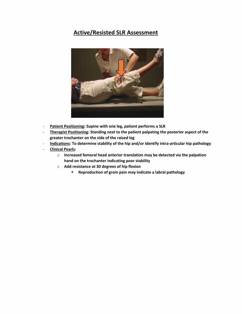
Active/Resisted SLR Assessment
- Patient Positioning: Supine with one leg, patient performs a SLR
- Therapist Positioning: Standing next to the patient palpating the posterior aspect of the
greater trochanter on the side of the raised leg
- Indications: To determine stability of the hip and/or identify intra-articular hip pathology
- Clinical Pearls:
o Increased femoral head anterior translation may be detected via the palpation
hand on the trochanter indicating poor stability
o Add resistance at 30 degrees of hip flexion
Reproduction of groin pain may indicate a labral pathology
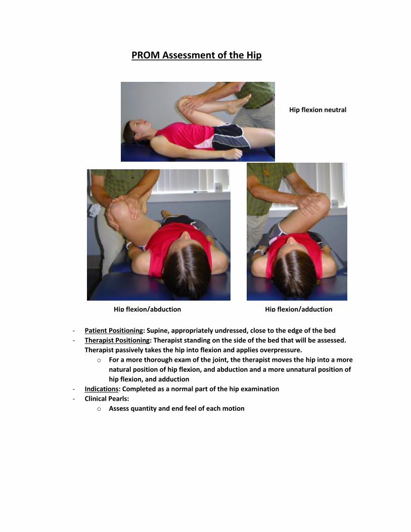
PROM Assessment of the Hip
- Patient Positioning: Supine, appropriately undressed, close to the edge of the bed
- Therapist Positioning: Therapist standing on the side of the bed that will be assessed.
Therapist passively takes the hip into flexion and applies overpressure.
o For a more thorough exam of the joint, the therapist moves the hip into a more
natural position of hip flexion, and abduction and a more unnatural position of
hip flexion, and adduction
- Indications: Completed as a normal part of the hip examination
- Clinical Pearls:
o Assess quantity and end feel of each motion
Hip flexion neutral
Hip flexion/abduction Hip flexion/adduction
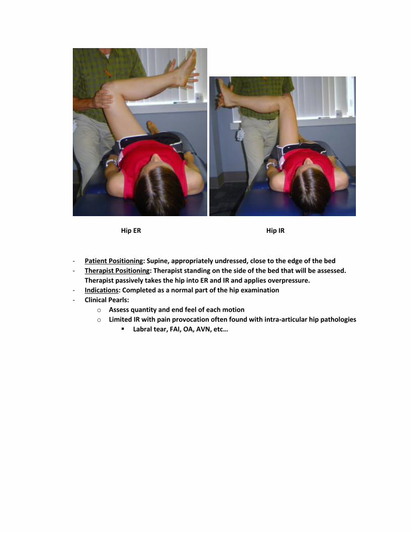
Hip ER Hip IR
- Patient Positioning: Supine, appropriately undressed, close to the edge of the bed
- Therapist Positioning: Therapist standing on the side of the bed that will be assessed.
Therapist passively takes the hip into ER and IR and applies overpressure.
- Indications: Completed as a normal part of the hip examination
- Clinical Pearls:
o Assess quantity and end feel of each motion
o Limited IR with pain provocation often found with intra-articular hip pathologies
Labral tear, FAI, OA, AVN, etc…

FABER Test
- Patient Positioning: Supine with the tested leg in a figure 4 position, ankle resting on the
opposite knee
- Test Performance:
o Therapist stabilizes the opposite side of the pelvis while applying overpressure to
the tested leg towards the table
- Indications: Performed to try and identify an intra-articular hip pathology. Also may help
diagnose an SIJ dysfunction
- Clinical Pearls:
o Positive test is provocation of pain
Location of the pain may indicate where the pathology is
o Amount of motion and end feel can also be valuable to help with diagnosis
Test leg is compared to opposite side
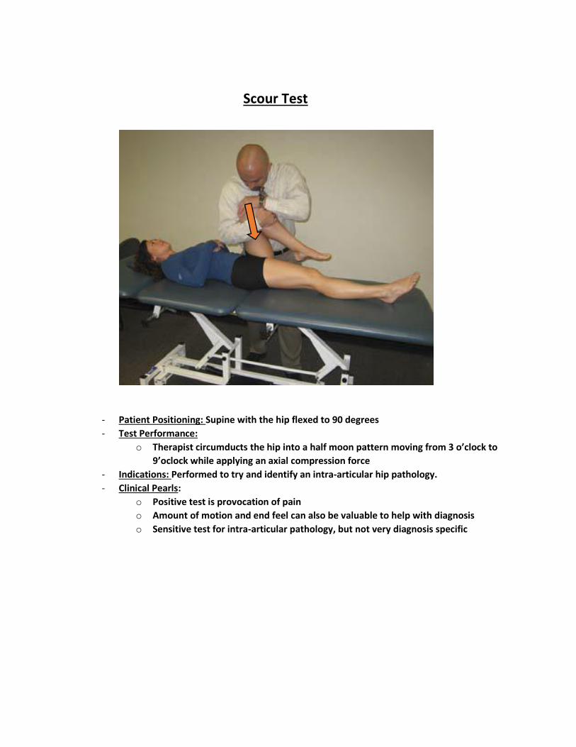
Scour Test
- Patient Positioning: Supine with the hip flexed to 90 degrees
- Test Performance:
o Therapist circumducts the hip into a half moon pattern moving from 3 o’clock to
9’oclock while applying an axial compression force
- Indications: Performed to try and identify an intra-articular hip pathology.
- Clinical Pearls:
o Positive test is provocation of pain
o Amount of motion and end feel can also be valuable to help with diagnosis
o Sensitive test for intra-articular pathology, but not very diagnosis specific
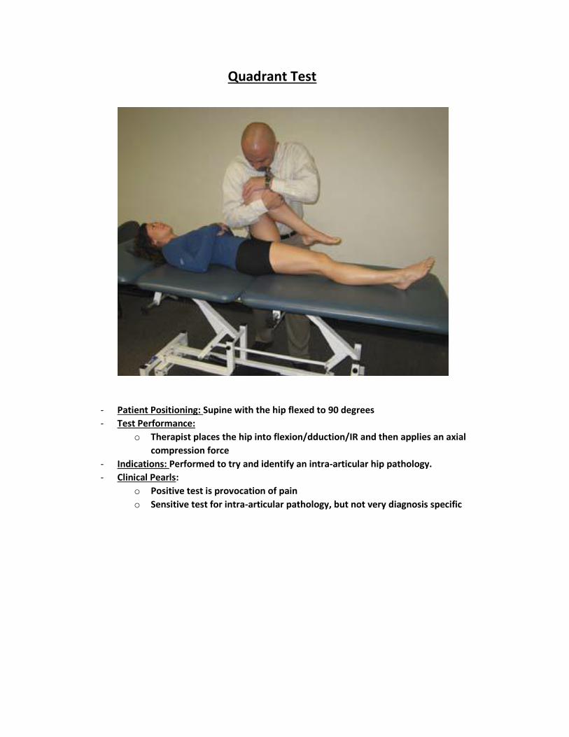
Quadrant Test
- Patient Positioning: Supine with the hip flexed to 90 degrees
- Test Performance:
o Therapist places the hip into flexion/dduction/IR and then applies an axial
compression force
- Indications: Performed to try and identify an intra-articular hip pathology.
- Clinical Pearls:
o Positive test is provocation of pain
o Sensitive test for intra-articular pathology, but not very diagnosis specific

Impingement Test/FADDIR
- Patient Positioning: Supine with the hip and knee flexed to 90 degrees
- Test Performance:
o Therapist places the hip into flexion/adduction and then IR with overpressure
- Indications: Performed to try and identify an intra-articular hip pathology.
- Clinical Pearls:
o Positive test is provocation of pain (in the groin)
o May be indicative of an anterior labral pathology
o Sensitive test for intra-articular pathology, but not very diagnosis specific

Log Roll Test
- Patient Positioning: Supine with leg extended out
- Test Performance:
o Therapist log rolls the patients entire LE by maximally IR/ER rotating the hip
- Indications: Performed to try and identify an intra-articular hip pathology.
- Clinical Pearls:
o Positive test is producing mechanical signs and symptoms
Clicking or popping
o Utilized as a screen for capsular laxity
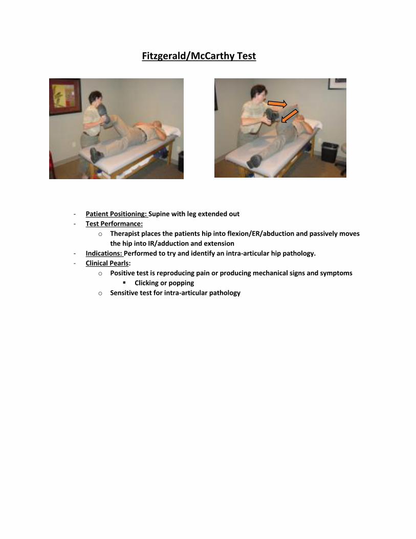
Fitzgerald/McCarthy Test
- Patient Positioning: Supine with leg extended out
- Test Performance:
o Therapist places the patients hip into flexion/ER/abduction and passively moves
the hip into IR/adduction and extension
- Indications: Performed to try and identify an intra-articular hip pathology.
- Clinical Pearls:
o Positive test is reproducing pain or producing mechanical signs and symptoms
Clicking or popping
o Sensitive test for intra-articular pathology
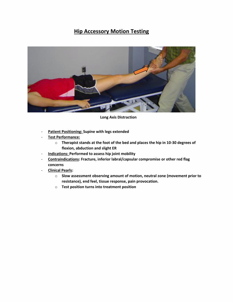
Hip Accessory Motion Testing
- Patient Positioning: Supine with legs extended
- Test Performance:
o Therapist stands at the foot of the bed and places the hip in 10-30 degrees of
flexion, abduction and slight ER
- Indications: Performed to assess hip joint mobility
- Contraindications: Fracture, inferior labral/capsular compromise or other red flag
concerns
- Clinical Pearls:
o Slow assessment observing amount of motion, neutral zone (movement prior to
resistance), end feel, tissue response, pain provocation.
o Test position turns into treatment position
Long Axis Distraction

- Patient Positioning: Supine at the edge of the bed
- Test Performance:
o Therapist stands at the side of the bed laterally to the patients hip.
o Therapist flexes the hip to 90 degrees and allows the knee to flex comfortably.
Therapist grasps around the thigh as close to the joint line as possible. Therapist
applies a lateral glide to the joint by pulling the proximal femur towards them,
while at the same time providing a counterforce at the patient knee with their
shoulder.
- Indications: Performed to assess hip joint mobility
- Contraindications: Fractur, labral compromise or other red flag concerns
- Clinical Pearls:
o Slow assessment observing amount of motion, neutral zone (movement prior to
resistance), end feel, tissue response, pain provocation.
o For a more effective assessment, stay in the plane of the joint which is
anteromedial/posterolateral
o Test position turns into treatment position Utilize mobilization belt for more effective treatment
Lateral Glide at 90 degrees Hip Flexion
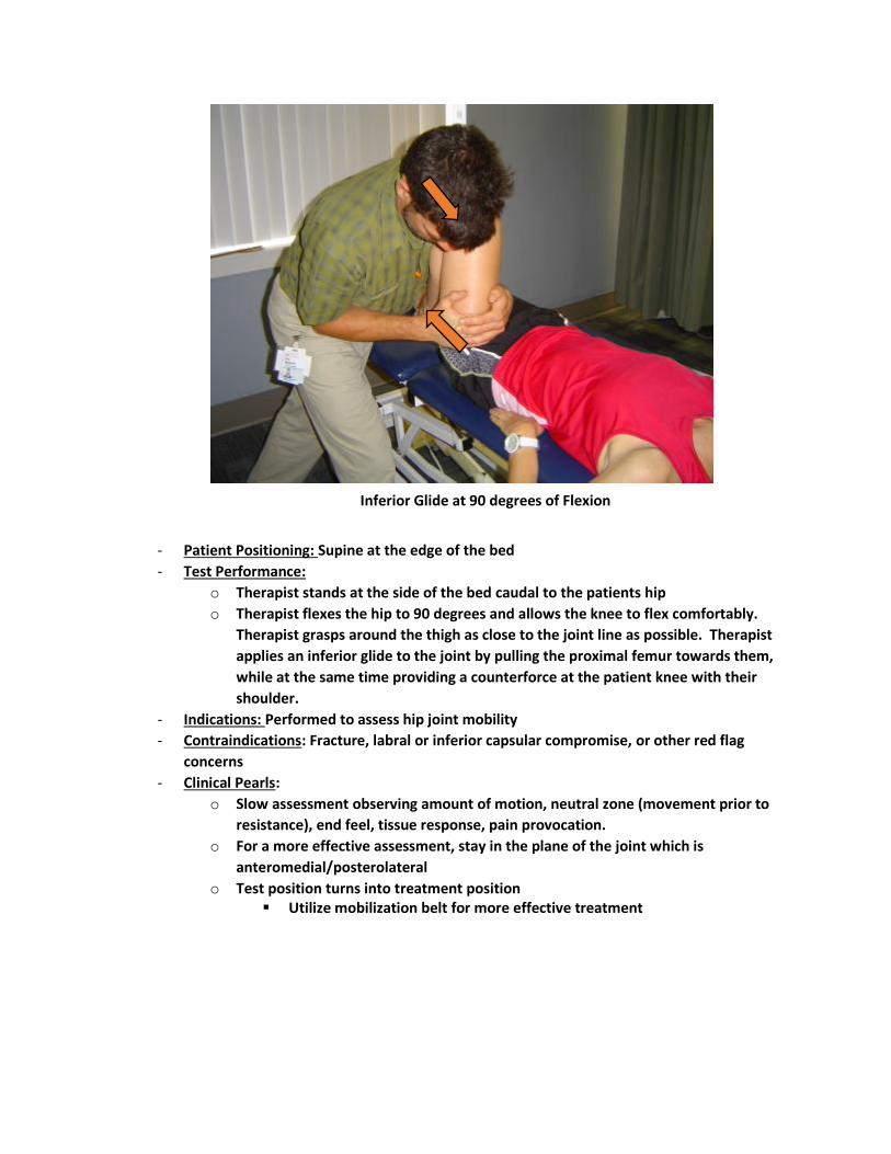
- Patient Positioning: Supine at the edge of the bed
- Test Performance:
o Therapist stands at the side of the bed caudal to the patients hip
o Therapist flexes the hip to 90 degrees and allows the knee to flex comfortably.
Therapist grasps around the thigh as close to the joint line as possible. Therapist
applies an inferior glide to the joint by pulling the proximal femur towards them,
while at the same time providing a counterforce at the patient knee with their
shoulder.
- Indications: Performed to assess hip joint mobility
- Contraindications: Fracture, labral or inferior capsular compromise, or other red flag
concerns
- Clinical Pearls:
o Slow assessment observing amount of motion, neutral zone (movement prior to
resistance), end feel, tissue response, pain provocation.
o For a more effective assessment, stay in the plane of the joint which is
anteromedial/posterolateral
o Test position turns into treatment position Utilize mobilization belt for more effective treatment
Inferior Glide at 90 degrees of Flexion
- Patient Positioning: Supine at the edge of the bed
- Test Performance:
o Therapist stands at the side of the bed caudal to the patients hip
o Therapist flexes the hip to 90 degrees and allows the knee to flex comfortably.
Therapist grasps around the thigh as close to the joint line as possible. Therapist
applies a posterior glide to the joint by pushing the proximal femur towards the
floor, while maintaining the hip flexion at 90 degrees
- Indications: Performed to assess hip joint mobility
- Contraindications: Fracture, labral or posterior capsule compromise or other red flag
concerns
- Clinical Pearls:
o Slow assessment observing amount of motion, neutral zone (movement prior to
resistance), end feel, tissue response, pain provocation.
o For a more effective assessment, stay in the plane of the joint which is
anteromedial/posterolateral
o Test position turns into treatment position
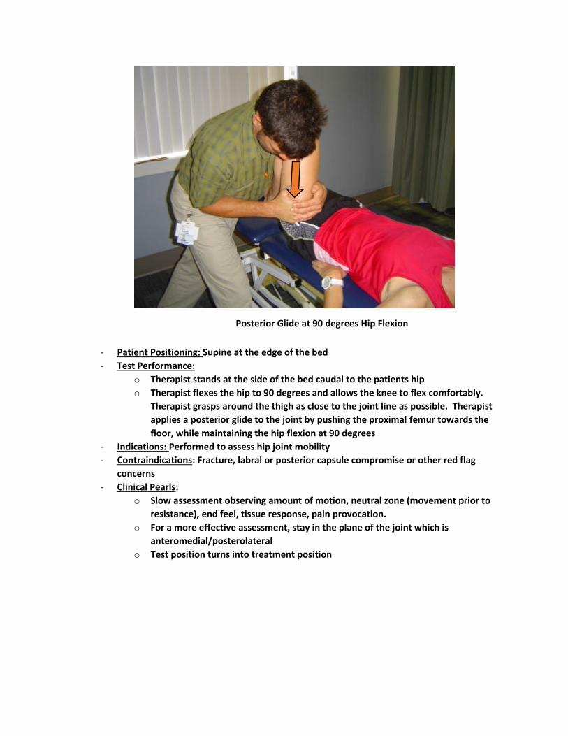
Posterior Glide at 90 degrees Hip Flexion
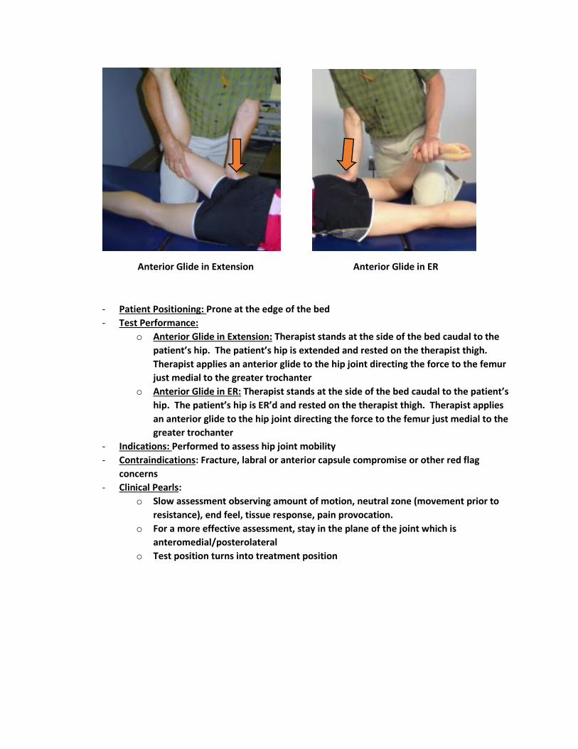
- Patient Positioning: Prone at the edge of the bed
- Test Performance:
o Anterior Glide in Extension: Therapist stands at the side of the bed caudal to the
patient’s hip. The patient’s hip is extended and rested on the therapist thigh.
Therapist applies an anterior glide to the hip joint directing the force to the femur
just medial to the greater trochanter
o Anterior Glide in ER: Therapist stands at the side of the bed caudal to the patient’s
hip. The patient’s hip is ER’d and rested on the therapist thigh. Therapist applies
an anterior glide to the hip joint directing the force to the femur just medial to the
greater trochanter
- Indications: Performed to assess hip joint mobility
- Contraindications: Fracture, labral or anterior capsule compromise or other red flag
concerns
- Clinical Pearls:
o Slow assessment observing amount of motion, neutral zone (movement prior to
resistance), end feel, tissue response, pain provocation.
o For a more effective assessment, stay in the plane of the joint which is
anteromedial/posterolateral
o Test position turns into treatment position
Anterior Glide in Extension Anterior Glide in ER
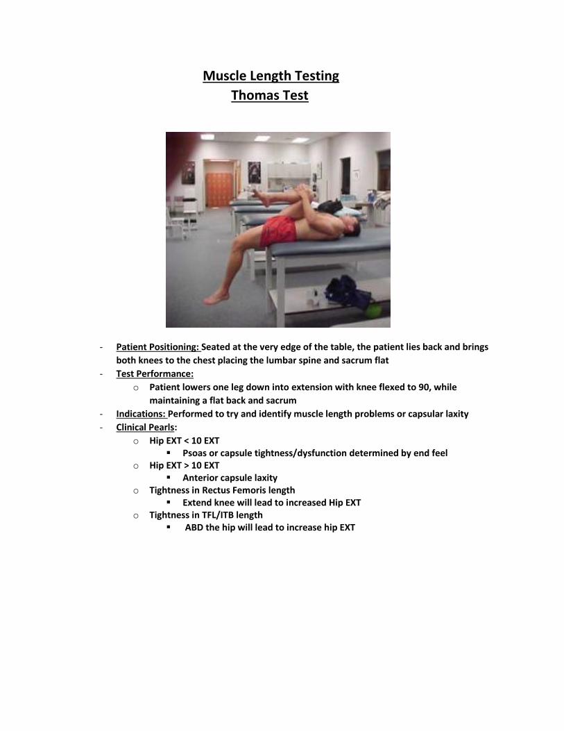
Muscle Length Testing
Thomas Test
- Patient Positioning: Seated at the very edge of the table, the patient lies back and brings
both knees to the chest placing the lumbar spine and sacrum flat
- Test Performance:
o Patient lowers one leg down into extension with knee flexed to 90, while
maintaining a flat back and sacrum
- Indications: Performed to try and identify muscle length problems or capsular laxity
- Clinical Pearls:
o Hip EXT < 10 EXT Psoas or capsule tightness/dysfunction determined by end feel
o Hip EXT > 10 EXT Anterior capsule laxity
o Tightness in Rectus Femoris length Extend knee will lead to increased Hip EXT
o Tightness in TFL/ITB length ABD the hip will lead to increase hip EXT
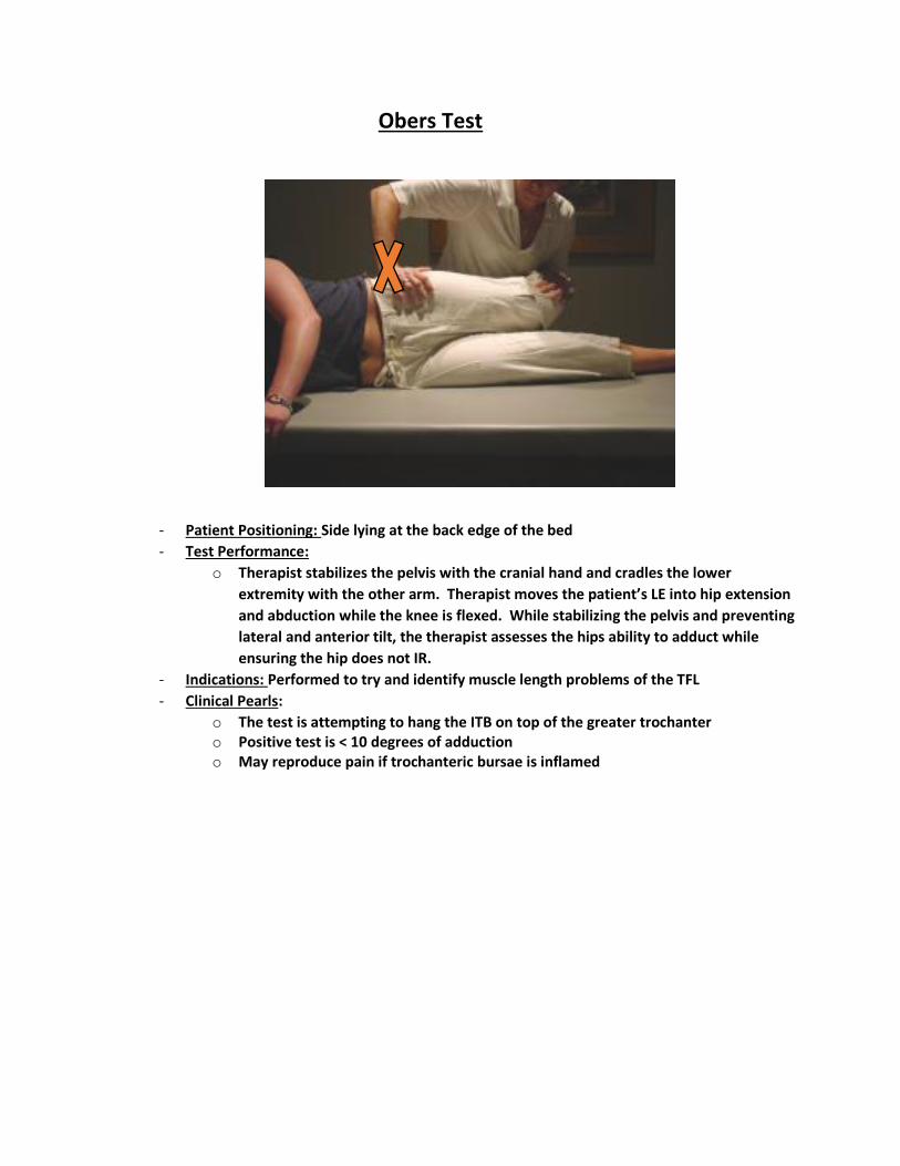
Obers Test
- Patient Positioning: Side lying at the back edge of the bed
- Test Performance:
o Therapist stabilizes the pelvis with the cranial hand and cradles the lower
extremity with the other arm. Therapist moves the patient’s LE into hip extension
and abduction while the knee is flexed. While stabilizing the pelvis and preventing
lateral and anterior tilt, the therapist assesses the hips ability to adduct while
ensuring the hip does not IR.
- Indications: Performed to try and identify muscle length problems of the TFL
- Clinical Pearls:
o The test is attempting to hang the ITB on top of the greater trochanter o Positive test is < 10 degrees of adduction o May reproduce pain if trochanteric bursae is inflamed
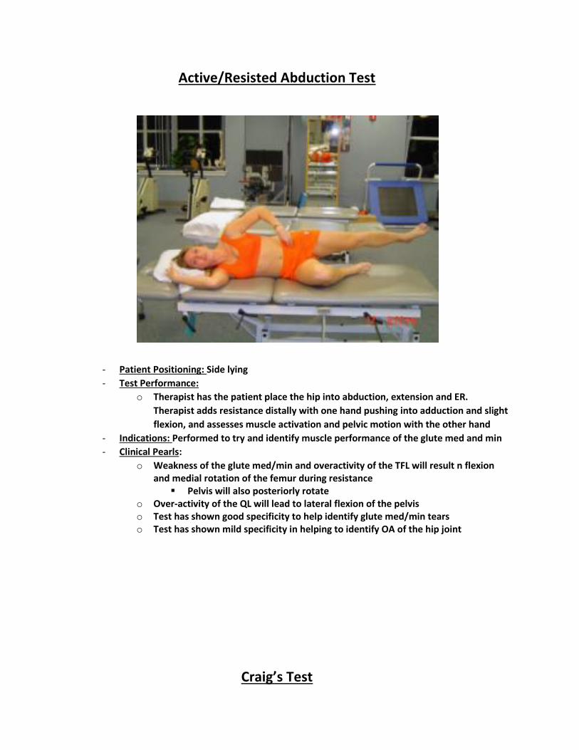
Active/Resisted Abduction Test
- Patient Positioning: Side lying
- Test Performance:
o Therapist has the patient place the hip into abduction, extension and ER.
Therapist adds resistance distally with one hand pushing into adduction and slight
flexion, and assesses muscle activation and pelvic motion with the other hand
- Indications: Performed to try and identify muscle performance of the glute med and min
- Clinical Pearls:
o Weakness of the glute med/min and overactivity of the TFL will result n flexion and medial rotation of the femur during resistance
Pelvis will also posteriorly rotate o Over-activity of the QL will lead to lateral flexion of the pelvis o Test has shown good specificity to help identify glute med/min tears o Test has shown mild specificity in helping to identify OA of the hip joint
Craig’s Test

- Patient Positioning: Prone with knee flexed to 90 degrees
- Test Performance:
o Therapist palpates the posterior aspect of the greater trochanter with one hand
and moves the hip into IR/ER with the other hand by moving the tibia. The
movement into IR/ER is stopped when the greater trochanter if felt most
prominent (parallel with the table). Measure the angle of the tibia from vertical
to estimate torsion of the femoral head.
- Indications: Performed to try and identify anteversion or retroversion of the femoral head
- Clinical Pearls:
o Anteverson of the hip is most common and more common in females than in males
o Normal anteversion of the hip is 8-15 degrees o IR/ER of the femur should be around 35 degrees each
Anteverted hips have excessive IR Retroverted hips have excessive ER Asymmetry is considered when there is a difference of greater than 10
degrees (between each side or between ER/IR)

Pubic Patella Percussion Test
- Patient Positioning: Supine
- Test Performance:
o Patient places stethoscope over their pubic bone and holds it in place. The
therapist either taps the patella or places a tuning fork over the patella. The
therapist listens to the sound that is transferred from the patella to the pubic
bone. That sound is compared to the symptomatic side.
- Indications: Performed to try and identify femoral fractures
- Clinical Pearls:
o The fracture creates a disruption in sound production and leads to a muffled sound
o Highly sensitive and specific
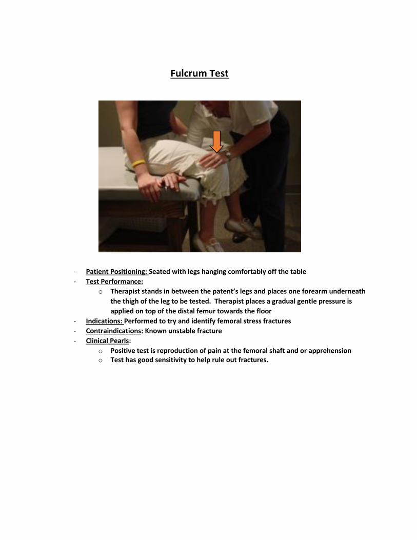
Fulcrum Test
- Patient Positioning: Seated with legs hanging comfortably off the table
- Test Performance:
o Therapist stands in between the patent’s legs and places one forearm underneath
the thigh of the leg to be tested. Therapist places a gradual gentle pressure is
applied on top of the distal femur towards the floor
- Indications: Performed to try and identify femoral stress fractures
- Contraindications: Known unstable fracture
- Clinical Pearls:
o Positive test is reproduction of pain at the femoral shaft and or apprehension o Test has good sensitivity to help rule out fractures.