high resolution mapping of epitopes on the c2 domain of factor
TRANSCRIPT
Regular Article
THROMBOSIS AND HEMOSTASIS
High-resolution mapping of epitopes on the C2 domain of factor VIII byanalysis of point mutants using surface plasmon resonancePhuong-Cac T. Nguyen,1 Kenneth B. Lewis,1 Ruth A. Ettinger,1 Jason T. Schuman,2 Jasper C. Lin,1 John F. Healey,3
Shannon L. Meeks,3 Pete Lollar,3 and Kathleen P. Pratt1,4,5
1Puget Sound Blood Center Research Institute, Seattle, WA; 2GE Health Sciences, Portland, OR; 3Aflac Cancer and Blood Disorders Center, Department of
Pediatrics, Emory University, Atlanta, GA; 4Division of Hematology, University of Washington, Seattle, WA; and 5Department of Medicine, Uniformed
Services University of the Health Sciences, Bethesda, MD
Key Points
• Amino acid residuescomprising B-cell epitopesrecognized by neutralizing anti-factor VIII antibodies (inhibitors)have been identified.
• Amino acids contributingsignificant antigen–antibodybinding avidity are candidatesfor mutagenesis in the designof less antigenic proteins.
Neutralizing anti-factor VIII (FVIII) antibodies that develop in patients with hemophilia A
and inmurinehemophiliaAmodels, clinically termed“inhibitors,” bind to several distinct
surfaces on the FVIII-C2 domain. Tomap these epitopes at high resolution, 60 recombinant
FVIII-C2 proteinswere generated, each having a single surface-exposed residuemutated
to alanine or a conservative substitution. The binding kinetics of these muteins to
11 monoclonal, inhibitory anti-FVIII-C2 antibodies were evaluated by surface plasmon
resonance and the results compared with those obtained for wild-type FVIII-C2. Clusters
of residues with significantly altered binding kinetics identified “functional” B-cell
epitopes, defined as those residues contributing appreciable antigen–antibody avidity.
These antibodies were previously shown to neutralize FVIII activity by interfering with
proteolytic activationof FVIII by thrombin or factor Xa, or with its binding to phospholipid
surfaces, von Willebrand factor, or other components of the intrinsic tenase complex.
Finemappingof epitopesby surfaceplasmon resonance also indicated surfaces through
which FVIII interacts with proteins and phospholipids as it participates in coagulation.
Mutations that significantly altered the dissociation times/half-lives identified functionally important interactions within antigen–
antibody interfaces and suggested specific sequence modifications to generate novel, less antigenic FVIII proteins with possible
therapeutic potential for treatment of inhibitor patients. (Blood. 2014;123(17):2732-2739)
Introduction
The development of neutralizing anti-factor VIII (FVIII) antibodies isa serious complication thatmaybeencounteredwhenFVIII replacementtherapy is administered to patients with hemophilia A (HA). It affects25% to 30% of the treated HA population, with a peak occurrence after;14 FVIII infusions.1-3 Autoimmune responses to FVIII can alsooccur,4 and although this happens only rarely, the resulting bleedingphenotype can be severe. Inhibitors can be difficult and extremelyexpensive to manage clinically. Interestingly, porcine FVIII has beenused effectively in the clinic as a “bypass” therapy; that is, a therapeuticprotein that can evade neutralization by anti-FVIII antibodies in manyallo- andautoimmune inhibitorpatients.5-7However, somepatientshaveor could develop antibodies that neutralize porcine FVIII as well,8
because of antigenic cross-reactivity9 or because regions in which theporcine sequence differs from the human FVIII sequence stimulateeffector T cells, leading to antibody production. Identification of thebinding sites (B-cell epitopes) on FVIII that are recognized by inhibitorswould allow rational design of novel therapeutic FVIII proteins that aremore similar to human FVIII and, hence, likely to be less immunogenic.
The most common epitopes recognized by hemophilic inhibitorsare on the FVIII A2 and C2 domains.10,11 The FVIII C2 domain
(FVIII-C2)mediates numerous functions that are essential for the fullprocoagulant cofactor activity of FVIII, including membrane bindingand assembly of the intrinsic tenase complex.12 The goal of thepresent study is to identify B-cell epitopes on FVIII-C2 that arerecognized by neutralizing anti-FVIII antibodies. In an earlier study,13
competition enzyme-linked immunosorbent assay (ELISA) assayswere employed to characterize 56 murine monoclonal antibodies(mAbs) that bound to FVIII-C2 and blocked FVIII procoagulantactivity. Results of these assays indicated there were 3 distinctepitopes on this domain, types A, B, and C, as well as inhibitoryantibodies that bound to partially overlapping epitopes AB and BC.A, B, and AB antibodies, termed “classical” anti-C2 antibodies,inhibit the assembly of the intrinsic tenase complex on negativelycharged phospholipid membranes. C and BC antibodies, termed“nonclassical” anti-C2 antibodies, inhibit the proteolytic activationof FVIII to FVIIIa by thrombin and/or by activated factor X (FXa).To identify the specific amino acid residues comprising these 5 typesof epitopes, 60 recombinant FVIII-C2 mutant proteins (muteins)plus the wild-type (WT) protein (WT-FVIII-C2) were generatedusing an Escherichia coli expression system, including 59 with an
Submitted September 24, 2013; accepted February 9, 2014. Prepublished
online as Blood First Edition paper, March 3, 2014; DOI 10.1182/blood-2013-
09-527275.
The online version of this article contains a data supplement.
There is an Inside Blood Commentary on this article in this issue.
The publication costs of this article were defrayed in part by page charge
payment. Therefore, and solely to indicate this fact, this article is hereby
marked “advertisement” in accordance with 18 USC section 1734.
2732 BLOOD, 24 APRIL 2014 x VOLUME 123, NUMBER 17
For personal use only.on April 16, 2018. by guest www.bloodjournal.orgFrom
alanine substitution at a surface-exposed amino acid side chain plusthe conservative substitution R2307Q. (The “legacy” numberingfor FVIII residues is employed in this study for consistency with theearlier study.13) Surface plasmon resonance (SPR) experiments werecarried out to measure binding kinetics of WT-FVIII-C2 and FVIII-C2 muteins to 10 representative mAbs from the series, characterizedearlier by competition ELISA and functional assays, as well as to thehuman-derived monoclonal anti-FVIII antibody BO2C11.14
Methods
Antibodies
TenmurinemAbswere selected from56mAbs characterized earlier usingELISAassays13 as representative of type A, AB, B, BC, and C inhibitors. Murine anti-FVIII C2 domain mAbs ESH4 and ESH8 were from American Diagnostica,whereas mAbs 3E6 (GMA-8013), I54, I109, 1B5 (GMA-8008), 3D12, 3G6(GMA-8014), 2-77 (GMA-8006), and 2-117 (GMA-8003) were prepared asdescribed previously13 or were kindly provided byWilliamChurch (GreenMountain Antibodies). The human anti-FVIII mAb BO2C11 was kindlyprovided by Marc Jacquemin (Department of Cardiovascular Sciences, KULeuven, Leuven, Belgium). Goat anti-mouse immunoglobulin G (IgG), Fc-g(115-005-071) was from Jackson ImmunoResearch.
FVIII-C2 proteins and SPR measurements
FVIII-C2 proteins were expressed in E coli and purified and analyzed by SPR,as described in the supplemental Methods, available at the Blood Web site.Briefly, SPR measurements were carried out on a Biacore T100 instrument (GEHealthcare Life Sciences) under standard conditions (25°C and 1 atm). Goat anti-mouse IgG specifically directed toward the Fc-g fragment was immobilizedcovalently on all channels of a CM5 chip by amine derivatization. Murine anti-FVIII mAb stock solutions were injected over the sensor in 3 flow channels,whereas the fourth channel served as a reference. BO2C11-Ag-binding fragment(Fab) was immobilized covalently by amine derivatization. Single-cycle kineticsexperiments15 were carried out in which WT or mutant FVIII-C2 proteins wereinjected serially over the biosensor surfaces at increasing concentrations withoutregenerating the biosensor surface after each injection, followed by a 30- to 60-minute buffer injection to measure dissociation rates. The association (ka) anddissociation (kd) rate constants forbindingofWT-FVIII-C2weremeasuredduringeach set of SPR runs, and the resulting kd values were used to compute thekd(mutein)/kd(WT) ratios for that set ofmuteins. FVIII-C2muteinswith a kdmorethan 2.0 times the kd for WT-FVIII-C2 were considered candidates for B-cellepitope residues. For each of the mAbs, the rate constants for the binding ofWT-FVIII-C2 were determined by averaging the results obtained from at least 3 SPRruns.Dissociation rate constants (kd), rather thanaffinities,werechosenas themostrelevant metric for identifying “functional B-cell epitopes” because the residencetime (1/kd for a bimolecular interaction) of an antibody–antigen complex indicatesits maximum potential lifetime in the circulation. Analysis of residence times iswidely used in lead optimization studies of potential inhibitorydrug targets.16-19
Visualization of B-cell epitopes
After the SPR data were collected, the crystal structures of FVIII-C220 andB-domain-deleted FVIII21,22 were visualized using the graphics programPyMOL23 to localize the sites showing altered binding kinetics to the mAbsanalyzed herein. The cutoff kd value was chosen empirically to minimize thenumber of potential epitope residues located distal from the primary clustersof candidate residues for this series of mAbs.
Results
FVIII-C2 proteins
WT-FVIII-C2 and 60 FVIII-C2 muteins were purified to more than95% homogeneity (supplemental Figure 1). Six additional FVIII-C2
muteins (S2193A, K2227A, V2232A, K2236A, K2279A, andK2281A) were not expressed in a soluble form and were thereforenot analyzed. Dynamic light scattering analyses of the purifiedproteins, carried out for aliquots of each protein preparation bothbefore and after freezing at 280°C, showed a single peak at theexpected size for monomeric FVIII-C2 (not shown). Some of themutant protein preparations showed evidence of higher molecularweight aggregates; these were not analyzed further and werediscarded. Multiple aliquots of each well-behaved FVIII-C2 proteinpreparation were stored frozen to avoid multiple freeze–thawcycles that could endanger protein structural integrity.
Altered binding kinetics resulting from amino acid
substitutions in FVIII-C2
The amino acid substitutions affected kd values (relative to the valuesforWT-FVIII-C2) more than ka values in almost all cases. Therefore,a cutoff value based on the ratio of measured kd values of the mutantvs WT protein was chosen to indicate whether the WT residues atthese positions should be considered as potential contributors tofunctional B-cell epitopes recognized by the corresponding anti-bodies. Setting this cutoff value at kd . 2.03 the measured kd forWT-FVIII-C2 resulted in the identification of 5 to 18 residues ascandidates for the B-cell epitopes recognized by the 10 murine and 1human (BO2C11) mAbs.24 Because all of the mAbs were attachedon the biosensor surface, the antigen–antibody binding interactionscould be modeled as 1:1 interactions (with each Fab region availableto bind a single FVIII-C2 protein).
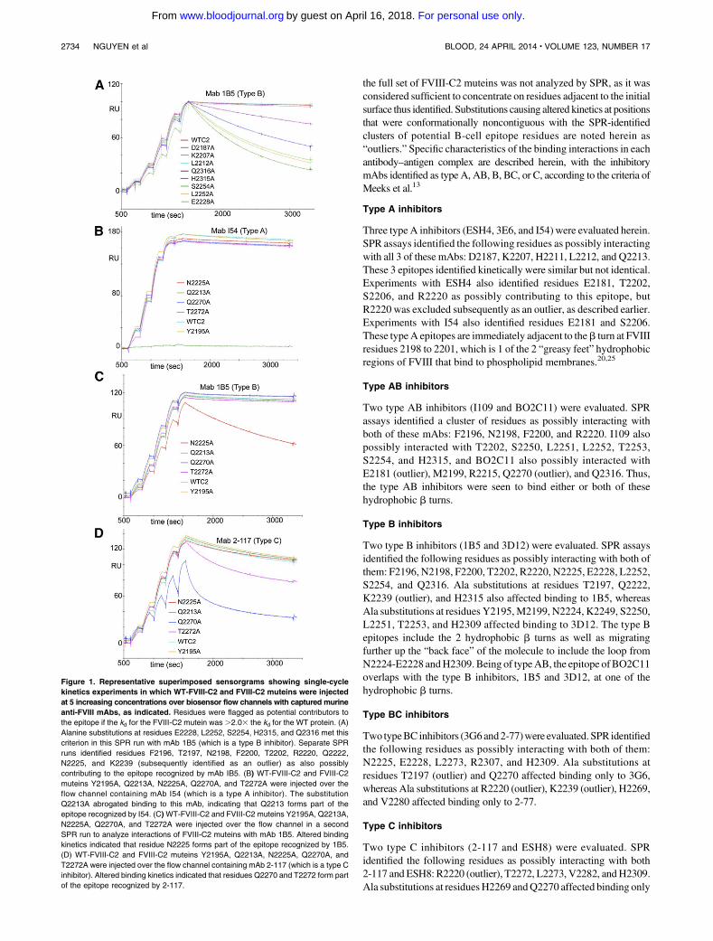
Representative sensorgrams are shown inFigure 1.Almost all of theFVIII-C2 muteins showed binding kinetics to some or all of the mAbsthat were highly similar to the binding of WT-FVIII-C2 (see summaryof results in Table 1 and kinetic constants in supplemental Table 2),indicating the substitutions did not cause global protein misfolding.Thirty-eight of the 60 muteins tested showed altered binding kineticsrestricted to a subset of the mAbs. FVIII-C2-F2200A showed alteredkinetics in binding to type AB and type BmAbs (I109, BO2C11, 1B5,and 3D12), with a kd. 2.03 that ofWT-FVIII-C2. FVIII-C2-R2220Ashowed altered binding to all mAbs except typeA antibodies 3E6 andI54 and type BC antibody 3G6. The ratio (kd[mutein]/kd[WT]) forbinding to the third type A mAb, ESH4, was 2.7, and with a morestringent cutoff value for this ratio, R2220 would not be identified aspart of the epitope recognized by this antibody. Removal of themostly buried R2220 side chain would be expected to destabilize themembrane-binding region of FVIII-C2. Therefore, its effect onbinding kinetics to ESH4 was judged to be a conformational effect,and it was not assigned to the epitope for this mAb. The kinetic datafrom analyzing several preparations of FVIII-C2-F2200A and FVIII-C2-R2220A were more variable and of poorer quality than thatof WT-FVIII-C2 and the other muteins, indicating that thesesubstitutions altered the protein stability. Nevertheless, the fact thatneighboring residues were also pinpointed as parts of epitopesrecognized by type AB and B antibodies supported their assignmentto theseepitopes.Qualitatively, several otherFVIII-C2muteins showedincreased antibody–antigendissociation rates relative toWT-FVIII-C2,although accurate kinetic constants could not be obtained. These resultsare indicated in Table 1 as qualitative fast dissociation (QFD).
Identification of B-cell epitopes
The residues identified by the cutoff criterion of kd(mutein) .2.03 kd(WT), when visualized using PyMOL,23 formed distinctclusters indicating the FVIII-C2 surfaces recognized by each mAb(Figure 2). Once a cluster was localized to a specific surface region,
BLOOD, 24 APRIL 2014 x VOLUME 123, NUMBER 17 B-CELL EPITOPES ON THE FACTOR VIII C2 DOMAIN 2733
For personal use only.on April 16, 2018. by guest www.bloodjournal.orgFrom
the full set of FVIII-C2 muteins was not analyzed by SPR, as it wasconsidered sufficient to concentrate on residues adjacent to the initialsurface thus identified. Substitutions causing altered kinetics at positionsthat were conformationally noncontiguous with the SPR-identifiedclusters of potential B-cell epitope residues are noted herein as“outliers.” Specific characteristics of the binding interactions in eachantibody–antigen complex are described herein, with the inhibitorymAbs identified as type A, AB, B, BC, or C, according to the criteria ofMeeks et al.13
Type A inhibitors
Three type A inhibitors (ESH4, 3E6, and I54) were evaluated herein.SPR assays identified the following residues as possibly interactingwith all 3 of these mAbs: D2187, K2207, H2211, L2212, and Q2213.These 3 epitopes identified kinetically were similar but not identical.Experiments with ESH4 also identified residues E2181, T2202,S2206, and R2220 as possibly contributing to this epitope, butR2220 was excluded subsequently as an outlier, as described earlier.Experiments with I54 also identified residues E2181 and S2206.These typeA epitopes are immediately adjacent to theb turn at FVIIIresidues 2198 to 2201, which is 1 of the 2 “greasy feet” hydrophobicregions of FVIII that bind to phospholipid membranes.20,25
Type AB inhibitors
Two type AB inhibitors (I109 and BO2C11) were evaluated. SPRassays identified a cluster of residues as possibly interacting withboth of these mAbs: F2196, N2198, F2200, and R2220. I109 alsopossibly interacted with T2202, S2250, L2251, L2252, T2253,S2254, and H2315, and BO2C11 also possibly interacted withE2181 (outlier), M2199, R2215, Q2270 (outlier), and Q2316. Thus,the type AB inhibitors were seen to bind either or both of thesehydrophobic b turns.
Type B inhibitors
Two type B inhibitors (1B5 and 3D12) were evaluated. SPR assaysidentified the following residues as possibly interacting with both ofthem: F2196, N2198, F2200, T2202, R2220, N2225, E2228, L2252,S2254, and Q2316. Ala substitutions at residues T2197, Q2222,K2239 (outlier), and H2315 also affected binding to 1B5, whereasAla substitutions at residues Y2195,M2199, N2224, K2249, S2250,L2251, T2253, and H2309 affected binding to 3D12. The type Bepitopes include the 2 hydrophobic b turns as well as migratingfurther up the “back face” of the molecule to include the loop fromN2224-E2228 andH2309.Being of typeAB, the epitope ofBO2C11overlaps with the type B inhibitors, 1B5 and 3D12, at one of thehydrophobic b turns.
Type BC inhibitors
Two typeBC inhibitors (3G6and2-77)were evaluated. SPR identifiedthe following residues as possibly interacting with both of them:N2225, E2228, L2273, R2307, and H2309. Ala substitutions atresidues T2197 (outlier) and Q2270 affected binding only to 3G6,whereas Ala substitutions at R2220 (outlier), K2239 (outlier), H2269,and V2280 affected binding only to 2-77.
Type C inhibitors
Two type C inhibitors (2-117 and ESH8) were evaluated. SPRidentified the following residues as possibly interacting with both2-117 and ESH8: R2220 (outlier), T2272, L2273, V2282, andH2309.Ala substitutions at residuesH2269 andQ2270 affected binding only
Figure 1. Representative superimposed sensorgrams showing single-cycle
kinetics experiments in which WT-FVIII-C2 and FVIII-C2 muteins were injected
at 5 increasing concentrations over biosensor flow channels with captured murine
anti-FVIII mAbs, as indicated. Residues were flagged as potential contributors to
the epitope if the kd for the FVIII-C2 mutein was .2.03 the kd for the WT protein. (A)
Alanine substitutions at residues E2228, L2252, S2254, H2315, and Q2316 met this
criterion in this SPR run with mAb 1B5 (which is a type B inhibitor). Separate SPR
runs identified residues F2196, T2197, N2198, F2200, T2202, R2220, Q2222,
N2225, and K2239 (subsequently identified as an outlier) as also possibly
contributing to the epitope recognized by mAb IB5. (B) WT-FVIII-C2 and FVIII-C2
muteins Y2195A, Q2213A, N2225A, Q2270A, and T2272A were injected over the
flow channel containing mAb I54 (which is a type A inhibitor). The substitution
Q2213A abrogated binding to this mAb, indicating that Q2213 forms part of the
epitope recognized by I54. (C)WT-FVIII-C2 and FVIII-C2 muteins Y2195A, Q2213A,
N2225A, Q2270A, and T2272A were injected over the flow channel in a second
SPR run to analyze interactions of FVIII-C2 muteins with mAb 1B5. Altered binding
kinetics indicated that residue N2225 forms part of the epitope recognized by 1B5.
(D) WT-FVIII-C2 and FVIII-C2 muteins Y2195A, Q2213A, N2225A, Q2270A, and
T2272A were injected over the flow channel containing mAb 2-117 (which is a type C
inhibitor). Altered binding kinetics indicated that residues Q2270 and T2272 form part
of the epitope recognized by 2-117.
2734 NGUYEN et al BLOOD, 24 APRIL 2014 x VOLUME 123, NUMBER 17
For personal use only.on April 16, 2018. by guest www.bloodjournal.orgFrom
to 2-117, whereas Ala substitutions at residues V2280 and Q2311affected only ESH8 binding. The conservative substitution R2307Qaffected binding to mAb 2-117, but not to ESH8.
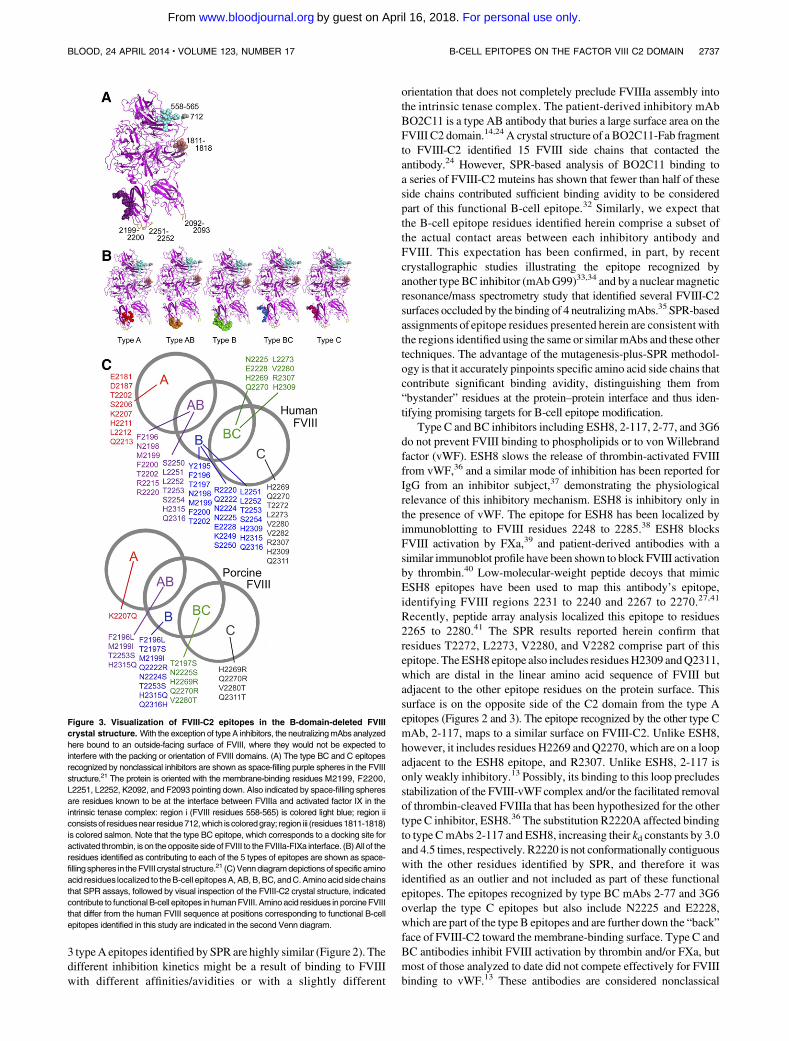
Modeling of epitopes in the FVIII structure
Figure 3A shows the BC and C epitope residues that comprisenonclassical inhibitor antibodies (eg, antibodies that prevent FVIIIactivation by thrombin and/or FXa). Figure 3B shows the location ofthe epitopes recognized by type A, AB, B, BC, and C antibodies.
Discussion
The SPR experiments identified 3 distinct clusters of surface-exposedside chains onFVIII-C2 that contributed significant binding avidity for
type A, B, and C FVIII-neutralizing antibodies, plus 2 clusterscontaining residues that comprised overlap regions for mAb typesAB and BC, respectively. SPR experiments were carried out for 3type A and 2 each of types AB, B, BC, and C mAbs. The resultingassignments of epitope residues were consistent within each mAbtype and were also consistent with the competition ELISA experi-ments13 and with peptide-based epitope mapping of FVIII-C2,26,27
as well as with ELISA assays evaluating binding of the mAbs tothe following FVIII muteins: F2196L, K2227E, M2199I/F2200L,V2223A/K2227E, andM2199I/F2200L/L2251V/L2252F.13 A recentanalysis of antibodies purified from a patient with an autoimmuneresponse to FVIII (acquired HA) indicated that these includedantibodieswith epitopes similar to those recognized byESH4 (typeA)and ESH8 (type C).28 The bleeding phenotype of acquired HA isoften more severe than that of congenital severe HA, possibly because
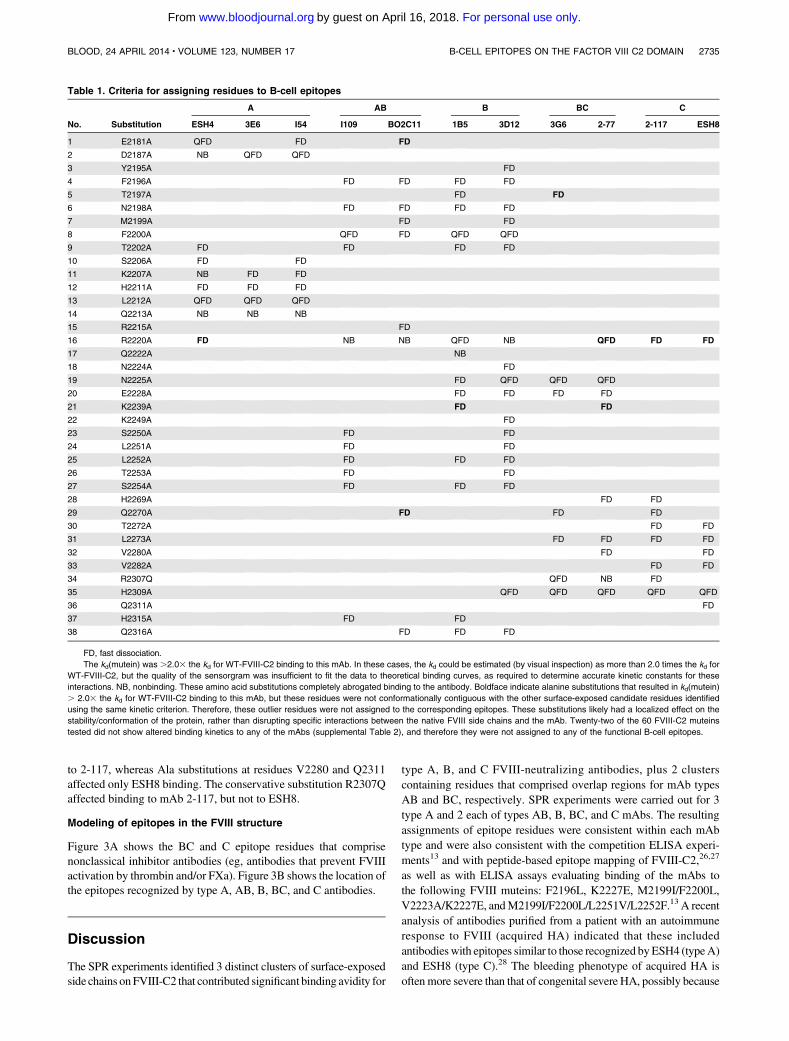
Table 1. Criteria for assigning residues to B-cell epitopes
No. Substitution
A AB B BC C
ESH4 3E6 I54 I109 BO2C11 1B5 3D12 3G6 2-77 2-117 ESH8
1 E2181A QFD FD FD
2 D2187A NB QFD QFD
3 Y2195A FD
4 F2196A FD FD FD FD
5 T2197A FD FD
6 N2198A FD FD FD FD
7 M2199A FD FD
8 F2200A QFD FD QFD QFD
9 T2202A FD FD FD FD
10 S2206A FD FD
11 K2207A NB FD FD
12 H2211A FD FD FD
13 L2212A QFD QFD QFD
14 Q2213A NB NB NB
15 R2215A FD
16 R2220A FD NB NB QFD NB QFD FD FD
17 Q2222A NB
18 N2224A FD
19 N2225A FD QFD QFD QFD
20 E2228A FD FD FD FD
21 K2239A FD FD
22 K2249A FD
23 S2250A FD FD
24 L2251A FD FD
25 L2252A FD FD FD
26 T2253A FD FD
27 S2254A FD FD FD
28 H2269A FD FD
29 Q2270A FD FD FD
30 T2272A FD FD
31 L2273A FD FD FD FD
32 V2280A FD FD
33 V2282A FD FD
34 R2307Q QFD NB FD
35 H2309A QFD QFD QFD QFD QFD
36 Q2311A FD
37 H2315A FD FD
38 Q2316A FD FD FD
FD, fast dissociation.
The kd(mutein) was .2.03 the kd for WT-FVIII-C2 binding to this mAb. In these cases, the kd could be estimated (by visual inspection) as more than 2.0 times the kd for
WT-FVIII-C2, but the quality of the sensorgram was insufficient to fit the data to theoretical binding curves, as required to determine accurate kinetic constants for these
interactions. NB, nonbinding. These amino acid substitutions completely abrogated binding to the antibody. Boldface indicate alanine substitutions that resulted in kd(mutein)
. 2.03 the kd for WT-FVIII-C2 binding to this mAb, but these residues were not conformationally contiguous with the other surface-exposed candidate residues identified
using the same kinetic criterion. Therefore, these outlier residues were not assigned to the corresponding epitopes. These substitutions likely had a localized effect on the
stability/conformation of the protein, rather than disrupting specific interactions between the native FVIII side chains and the mAb. Twenty-two of the 60 FVIII-C2 muteins
tested did not show altered binding kinetics to any of the mAbs (supplemental Table 2), and therefore they were not assigned to any of the functional B-cell epitopes.
BLOOD, 24 APRIL 2014 x VOLUME 123, NUMBER 17 B-CELL EPITOPES ON THE FACTOR VIII C2 DOMAIN 2735
For personal use only.on April 16, 2018. by guest www.bloodjournal.orgFrom

these antibodies bind to a somewhat different set of immunodominantB-cell epitopes and block FVIII functionality more effectively.
The strategy chosen to identify specific residues as members of aB-cell epitope by SPRwas to compare the experimental dissociationrate constant kd for a given FVIII-C2 mutein with the kd for WT-FVIII-C2 dissociating from the same antibody, noting whichsubstitutions increased the kd tomore than 2.03 that ofWT-FVIII-C2. Once 1 or more muteins with this property were identified, theFVIII-C2 structure was visualized using PyMOL.23 Care was takento analyze the FVIII-C2 muteins with substitutions in closeproximity to the initial cluster of surface-exposed residues identifiedby their altered kd values, but it was not considered essential toanalyze the entire series of muteins for each antibody. The precisemapping of this series of B-cell epitopes, combined with the earlieranalysis of the biochemical events (intrinsic tenase assembly, pro-teolytic activation of FVIII, etc) that were blocked by each type ofinhibitor,13 pinpointed specific surfaces on the FVIII protein thatinteract with its partners in promoting blood coagulation. The lists ofresidues comprising these epitopes (Table 1) are not comprehensivebecause not all surface residuesweremutated and several substitutionsaffected protein stability, and sowere not analyzed further. However,the coverage of the protein surface was sufficient to definitively
identify distinct clusters of residues contributing to specific antigen–antibody binding avidities.
The type A, AB, and B inhibitors interfere with FVIII or FVIIIabinding to phosphatidylserine-containing phospholipid mem-branes.13 As expected, the epitopes recognized by the AB andB mAbs included the hydrophobic b hairpin turns, as well as residuesH2315-Q2316, which are known to participate in membranebinding.25,29 ELISA experiments reported earlier13 showed that theepitope recognized by type AB inhibitor I109 includes residuesM2199 and F2200, and the SPR results confirm that this mAb alsobinds to the second hairpin turn containing L2251 and L2252.Interestingly, the epitopes recognized by the type A inhibitors ESH4,I54, and 3E6 are poised just above these projecting hairpin turns, andtheir inclusion of charged residues (E2181, D2187, K2206, K2207)indicates that these inhibitors block electrostatic interactions thatcould otherwise form between the positively charged FVIII sidechains and negatively charged membrane surfaces. The kinetics ofFVIII neutralization by inhibitory antibodies have long been classifiedas type 1 or type 2.30,31 Type 1 inhibitors completely block FVIIIactivity at saturating concentrations, whereas type 2 inhibitors do notcompletely inhibit clotting, even at saturating levels. Mabs I54 and3E6 are type 1 inhibitors, whereas ESH4 is a type 2 inhibitor. The
Figure 2. The B-cell epitopes indicated by the SPR
experiments are visualized using space-filling depic-
tions of the FVIII-C2 domain crystal structure in
standard orientation, with the membrane-interact-
ing loops pointing downward. The FVIII-C2 structure
is also shown rotated 180° about the vertical axis for
type AB and type B mAbs to visualize both sides of the
molecule. The B-cell epitopes identified on the basis of
altered binding kinetics are color-coded according to
FVIII inhibitor type; that is, A (red/salmon), AB (orange/
yellow), B (dark/light green), BC (dark/light blue), and C
(dark/light magenta). The darker colors indicate resi-
dues for which amino acid substitutions increased the
residence time by at least 10 times compared with that
for WT-FVIII-C2 binding to this mAb. Substitutions
abrogating binding were also colored darker. Substitu-
tions for which accurate kd values could not be obtained
were not colored darker because their effects on kinetics
may have been in part a result of effects on protein
stability. Several outlier residues identified as candidates
using the cutoff criterion of kd(mutein). 2.0 kd(WT) are
not shown, as they were eliminated after visualization
of the FVIII-C2 crystal structure.
2736 NGUYEN et al BLOOD, 24 APRIL 2014 x VOLUME 123, NUMBER 17
For personal use only.on April 16, 2018. by guest www.bloodjournal.orgFrom
3 typeA epitopes identified by SPR are highly similar (Figure 2). Thedifferent inhibition kinetics might be a result of binding to FVIIIwith different affinities/avidities or with a slightly different
orientation that does not completely preclude FVIIIa assembly intothe intrinsic tenase complex. The patient-derived inhibitory mAbBO2C11 is a type AB antibody that buries a large surface area on theFVIII C2 domain.14,24 A crystal structure of a BO2C11-Fab fragmentto FVIII-C2 identified 15 FVIII side chains that contacted theantibody.24 However, SPR-based analysis of BO2C11 binding toa series of FVIII-C2 muteins has shown that fewer than half of theseside chains contributed sufficient binding avidity to be consideredpart of this functional B-cell epitope.32 Similarly, we expect thatthe B-cell epitope residues identified herein comprise a subset ofthe actual contact areas between each inhibitory antibody andFVIII. This expectation has been confirmed, in part, by recentcrystallographic studies illustrating the epitope recognized byanother type BC inhibitor (mAbG99)33,34 and by a nuclear magneticresonance/mass spectrometry study that identified several FVIII-C2surfaces occluded by the binding of 4 neutralizingmAbs.35 SPR-basedassignments of epitope residues presented herein are consistent withthe regions identified using the same or similar mAbs and these othertechniques. The advantage of the mutagenesis-plus-SPR methodol-ogy is that it accurately pinpoints specific amino acid side chains thatcontribute significant binding avidity, distinguishing them from“bystander” residues at the protein–protein interface and thus iden-tifying promising targets for B-cell epitope modification.
Type C and BC inhibitors including ESH8, 2-117, 2-77, and 3G6do not prevent FVIII binding to phospholipids or to von Willebrandfactor (vWF). ESH8 slows the release of thrombin-activated FVIIIfrom vWF,36 and a similar mode of inhibition has been reported forIgG from an inhibitor subject,37 demonstrating the physiologicalrelevance of this inhibitory mechanism. ESH8 is inhibitory only inthe presence of vWF. The epitope for ESH8 has been localized byimmunoblotting to FVIII residues 2248 to 2285.38 ESH8 blocksFVIII activation by FXa,39 and patient-derived antibodies with asimilar immunoblot profile have been shown to block FVIII activationby thrombin.40 Low-molecular-weight peptide decoys that mimicESH8 epitopes have been used to map this antibody’s epitope,identifying FVIII regions 2231 to 2240 and 2267 to 2270.27,41
Recently, peptide array analysis localized this epitope to residues2265 to 2280.41 The SPR results reported herein confirm thatresidues T2272, L2273, V2280, and V2282 comprise part of thisepitope. TheESH8 epitope also includes residuesH2309 andQ2311,which are distal in the linear amino acid sequence of FVIII butadjacent to the other epitope residues on the protein surface. Thissurface is on the opposite side of the C2 domain from the type Aepitopes (Figures 2 and 3). The epitope recognized by the other type CmAb, 2-117, maps to a similar surface on FVIII-C2. Unlike ESH8,however, it includes residues H2269 andQ2270, which are on a loopadjacent to the ESH8 epitope, and R2307. Unlike ESH8, 2-117 isonly weakly inhibitory.13 Possibly, its binding to this loop precludesstabilization of the FVIII-vWF complex and/or the facilitated removalof thrombin-cleaved FVIIIa that has been hypothesized for the othertype C inhibitor, ESH8.36 The substitution R2220A affected bindingto type CmAbs 2-117 and ESH8, increasing their kd constants by 3.0and 4.5 times, respectively. R2220 is not conformationally contiguouswith the other residues identified by SPR, and therefore it wasidentified as an outlier and not included as part of these functionalepitopes. The epitopes recognized by type BC mAbs 2-77 and 3G6overlap the type C epitopes but also include N2225 and E2228,which are part of the type B epitopes and are further down the “back”face of FVIII-C2 toward the membrane-binding surface. Type C andBC antibodies inhibit FVIII activation by thrombin and/or FXa, butmost of those analyzed to date did not compete effectively for FVIIIbinding to vWF.13 These antibodies are considered nonclassical
Figure 3. Visualization of FVIII-C2 epitopes in the B-domain-deleted FVIII
crystal structure. With the exception of type A inhibitors, the neutralizingmAbs analyzed
here bound to an outside-facing surface of FVIII, where they would not be expected to
interfere with the packing or orientation of FVIII domains. (A) The type BC and C epitopes
recognized by nonclassical inhibitors are shown as space-filling purple spheres in the FVIII
structure.21 The protein is oriented with the membrane-binding residues M2199, F2200,
L2251, L2252, K2092, and F2093 pointing down. Also indicated by space-filling spheres
are residues known to be at the interface between FVIIIa and activated factor IX in the
intrinsic tenase complex: region i (FVIII residues 558-565) is colored light blue; region ii
consistsof residuesnear residue712,which iscoloredgray; region iii (residues1811-1818)
is colored salmon. Note that the type BC epitope, which corresponds to a docking site for
activated thrombin, is on theopposite sideof FVIII to theFVIIIa-FIXa interface. (B)All of the
residues identified as contributing to each of the 5 types of epitopes are shown as space-
filling spheres in theFVIII crystal structure.21 (C)Venndiagramdepictionsof specific amino
acid residues localized to theB-cell epitopesA,AB,B,BC, andC.Aminoacid sidechains
that SPR assays, followed by visual inspection of the FVIII-C2 crystal structure, indicated
contribute to functional B-cell epitopes in humanFVIII. Aminoacid residues in porcineFVIII
that differ from the human FVIII sequence at positions corresponding to functional B-cell
epitopes identified in this study are indicated in the second Venn diagram.
BLOOD, 24 APRIL 2014 x VOLUME 123, NUMBER 17 B-CELL EPITOPES ON THE FACTOR VIII C2 DOMAIN 2737
For personal use only.on April 16, 2018. by guest www.bloodjournal.orgFrom

inhibitors because their identification pointed to a previously under-appreciated and important role for the FVIII-C2 domain in proteolyticactivation of FVIII. Their localization at a surface distinct from theother types of epitopes, and also on an outer surface of the FVIIIprotein (ie, not at an interdomain interface), is shown in Figure 3A-B.
The mapping of this series of FVIII-C2 domain epitopes willfacilitate additional studies to model the domain orientations inFVIII (which may well differ among the solution, vWF-bound, andmembrane-bound FVIII structures),21,22,42 as well as the interactionsbetween FVIIIa and the other components of the intrinsic tenasecomplex. The high-resolution definition of these physiologicallyrelevant andmedically important epitopes also suggests specific sitesat which the FVIII sequence could be modified to generate lessantigenic FVIII variants. We propose that next-generation therapeuticFVIII proteins could include rationally designed variants of humanFVIII that, similar to porcine FVIII, could provide at least short-termhemostatic support in patients with high-titer inhibitors.5,6 The C2domain of porcine FVIII contains 32 residues that differ from thehuman FVIII sequence.43 Soluble FVIII-C2 proteins with alaninesubstitutions at 20 of these sites were characterized by SPR. Thehuman residues at 14 of these sites were identified as contributing toB-cell epitopes recognized by neutralizing anti-FVIII antibodies(Figure 3C).
Both antigen–antibody affinities44,45 and residence times16,19
(which reflect avidity of binding) of these complexes are of interest incharacterizing inhibitor responses. Amino acid substitutions at theantigen–antibody interfaces decreased some of the binding affinities(ΔΔG values) by 0 to 18 kJ/mol (supplemental Table 2). Because kdsare directly related to residence times and dissociation half-lives(half-life, ln 2/kd), and hence the expected duration of FVIIIneutralization by inhibitor antibodies, functional B-cell epitopeswere identified on the basis of the effects of amino acidsubstitutions on kd constants. This experimental approach may beused to target functional B-cell epitopes, including critical residues inantigenic loops in the FVIII A2 domain and in other regions ofFVIII,46-52 in designing novel FVIII muteins that could provideuseful bypass therapy options for inhibitor patients. Because theirsequences would be closer to that of the FVIII used to treat theoriginal bleeding disorder, the risk of provoking new T-cellresponses and subsequent new inhibitors53-58 to such rationallydesigned therapeutic FVIII muteins might also be lowered, incomparison with porcine FVIII used as bypass therapy. We expectthat sequence modifications to neutralize immunodominant B-celland T-cell epitopes will eventually be a feature of therapeutic FVIII
proteins targeted to patients with refractory inhibitor responses, aswell as to patients with poor prognostic factors such as high-risk F8gene mutations or a family history of inhibitors.59
Acknowledgments
The authors thank Emily Spence, Amjad Hussain, Rebecca Lefavor,and Shelley Nakaya Fletcher for generating FVIII-C2 muteins.
This work was supported by unrestricted research fundsfrom Bayer (K.P.P.) and CSL Behring (K.P.P.), a Pfizer ASPIREhemophilia award (K.P.P.), grants from the National Institutes ofHealth, Heart, Lung and Blood Institute (1RC2-HL101851) (K.P.P.);(U54-HL112309, K08-HL102262) (S.L.M.); and (U54-HL112309,R01-HL082609, and R01-HL040921) (P.L.), and Hemophilia ofGeorgia, Inc (S.L.M., P.L.).
The opinions or assertions contained herein are the private ones ofthe authors and are not to be construed as official or reflecting theviews of the Department of Defense or the Uniformed ServicesUniversity of the Health Sciences.
Authorship
Contribution: P.-C.T.N., K.B.L., R.A.E., J.T.S., and J.C.L. carriedout the experiments; K.B.L., P.-C.T.N., J.T.S., R.A.E., J.C.L., andK.P.P. designed the experiments; J.F.H., S.L.M., and P.L. performedmAb isolation and characterization; and P.-C.T.N., K.B.L., R.A.E.,J.T.S., J.C.L., andK.P.P. analyzed the data andwrote themanuscript,which was reviewed and approved by all authors.
Conflict-of-interest disclosure: K.P.P. and K.B.L. are inventorson a patent (International Patent Application No. PCT/US2012/61/553 660) that describes characterization of B-cell epitopes usingSPR. J.T.S. is an employee of GE Healthcare. K.P.P. has receivedunrestricted research funding from Bayer, Pfizer, and CSL Behringand honoraria from Bayer, Pfizer, and Novo Nordisk. The remainingauthors declare no competing financial interests.
Correspondence: Kathleen P. Pratt, Department of Medicine(MED) A3075, Uniformed Services University of the HealthSciences, 4301 Jones Bridge Rd, Bethesda, MD 20814; e-mail:[email protected].
References
1. Ehrenforth S, Kreuz W, Scharrer I, et al. Incidenceof development of factor VIII and factor IXinhibitors in haemophiliacs. Lancet. 1992;339(8793):594-598.
2. Kreuz W, Ettingshausen CE, Zyschka A, et al.Inhibitor development in previously untreatedpatients with hemophilia A: a prospective long-term follow-up comparing plasma-derived andrecombinant products. Semin Thromb Hemost.2002;28(3):285-290.
3. Gouw SC, Fijnvandraat K. Unraveling the geneticsof inhibitors in hemophilia. Blood. 2013;121(8):1250-1251.
4. Cugno M, Gualtierotti R, Tedeschi A, Meroni PL.Autoantibodies to coagulation factors: Frompathophysiology to diagnosis and therapy.Autoimmun Rev. 2014;13(1):40-48.
5. Aledort LM. The role of porcine factor VIII in themanagement of unexpected bleeding episodes.
Haemophilia. 2002;8(Suppl 1):17-19, discussion28-32.
6. Toschi V. OBI-1, porcine recombinant FactorVIII for the potential treatment of patients withcongenital hemophilia A and alloantibodiesagainst human Factor VIII. Curr Opin Mol Ther.2010;12(5):617-625.
7. Kulkarni R, Aledort LM, Berntorp E, et al.Therapeutic choices for patients with hemophiliaand high-titer inhibitors. Am J Hematol. 2001;67(4):240-246.
8. Kempton CL, Abshire TC, Deveras RA, et al.Pharmacokinetics and safety of OBI-1,a recombinant B domain-deleted porcine factorVIII, in subjects with haemophilia A. Haemophilia.2012;18(5):798-804.
9. Barrow RT, Lollar P. Neutralization of antifactorVIII inhibitors by recombinant porcine factor VIII.J Thromb Haemost. 2006;4(10):2223-2229.
10. Arai M, Scandella D, Hoyer LW. Molecular basis offactor VIII inhibition by human antibodies. Antibodiesthat bind to the factor VIII light chain prevent theinteraction of factor VIII with phospholipid. J ClinInvest. 1989;83(6):1978-1984.
11. Scandella DH, Nakai H, Felch M, et al. Inhemophilia A and autoantibody inhibitor patients:the factor VIII A2 domain and light chain are mostimmunogenic. Thromb Res. 2001;101(5):377-385.
12. Pratt KP. Relating structure to function: the role ofthe C2 domain in Factor VIII. Curr Opin DrugDiscov Devel. 2000;3(5):516-526.
13. Meeks SL, Healey JF, Parker ET, Barrow RT,Lollar P. Antihuman factor VIII C2 domainantibodies in hemophilia A mice recognizea functionally complex continuous spectrum ofepitopes dominated by inhibitors of factor VIIIactivation. Blood. 2007;110(13):4234-4242.
2738 NGUYEN et al BLOOD, 24 APRIL 2014 x VOLUME 123, NUMBER 17
For personal use only.on April 16, 2018. by guest www.bloodjournal.orgFrom
14. Jacquemin MG, Desqueper BG, Benhida A, et al.Mechanism and kinetics of factor VIII inactivation:study with an IgG4 monoclonal antibody derivedfrom a hemophilia A patient with inhibitor. Blood.1998;92(2):496-506.
15. Karlsson R, Katsamba PS, Nordin H, Pol E,Myszka DG. Analyzing a kinetic titration seriesusing affinity biosensors. Anal Biochem. 2006;349(1):136-147.
16. Copeland RA, Pompliano DL, Meek TD. Drug-target residence time and its implications for leadoptimization. Nat Rev Drug Discov. 2006;5(9):730-739.
17. Wood ER, Truesdale AT, McDonald OB, et al.A unique structure for epidermal growth factorreceptor bound to GW572016 (Lapatinib):relationships among protein conformation,inhibitor off-rate, and receptor activity in tumorcells. Cancer Res. 2004;64(18):6652-6659.
18. Mondal K, Regnstrom K, Morishige W, et al.Thermodynamic and kinetic characterizationof hydroxyethylamine b-secretase-1 inhibitors.Biochem Biophys Res Commun. 2013;441(2):291-296.
19. Tummino PJ, Copeland RA. Residence time ofreceptor-ligand complexes and its effect onbiological function. Biochemistry. 2008;47(20):5481-5492.
20. Pratt KP, Shen BW, Takeshima K, Davie EW,Fujikawa K, Stoddard BL. Structure of the C2domain of human factor VIII at 1.5 A resolution.Nature. 1999;402(6760):439-442.
21. Ngo JC, Huang M, Roth DA, Furie BC, Furie B.Crystal structure of human factor VIII: implicationsfor the formation of the factor IXa-factor VIIIacomplex. Structure. 2008;16(4):597-606.
22. Shen BW, Spiegel PC, Chang CH, et al. Thetertiary structure and domain organization ofcoagulation factor VIII. Blood. 2008;111(3):1240-1247.
23. PyMOL. The PyMOL Molecular Graphics System,Version 1.5.0.4 Schrodinger, LLC.; 2013.
24. Spiegel PC Jr, Jacquemin M, Saint-Remy JM,Stoddard BL, Pratt KP. Structure of a factor VIIIC2 domain-immunoglobulin G4kappa Fabcomplex: identification of an inhibitory antibodyepitope on the surface of factor VIII. Blood. 2001;98(1):13-19.
25. Lewis DA, Moore KD, Ortel TL. Binding of factorVIII inhibitors to discrete regions of the factor VIIIC2 domain disrupt phospholipid binding. BloodCoagul Fibrinolysis. 2003;14(4):361-368.
26. Lebreton A, Moreau V, Lapalud P, et al.Discontinuous epitopes on the C2 domain ofcoagulation Factor VIII mapped by computer-designed synthetic peptides. Br J Haematol.2011;155(4):487-497.
27. Villard S, Lacroix-Desmazes S, Kieber-EmmonsT, et al. Peptide decoys selected by phage displayblock in vitro and in vivo activity of a human anti-FVIII inhibitor. Blood. 2003;102(3):949-952.
28. Matsumoto T, Nogami K, Ogiwara K, Shima M.A putative inhibitory mechanism in the tenasecomplex responsible for loss of coagulationfunction in acquired haemophilia A patients withanti-C2 autoantibodies. Thromb Haemost. 2012;107(2):288-301.
29. Liu Z, Lin L, Yuan C, et al. Trp2313-His2315 offactor VIII C2 domain is involved in membrane
binding: structure of a complex between the C2domain and an inhibitor of membrane binding.J Biol Chem. 2010;285(12):8824-8829.
30. Biggs R, Austen DE, Denson KW, Rizza CR,Borrett R. The mode of action of antibodies whichdestroy factor VIII. I. Antibodies which havesecond-order concentration graphs. Br JHaematol. 1972;23(2):125-135.
31. Biggs R, Austen DE, Denson KW, Borrett R,Rizza CR. The mode of action of antibodies whichdestroy factor VIII. II. Antibodies which givecomplex concentration graphs. Br J Haematol.1972;23(2):137-155.
32. Lin JC, Schuman JT, Nakaya SM, et al. Six aminoacid residues in a 1200 A2 interface mediatebinding to an IgG4-kappa inhibitory antibody[abstract]. Blood. 2008;112(11):Abstract 3380.
33. Walter JD, Werther RA, Polozova MS, et al.Characterization and solution structure of thefactor VIII C2 domain in a ternary complex withclassical and non-classical inhibitor antibodies.J Biol Chem. 2013;288(14):9905-9914.
34. Walter JD, Werther RA, Brison CM, et al.Structure of the factor VIII C2 domain in a ternarycomplex with 2 inhibitor antibodies revealsclassical and nonclassical epitopes. Blood. 2013;122(26):4270-4278.
35. Sevy AM, Healey JF, Deng W, Spiegel PC, MeeksSL, Li R. Epitope mapping of inhibitory antibodiestargeting the C2 domain of coagulation factor VIIIby hydrogen-deuterium exchange massspectrometry. J Thromb Haemost. 2013;11(12):2128-2136.
36. Saenko EL, Shima M, Gilbert GE, Scandella D.Slowed release of thrombin-cleaved factor VIIIfrom von Willebrand factor by a monoclonal anda human antibody is a novel mechanism for factorVIII inhibition. J Biol Chem. 1996;271(44):27424-27431.
37. Jacquemin M, Benhida A, Peerlinck K, et al. Ahuman antibody directed to the factor VIII C1domain inhibits factor VIII cofactor activity andbinding to von Willebrand factor. Blood. 2000;95(1):156-163.
38. Scandella D, Gilbert GE, Shima M, et al.Some factor VIII inhibitor antibodies recognizea common epitope corresponding to C2 domainamino acids 2248 through 2312, which overlapa phospholipid-binding site. Blood. 1995;86(5):1811-1819.
39. Nogami K, Shima M, Hosokawa K, et al. Role offactor VIII C2 domain in factor VIII binding tofactor Xa. J Biol Chem. 1999;274(43):31000-31007.
40. Nogami K, Shima M, Hosokawa K, et al. FactorVIII C2 domain contains the thrombin-binding siteresponsible for thrombin-catalyzed cleavage atArg1689. J Biol Chem. 2000;275(33):25774-25780.
41. Albert T, Egler C, Jakuschev S, et al. The B-cellepitope of the monoclonal anti-factor VIII antibodyESH8 characterized by peptide array analysis.Thromb Haemost. 2008;99(3):634-637.
42. Stoilova-McPhie S, Lynch GC, Ludtke S, PettittBM. Domain organization of membrane-boundfactor VIII. Biopolymers. 2013;99(7):448-459.
43. Healey JF, Lubin IM, Lollar P. The cDNA andderived amino acid sequence of porcine factorVIII. Blood. 1996;88(11):4209-4214.
44. Cunningham BC, Wells JA. Comparison ofa structural and a functional epitope. J Mol Biol.1993;234(3):554-563.
45. Sundberg EJ, Mariuzza RA. Molecular recognitionin antibody-antigen complexes. Adv ProteinChem. 2002;61:119-160.
46. Barrow RT, Healey JF, Gailani D, Scandella D,Lollar P. Reduction of the antigenicity of factor VIIItoward complex inhibitory antibody plasmas usingmultiply-substituted hybrid human/porcine factorVIII molecules. Blood. 2000;95(2):564-568.
47. Fay PJ, Scandella D. Human inhibitor antibodiesspecific for the factor VIII A2 domain disrupt theinteraction between the subunit and factor IXa.J Biol Chem. 1999;274(42):29826-29830.
48. Healey JF, Lubin IM, Nakai H, et al. Residues484-508 contain a major determinant of theinhibitory epitope in the A2 domain of humanfactor VIII. J Biol Chem. 1995;270(24):14505-14509.
49. Lollar P. Mapping factor VIII inhibitor epitopesusing hybrid human/porcine factor VIII molecules.Haematologica. 2000;85(10 Suppl):26-28,discussion 28-30.
50. Sawamoto Y, Shima M, Tanaka I, et al. Anti-factorVIII inhibitor alloantibodies recognizing the A2domain in the human factor VIII heavy chainpoorly bind to porcine factor VIII. Int J Hematol.1997;65(2):151-158.
51. Lubin IM, Healey JF, Scandella D, Runge MS,Lollar P. Elimination of a major inhibitor epitope infactor VIII. J Biol Chem. 1994;269(12):8639-8641.
52. Lubin IM, Healey JF, Barrow RT, Scandella D,Lollar P. Analysis of the human factor VIIIA2 inhibitor epitope by alanine scanningmutagenesis. J Biol Chem. 1997;272(48):30191-30195.
53. Jacquemin M, Vantomme V, Buhot C, et al. CD41T-cell clones specific for wild-type factor VIII:a molecular mechanism responsible for a higherincidence of inhibitor formation in mild/moderatehemophilia A. Blood. 2003;101(4):1351-1358.
54. Ettinger RA, James EA, Kwok WW, ThompsonAR, Pratt KP. HLA-DR-restricted T-cell responsesto factor VIII epitopes in a mild haemophilia Afamily with missense substitution A2201P.Haemophilia. 2010;16(102):44-55.
55. Ettinger RA, James EA, Kwok WW, ThompsonAR, Pratt KP. Lineages of human T-cell clones,including T helper 17/T helper 1 cells, isolatedat different stages of anti-factor VIII immuneresponses. Blood. 2009;114(7):1423-1428.
56. James EA, Kwok WW, Ettinger RA, ThompsonAR, Pratt KP. T-cell responses over time ina mild hemophilia A inhibitor subject: epitopeidentification and transient immunogenicity of thecorresponding self-peptide. J Thromb Haemost.2007;5(12):2399-2407.
57. Pratt KP, Thompson AR. B-cell and T-cellepitopes in anti-factor VIII immune responses.Clin Rev Allergy Immunol. 2009;37(2):80-95.
58. Scott DW, Pratt KP, Miao CH. Progress towardinducing immunologic tolerance to factor VIII.Blood. 2013;121(22):4449-4456.
59. Gouw SC, van den Berg HM, Oldenburg J, et al.F8 gene mutation type and inhibitor developmentin patients with severe hemophilia A: systematicreview and meta-analysis. Blood. 2012;119(12):2922-2934.
BLOOD, 24 APRIL 2014 x VOLUME 123, NUMBER 17 B-CELL EPITOPES ON THE FACTOR VIII C2 DOMAIN 2739
For personal use only.on April 16, 2018. by guest www.bloodjournal.orgFrom

online March 3, 2014 originally publisheddoi:10.1182/blood-2013-09-527275
2014 123: 2732-2739
F. Healey, Shannon L. Meeks, Pete Lollar and Kathleen P. PrattPhuong-Cac T. Nguyen, Kenneth B. Lewis, Ruth A. Ettinger, Jason T. Schuman, Jasper C. Lin, John analysis of point mutants using surface plasmon resonanceHigh-resolution mapping of epitopes on the C2 domain of factor VIII by
http://www.bloodjournal.org/content/123/17/2732.full.htmlUpdated information and services can be found at:
(1144 articles)Thrombosis and Hemostasis Articles on similar topics can be found in the following Blood collections
http://www.bloodjournal.org/site/misc/rights.xhtml#repub_requestsInformation about reproducing this article in parts or in its entirety may be found online at:
http://www.bloodjournal.org/site/misc/rights.xhtml#reprintsInformation about ordering reprints may be found online at:
http://www.bloodjournal.org/site/subscriptions/index.xhtmlInformation about subscriptions and ASH membership may be found online at:
Copyright 2011 by The American Society of Hematology; all rights reserved.of Hematology, 2021 L St, NW, Suite 900, Washington DC 20036.Blood (print ISSN 0006-4971, online ISSN 1528-0020), is published weekly by the American Society
For personal use only.on April 16, 2018. by guest www.bloodjournal.orgFrom