high expression of somatic embryogenesis … cultures of immature cotyledonary-stage ... the cells...
TRANSCRIPT
ORIGINAL ARTICLE
High expression of SOMATIC EMBRYOGENESISRECEPTOR-LIKE KINASE coincides with initiation of various
Maria Pilarska1 & PrzemysławMalec2 & Jan Salaj3 & Filip Bartnicki2 & Robert Konieczny4
Received: 29 December 2014 /Accepted: 26 March 2015 /Published online: 16 April 2015# The Author(s) 2015. This article is published with open access at Springerlink.com
Abstract The aim of this study was to identify and examinethe expression pattern of the ortholog of SOMATIC EM-BRYOGENESIS RECEPTOR-LIKE KINASE gene from Tri-folium nigrescens (TnSERK) in embryogenic and non-regenerative cultures of immature cotyledonary-stage zygoticembryos (CsZEs). In the presence of 1-naphthaleneacetic ac-id and N6-[2-isopentenyl]-adenine, the CsZE regenerated em-bryoids directly and in a lengthy culture produced calluswhich was embryogenic or remained non-regenerative. Asrevealed by semi-quantitative reverse transcription polymer-ase chain reaction (RT-PCR), the TnSERK was expressed inboth embryogenic and non-regenerative cultures, but the ex-pression level was significantly higher in embryogenic ones.An in situ RNA hybridization assay revealed that the expres-sion of TnSERK preceded the induction of cell division inexplants, and then, it was maintained exclusively in actively
dividing cells from which embryoids, embryo-like structures(ELSs), callus or tracheary elements were produced. Howev-er, the cells involved in different morphogenic events dif-fered in intensity of hybridization signal which was thehighest in embryogenic cells. The TnSERK was up-regulated during the development of embryoids, but in coty-ledonary embryos, it was preferentially expressed in the re-gions of the apical meristems. The occurrence of morpholog-ical and anatomical abnormalities in embryoid developmentwas preceded by a decline in TnSERK expression, and thiscoincided with the parenchymatization of the ground tissuein developing ELSs. TnSERKwas also down-regulated duringthe maturation of parenchyma and xylem elements in CsZEand callus. Altogether, these data suggest the involvement ofTnSERK in the induction of various developmental programsrelated to differentiation/transdifferentiation and totipotentstate of cell(s).
Keywords Gene expression . Differentiation . Pluripotency .
Somatic embryogenesis . Totipotency . Transdifferentiation
Abbreviations2iP N6-[2-isopentenyl]-adenineCsZE Cotyledonary-stage zygotic embryoEC Embryogenic callusELS Embryo-like structureISH In situ hybridizationNAA 1-Naphthaleneacetic acidNRC Non-regenerative callusSE Somatic embryogenesisSERK SOMATIC EMBRYOGENESIS
RECEPTOR-LIKE KINASE
Handling Editor: Peter Nick
Electronic supplementary material The online version of this article(doi:10.1007/s00709-015-0814-5) contains supplementary material,which is available to authorized users.
* Robert [email protected]
1 The Franciszek Górski Institute of Plant Physiology, Polish Academyof Sciences, Niezapominajek 21, 30-239 Kraków, Poland
2 Department of Plant Physiology and Biochemistry, Faculty ofBiochemistry, Biophysics and Biotechnology, JagiellonianUniversity, Gronostajowa 7, 30-387 Kraków, Poland
3 Institute of Plant Genetics and Biotechnology, Slovak Academy ofSciences, Akademicka 2, 950-07 Nitra, Slovak Republic
4 Department of Plant Cytology and Embryology, Institute of Botany,Jagiellonian University, Gronostajowa 9, 30-387 Kraków, Poland
Protoplasma (2016) 253:345–355DOI 10.1007/s00709-015-0814-5
developmental pathways in in vitro culture of Trifolium nigrescens

Introduction
Somatic embryogenesis (SE) is a process whereby a singleplant cell or group of cells from somatic tissue forms an em-bryo. SE is commonly exploited as a model for studying thestructural and molecular events which underlie plant differenti-ation. In recent years, extensive research has been carried outon genes with a specific role in the induction and maintenanceof SE (for a review, see Ikeda et al. 2006; Elhiti et al. 2013).One of such genes is SOMATIC EMBRYOGENESISRECEPTOR-LIKE KINASE 1 (SERK1) which encodes a trans-membrane protein kinase belonging to the family of leucine-rich repeat protein receptor-like kinases (Hecht et al. 2001). TheSERK gene was initially isolated from carrot embryogenic cal-lus where a high expression of it marked cells subsequentlydeveloping into somatic embryos (Schmidt et al. 1997). InArabidopsis thaliana, five genes belonging to the SERK familywere identified and overexpression of SERK1 was reported tolead to a significant increase in embryogenic competence intransgenic lines (Hecht et al. 2001). So far, apart from carrotand A. thaliana, the SERK1 gene has been found to beexpressed during the induction of SE in monocotyledonousand dicotyledonous species, i.e. Dactylis glomerata (Somlevaet al. 2000), Ocotea catharinensis (Santa-Catarina et al. 2004),Theobroma cacao (de Oliveira Santos et al. 2005),Vitis vinifera(Schellenbaum et al. 2008), Cocos nucifera (Pérez-Núňez et al.2009) or Ananas comosus (Ma et al. 2012). Although the pre-cise expression pattern varies among species, these results dem-onstrated that in different plants, up-regulation of specificSERKs can be attributed to the induction of totipotency. Furtherstudies, however, showed that SERKs also play a part in otherdevelopmental events such as induction and maintenance ofshoot and root apical meristems during organogenesis in vitroand in vivo (Nolan et al. 2003, 2009; Thomas et al. 2004;Savona et al. 2012; Du et al. 2012), male sporogenesis(Albrecht et al. 2005), apomixes (Albertini et al. 2005) as wellas in defense responses (Santos et al. 2009; Huang et al. 2009).Recent studies showed that AtSERK proteins play a crucial rolein brass inos te ro id s igna l ing as co- receptors ofBRASSINOSTEROID-INSENSITIVE1 (BRI1) (Albrechtet al. 2008; Gou et al. 2012). This multifunctionality of SERKsimplies a need for a detailed expressional analysis of each newidentified gene to properly determine its possible function in thebiology of a particular plant.
In situ hybridization (ISH) of RNA is one of the mostpowerful techniques developed for localizing the expressionsite of a gene at the cell, tissue and organ levels. ISH has beensuccessfully employed in analyzing the spatio-temporal pat-tern of SERK1 expression in sunflower (Thomas et al. 2004),Medicago truncatula (Nolan et al. 2009), coconut (Pérez-Núňez et al. 2009) and, more recently, Cyclamen persicum(Savona et al. 2012). In the genus Trifolium, regeneration viaSE has been described in many species (see Konieczny et al.
2010). However, most of the published reports focus on theimprovement of culture conditions for efficient SE induction,plant development and acclimatization, whilst the molecularbasis of somatic embryo formation still remains largely un-known. So far, in this genus, the sequence and expression hasnot been analyzed for any of the genes from the SERK family.
Trifolium nigrescens (Viv.) is a self-incompatible diploid(2n=2x=16) used as a forage legume for pasture and soil im-provement (Hoveland and Evers 1995). Due to its high seedproduction under hard grazing and its resistance to southern rootknot (Meloidogyne incognita) and clover cyst (Heteroderatrifolii) nematodes (Pederson andWindham1989),T. nigrescensis investigated for its potential as a germplasm for the improve-ment of other pasture clovers, e.g. Trifolium repens throughinterspecific hybridization (Marshall et al. 2002, 2008). Earlier,we reported that somatic embryos of T. nigrescens can be pro-duced from cotyledonary-stage zygotic embryos (CsZEs) di-rectly or via callus formation (Konieczny et al. 2010). Recently,we analyzed some structural aspects of T. nigrescens somaticembryo formation and the involvement of cell wall componentsin SE (Pilarska et al. 2013). In this study, we used a T. nigrescensculture system to determine if an ortholog of the SERK gene ispresent in the T. nigrescens genome and to gain an insight intoits role in the induction and during the course of SE and callusgrowth. We applied ISH to reveal the expression pattern of aT. nigrescens SERK ortholog (TnSERK) during direct and indi-rect SE and in non-regenerative callus, and these data are sup-plemented with reverse transcription polymerase chain reaction(RT-PCR) analyses of the TnSERK expression level and thehistology of SE and callogenesis.
Material and methods
Plant material and culture conditions
Seeds of T. nigrescens (Viv.) ssp. nigrescenswere obtained fromthe Institute of Plant Genetics and Crop Plant Research,Gatersleben, Germany. The procedure for donor plant growth,explant preparation and culture conditions for SE induction wasdescribed previously (Konieczny et al. 2010). Briefly, SE wasinitiated from immature zygotic embryos (cotyledonary stage,ca. 3 mm in length) on a Murashige and Skoog (1962) basalmedium supplemented with 30 g l−1 sucrose, 8 g l−1 agar (Difco,USA), 0.5 mg l−1 1-naphthaleneacetic acid (NAA) and2.0 mg l−1 N6-[2-isopentenyl]-adenine (2iP), pH=5.7. Cultureswere maintained in the 16:8-h photoperiod of light and darkness.
Histological studies
Material for histological studies was collected after 3, 5, 7, 10and 15 days of culture. The specimens were fixed in 4 % (v/v)glutaraldehyde in a 0.1 M phosphate buffer (pH 7.2) for 2 h at
346 M. Pilarska et al.

room temperature, dehydrated in a graded series of ethanoland embedded in Technovit 7100 (Heraeus Kulzer, Germany)according to the manufacturer’s instructions. Sections of 5 μmin thickness were routinely stained with a 0.1 % (w/v) aquaticsolution of toluidine blue O.
DNA extraction, amplification, cloning and sequencing
Genomic DNAwas extracted from the leaves of mature plants(100 mg of the fresh weight from a 2-month-old plant) using aGenomic DNA Isolation Kit (A&A Biotechnology, Poland)according to the manufacturer’s protocol. The DNA concen-tration and purity were controlled spectrophotometrically.Conserved regions of SERK1 from A. thaliana (GenBank ac-cession number NM_105841.4) andM. truncatula (GenBankaccession number AY162176.1) were used to design primersfor amplifying a SERK-specific fragment from T. nigrescens.The PCR amplification (reaction final volume 50 μl) wascarried out with Pwo polymerase (A&A Biotechnology, Po-land), 25 ng of high-purity genomic DNA as a template and aprimer pair (forward: 5′ AGCCGAAGAAGATCCAGAAGTTCA 3′ and reverse: 5′ TCCATTGGCCATGTAAGGAT3′) with a PTC-150 MiniCycler (MJ Research, USA) and thefollowing program: 94 °C for 1 min, 30 cycles of 94 °C for45 s, 45 °C for 1 min, 72 °C for 2 min and 72 °C for 5 min. ThePCR products were separated by electrophoresis on 2 % aga-rose gel and identified under UV light. The resulting PCRproduct, ca. 330 bp in length, was excised from the gel, puri-fied with a Gel-Out kit (A&A Biotechnology, Poland),subcloned into pJET1.2/blunt plasmid (CloneJET PCR Clon-ing Kit; Fermentas Thermo Scientific, USA) and subjected tosequencing. Plasmids containing a TnSERK fragment insertedboth in sense and antisense orientation were isolated, propa-gated and maintained in an Escherichia coli DH5α strain,using standard procedures (Ausubel et al. 1995). ThisTnSERK fragment was further used to design gene-specificprimers suitable for the isolation of the partial genomic se-quence of TnSERK and RT-PCR as well as to construct aprobe for in situ hybridization.
To identify the genomic sequence downstream of the ini-tially isolated TnSERK fragment, the following primer pairwas used: forward: 5′ATGGAGGAGACAAAGTTCTGTGC 3′ and reverse: 5′TCATCTTGGACCAGATAATTCGAC 3′. The 1250-bp product was amplified by PCR as above,cloned and analyzed by sequencing.
Sequence analysis
The nucleotide sequences obtained were analyzed and com-pared with GenBank data using the nucleotide database ofBasic Alignment Search Tool (BLAST) software from theNational Center of Biotechnology Information (NCBI;www.ncbi.nlm.nih.gov/BLAST) (Schaffer et al. 2001). The
amino acid sequences deduced were compared by proteinBLASTP (Swiss Institute of Bioinformatics; http://web.expasy.org/cgi-bin/blast+/BLAST_new.pl) with ProteinKnowledgebase (UniProtKB/TrEMBL). Amino acidsequence alignment was performed using ClustalW (Chennaet al. 2003). Phylogenetic analysis was carried out withPhylogeny.fr software (Dereeper et al. 2008). Conservedregions were scanned by PROSITE (de Castro et al. 2006).
RNA extraction and cDNA synthesis
RNAwas extracted from mature leaf blades, non-regenerativecallus (NRC) and embryogenic callus (EC) with somatic em-bryos after 14 days of culture. Total RNA extraction wascarried out using the Spectrum™ Plant Total RNA Kit(Sigma-Aldrich, Germany) according to the manufacturer’sinstruction. To avoid DNA contamination, samples weretreated with DNase I (Fermentas Thermo Scientific, USA)for 30 min at room temperature. To produce a single-stranded cDNA population, 1 μg of total RNAwas reverselytranscribed using a RevertAid™ First Strand cDNA Synthe-sis Kit (Fermentas Thermo Scientific, USA), using the ran-dom primer technique, according to the manufacturer’sprotocol.
Analysis of TnSERK expression
For semi-quantitative RT-PCR, the following gene-specificprimers for TnSERK were used (forward: 5′ GCAAGTCGCAACCGATACTT and reverse: 5′ CCACGTAAACGGAGGAGATT). Primers designed to recognize a conserved do-main of ELONGATION FACTOR-1α gene (EF-1α) fromT. repens (GenBank accession number KC710340.1) wereused to amplify the EF-1α fragment that served as internalcontrol (forward: 5′ ACGCTCTTCTTGCTTCCACC 3′ andreverse: 5′ GTTGTCTCCCTCAAAACCGGA 3′). The PCRreaction was performed with Pwo polymerase (A&ABiotech-nology, Poland) using equal volumes of cDNA obtained from1 μg of total RNA as a template, with the following program:95 °C for 4 min, 27 cycles (denaturation at 95 °C for 1 min,annealing at 55 °C for 45 s, elongation at 72 °C for 1 min) andtermination at 72 °C for 2 min. PCR products were separatedin 2 % agarose gel stained with ethidium bromide. The bandintensities were measured and quantified by densitometryusing the UVP BioSpectrum Imaging System withVisionWorksLS version 6.8 software (UVP, USA). The rela-tive EF-1α band intensity (a single 194-bp product) was ac-knowledged as 100 % (Serazin-Leroy et al. 1998). The signif-icance of results from three biological replicates was checkedby one-way ANOVA and Duncan’s test (P≤0.05). Statisticafor Windows ver. 8.0 (StatSoft, Inc., Tulsa, USA) was used.
Expression of SERK in in vitro culture of Trifolium nigrescens 347

In situ hybridization
The material for ISH was sampled after 3, 5, 7, 10 and15 days of in vitro culture. Samples were fixed for 2 h in4 % (v/v) paraformaldehyde+0.25 % glutaraldehyde (v/v) ina 0.1 M sodium phosphate buffer (pH 7.2), dehydrated in aseries of alcohol solutions and embedded in Paraplast (Sig-ma-Aldrich, Germany). Sections of 6 μm in thickness weremounted on poly-lysine-coated slides, rehydrated and incu-bated 30 min at 37 °C with proteinase K to remove pro-teins. The pJET1.2/blunt plasmid containing the 322-bpsequence fragment of TnSERK (partial DNA fromT. nigrescens) was used to synthesize both sense and anti-sense digoxigenin-labeled RNA probes with T7 polymerase(DIG RNA Labelling Kit SP6/T7; Roche Applied Science,Germany). Hybridization was performed overnight at 54 °Cwith riboprobes diluted in a hybridization buffer [50 %dextran sulfate (w/v), 50 % deionized formamide (v/v),1 % Denhardt’s solution (v/v), 5 M NaCl, 10 μg μl−1
tRNA, 20 μg μl−1 polyA]. After posthybridization washesin a saline sodium citrate (SSC) buffer (2×, 1× and 0.5×SSC) at 50 °C, the slides were treated with RNAse A(100 μg ml−1) for 60 min at 37 °C in order to removeall non-hybridized RNA. Then, sections were washed inan SSC buffer and incubated 30 min in a blocking solution[100 mM Tris-HCl, pH 7.5, 150 mM NaCl, 2 % bovineserum albumins (w/v), 0.3 % Triton X-100 (v/v)] and120 min with an anti-DIG antibody coupled to alkalinephosphatase (diluted 1:200; Roche Applied Science, Ger-many). After washing, the sections were incubated in asolution of p-nitro blue tetrazolium/5-bromo-4-chloro-3-indolyl phosphate (Roche Applied Science, Germany) todevelop the staining reaction. The purple precipitates wereobserved under an Axioplan 2 Zeiss microscope andphotographed using a CCD camera (SONY DXC-S500).
Results
Cloning of TnSERK partial genomic sequence
Using high-purity genomic DNA from T. nigrescens as atemplate, a PCR with primers complementary to conservedregions in the ninth exon of the MtSERK1 and AtSERK1genes enabled a successful amplification of 330-bp product.BLASTN analysis revealed the high similarity of this nucle-otide sequence to corresponding regions of known plantSERK genes deposited in the NCBI database. In particular,the highest similarity was found to SERK1 orthologs inFabaceae: Medicago sativa MsSERK1 (95 %; GenBank ac-cession number EU421842.1), M. truncatula MtSERK1(93 %; GenBank accession number AY162177.1 andAY162176.1) and Glycine max GmSERK1 (88 %; GenBank
accession number NM_001251345.1). The 278-bp-long par-tial sequence from T. nigrescens was designated TnSERK1and deposited in the GenBank database under accessionnumber GU139549.1.
Subsequent PCR with a new pair of primers, including aforward primer complementary to the 5′ end of a knownTnSERK fragment and a reverse primer complementary toa conservative domain present in a known plant SERKs atthe end of the coding region, resulted in the amplificationand cloning of a 1250-bp-long genomic fragment ofTnSERK. On a nucleotide level, this gene fragment showedthe highest identity with the region of 2880–4133 bp ofMtSERK1 (91 %; see Online Resource 1) as well as withMsSERK1 and GmSERK1 (data not shown). An analysisperformed with BLASTP vs. the UniProtKB protein data-base revealed the high similarity (E value ~0.0) of the de-duced TnSERK amino acid sequence (339 residues) with C-terminal parts of over 100 known plant proteins representingSERKs. The alignment of the deduced TnSERK proteinwith selected sequences that produced the highest identityis presented in Online Resource 2. It shows that the primarystructure of putative TnSERK corresponds closely to thatfound in SERK proteins from other plant species. The highlevel of amino acid homology observed confirms that thecloned TnSERK fragment covers exons 9, 10 and 11 con-served in other SERKs, which encode subdomains of thecatalytic Ser/Thr kinase domain (Nolan et al. 2003). In fact,the PROSITE scan demonstrated the presence of both theprotein kinase ATP-binding region signature between resi-dues 25–47 and the serine/threonine protein kinase active-site signature between residues 142–154 in the predictedamino acid TnSERK sequence (not shown).
To compare the similarity between TnSERK and SERKsfrom other plant species, an unrooted phylogenetic tree wasconstructed based on the amino acid sequence of the putativeTnSERK and those of selected plants. As shown in Fig. 1,
Fig. 1 Phylogenetic relationship of TnSERK and representative plantSERKs. The phylogram was generated using the neighbour-joiningmethod with the Phylogeny.fr program. The putative TnSERK isclustered together with MtSERK1 (Medicago truncatula) andGmSERK1 (Glycine max). Bootstrap values and a relative distance areindicated
348 M. Pilarska et al.
TnSERK is clustered with MtSERK1 and closely associatedwith GmSERK1.
Semi-quantitative RT-PCR showed the highest expressionlevel of TnSERK in embryogenic callus
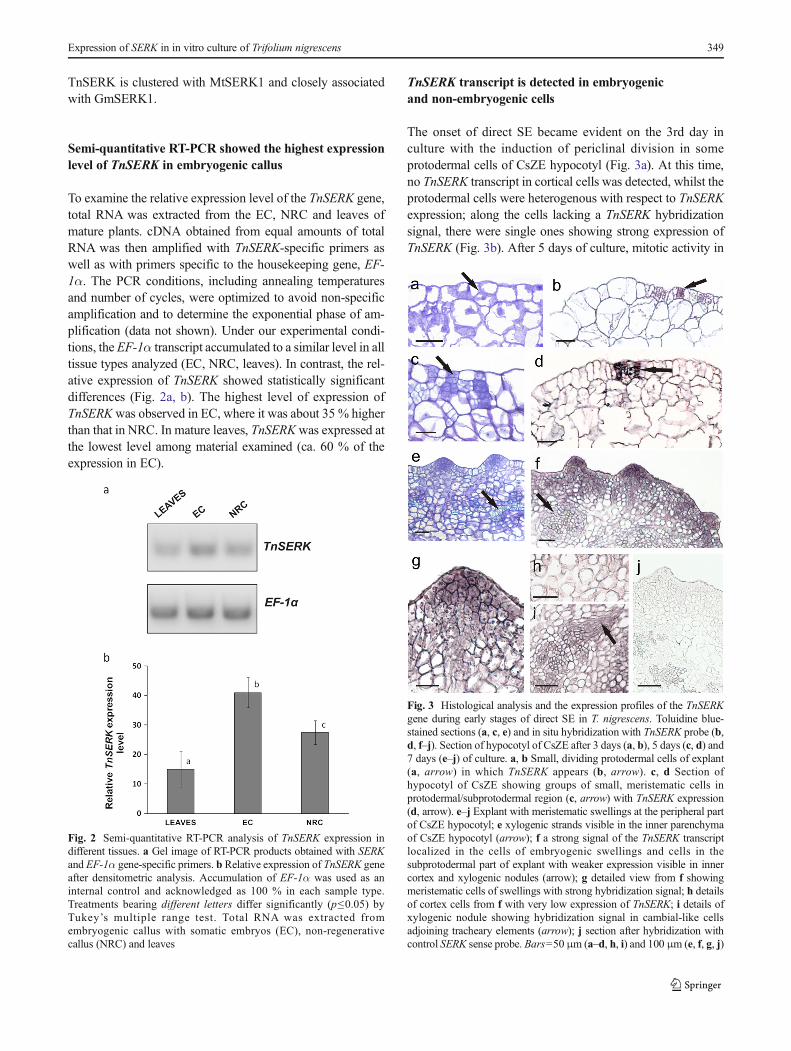
To examine the relative expression level of the TnSERK gene,total RNA was extracted from the EC, NRC and leaves ofmature plants. cDNA obtained from equal amounts of totalRNA was then amplified with TnSERK-specific primers aswell as with primers specific to the housekeeping gene, EF-1α. The PCR conditions, including annealing temperaturesand number of cycles, were optimized to avoid non-specificamplification and to determine the exponential phase of am-plification (data not shown). Under our experimental condi-tions, the EF-1α transcript accumulated to a similar level in alltissue types analyzed (EC, NRC, leaves). In contrast, the rel-ative expression of TnSERK showed statistically significantdifferences (Fig. 2a, b). The highest level of expression ofTnSERKwas observed in EC, where it was about 35 % higherthan that in NRC. In mature leaves, TnSERKwas expressed atthe lowest level among material examined (ca. 60 % of theexpression in EC).
TnSERK transcript is detected in embryogenicand non-embryogenic cells
The onset of direct SE became evident on the 3rd day inculture with the induction of periclinal division in someprotodermal cells of CsZE hypocotyl (Fig. 3a). At this time,no TnSERK transcript in cortical cells was detected, whilst theprotodermal cells were heterogenous with respect to TnSERKexpression; along the cells lacking a TnSERK hybridizationsignal, there were single ones showing strong expression ofTnSERK (Fig. 3b). After 5 days of culture, mitotic activity in
Fig. 2 Semi-quantitative RT-PCR analysis of TnSERK expression indifferent tissues. a Gel image of RT-PCR products obtained with SERKand EF-1α gene-specific primers. b Relative expression of TnSERK geneafter densitometric analysis. Accumulation of EF-1α was used as aninternal control and acknowledged as 100 % in each sample type.Treatments bearing different letters differ significantly (p≤0.05) byTukey’s multiple range test. Total RNA was extracted fromembryogenic callus with somatic embryos (EC), non-regenerativecallus (NRC) and leaves
Fig. 3 Histological analysis and the expression profiles of the TnSERKgene during early stages of direct SE in T. nigrescens. Toluidine blue-stained sections (a, c, e) and in situ hybridization with TnSERK probe (b,d, f–j). Section of hypocotyl of CsZE after 3 days (a, b), 5 days (c, d) and7 days (e–j) of culture. a, b Small, dividing protodermal cells of explant(a, arrow) in which TnSERK appears (b, arrow). c, d Section ofhypocotyl of CsZE showing groups of small, meristematic cells inprotodermal/subprotodermal region (c, arrow) with TnSERK expression(d, arrow). e–j Explant with meristematic swellings at the peripheral partof CsZE hypocotyl; e xylogenic strands visible in the inner parenchymaof CsZE hypocotyl (arrow); f a strong signal of the TnSERK transcriptlocalized in the cells of embryogenic swellings and cells in thesubprotodermal part of explant with weaker expression visible in innercortex and xylogenic nodules (arrow); g detailed view from f showingmeristematic cells of swellings with strong hybridization signal; h detailsof cortex cells from f with very low expression of TnSERK; i details ofxylogenic nodule showing hybridization signal in cambial-like cellsadjoining tracheary elements (arrow); j section after hybridization withcontrol SERK sense probe. Bars=50 μm (a–d, h, i) and 100 μm (e, f, g, j)
Expression of SERK in in vitro culture of Trifolium nigrescens 349

protodermal/subprotodermal region of hypocotyl gave rise tonumerous few-celled clusters consisting of small meristematiccells (Fig. 3c) with strong TnSERK expression (Fig. 3d). The-se cell clusters were maintained to day 7 followed by theformation of a few layers of meristematic tissue at the periph-ery of hypocotyl. Next, at the periphery of hypocotyl, numer-ous cone- or dome-shaped protrusions were produced(Fig. 3e). Histologically, the cells of these protrusions differedfrom subtending cells of a meristematic nature by having amore densely stained nucleus and a cytoplasm and a muchstronger signal of TnSERK expression. This was apparent inthe cytoplasm and nucleus and at the plasma membrane-cellwall interface (Fig. 3f, g). The hybridization signal was eithervery weak or not there in the non-dividing cells of the innercortex of the CsZE (Fig. 3h). Along with formation of meri-stematic protrusions at the periphery of the CsZE, somexylogenic nodules randomly distributed in inner regions ofthe explant were also produced (Fig. 3f). The expression ofTnSERK was not detected in the vascular core, but in theelongated cambial-like cells adjoining the tracheary elements(Fig. 3i). The use of a sense TnSERK riboprobe did not revealany unspecific signal throughout the explant (Fig. 3j).
Globular embryoids differentiated directly from theprotodermal/subprotodermal cells of meristematic protrusionsafter 10 days of culture. SEwas asynchronous, and embryos atdifferent stages of development were observed at this time ofculture. The embryoids were broadly attached to the mothertissue and covered by the epidermis of the initial explant(Fig. 4a). The embryoids developed according to zygotic em-bryogenesis and finally formed differentiated separate cotyle-donary primordia, distinct root and shoot meristems and pro-vascular strands passing to two cotyledons (Fig. 4a–c). ISHanalysis revealed that the TnSERKmRNAwas uniformly dis-tributed, and there were no differences in the strength of thehybridization signal between different cells of globular em-bryos (Fig. 4d). Further development of globular embryoswas accompanied by a differential expression pattern ofTnSERK, in which a more abundant accumulation of theTnSERK transcript was observed in cotyledons and at theshoot and root pole than in the ground tissues of the embryoaxis (Fig. 4e, f).
TnSERK is down-regulated during maturationof embryo-like structures
Aside from somatic embryos of typical morphology, in em-bryogenic cultures of CsZEs, embryo-like structures (ELSs)were also produced (Online Resource 3). Embryo-like struc-tures were initiated as elongated meristematic outgrowths, butwith age, their cells enlarged and the cytoplasm and nucleusbecame only faintly stained or even unstained with toluidineblue (Fig. 5a, b). Cells meristematic in appearance were some-times found at the tip of actively growing ELS, but shoot and
root meristems were never formed (Fig. 5b). ISH analysisconfirmed a strong signal of TnSERK expression, which wasuniformly distributed throughout early-staged ELS, whilst inmature ones, the hybridization signal was either not detectedor restricted to the group of meristematic cells at the tip of theregenerated structure (Fig. 5c, d). Mature ELSs were rod likein appearance; cotyledons were not produced. They did notdevelop into true organs or embryos, and with continued cul-ture, they became necrotic and decayed.
TnSERK is expressed in both embryogenicand non-regenerative callus
After 10 days of culture, direct SE was accompanied by localrupture of the CsZE epidermis and an outgrowth of callus onthe surface of the explant. On 15 day, the EC was composedof compact meristematic tissue interspersed with large paren-chymatous cells with the cytoplasm and nucleus undetectableby toluidine blue. Meristematic cells at the periphery of ECwere mostly organized into multi-celled clumps (Fig. 5e) fromwhich somatic embryos were induced (Fig. 5f). ISH analysisrevealed a strong signal of TnSERK expression in all the
Fig. 4 Histological analysis and the expression profiles of the TnSERKgene during late stages of SE in T. nigrescens. Toluidine blue-stainedsections (a–c) and in situ hybridization with TnSERK probe (d–f).Somatic embryo at globular (a, d), pre-torpedo (b), torpedo (e) andcotyledonary stage (c, f). d Globular somatic embryo with uniformexpression of TnSERK. e Intense hybridization signal visible in thecotyledon (arrow). f Mature embryo with high expression of TnSERKin cotyledons and root pool. Bars=100 μm (a–f)
350 M. Pilarska et al.
meristematic cells of EC, but the intensity of hybridizationwas the strongest in peripherally located ones. The TnSERKtranscript was confirmed either in the cytoplasm (meristematiccells of callus except of multi-celled clumps) or in the cyto-plasm and nucleus and at the cell wall-plasma membrane in-terface (multi-celled clumps) (Fig. 5g, h). Large, parenchyma-tous cells at the periphery of EC and, also, these located insidethe callus lacked any detectable TnSERK riboprobe hybridi-zation signal (Fig. 5g). The development of callus-derivedsomatic embryos and ELS as well as spatio-temporal patternof TnSERK expression within the embryo body during suc-cessive stages of differentiation were similar to those de-scribed above for direct SE (data not shown).
The formation of NRC was observed from CsZEs whichdid not display direct SE. Unlike EC, the NRC consistedmostly of large, loosely attached cells of a parenchymatousnature (Fig. 5i). The inner regions of callus contained smallgroups of relatively small cells with the cytoplasm somewhatmore darkly stained than that of adjacent parenchyma
(Fig. 5i). The hybridization signal was very weak or undetect-able in the non-dividing parenchymatous cells of NRC, whilstsmall cells, more meristematic in appearance, showed the ex-pression of TnSERK in both the cytoplasm and nucleus(Fig. 5j). Organogenesis or SE within NRC was not observed.
Within the time of culture, xylogenic nodules/strands wereproduced in both EC and NRC. The expression pattern ofTnSERK in callus-derived xylogenic nodules was similar tothat produced in the original explant during direct SE (Fig. 5j).When the sense TnSERK riboprobe was used, no signal wasdetected in any region of callus (Fig. 5k).
Discussion
TnSERK shows sequence similarity to other SERKproteins
In this study, for the first time, we have identified a sequencein the T. nigrescens genome that shows a similarity to genesencoding SERK proteins, e.g. MtSERK1. The members ofSERK family were recently found to be both structurally re-lated and involved in the regulation of plant responses to var-ious biotic and abiotic stimuli (Karlova et al. 2006; Santos andAragão 2009). Further analysis of the amino acid sequencededuced revealed a high identity of the cloned T. nigrescensfragment to known plant SERKs’ C-terminal coding region,including predominantly MtSERK1 (Nolan et al. 2003) andGmSERK1 (Yang et al. 2011). This result also suggests thatcorresponding regions of the putative TnSERK would play asimilar role. In particular, the protein kinase active-site signa-ture was identified in the putative TnSERK primary structure.The near 100 % identity of the corresponding region inTnSERK with the MtSERK1 indicates that this region in bothproteins serves the same function. Taken together, the similar-ity between TnSERK and MtSERK1 in terms of amino acidstructure suggests that TnSERK is an ortholog of the SERK1kinases present in other plant species. Phylogenetic analysisbased on predicted amino acid sequences also supports theortholog interpretation.
High expression of TnSERK accompanies induction of SE
Previously, we reported that the CsZEs of T. nigrescens canproduce somatic embryos by culturing on media containingdifferent combinations of auxins and cytokinins: 2,4-D andkinetin or NAA and 2iP (Konieczny et al. 2010). In this study,we used histological and ISH approaches to reveal the patternof TnSERK expression in CsZEs and CsZE-derived callusmaintained in the presence of NAA and 2iP. In agreement withan earlier report (Konieczny et al. 2010), the embryogenicresponse of CsZEs was efficient and initially confined to thehypocotyl of explants followed by the formation of hypocotyl-
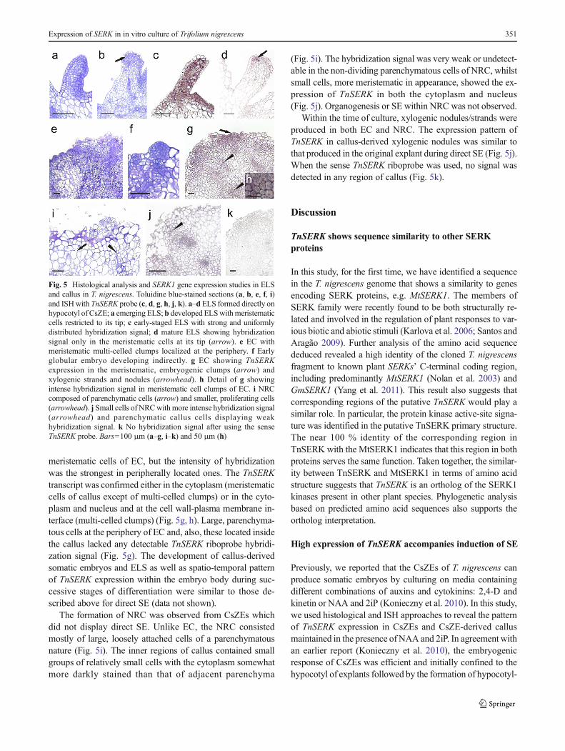
Fig. 5 Histological analysis and SERK1 gene expression studies in ELSand callus in T. nigrescens. Toluidine blue-stained sections (a, b, e, f, i)and ISHwith TnSERK probe (c, d, g, h, j, k). a–d ELS formed directly onhypocotyl of CsZE; a emerging ELS; b developed ELSwith meristematiccells restricted to its tip; c early-staged ELS with strong and uniformlydistributed hybridization signal; d mature ELS showing hybridizationsignal only in the meristematic cells at its tip (arrow). e EC withmeristematic multi-celled clumps localized at the periphery. f Earlyglobular embryo developing indirectly. g EC showing TnSERKexpression in the meristematic, embryogenic clumps (arrow) andxylogenic strands and nodules (arrowhead). h Detail of g showingintense hybridization signal in meristematic cell clumps of EC. i NRCcomposed of parenchymatic cells (arrow) and smaller, proliferating cells(arrowhead). j Small cells of NRCwith more intense hybridization signal(arrowhead) and parenchymatic callus cells displaying weakhybridization signal. k No hybridization signal after using the senseTnSERK probe. Bars=100 μm (a–g, i–k) and 50 μm (h)
Expression of SERK in in vitro culture of Trifolium nigrescens 351
derived embryogenic callus. In T. nigrescens, the cells fromwhich somatic embryos were differentiated displayed a num-ber of common features, such as small size, dense cytoplasmiccontent and large nuclei with prominent nucleoli, and alsostrong expression of TnSERK. This was also confirmed inthe SE of several dicots and monocots, e.g. D. glomerata(Somleva et al. 2000), A. thaliana (Salaj et al. 2008),M. truncatula (Nolan et al. 2009) or C. persicum (Savonaet al. 2012). In some species, like carrot (Schmidt et al.1997) or A. thaliana (Salaj et al. 2008), the expression ofSERK1 was found to mark the competence of single cells tofollow an embryogenic pathway. The results of our study arethe first to show the up-regulation of a gene from the SERKfamily in the initial cells of SE in clovers. However, the ex-pression of TnSERK in culture was not only related to theproduction of embryoids; cells showing accumulation of theTnSERK transcript were also involved in xylogenesis as wellas in the induction and proliferation of callus. A comparison ofhybridization signal intensity in cells differing inmorphogenicpotential revealed, however, that cells committed to SE weremore abundant in the TnSERK transcript than in the non-embryogenic cells of CsZE or callus. Thus, whilst the up-regulation of TnSERK seems to underlie different morphogen-ic events in T. nigrescens, the expression level of this gene isstrongly developmentally regulated and possibly confers spe-cific morphogenic competence of a particular cell.
Differential pattern of TnSERK expression accompaniesdevelopment of embryoids and embryo-like structures
Once induced, the expression of TnSERK continued through-out SE. As in somatic embryos ofM. truncatula (Nolan et al.2009), all cells of embryoids in T. nigrescens expressed SERK.However, with embryoid maturation, the pattern of gene ex-pression changed from uniform in globular embryos to tissue-specific in cotyledonary-staged ones. In these advanced em-bryoids, the TnSERK was preferentially up-regulated in mer-istematic cells in shoot and root poles, which supports thenotion of the involvement of SERK in defining and maintain-ing apical meristems in developing somatic embryos (Savonaet al. 2012). Indeed, the ELSs which were regularly induced inthe culture of T. nigrescens did not produce either shoot or rootmeristems in mature form and most of their cells lacked de-tectable TnSERK mRNA. Although a zone of meristematiccells showing a weak signal of TnSERK riboprobe hybridiza-tion occurred at the tip of some developing ELSs, this seemsto be the result of unorganized cell proliferation rather than astep towards organized structure.
In our experimental system, both ELSs and normal zygotic-like embryoids were initiated from small, densely stained nu-cleus and rich in TnSERK transcript cells. However, unlikezygotic-like SE, abnormal development and eventual ELS for-mation were associated with a decline in TnSERK expression,
and this was accompanied by the parenchymatization ofground tissue in the developing structure. In the literature, thereare only sparse data on the expression of SERK genes in ELSs.In a culture of mangosteen, there were globular structureswhich did not develop into true somatic embryos (Rohaniet al. 2012). These were identified as embryoids based on thepresence of the SERK1 transcript, but the expression of thisgene was not monitored beyond the globular stage. InT. nigrescens, the occurrence of morphological and anatomicalabnormalities in somatic embryos has already been reportedand ascribed to a disturbance of polar auxin transport (PAT)in an early stage of development, when cotyledon primordiaand embryo polarity are induced (Konieczny et al. 2012). Inthat study, ELSs were induced in the presence of auxinic her-bicide, 2,4-D, and they shared several features, such as an elon-gated embryo axis, excessive parenchymatization and a lack ofapical meristems, like the ELSs observed in the currentexperiments. The relationships between SERKs and PAT areonly poorly understood. Du et al. (2012) revealed that aserk1bak1bkk1 triple mutant of A. thaliana displayed drastical-ly reduced expression of a number of the genes critical to PAT,cell cycle maintenance and meristem differentiation. On theother hand, different auxins were shown to up-regulate variousSERKs (Nolan et al. 2003; Ge et al. 2010; Zhang et al. 2011). Inour experimental system, the occurrence of morphological ab-errations in SE preceded the decline in SERK expression, as thecells of early-stage ELSs displayed a very strong signal ofriboprobe hybridization, typical for embryonic cells. The ques-tion as to whether the down-regulation of TnSERK over thefurther development of ELSs was related or not to the distur-bance of PAT still needs to be elucidated.
Expression of TnSERK meets initiation of differentdevelopmental pathways
After 10 days of culture, the CsZEs of T. nigrescens started toproduce callus, which either regenerated somatic embryos orremained non-regenerative throughout the culture. The ex-pression level of TnSERK differed according to the regenera-tive potential of tissue, and it was revealed by RT-PCR to besignificantly higher in EC than in NRC. This is in line withstudies on several species (e.g. Pérez-Núňez et al. 2009; Nolanet al. 2009; Ma et al. 2012) and adds further evidence for theinvolvement of SERK in SE in T. nigrescens. ISH analysesshowed that aside from being expressed in embryogenic cells,TnSERK mRNA was also observed in populations of non-embryogenic but actively dividing cells of both EC andNRC. In contrast, the hybridization signal was either not de-tected or was very weak in the large and non-dividing paren-chymatous cells of calluses. The dividing cells were onlysparse in NRC, whilst they constituted the majority of ECwhich partly accounts for differences between EC and NRCin RT-PCR results. A positive correlation between the
352 M. Pilarska et al.

expression of SERK and mitotic activity was also reported inthe callus of coconut (Pérez-Núňez et al. 2009) andC. persicum (Savona et al. 2012). Nolan et al. (2009) observedinM. truncatula that up-regulation of SERK1 was not limitedto dividing cells, but that it also occurred in those cells whichbecame competent to divide. It was recently shown thatcallogenesis at its initial steps recapitulates the lateral rootdevelopment program, and therefore, it can be interpreted asa transdifferentiation of specific somatic cell in the explant(Opatrny 2014). In a culture of T. nigrescens, the first celldivisions leading to morphogenesis occurred exclusively inSERK-expressing cells of the CsZE protoderm, and these di-visions were induced de novo, as no dividing cells were foundin the hypocotyl of CsZE before explantation (Koniecznyet al. 2012). The expression of this gene continued in resultingmass of small actively dividing cells, but this ceased as thecell’s fate became established, e.g. in the course of the differ-entiation of parenchyma in developing ELSs and callus orduring xylem element maturation. These observations indicatea close relationship between the level of TnSERK expressionand the transdifferentiation/differentiation processes.
It is believed that under appropriate signal(s), plant cellscan follow differentiation and transdifferentiation or acquiretotipotency whichmanifests itself in the production of somaticembryos (Verdeil et al. 2007; Grafi et al. 2011). InT. nigrescens, cells expressing SERK were involved in differ-ent morphogenic events, but the cells of the embryogenic lineshowed a much stronger signal of TnSERK riboprobe hybrid-ization than those cells associated with other developmentalpathways, such as callus proliferation or xylogenesis. Thiswas also observed in a culture of C. persicumwhere the initialcells for SE were said to result from pluripotentiality beingmaintained over time in some cells of microcallus (Savonaet al. 2012). In regenerative explants of T. nigrescens, differentmorphogenic processes occurred in separate locations; SEwasconfined to the outermost cells of meristematic protrusions,whilst the production of meristematic but non-embryogeniccells occurred regularly in inner regions of explant/callus. Thissuggests a possible effect of positional information on expres-sion level of TnSERK and, thereby, on cell fate. The signal(s)that would direct the peripheral and inner cells of CsZE andcallus to different developmental fates remains unknown.Thorpe (1980) suggested that differences in morphogenic ca-pacity between the inner and outer regions of the explant mayreflect differences in the physiological gradient of the sub-stances, e.g. of growth regulators from the medium into thetissues. On the other hand, it has also been pointed out that theability of a cell to express totipotency and follow SE may berelated to the removal of the embryogenesis-repressive and/ordifferentiation-inductive effects of neighbouring cells and tis-sues (Williams and Maheswaran 1986). Superficially locatedcells have different surroundings than those inside tissue asthey are also in contact with the external environment which
potentially reduces the capacity for inter-cell communicationwith adjacent tissues (Kurczynska et al. 2012).
In summary, the TnSERK gene from T. nigrescens waspartly cloned and its expression pattern was monitored inin vitro cultured embryogenic and non-regenerative tissues.The down-regulation of TnSERK during cell specialization(the maturation of parenchyma and xylem elements) and itsup-regulation in the actively dividing cells involved in differ-ent morphogenic processes (SE, callus production,xylogenesis, formation of apical meristems in developing em-bryoids) suggest broad role for it in plant development, pos-sibly related to differentiation and transdifferentiation process-es as well as induction and maintenance of totipotent state incell(s). Unfortunately, T. nigrescens has not been the subject ofgenomic mapping or sequencing efforts so far. Consequently,the structure of T. nigrescens genome is unknown, which, inturn, hampers the application of molecular approaches forstudying the gene function. However, an increasing interestin this species in food, agricultural and pharmaceutical indus-tries (see: Konieczny et al. 2010, 2012; Demirkiran et al.2013) allows for envision that the sequencing of its genomeas well as the development of transformation techniques willbe initiated in the nearest future.
Acknowledgments The Institute of Genetics and Crop Plant Research(Gatersleben, Germany) is kindly acknowledged for providingT. nigrescens seeds. Financial support by The National Scholarship Pro-gram of the Slovak Republic through the scholarship to MP and RK isgratefully acknowledged.
Conflict of interest The authors declare that they have no conflict ofinterest.
Open Access This article is distributed under the terms of the CreativeCommons At t r ibut ion 4 .0 In te rna t ional License (h t tp : / /creativecommons.org/licenses/by/4.0/), which permits unrestricted use,distribution, and reproduction in any medium, provided you giveappropriate credit to the original author(s) and the source, provide a linkto the Creative Commons license, and indicate if changes were made.
References
Albertini E, Marconi G, Reale L, Barcaccia G, Porceddu A, Ferranti F,Falcinelli M (2005) SERK and APOSTART. Candidate genes forapomixis in Poa pratensis. Plant Physiol 138:2185–2199
Albrecht C, Russinova E, Hecht V, Baaijens E, de Vries S (2005) TheArabidopsis thaliana SOMATIC EMBRYOGENESIS RECEPTOR-LIKE KINASES1 and 2 control male sporogenesis. Plant Cell 17:3337–3349
Albrecht C, Russinova E, Kemmerling B, Kwaaitaal M, de Vries SC(2008) Arabidopsis SOMATIC EMBRYOGENESIS RECEPTORKINASE proteins serve brassinosteroid-dependent and-independentsignaling pathways. Plant Physiol 148:611–619
Ausubel F, Brent R,Kingston RE,MooreDD, Seidman JG, Smith JA, StruhlK (1995) Short protocols in molecular biology. Wiley, New York
Expression of SERK in in vitro culture of Trifolium nigrescens 353

Chenna R, Sugawara H, Koike T, Lopez R, Gibson TJ, Higgins DG,Thompson JD (2003) Multiple sequence alignment with theClustal series of programs. Nucleic Acids Res 31:3497–3500
de Castro E, Sigrist CJA, Gattiker A, Bulliard V, Langendijk-GenevauxPS, Gasteiger E, Bairoch A,Hulo N (2006) ScanProsite: detection ofPROSITE signature matches and ProRule-associated functional andstructural residues in proteins. Nucleic Acids Res 34:W362–W365
de Oliveira SantosM, Romano E, Yotoko KSC, Tinoco MLP, Dias BBA,Aragao FJL (2005) Characterisation of the cacao somatic embryo-genesis receptor-like kinase (SERK) gene expressed during somaticembryogenesis. Plant Sci 168(3):723–729
Demirkiran O, Sabudak T, Ozturk M, Topcu G (2013) Antioxidant andtyrosinase inhibitory activities of flavonoids from Trifoliumnigrescens subsp. petrisavi. J Food Chem 61:12598–12603
Dereeper A, Guignon V, Blanc G, Audic S, Buffet S, Chevenet F,Dufayard JF, Guindon S, Lefort V, Lescot M, Claverie JM,Gascuel O (2008) Phylogeny.fr: robust phylogenetic analysis forthe non-specialist. Nucleic Acids Res 36:W465–W469
Du J, Yin H, Zhang S,Wei Z, Zhao B, Zhang J, Gou X, Lin H, Lin H, Li J(2012) Somatic embryogenesis receptor kinases control root devel-opment mainly via brassinosteroid-independent actions inArabidopsis thaliana. J Integr Plant Biol 54:388–399
Elhiti M, Stasolla C, Wang A (2013) Molecular regulation of plant so-matic embryogenesis. In Vitro Cell Dev Biol Plant 49:631–642
Ge XX, Gai EF, Chai LJ, Guo WW (2010) Cloning, molecular charac-t e r i z a t i on and exp re s s i on ana l y s i s o f a SOMATICEMBRYOGENESIS RECEPTOR-LIKE KINASE gene (CitSERK1-like) in Valencia sweet orange. Acta Physiol Plant 32:1197–1207
GouX, YinH, HeK,Du J, Yi J, Xu S, Lin H, Clouse S, Li J (2012) Geneticevidence for an indispensable role of somatic embryogenesis receptorkinases in brassinosteroid signaling. PLoS Genet 8, e1002452
Grafi G, Chalifa-Caspi V, Nagar T, Plaschkes I, Barak S, Ransbotyn V(2011) Plant response to stress meets dedifferentiation. Planta 233:433–438
Hecht V, Vielle-Calzada J, Hartog M, Schmidt E, Grossniklaus U, deVries S (2001) The Arabidopsis somatic embryogenesis receptorkinase 1 gene is expressed in developing ovules and embryos andenhances embryogenic competence in culture. Plant Physiol 127:803–816
Hoveland CS, Evers GW (1995) Arrowleaf, crimson and other annualclovers. In: Barnes RF, Miller DA, Nelson CJ (eds) Forages, vol 1,5th edn, An introduction to grassland agriculture. Iowa StateUniversity Press, Ames, pp 249–260
Huang X, Lu XY, Zhao JT, Chen JK, Dai XM, Xiao W, Chen YP, ChenYF, Huang XL (2009) MaSERK1 gene expression associated withsomatic embryogenic competence and disease resistance response inbanana (Musa spp.). Plant Mol Biol Report 28:309–316
Ikeda M, Umehara M, Kamada H (2006) Embryogenesis-related genes;its expression and roles during somatic and zygotic embryogenesisin carrot and Arabidopsis. Plant Biotechnol J 23:153–161
Karlova R, Boeren S, Russinova E, Aker J, Vervoort J, de Vries S (2006)The Arabidopsis SOMATIC EMBRYOGENESIS RECEPTOR-LIKE KINASE1 protein complex includes BRASSINOSTEROID-INSENSITIVE1. Plant Cell 18:626–638
Konieczny R, Pilarska M, Tuleja M, Salaj T, Ilnicki T (2010) Somaticembryogenesis and plant regeneration in zygotic embryos ofTrifolium nigrescens (Viv.). Plant Cell Tissue Org Cult 100:123–130
Konieczny R, Sliwinska E, Tuleja M, Pilarska M (2012)Morphohistological and flow cytometric analyses of somatic em-bryogenesis in Trifolium nigrescens Viv. Plant Cell Tissue OrgCult 109:131–141
Kurczynska EU, Potocka I, Dobrowolska I, Kulinska-Lukaszek K, SalaK, Wrobel J (2012) Cellular markers for somatic embryogenesis. In:Sato KI (ed) Embryogenesis. InTech, ISBN: 978-953-51-0466-7.doi: 10.5772/37617. Available from: http://www.intechopen.com/books/embryogenesis/cellular-markers-for-somatic-embryogenesis
Ma J, He Y, Wu C, Liu H, Hu Z, Sun G (2012) Cloning and molecularcharacterization of a SERK gene transcriptionally induced duringsomatic embryogenesis in Ananas comosus cv. Shenwan. PlantMol Biol Report 30:195–203
Marshall AH, Michaelson-Yeates TPT, Abberton MT, Williams A,Powell HG (2002) Variation for reproductive and agronomic traitsamong T. repens×T. nigrescens third generation backcross hybridsin the field. Euphytica 126:195–201
Marshall AH, Michaelson-Yeates TPT, Abberton MT (2008)Introgression of reproductive traits from Trifolium nigrescens in-creases the seed yield of white clover (T. repens). Plant Breed 12:597–601
Murashige T, Skoog F (1962) A revised medium for rapid growth andbioassay with tobacco tissue cultures. Physiol Plant 15:473–497
Nolan KE, Irwanto RR, Rose RJ (2003) Auxin up-regulates MtSERK1expression in both Medicago truncatula root-forming and embryo-genic cultures. Plant Physiol 133:218–230
Nolan KE, Kurdyukov S, Rose RJ (2009) Expression of the SOMATICEMBRYOGENESIS RECEPTOR-LIKE KINASE1 (SERK1) gene isassociated with developmental change in the life cycle of the modellegume Medicago truncatula. J Exp Bot 60:1759–1771
Pederson GA,Windham GL (1989) Resistance toMeloidogyne incognitain Trifolium interspecific hybrids and species related to white clover.Plant Dis 73:567–569
Pérez-Núňez MT, Souza R, Sáenz L, Chan JL, Zúňiga-Aguilar JJ,Oropeza C (2009) Detection of a SERK-like gene in coconut andanalysis of its expression during the formation of embryogenic cal-lus and somatic embryos. Plant Cell Rep 28:11–19
Pilarska M, Knox JP, Konieczny R (2013) Arabinogalactan-protein andpectin epitopes in relation to an extracellular matrix surface networkand somatic embryogenesis and callogenesis in Trifolium nigrescensViv. Plant Cell Tissue Org Cult 115:35–44
Rohani ER, Ismanizan I, Noor NM (2012) Somatic embryogenesis ofmangosteen. Plant Cell Tissue Org Cult 110:251–259
Salaj J, von Recklinghausen IR, Hecht V, de Vries SC, Schela JHN, vanLammeren AAM (2008) AtSERK1 expression precedes and coin-cides with early somatic embryogenesis in Arabidopsis thaliana.Plant Physiol Biochem 46:709–714
Santa-Catarina C, Hanai LR, Dornelas MC, Viana AM, Floh EIS (2004)SERK gene homolog expression, polyamines and amino acids asso-ciated with somatic embryogenic competence of Ocoteacatharinensis Mez. (Lauraceae). Plant Cell Tissue Org Cult 79:53–61
Santos MO, Aragão FJL (2009) Role of SERK genes in plant environ-mental response. Plant Signal Behav 4:1111–1113
Santos MO, Romano E, Vieira LS, Baldoni AB, Aragão FJL (2009)Suppression of SERK gene expression affects fungus tolerance andsomatic embryogenesis in transgenic lettuce. Plant Biol 11:83–89
Savona M, Mattioli R, Nigro S, Falasca G, Della Rovere F, Costantino P,de Vreis SC, Ruffoni B, Trovato M, Altamura MM (2012) TwoSERK genes are markers of pluripotency in Cyclamen persicumMill. J Exp Bot 63:471–488
Schaffer AA, Aravind L, Madden TL, Shavirin S, Spouge JL, Wolf YI,Koonin EV, Altschul SF (2001) Improving the accuracy of PSI-BLAST protein database searches with composition-based statisticsand other refinements. Nucleic Acids Res 29:2994–3005
Schellenbaum P, Jacques A, Maillot P, Bertsch C, Mazet F, Farine S,Walter B (2008) Characterization of VvSERK1, VvSERK2,VvSERK3 and VvL1L genes and their expression during somaticembryogenesis of grapevine (Vitis vinifera L.). Plant Cell Rep 27:1799–1809
Schmidt E, Guzzo F, Toonen M, de Vries S (1997) A leucine-rich repeatcontaining receptor-like kinase marks somatic cells competent toform embryos. Development 124:2049–2062
Serazin-Leroy V, Denis-Henriot D, Morot M, deMazancourt P, GiudicelliY (1998) Semi-quantitative RT-PCR for comparison of mRNAs in
354 M. Pilarska et al.

cells with different amounts of housekeeping gene transcripts. MolCell Probes 12:283–291
Somleva MN, Schmidt EDL, de Vries SC (2000) Embryogenic cells inDactylis glomerata L. (Poaceae) explants identified by cell trackingand by SERK expression. Plant Cell Rep 19:718–726
Thomas C, Meyer D, Himber C, Steinmetz A (2004) Spatial expressionof a sunflower SERK gene during induction of somatic embryogen-esis and shoot organogenesis. Plant Physiol Biochem 42:35–42
Thorpe TA (1980) Organogenesis in vitro: structural, physiological andbiochemical aspects. Int Rev Cytol Suppl 112A:71–111
Verdeil JL, Alemanno L, Niemenak N, Tranbarger TJ (2007) Pluripotentversus totipotent plant stem cells: dependence versus autonomy?Trends Plant Sci 12(6):245–252
Williams EG, Maheswaran G (1986) Somatic embryogenesis: factorsinfluencing coordinated behaviour of cells as an embryogenicgroup. Ann Bot 57(4):443–462
Yang C, Zhao TJ, Yu DY, Gai JY (2011) Isolation and functional charac-terization of a SERK gene from soybean (Glycine max (L.) Merr.).Plant Mol Biol Report 29:334–344
Zhang SZ, Liu XG, Lin YA, Xie GN, Fu FL, Liu HL, Wang J, Gao SB,Lan H, Rong TZ (2011) Characterization of a ZmSERK gene and itsrelationship to somatic embryogenesis in a maize culture. Plant CellTissue Org Cult 105:29–37
Expression of SERK in in vitro culture of Trifolium nigrescens 355