high-efficiency triple-helix-mediated photo-cross-linking at a targeted site within a selectable...
TRANSCRIPT

High-Efficiency Triple-Helix-Mediated Photo-Cross-Linking at a Targeted Sitewithin a Selectable Mammalian Gene†
Karen M. Vasquez,‡ Theodore G. Wensel,‡ Michael E. Hogan,§ and John H. Wilson*,‡
Verna and Marrs McLean Department of Biochemistry and Department of Molecular Physiology and Biophysics,Baylor College of Medicine, One Baylor Plaza, Houston, Texas 77030
ReceiVed April 12, 1996; ReVised Manuscript ReceiVed June 17, 1996X
ABSTRACT: Targeting damage to specific sites in the genome represents an attractive approach tomanipulating gene function in mammalian cells. To test the applicability of triple-helix formation as ameans for achieving precisely timed site-specific damage within a mammalian gene, a triplex-formingoligodeoxyribonucleotide (TFO) that binds with high affinity to a specific site within the hamster adeninephosphoribosyltransferase (APRT) gene was modified with the photochemically reactive psoralen derivative4′-(hydroxymethyl)-4,5′,8-trimethylpsoralen (HMT). The modified TFO, psorTFO1, bound with highaffinity to a target site within intron 1 of the APRT gene. Upon irradiation, photomonoadducts (i.e.,covalent adducts of psorTFO1 to one strand of the target duplex) were formed with high efficiency (∼50%).Introduction of 5′-TpA sequences (the preferred site for psoralen-induced photo-cross-links) at or nearthe triplex junction leads to increased efficiency of total photoadduct formation and to efficient formationof products that had the electrophoretic mobility on denaturing PAGE expected for three-stranded photo-cross-links (i.e., products containing psorTFO1 covalently linked to both strands of the duplex). Theiridentities as cross-links were verified by (1) identical electrophoretic mobility of products formed witheither duplex strand radiolabeled and (2) coprecipitation of the radiolabeled duplex strand with itscomplementary biotinylated strand following denaturation. In addition, the cross-links were completelyreversible upon irradiation at 254 nm, as expected for psoralen-mediated cross-links. The yield anddistribution of photoadducts depended on 5′-TpA position. The most efficient photoadduct formation(∼90%) and photo-cross-link formation (∼90% of total photoadducts) were observed for a 5′-TpA adjacentto the triplex junction, with significant, but lower, cross-linking efficiency within three base pairs of thejunction. Molecular models of the psoralen-conjugated triplex with its six-carbon linker suggested a simpleexplanation for this distance dependence: facile intercalation near the triplex/duplex junction, withincreasing strain required for intercalation at more distant sites. These results indicate that psorTFO1allows for DNA damage with high precision and high efficiency, and the likely proportion of monoadductsand cross-links can be estimated from the target sequence.
Damage to the mammalian genome can dramaticallyincrease the rates of recombination and mutation (Friedberget al., 1995). Targeting such damage to specific sites hasthe potential for controlled gene manipulation, allowingtargeted replacement, repair, mutation, or inactivation ofspecific genes. A promising approach to site-selective DNAdamage is the use of modified triplex-forming oligonucle-otides (TFOs)1 designed to bind to unique sites on double-stranded DNA within a mammalian genome. Induction ofintracellular triplex formation by extracellular application ofTFOs has been successfully used to suppress transcriptionin cells (Postel et al., 1991; Orson et al., 1991; McShan et
al., 1992; Grigoriev et al., 1993; Ing et al., 1993; Okada etal., 1994; Roy, 1994; Scaggiante et al., 1994; Tu et al., 1995;Kovacs et al., 1996), to inhibit replication (Birg et al., 1990),and to induce site-specific mutations in an extrachromosomalvector transfected into mammalian cells (Wang et al., 1995,1996).
Of the several classes of DNA-DNA triplexes, thoseformed by GT-rich TFOs (Cooney et al., 1988; Hogan etal., 1989; Beal & Dervan, 1991) bind in the major grooveof the underlying duplex in an antiparallel fashion througha reverse Hoogsteen hydrogen-bonding motif and form stablethree-stranded DNA complexes at physiological pH. Theaffinities with which such triplexes form can be quite high;Kd values< 10-9 M have been observed, and the meanbound lifetimes for these triplexes can be many hours(Vasquez et al., 1995). In addition, TFOs with a 3′-propanolamine modification, have been shown to be stablein ViVo for many hours (Zendegui et al., 1992).Chemical and photochemical DNA damaging agents have
been used extensively to study the effects of random DNAdamage. Combining the site specificity conferred by triplexformation with temporal control through use of photoacti-vatable DNA damaging agents attached to TFOs has thepotential to optimize site-specific DNA damage without
† This work was funded by grants from the NIH to J.H.W.(GM38219) and M.E.H. (A132804), a grant from the Welch Foundationto T.G.W. (Q1179), a grant to J.H.W. and T.G.W. from the TexasAdvanced Technology Program (004949-083), and by Aronex Phar-maceutical Corp.* Address correspondence to this author. Phone: (713) 798-5760.
FAX: (713) 795-5487. email: [email protected].‡ Verna and Marrs McLean Department of Biochemistry.§ Department of Molecular Physiology and Biophysics.X Abstract published inAdVance ACS Abstracts,August 1, 1996.1 Abbreviations: APRT, adenine phosphoribosyltransferase; CHO,
Chinese hamster ovary; TFO, triplex-forming oligodeoxyribonucleotide;HMT, 4′-(hydroxymethyl)-4,5′,8-trimethylpsoralen; psorTFO1, theconjugate of HMT with the specific TFO targeted to APRT intron 1.
10712 Biochemistry1996,35, 10712-10719
S0006-2960(96)00881-1 CCC: $12.00 © 1996 American Chemical Society
+ +
+ +
detriment to the rest of the genome. Triplex-mediated DNAdamage has been usedin Vitro to induce site-specific DNAnicking, cleavage, and cross-linking (Moser & Dervan, 1987;Perrouault et al., 1990; Strobel et al., 1991; Takasugi et al.,1991).The most widely used class of photoactivatable DNA
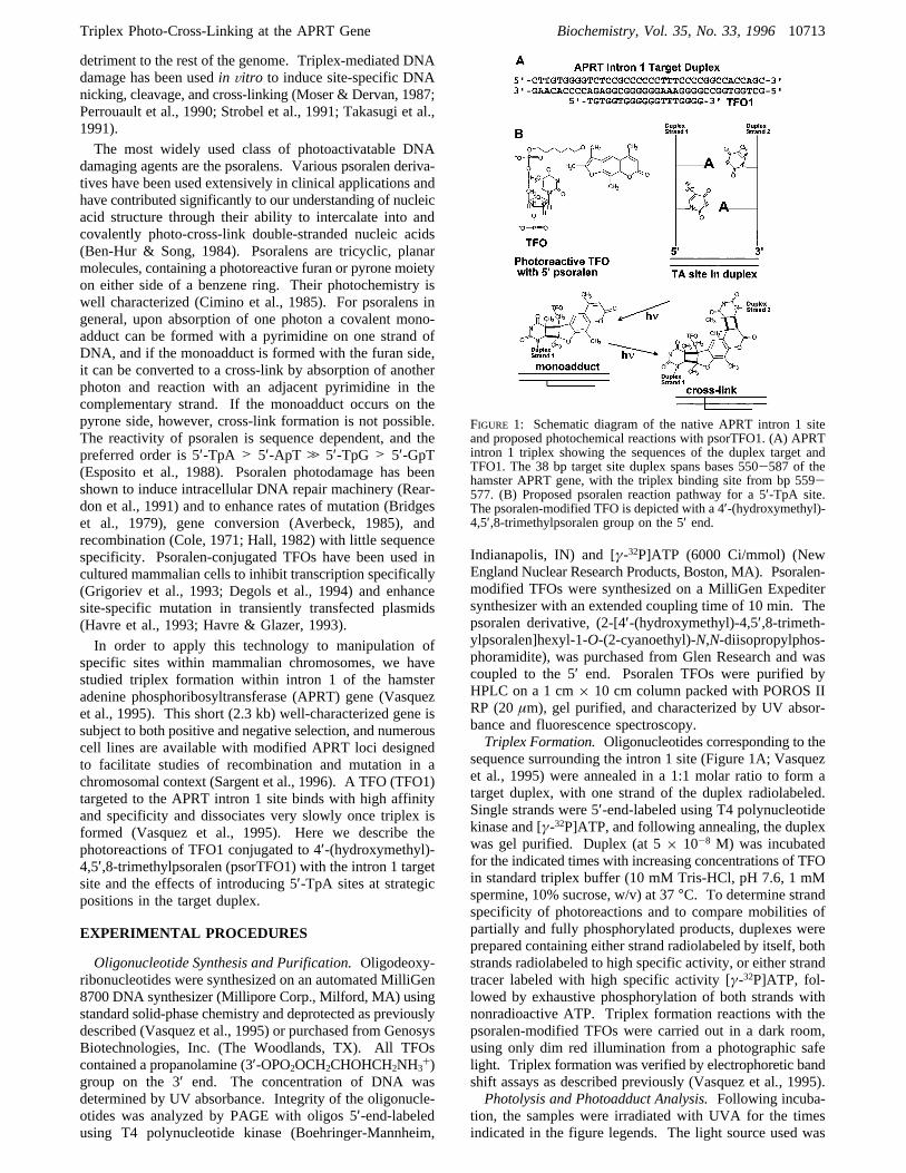
damaging agents are the psoralens. Various psoralen deriva-tives have been used extensively in clinical applications andhave contributed significantly to our understanding of nucleicacid structure through their ability to intercalate into andcovalently photo-cross-link double-stranded nucleic acids(Ben-Hur & Song, 1984). Psoralens are tricyclic, planarmolecules, containing a photoreactive furan or pyrone moietyon either side of a benzene ring. Their photochemistry iswell characterized (Cimino et al., 1985). For psoralens ingeneral, upon absorption of one photon a covalent mono-adduct can be formed with a pyrimidine on one strand ofDNA, and if the monoadduct is formed with the furan side,it can be converted to a cross-link by absorption of anotherphoton and reaction with an adjacent pyrimidine in thecomplementary strand. If the monoadduct occurs on thepyrone side, however, cross-link formation is not possible.The reactivity of psoralen is sequence dependent, and thepreferred order is 5′-TpA > 5′-ApT . 5′-TpG > 5′-GpT(Esposito et al., 1988). Psoralen photodamage has beenshown to induce intracellular DNA repair machinery (Rear-don et al., 1991) and to enhance rates of mutation (Bridgeset al., 1979), gene conversion (Averbeck, 1985), andrecombination (Cole, 1971; Hall, 1982) with little sequencespecificity. Psoralen-conjugated TFOs have been used incultured mammalian cells to inhibit transcription specifically(Grigoriev et al., 1993; Degols et al., 1994) and enhancesite-specific mutation in transiently transfected plasmids(Havre et al., 1993; Havre & Glazer, 1993).In order to apply this technology to manipulation of
specific sites within mammalian chromosomes, we havestudied triplex formation within intron 1 of the hamsteradenine phosphoribosyltransferase (APRT) gene (Vasquezet al., 1995). This short (2.3 kb) well-characterized gene issubject to both positive and negative selection, and numerouscell lines are available with modified APRT loci designedto facilitate studies of recombination and mutation in achromosomal context (Sargent et al., 1996). A TFO (TFO1)targeted to the APRT intron 1 site binds with high affinityand specificity and dissociates very slowly once triplex isformed (Vasquez et al., 1995). Here we describe thephotoreactions of TFO1 conjugated to 4′-(hydroxymethyl)-4,5′,8-trimethylpsoralen (psorTFO1) with the intron 1 targetsite and the effects of introducing 5′-TpA sites at strategicpositions in the target duplex.
EXPERIMENTAL PROCEDURES
Oligonucleotide Synthesis and Purification.Oligodeoxy-ribonucleotides were synthesized on an automated MilliGen8700 DNA synthesizer (Millipore Corp., Milford, MA) usingstandard solid-phase chemistry and deprotected as previouslydescribed (Vasquez et al., 1995) or purchased from GenosysBiotechnologies, Inc. (The Woodlands, TX). All TFOscontained a propanolamine (3′-OPO2OCH2CHOHCH2NH3
+)group on the 3′ end. The concentration of DNA wasdetermined by UV absorbance. Integrity of the oligonucle-otides was analyzed by PAGE with oligos 5′-end-labeledusing T4 polynucleotide kinase (Boehringer-Mannheim,
Indianapolis, IN) and [γ-32P]ATP (6000 Ci/mmol) (NewEngland Nuclear Research Products, Boston, MA). Psoralen-modified TFOs were synthesized on a MilliGen Expeditersynthesizer with an extended coupling time of 10 min. Thepsoralen derivative, (2-[4′-(hydroxymethyl)-4,5′,8-trimeth-ylpsoralen]hexyl-1-O-(2-cyanoethyl)-N,N-diisopropylphos-phoramidite), was purchased from Glen Research and wascoupled to the 5′ end. Psoralen TFOs were purified byHPLC on a 1 cm× 10 cm column packed with POROS IIRP (20µm), gel purified, and characterized by UV absor-bance and fluorescence spectroscopy.Triplex Formation.Oligonucleotides corresponding to the
sequence surrounding the intron 1 site (Figure 1A; Vasquezet al., 1995) were annealed in a 1:1 molar ratio to form atarget duplex, with one strand of the duplex radiolabeled.Single strands were 5′-end-labeled using T4 polynucleotidekinase and [γ-32P]ATP, and following annealing, the duplexwas gel purified. Duplex (at 5× 10⁻⁸ M) was incubatedfor the indicated times with increasing concentrations of TFOin standard triplex buffer (10 mM Tris-HCl, pH 7.6, 1 mMspermine, 10% sucrose, w/v) at 37°C. To determine strandspecificity of photoreactions and to compare mobilities ofpartially and fully phosphorylated products, duplexes wereprepared containing either strand radiolabeled by itself, bothstrands radiolabeled to high specific activity, or either strandtracer labeled with high specific activity [γ-32P]ATP, fol-lowed by exhaustive phosphorylation of both strands withnonradioactive ATP. Triplex formation reactions with thepsoralen-modified TFOs were carried out in a dark room,using only dim red illumination from a photographic safelight. Triplex formation was verified by electrophoretic bandshift assays as described previously (Vasquez et al., 1995).Photolysis and Photoadduct Analysis.Following incuba-
tion, the samples were irradiated with UVA for the timesindicated in the figure legends. The light source used was
FIGURE 1: Schematic diagram of the native APRT intron 1 siteand proposed photochemical reactions with psorTFO1. (A) APRTintron 1 triplex showing the sequences of the duplex target andTFO1. The 38 bp target site duplex spans bases 550-587 of thehamster APRT gene, with the triplex binding site from bp 559-577. (B) Proposed psoralen reaction pathway for a 5′-TpA site.The psoralen-modified TFO is depicted with a 4′-(hydroxymethyl)-4,5′,8-trimethylpsoralen group on the 5′ end.
Triplex Photo-Cross-Linking at the APRT Gene Biochemistry, Vol. 35, No. 33, 199610713
+ +
+ +

a high-intensity (150 W, Oriel) xenon/mercury arc lamp (∼12J/cm2 per minute of irradiation, based on the manufacturer’sspecifications) focused on the sample, using a NaNO3
solution filter to eliminate infrared. For the irradiation dosecorresponding to a time point of 0.6 s (0.12 J/cm2), sampleswere irradiated for 6 s using a 1.0 OD neutral density filterin conjunction with the NaNO3 filter. After irradiation,reaction products were denatured at 97°C in 50% (v/v)formamide and analyzed by denaturing PAGE. Electro-phoresis was through a 7 M urea-15% polyacrylamide gelcontaining 89 mM Tris, 89 mM boric acid, pH 8.0, and 2mM EDTA (TBE, unless otherwise noted). Gels were runfor 1-2 h at 60 W at 55°C, dried, and exposed to film forautoradiography. For quantitation, radioactivity was mea-sured using a Betagen Beta Scope 603 blot analyzer.Photoadduct yields for each duplex varied somewhat as afunction of lamp configurations and age of psorTFO1 stocks;under Results, both the yields obtained in the experimentsshown and the maximum yields obtained are given.PhotoreVersibility of Cross-Links. Radiolabeled cross-
linked bands were gel purified from a 15% polyacrylamidegel containing 7 M urea. Bands were electroeluted in TBEand concentrated by centrifugal filtration. The purified cross-links were then irradiated at 254 nm using a xenon/mercuryarc lamp filtered to select either 254 or 334 nm light only.The reaction products were then subjected to denaturingPAGE on a 15% polyacrylamide gel, which was dried priorto autoradiography.Verification of Cross-Links. Biotinylated oligonucleotides
were purchased from Genosys Biotechnologies, Inc. (TheWoodlands, TX). Duplexes were annealed and gel purifiedwith one strand containing 5′-biotin and the complementarystrand radiolabeled on the 5′ end. Biotinylated vesicles weremade by mixing a solution of 98% phosphatidylcholine with2% biotin X-dipalmitoylphosphatidylethanolamine (Molec-ular Probes, Inc., Eugene, OR). The mixture was dried undervacuum and resuspended in TE (10 mM Tris, pH 7.4, 1 mMEDTA). The phospholipid mixture was then extruded tentimes through two 0.2µM polycarbonate filters, using aLipex extrusion device. TFO (10-6 M) was added to targetduplex (5× 10⁻⁸ M) in standard triplex binding buffer andincubated for 2 h at 37°C to allow formation of triplex.Samples were irradiated with UVA (as described above) for6 s. The reaction products were heat denatured (97°C, 10min), and a 50-fold excess of unlabeled strand was added tocompete with the labeled strand of the duplex. Streptavidin(50 µg/mL) was added to the sample, and the reactionmixture was vortexed and incubated for 30 min on a shakerat room temperature. Following the incubation period, biotinvesicles were added (350 mM), and the reaction mixture wasagain vortexed and shaken for 30 min at room temperature.To pellet the vesicles, the samples were centrifuged in anEppendorf for 20 min, the supernatant was removed, andTE was added to wash the pellet, which was then centrifugedfor another 20 min. The supernatant, pellet, and washfractions either were added to scintillation fluid for radio-activity measurements by scintillation counting or weredenatured, phenol/chloroform extracted, and analyzed bydenaturing PAGE.
RESULTS
Photoadduct Formation by PsorTFO1 at the NatiVe APRTIntron 1 Site.When psorTFO1 binding to the APRT intron
1 site target duplex was measured using standard band-shiftanalysis (Vasquez et al., 1995), neither a significant increasenor a decrease in affinity was observed as compared to theunmodified TFO (Kd e 10-9 M; data not shown). As shownin Figure 2, irradiation of the preformed triplex withultraviolet light led to fairly efficient formation of photo-adducts, as evidenced by the appearance of new species withgreatly altered electrophoretic mobility. On the basis ofestablished psoralen photochemistry [e.g., Gasparro et al.(1994)], two types of photoadducts are expected uponirradiation: monoadducts consisting of psorTFO1 covalentlylinked to one strand of the duplex (either of the comple-mentary strands is a potential target) and cross-links, in whichboth duplex strands are covalently linked to psorTFO1,resulting in a covalent three-stranded structure (see Figure1B). The distribution of photoadducts shown in Figure 2reveals that the major products had electrophoretic mobilitiesconsistent with monoadduct formation. The multiplicity ofbands that migrate as monoadducts is not surprising giventhat each strand of the duplex has a distinct electrophoreticmobility, and there may be more than one site of reactionwith psoralen. Quantitation of radioactivity of the productsindicated a total photoadduct yield of 42% ((3%) for theexperiment shown in Figure 2 (maximum of 55% in allexperiments; see Experimental Procedures). Products withsufficiently slow mobility to be cross-links accounted forless than 3% of the total photoadduct radioactivity.
FIGURE 2: Electrophoretic analysis of photoadducts formed bypsorTFO1 at the native APRT intron 1 site. End-labeled targetduplex (5× 10⁻⁸ M) was incubated for 2 h with either psorTFO1or a control psoralen TFO (psorTFOC), at a concentration of 1×10⁻⁶ M in standard triplex binding buffer (see ExperimentalProcedures). The control oligonucleotide, psorTFOC, has the samebase composition as psorTFO1 but a scrambled sequence that doesnot bind the intron 1 site. The preformed triplex was irradiated forvarious times, as indicated, using a Xe/Hg arc lamp (12 J/cm2 permin) and then subjected to denaturing PAGE and autoradiography.Positions on the gel are indicated for the labeled duplex strandsand for the photoadducts whose mobilities correspond to monoad-ducts (two connected lines) or cross-links (three connected lines).The label py stands for pyrimidine-rich strand (top strand in Figure1A) and pu for purine-rich strand (bottom strand in Figure 1A).
10714 Biochemistry, Vol. 35, No. 33, 1996 Vasquez et al.
+ +
+ +
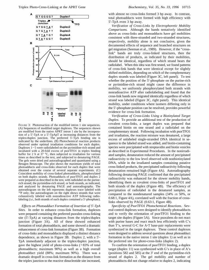
Effects on Photoadduct Formation of Insertion of 5′-TpASites. In order to enhance cross-link formation, duplexeswere prepared containing the preferred psoralen cross-linkingsite (5′-TpA) at varying distances from the triplex/duplexjunction (Figure 3A). All of these showed enhancedformation of total photoadducts, and some showed a dramaticenhancement of cross-link formation (Figure 3B). Formationof cross-links and monoadducts displayed a distinct distancedependence, as shown in Figure 3B. Duplex 2, with a 5′-TpA immediately adjacent to the triplex/duplex junction,gave the highest yield of photo-cross-links (>83% of totalphotoadducts; maximum 90%) as well as a high overallphotoadduct yield (88%; maximum 90%). There was adramatic dropoff in cross-link formation as the distance fromthe triplex junction to the reactive dinucleotide site increased,
with almost no cross-links formed 3 bp away. In contrast,total photoadducts were formed with high efficiency with5′-TpA even 3 bp away.Verification of Cross-Links by Electrophoretic Mobility
Comparisons. Although the bands tentatively identifiedabove as cross-links and monoadducts have gel mobilitiesconsistent with three-stranded and two-stranded structures,respectively, mobility alone is not conclusive, given thedocumented effects of sequence and branched structures ongel migration (Seeman et al., 1989). However, if the “cross-link” bands are truly cross-linked structures, then thedistribution of products, as indicated by their mobilities,should be identical, regardless of which strand bears theradiolabel. When this idea was first tested, we found patternsof cross-link bands that were identical except for slightlyshifted mobilities, depending on which of the complementaryduplex strands was labeled (Figure 3C, left panel). To testwhether the position of the 5′-phosphate on the purine-richor pyrimidine-rich strand might cause the difference inmobility, we uniformly phosphorylated both strands withnonradioactive ATP after radiolabeling and found that thecross-link bands now migrated identically regardless of whichstrand was labeled (Figure 3C, right panel). This identicalmobility, under conditions where isomers differing only inthe 5′-phosphate position can be resolved, provides powerfulevidence for cross-link formation.Verification of Cross-Links Using a Biotinylated Target
Duplex. To provide an additional test of the production ofcovalent cross-links, a target duplex was prepared thatcontained biotin on one strand and a radiolabel on thecomplementary strand. Following incubation with psorTFO1and irradiation, the reaction mixture was denatured, a largeexcess of unlabeled single-stranded DNA identical in se-quence to the labeled strand was added, and biotin-containingspecies were precipitated with streptavidin and biotin vesiclesas described in Experimental Procedures. In the nonirradi-ated samples, denaturation reduced the amount of precipitatedradioactivity to the low level observed with nonbiotinylatedDNA, while in the irradiated samples containing putativecross-linked products, the precipitated radioactivity followingdenaturation remained high (Figure 4A). Autoradiographyfollowing denaturing PAGE confirmed that the precipitatedradioactivity was enhanced for the slower mobility bands,identifying them as covalent cross-links of psorTFO1 andboth strands of the duplex (Figure 4B). The efficiency ofprecipitation of radiolabel in the denatured samples, ascompared to the nondenatured samples (34%Vs 50%, or0.68:1, Figure 4A), correlated well with the fraction of cross-links observed by PAGE (0.63:1, Figure 4B).Specificity of PsorTFO1 Photochemical Reactions. Sev-
eral control duplexes were designed to identify photoadductsand to verify the orientation of psorTFO1 binding to thetarget site duplex (Figure 5A). Since psoralens do not reactwith purine bases and react much less efficiently with C’sthan T’s, several G‚C for A‚T base-pair replacements weresynthesized in the target duplexes. These control duplexeswere designed to address several questions about photoadductformation in the native intron 1 site (duplex 1) as well as inthe preferred site for photo-cross-links (duplex 2).To confirm the orientation of psorTFO1 binding, a duplex
target (duplex 11) was synthesized with G’s in place of A’s(positions 582 and 585) at the 3′ end of the pyrimidine-richstrand of duplex 2. The gel mobility and number ofphotoadducts did not change relative to duplex 2, indicating
FIGURE 3: Photoreaction of the modified intron 1 site sequences.(A) Sequences of modified target duplexes. The sequences shownare modified from the native APRT intron 1 site by the incorpora-tion of a 5′-TpA or a 5′-TpApT at increasing distances from thetriplex/duplex junction. The preferred 5′-TpA binding site isindicated by the underlines. (B) Photochemical reaction productsobserved under optimal irradiation conditions for each duplex.Duplexes 1-5 were radiolabeled on the pyrimidine-rich strand andincubated with a 20-fold excess of psorTFO1 in triplex bindingbuffer for 2 h at 37°C, then subjected to irradiation for varioustimes as described in the text, and subjected to denaturing PAGE.The gels were dried and autoradiographed and quantitated using aBetagen Betascope. The plot shows the maximum yields of totalphotoadducts and cross-links observed for each duplex that wereobtained over the course of several separate experiments. (C)Coincident mobility of cross-linked photoadducts, phosphorylatedon both duplex strands. Photoadducts of psorTFO1 and duplex 2were prepared as described in the text, with radiolabel on the purine-rich strand, the pyrimidine-rich strand, or both strands, as indicated,and analyzed by denaturing PAGE and autoradiography. Theautoradiogram on the left represents duplexes trace labeled with32P only; the autoradiogram on the right shows strands that wereexhaustively labeled with nonradioactive ATP following radio-labeling (i.e., both strands of each duplex contained a 5′-phosphate).
Triplex Photo-Cross-Linking at the APRT Gene Biochemistry, Vol. 35, No. 33, 199610715
+ +
+ +

that none of the photoadducts resulted from psorTFO1reaction with T’s at the 3′ end of the duplex (data not shown).This result, along with the high-efficiency cross-linking toT’s at the 5′ end of the duplex (Figure 3A,B), confirms thatpsorTFO1 binds in the expected antiparallel orientationrelative to the purine-rich strand of the target duplex.To determine the distance restrictions on psorTFO1
photoreactions, a series of duplexes (duplex 6-duplex 10)were made by replacing T’s with G’s or C’s, 5, 7, or 8 basesaway from the triplex junction. As shown for duplex 9 inFigure 5B (lanes 5 and 6), the gel patterns remained the same(compare with Figure 3C), indicating that HMT is limitedto reactions<5 bp away from the junction. This result isnot surprising given that the linker length is<20 Å, whichwould allow reaction within a 3 bp range. A molecularmodel of the 38 bp target duplex with psorTFO1 bound isshown in Figure 6; the psoralen moiety is shown notintercalated to illustrate the dimensions of the photoreactivegroup and its linker. The structural model supports a spanof HMT reaction within a 3 bp range, consistent with ourresults.To confirm the role of the 5′-TpA in cross-link formation,
a duplex (duplex 12) was synthesized with a 5′-ApT in placeof the 5′-TpApT at the junction, reducing the number ofavailable cross-linking sites. As shown in Figure 5B (lanes1-4), duplex 12 shows only one band migrating as a cross-linked product as compared to the three-band pattern shownwith duplex 9 (compare with lanes 5 and 6), again providingevidence for cross-link formation by psorTFO1.Kinetics of Photoadduct Formation. In the course of
determining optimal conditions for photoadduct formation,we monitored the kinetics of the photoreactions for each
duplex. Figure 7A shows typical autoradiograms corre-sponding to time courses for duplexes 4 and 5. Figure 7Bshows the results of quantitation of photoadducts as afunction of time for duplexes 1-5 in an experiment in whichall duplexes were treated with the same preparation ofpsorTFO1 under identical conditions. In each case, totalphotoadduct formation reached nearly its maximal extentwithin about 1 min; under brighter illumination the reactionwas even faster, reaching completion in a few seconds (datanot shown). Monoadduct formation was relatively rapid,reaching its maximal extent in about 1 min for duplexes 1,4, and 5 and in about 6 s for duplexes 2 and 3. For duplexes2 and 3, monoadducts likely continued to form up to 1 minbut were lost to cross-links, so that the steady-state level ofmonoadducts remained nearly constant from 6 to 60 s.Cross-link formation, in contrast to monoadduct formation,displayed a lag in every case, presumably as a result of thedelay required for sufficient monoadducts to accumulate toserve as precursors for the cross-links. Interestingly, the twoduplexes that formed cross-links most efficiently (duplex 2and duplex 3) reached their maximal cross-linking extent inabout 1 min, after which the cross-links began to break down,presumably by photoreversal (see below). In contrast,duplexes 1, 4, and 5 did not display maximum cross-linkinguntil about 10 min, and very little loss of cross-links wasobserved up to 30 min, possibly because of a balancebetween breakdown and continued slow cross-link formation.These results can be rationalized in terms of facile
intercalation at sites at or 1 bp away from the triplex/duplex
FIGURE 4: Verification of cross-links with a biotinylated duplex.(A) Radioactivity corresponding to the nonbiotinylated radiolabeledstrand of the duplex target coprecipitating with its biotinylatedcomplement, following triplex formation with psorTFO1 andindicated treatments. Denaturation (Denat.) was accomplished byheating at 97°C for 10 min, followed by addition of a 50-foldexcess of unlabeled complementary strand; UV refers to standardirradiation conditions after triplex formation as described in thetext. Pelleted samples (see Experimental Procedures) were quan-titated by scintillation counting and plotted as radioactivity (percentof total counts). (B) Autoradiogram of samples on a denaturinggel from an experiment parallel to that described for panel A.Deduced structures of the photoadducts are shown schematicallyto the right of the corresponding bands, with B referring to thebiotin label and P referring to32P.
FIGURE 5: Photoreactions of psorTFO1 with duplexes designed totest specificity of photoadduct formation. (A) Sequences of duplexestested. (B) Products of photoreaction of psorTFO1 with duplex 12(lanes 1-4) with a 5′-ApT at the triplex junction or duplex 9 (lanes5 and 6) with a 5′-TpApT at the triplex junction. Samples weresubjected to standard irradiation conditions for 6 s, run on adenaturing 15% PAG, and then visualized by autoradiography.
10716 Biochemistry, Vol. 35, No. 33, 1996 Vasquez et al.
+ +
+ +

junction and the well-known selective reactivity of T towardpsoralen. The high yield of photoadducts observed withduplex 3 can also be attributed to its high T content near thejunction, and its higher cross-linking efficiency, as comparedto duplex 1, can be attributed to the presence of a nearby5′-TpA. However, the increased distance from the junctionof the 5′-TpA in duplex 3, as compared to duplex 2, appearsto lower the cross-linking efficiency as shown in Figure 7B.The trend of less efficient cross-linking as the 5′-TpA wasmoved further from the junction continued with duplex 4and duplex 5. Duplex 4, whose cross-linking target site was2 bp from the junction, gave only moderate cross-link yields(15%), while moving the 5′-TpA 3 bp from the junction(duplex 5) virtually eliminated cross-linking. Both duplex4 and duplex 5 required 10 min to reach maximumphotoadduct formation, likely as a result of a requirementfor rarely sampled torsionally strained conformations toachieve reaction with the more distant T residues. It isinteresting that duplex 1 and duplex 5 gave comparable
photoadduct yields on a similar time scale with no changein band patterns on denaturing gels, yet duplex 5 containedan extra T 3 bp away from the junction (see Figure 3A).This comparison suggests that there may be no reaction ofpsorTFO1 with the more distant T residue. As previouslydiscussed, both the distribution of products and the kineticsare consistent with the length of the HMT linker.PhotoreVersibility of Photoadducts. A well-known feature
of covalent psoralen-DNA photoadducts is that they arefully reversible by irradiation at 240-310 nm (Cimino etal., 1986; Shi & Hearst, 1987). To confirm that thephotoadducts were actually typical covalent psoralen prod-ucts, we irradiated gel-purified cross-linked products at 254nm. Figure 8 shows that the cross-linked products were fullyreversible (compare lanes 2 and 3), consistent with covalentHMT-DNA photoadducts.If the gel-purified products had been covalent furan-sided
monoadducts, instead of cross-links, then upon a secondirradiation at 334 nm, cross-links would have been expectedto result. As shown in Figure 8, the mobility of the gel-purified products was identical with that of the productresulting from a second round of irradiation at 334 nm(compare lanes 2 and 4). This result also supports ourconclusion that the initial products were covalent HMT-DNA cross-links.
DISCUSSION
Triplex technology offers an alternative approach togenome manipulation that could overcome a number oflimitations to the currently available methodology: unknownbiological consequences of using viral vectors, mutationsassociated with random integration, and low frequencies oftargeted recombination. These studies were designed to testthe feasibility of using triplex-mediated photochemistry asa key element in a gene manipulation strategy that wouldallow targeting of specific sites within any gene with highefficiency, followed by controlled activation of the genemodification process. Triplex technology potentially offersa number of important advantages: high-affinity bindingdirected at only one or two targets per cell, direct inactivationof a gene, and sensitization of a gene for targeted recombina-tion. Photochemically damaging DNA at a specific siteshould enhance the rate of recombination at that site and,therefore, increase the efficiency of gene therapeutics.Psoralen activation by UVA may be particularly useful forstudies in cultured cells and for treatment of accessibletissues. However, visible light activation of psoralen (Gas-parro et al., 1993) may allow deeper tissue penetration andmay be less mutagenic than UVA.PsorTFO1 Binding to the NatiVe APRT Intron 1 Site.We
have shown here that a psoralen-modified TFO (psorTFO1)forms triplex with high affinity by binding the underlyingduplex in an antiparallel fashion. Surprisingly, the psoralenmodification does not appear to enhance binding affinity, incontrast to other studies with intercalator-modified TFOs (LeDoan et al., 1987; Grigoriev et al., 1992; Mouscadet et al.,1994).Cross-LinksVs Monoadducts. Although the effects on
chromosomal recombination and mutation of triplex-medi-ated photoreactions will have to be determined by experi-ments in living cells, there is already considerable informa-tion about the effects of psoralen monoadducts and cross-links on recombination and mutation in mammalian cells.
FIGURE 6: Molecular models of triplex formed by duplex 1 andpsorTFO1, illustrating the geometric relationship of the psoralenmoiety and the triplex junction: space-filling model (right); wire-frame model (left), enlarged to show the region of the triplex/duplexjunction. The pyrimidine-rich strand of the duplex is shown in green,the purine-rich strand in blue, the TFO in red, and the psoralenmoiety in yellow. The psoralen ring system is shown withoutintercalation to illustrate distance constraints; the photoadducts aremost likely formed after intercalation. The model building withenergy minimization was carried out using Biosym/MSI’s InsightII molecular modeling program in a way similar to that describedby Kessler et al. (1993).
Triplex Photo-Cross-Linking at the APRT Gene Biochemistry, Vol. 35, No. 33, 199610717
+ +
+ +

The relatively greater potency of cross-links in stimulatingthese processes has been tied to the mechanisms by whichthey are repaired (Sladek et al., 1989; Cheng et al., 1988). Itcan be argued that monoadducts are also mutagenic as theyhave been implicated in enhancing the frequency of mutationin mammalian cells (Gunther et al., 1995). Thus, establishingthe sequence requirements for efficient formation of bothmonoadducts and cross-links is essential for analyzing thein ViVo effects of these photoadducts. The results describedhere make it clear that the distance of 5′-TpA sites from the
triplex/duplex junction is the most important determinant ofboth the efficiency of product formation and the proportionsof monoadducts and cross-links. This sensitivity to positionmight be expected since the conformation of the triplex/duplex junction differs from either B-form duplex DNA orA-form triplex DNA (Radhakrishnan & Patel, 1994). Thetriplex/duplex junction has been shown to be more reactiveto intercalating agents (Collier et al., 1991; Sun et al., 1991),and therefore, it might be expected that psoralen would havegreater potential for reaction at the junction. In fact, forpsoralen-linked triplexes, simple considerations of linkerlength, sequence, and sequence context (i.e., proximity tothe triplex/duplex junction) can be used to estimate theoutcome fairly accurately.Photoreaction Kinetics.Additional features of the pho-
toreactions with great significance for intracellular applica-tions are the dependence on time and radiation dose.Obviously, a requirement for prolonged irradiation with UVlight would preclude studies in viable cells. Our results showthat the ultimate yield of photoadducts after irradiation isdependent on sequence with a 5′-TpApT being favored forformation of cross-links and an overall high yield ofphotoadducts. The distance of T’s from the triplex/duplexjunction is critical in the formation of both cross-links andmonoadducts. The kinetics of photoadduct formation imply,as expected, that there is an initial increase in monoadductformation with a subsequent increase in cross-links. It alsoappears that the further the 5′-TpA is from the junction, themore time required for cross-link formation (see Figure 7).Implications for Gene Targeting.The efficiency of
psoralen photoadduct formation at the native intron 1 site(∼50%) is encouraging, although it appears that the productsare predominantly the result of psorTFO1 covalently cross-linking to one strand of the target duplex, and not both. The
FIGURE 7: Kinetics of photoadduct formation. (A) Time courses of photoadduct formation for duplexes 4 and 5. Following incubation ofpsorTFO1 with 5′-end-labeled duplex, the samples were incubated for 0-60 min, and then products were separated by denaturing gelelectrophoresis on a 15% polyacrylamide gel and subjected to autoradiography. (B) Similar samples were prepared for duplexes 1-5, andthe radioactivity was quantitated using a Betagen Betascope. The results are plotted as a percent of the total radioactivity in cross-linkedadducts (X-link), monoadducts (Mono), or their sum (Total)Vs irradiation time. Time is plotted on a log scale to show the full range ofkinetic behavior observed.
FIGURE 8: Photoreversibilty of photoadducts. Following standardincubation and irradiation procedures for duplex 2, the photoadductswere separated by PAGE on a 15%, 7 M urea gel buffered withTBE. After irradiation cross-linked bands were gel purified andthen irradiated a second time with UV of either 254 or 334 nmand then again subjected to PAGE, along with control samples.Lane 1 contains a control reaction mixture that was not subjectedto the initial irradiation; lane 2 contains the gel-purified photo-cross-linked products without a second irradiation; lane 3 containsthe same sample as in lane 2 after a second irradiation at 254 nm;lane 4 contains the same sample as in lane 2 after a secondirradiation at 334 nm; lane 5 contains the irradiated mixture fromwhich the cross-linked species in lane 2 was purified.
10718 Biochemistry, Vol. 35, No. 33, 1996 Vasquez et al.
+ +
+ +

inability of psorTFO1 to cross-link to both strands of thetarget duplex was not unexpected: the target site contains a5′-GpT at the triplex junction, a sequence that does not formphoto-cross-links efficiently with psoralen. A comparisonof the effects of monoadductsVscross-links on chromosomalrecombination seems a requirement for maximizing the useof site-specific DNA damage as a tool for precise genomemanipulation. We are currently testing the effects ofmonoadduct formation on recombination and mutationfrequencies in cells using the native APRT intron 1 site inCHO cells.Perhaps the most important conclusion for immediate
application of triplex-mediated psoralen photochemistry togenome manipulation in living cells is that the sequenceimmediately adjacent to the triplex binding site in the targetduplex is critically important. Specifically, at least onethymine residue must be within reach of the psoralen forefficient photoadduct formation, and rapid and efficientphoto-cross-linking requires a 5′-TpA sequence immediatelyadjacent to the triplex/duplex junction. Because duplex 2shows very high efficiency cross-link formation, this se-quence is a promising site to target into the APRT gene inCHO cells to test its effect on recombination and mutation.We are currently testing the applicability of these now well-definedin Vitro rules, to the conditions present in the nucleiof mammalian cells, using modified APRT genes engineeredinto hamster chromosomes.
ACKNOWLEDGMENT
We thank Dr. Veeraiah Bodepudi and Robert Tinder forpreparing psorTFO1, Sean Smith for the molecular modelingstudies of the psorTFO1 triplex, and Mark Brenneman, AprilKilburn, Kathleen Marburger, Ray Merrihew, and GeoffSargent for helpful discussions.
REFERENCES
Averbeck, D. (1985)Mutat. Res. 151, 217-233.Beal, P. A., & Dervan, P. B. (1991)Science 251, 1360-1363.Ben-Hur, E., & Song, P. S. (1984)AdV. Radiat. Biol. 11, 131-157.
Birg, F., Praseuth D., Zerial, A., Thuong, N. T., Asseline, U.,LeDoan, T., & Helene, C. (1990)Nucleic Acids Res. 18, 2901-2908.
Bridges, B. A., Mottershead, R. P., & Knowles, A. (1979)Chem.-Biol. Interact. 27, 221-233.
Cheng, S., Van Houten, B., Gamper, H. B., Sancar, A., & Hearst,J. E. (1988)J. Biol. Chem. 263, 15110-15117.
Cimino, G. D., Gamper, H. B., Isaacs, S. T., & Hearst, J. E. (1985)Annu. ReV. Biochem. 54, 1151-1193.
Cimino, G. D., Shi, Y., & Hearst, J. E. (1986)Biochemistry 25,3013-3020.
Cole, R. S. (1971)Biochim. Biophys. Acta 254, 30-39.Colier, D. A., Mergny, J. L., Thuong, N. T., & Helene, C. (1991)Nucleic Acids Res. 19, 4219-24.
Cooney, M., Czernuszewicz, G., Postel, E. H., Flint, S. J., & Hogan,M. E. (1988)Science 241, 456-459.
Degols, G., Clarenc, J. P., Lebleu, B., & Leonetti, J. P. (1994)J.Biol. Chem. 269, 16933-16937.
Esposito, F., Brankamp, R. G., & Sinden, R. R. (1988)J. Biol.Chem. 263, 11466-11472.
Friedberg, E. C., Walker, G. C., & Siede, W. (1995)DNA Repairand Mutagenesis, pp 523-576, ASM Press, Washington DC.
Gasparro, F. P., Gattolin, P., & Olack, G. A. (1993)Photochem.Photobiol. 57, 1007-1010.
Gasparro, F. P., Havre, P. A., Olack, G. A., Gunther, E. J., & Glazer,P. M. (1994)Nucleic Acids Res. 22, 2845-2852.
Grigoriev, M., Praseuth, D., Robin, P., Hemar, A., Saison-Behmoaras, T., Dautry-Varsat, A., Thuong, N. T., Helene, C.,& Harel-Bellan, A. (1992)J. Biol. Chem. 267, 3389-3395.
Grigoriev, M., Praseuth, D., Guieysse, A. L., Robin, P., Thuong,N. T., Helene, C., & Harel-Bellan, A. (1993)Proc. Natl. Acad.Sci. U.S.A. 90, 3501-3505.
Gunther, E. J., Yeasky, T. M., Gasparro, F. P., & Glazer, P. M.(1995)Cancer Res. 55, 1283-1288.
Hall, J. D. (1982)Mol. Gen. Genet. 188, 135-138.Havre, P. A., & Glazer, P. M. (1993)J. Virol. 67, 7324-7331.Havre, P. A., Gunther, E. J., Gasparro, F. P., & Glazer, P. M. (1993)Proc. Natl. Acad. Sci. U.S.A. 90, 7879-7883.
Ing, N. H., Beekman, J. M., Kessler, D. J., Murphy, M., Jayaraman,K., Zendegui, J. G., Hogan, M. E., O’Malley, B. W., & Tsai,M. (1993)Nucleic Acids Res. 21, 2789-2796.
Kessler, D. J., Pettitt, B. M., Cheng, Y.-K., Smith, S. R., Jayaraman,K., Vu, H. M., & Hogan, M. E. (1993)Nucleic Acids Res. 21,4810-4815.
Kovacs, A., Kandala, J. C., Weber, K. T., & Guntaka, R. V. (1996)J. Biol. Chem. 271, 1805-1812.
Le Doan, T., Perrouault, L., Praseuth, D., Habhoub, N., Decout, J.L., Thuong, N. T., Lhomme, J., & Helene, C. (1987)NucleicAcids Res. 15, 7749-7760.
McShan, W. M., Rossen, R. D., Laughter, A. H., Trial, J., Kessler,D. J., Zendegui, J. G., Hogan, M. E., & Orson, F. M. (1992)J.Biol. Chem. 267, 5712-5721.
Moser, H. E., & Dervan, P. B. (1987)Science 238, 645-650.Mouscadet, J. F., Ketterle, C., Goulaouic, H., Carteau, S., Subra,F., Le Bret, M., & Auclair, C. (1994)Biochemistry 33, 4187-4196.
Okada, T., Yamaguchi, K., & Yamashita, Y. (1994)Growth Factors11, 259-270.
Orson, F. M., Thomas, D. W., McShan, W. M., Kessler, D. J., &Hogan, M. E. (1991)Nucleic Acids Res. 19, 3435-3441.
Perrouault, L., Asseline, U., Rivalle, C., Thuong, N. T., Bisagni,E., Giovannangeli, C., Le Doan, T., & Helene, C. (1990)Nature344, 358-360.
Postel, E. H., Flint, S. J., Kessler, D. J., & Hogan, M. E. (1991)Proc. Natl. Acad. Sci. U.S.A. 88, 8227-8231.
Radhakrishnan, I., & Patel, D. J. (1994)Biochemistry 33, 11405-11416.
Reardon, J. T. R., Spielman, P., Huang, J. C., Sastry, S., Sancar,A., & Hearst, J. E. (1991)Nucleic Acids Res. 19, 4623-4629.
Roy, C. (1994)Eur. J. Biochem. 220, 493-503.Sargent, R. G., Merrihew, R. V., Nairn, R., Adair, G., Meuth, M.,& Wilson, J. H. (1996)Nucleic Acids Res. 24, 746-753.
Scaggiante, B., Morassutti, C., Tolazzi, G., Michelutti, A., Bacca-rani, M., & Quadrifoglio, F. (1994)FEBS Lett. 352, 380-384.
Seeman, N. C., Chen, J. H., & Kallenbach, N. R. (1989)Electro-phoresis 10, 345-354.
Shi, Y., & Hearst, J. E. (1987)Biochemistry 26, 3786-3792.Sladek, F. M., Melian, A., & Howard-Flanders, P. (1989)Proc.Natl. Acad. Sci. U.S.A. 86, 3982-3986.
Sun, J. S., Lavery, R., Chomilier, J., Zakrzewska, K., Montenay-Garstier, T., & Helene, C. (1991)J. Biomol. Struct. Dyn. 9, 425-436.
Takasugi, M., Guendouz, A., Chassignol, M., Decout, J. L.,Lhomme, J., Thuong, N. T., & Helene, C. (1991)Proc. Natl.Acad. Sci. U.S.A. 88, 5602-5606.
Tu, G. C., Cao, Q. N., & Israel, Y. (1995)J. Biol. Chem. 270,28402-28407.
Vasquez, K. M., Wensel, T. G., Hogan, M. E., & Wilson, J. H.(1995)Biochemistry 34, 7243-251.
Wang, G., Levy, D. D., Seidman, M. M., & Glazer, P. M. (1995)Mol. Cell. Biol. 15, 1759-1768.
Wang, G., Levy, D. D., Seidman, M. M., & Glazer, P. M. (1996)Science 271, 802-805.
Zendegui, J. G., Vasquez, K. M., Tinsley, J. H., Kessler, D. J., &Hogan, M. E. (1992)Nucleic Acids Res. 20, 307-314.
BI960881F
Triplex Photo-Cross-Linking at the APRT Gene Biochemistry, Vol. 35, No. 33, 199610719
+ +
+ +