hi vision preirus - alpha imaging · hi vision preirus – advanced product features discover new...
TRANSCRIPT
Upda
te H
V Pr
eiru
s | e
| 04
.201
2 | a
+w |
HV P
reiru
s 20
14 |
e | 1
0.20
14 |
a+w
Hitachi Medical CorporationAkihabara UDX, 4-14-1Soto-KandaChiyoda-kuTokyo, 101-0021, JapanPhone +81 3 3526 8410Fax +81 3 3526 8409
Hitachi Medical Systems America, Inc.1959 Summit Commerce ParkTwinsburg, Ohio 44087, USAPhone +1 330 425 1313Fax +1 330 425 1410
Hitachi Medical Systems (Beijing) CorporationRm.609 Winterless Centre B-area, No1 Xi-Da-Wang Road,Chaoyang District,Beijing, P.R.China 100026Phone +86 10 6538 8881
Hitachi Medical Systems (S) Pte Ltd 7 Tampines Grande Hitachi Square #04-01Singapore 528736 Phone +65 6602 0110Fax +65 6602 0111
Hitachi Medical Systems Europe Holding AGSumpfstrasse 13CH-6300 ZugPhone +41 41 748 63 33Fax +41 41 748 63 32Export Division· Ultrasound Phone +41 41 748 63 47 Fax +41 41 748 63 32· MR/CT Phone +41 41 748 63 49 Fax +41 41 748 63 32
Hitachi Aloka Medical Ltd.6-22-1, Mure, MitakaTokyo 181-8622, JapanPhone +81 422 45-6049Fax +81 422 45-4058
Hitachi Aloka Medical Ltd.10 Fairfield BoulevardWallingford, Connecticut 06492, USAPhone +1 203 269 5088
Hitachi Aloka Medical Ltd.South, 6th Floor, No. 456 Fute North Road, Waigaoqiao Shanghai, ChinaPhone +86 21 5866 5820
Hitachi Aloka Medical Ltd. 1 Maritime Square #10-32/32AHarbour Front Centre099253 SingaporePhone +65 6271 1960
www.hitachi-medical-systems.com
Hitachi Medical Systems GmbH Technology AcademyWanheimer Strasse 59D-40472 DüsseldorfPhone +49 211 1665 10Fax +49 211 1665 169
Hitachi Medical Systems GmbHOtto-von-Guericke-Ring 3D-65205 WiesbadenPhone +49 6122 7036 0Fax +49 6122 7036 10
Hitachi Medical Systems GesmbHIZ NÖ-Süd, Strasse 2a, Objekt M39/IIA-2351 Wiener NeudorfPhone +43 2236 677 750 Fax +43 2236 677 75049
Hitachi Medical Systems Kft.Damjanich u. 11-15Ligetváros Irodaház I. em. 102H-1071 BudapestPhone +36 1 478 0090Fax +36 1 478 0091
Hitachi Medical Systems BVEdisonstraat 1aNL-2811 EM ReeuwijkPhone +31 182 39 77 77Fax +31 182 39 77 79
Hitachi Medical Systems N.V./S.A.Mechelen Noord IIWayenborgstraat 8B-2800 MechelenPhone +32 15 20 22 55Fax +32 15 20 01 92
Hitachi Medical Systems UK Ltd1 Davy ClosePark Farm Industrial EstateWellingboroughNorthamptonshire NN8 6XX UKPhone +44 844 800 4294 Fax +44 1933 4058 59
Hitachi Medical Systems S.A.S.Espace Porte de l´Est 39, avenue Urbain le VerrierF-69800 Saint-PriestPhone +33 4 72 14 59 72Fax +33 4 72 81 96 06
Hitachi Medical Systems S.p.A.Via Edison 6I-20090 Assago MIPhone +39 02 97166Fax +39 02 97166127
Hitachi Medical Systems S.L.Avda. de Manoteras, 22 Local 70 y 87 SP-28050 MadridPhone +34 91 358 93 50Fax +34 91 358 96 03
Plus representations in various European countries.
Hitachi Medical Corporation Medical SystemOperations Group, Kashiwa, is certified ascomplying with the International Standard ofSystem Quality Assurance (ISO 9001), MedicalDevice Special Requirements (ISO 13485).
Hitachi Medical Corporation Medical SystemOperations Group, Kashiwa, has been certifiedto ISO 14001 (Environmental ManagementSystems).
The legal manufacturer of PENTAX ultrasoundendoscopes is Hoya Corporation, Tokyo, Japan.They are distributed by Hitachi Medical SystemsEurope Holding AG, Zug, Switzerland and itssubsidiaries in the assigned geographical areasin Europe.
Specifications and physical appearance maybe changed without prior notice in order toimprove performance. Some features describedare optional. Please read instruction manualto ensure correct operation of the equipment.
This brochure is printed on biodegradable FSC®-certified paper produced from responsibleforestry. FSC® stands for “Forest StewardshipCouncil®”.
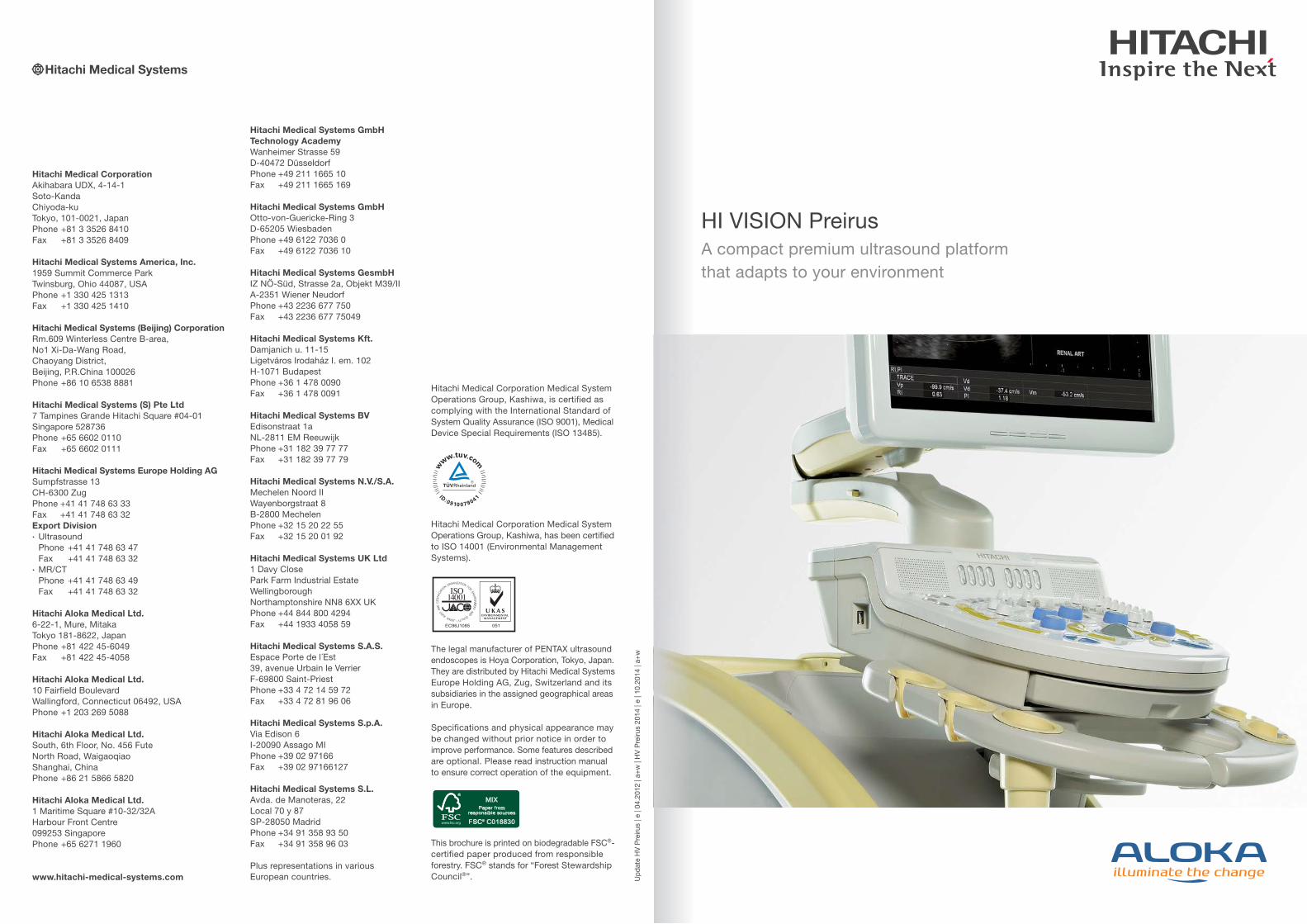
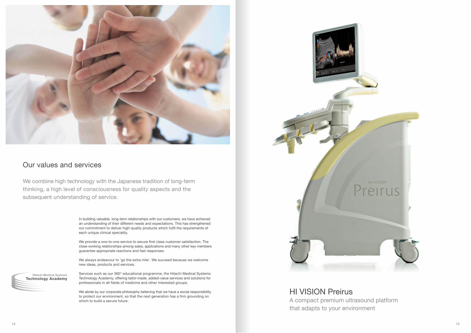
HI VISION PreirusA compact premium ultrasound platform that adapts to your environment
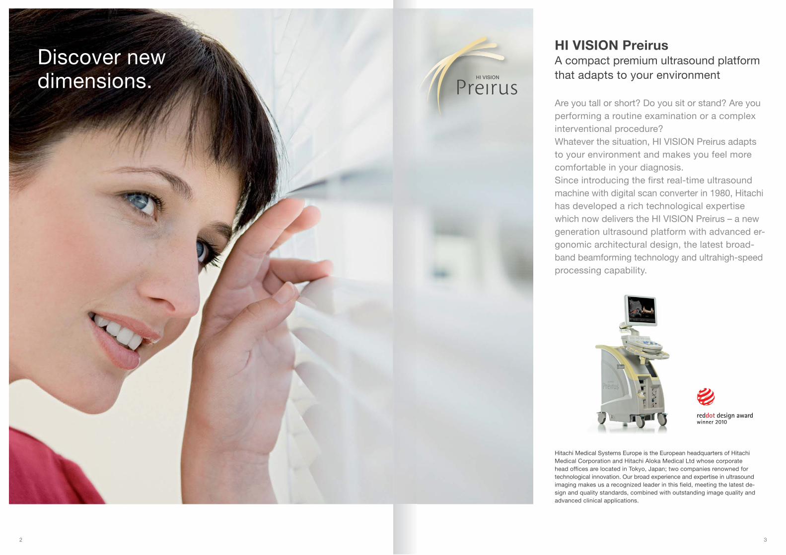
HI VISION PreirusA compact premium ultrasound platformthat adapts to your environment
Are you tall or short? Do you sit or stand? Are youperforming a routine examination or a complexinterventional procedure?Whatever the situation, HI VISION Preirus adaptsto your environment and makes you feel morecomfortable in your diagnosis.Since introducing the first real-time ultrasoundmachine with digital scan converter in 1980, Hitachihas developed a rich technological expertisewhich now delivers the HI VISION Preirus – a newgeneration ultrasound platform with advanced er-gonomic architectural design, the latest broad-band beamforming technology and ultrahigh-speedprocessing capability.
2 3
Discover new dimensions.
Hitachi Medical Systems Europe is the European headquarters of HitachiMedical Corporation and Hitachi Aloka Medical Ltd whose corporatehead offices are located in Tokyo, Japan; two companies renowned fortechnological innovation. Our broad experience and expertise in ultrasoundimaging makes us a recognized leader in this field, meeting the latest de-sign and quality standards, combined with outstanding image quality andadvanced clinical applications.

HI VISION Preirus – Advanced Product Features
Discover new dimensions in ergonomic design – increased machineflexibility means it does the twisting and turning, so you don't have to.
Ergonomic Design
The unique design of the HI VISION Preirus platform allows precise adjustment tofacilitate the performance of any examination by any operator. With its superslim-line footprint, and operator console with large digital LCD viewing monitormoving together in a rotational arc, positioning at the bedside can be optimisedto make diagnosis a more comfortable experience for you and your patient.
Second Generation Graphical User Interface with Smart Touch Technology
By integrating the system's operator controls on a 19” digital LCD monitor usinga series of Touch Panel keys and Smart Tab menus, the HI VISION Preirus allowsintuitive scan parameter adjustment without having to reduce concentration onthe diagnostic image. A thumbnail image gallery displays current and storedimages for easy comparison. Opening of menus and toolbars, including accessto a user manual, is controlled by the operator leaving a full-screen, unclutteredimage display for maximum diagnostic capability.
54
6 7
Patient Scanning Selector (PSS)
Detailed adjustment of all imaging parameters is essential to optimise diagnosticcapability for each anatomical area and for each individual patient. The PatientScanning Selector (PSS) gives flexibility within a chosen clinical application tocustomise, save and later recall examination specific combinations of imagingparameters at the touch of a button.
Advanced Imaging Technology
Ultrahigh-speed image processing on the HI VISION Preirus platform enhancesthe performance of established image quality improvement technologies such asHI Rez+ (tissue adaptive filtering), HdTHI (High definition dynamic Tissue HarmonicImaging), HI Com (frequency and spatial compounding), and Coded Imaging.Flexible pitch scanning using a new proprietary ASIC gives the operator morecontrol over frame rate and line density resulting in faster frame rates and main-tained high quality B-mode imaging even in Colour Doppler and Elastography(HI-RTE) modes.
New image formats – Imaging outside the box
Examinations of superficial structures using high frequency linear array transducersare transformed using a new trapezoid display format to 'image outside the box' –expanded field of view for B-mode and Colour Doppler imaging, and a steeringB-mode option to optimise beam-to-vessel angle, enhance anatomical and vasculardisplay. High resolution zoom (HI Zoom) and image magnification (Pan Zoom)functions are available at the touch of a button for optimisation of line density andfrozen image size adjustment, respectively.
HI VISION Preirus – HI VISION Imaging
The art of effective imaging – customised scanning parameters combinedwith high speed image processing for improved diagnostic confidence.Proven innovative technologies confer superior penetration, temporal,spatial and contrast resolution giving high quality images for every patientevery time.
Double-De-codeFilter
Double-De-codeFilter
Sum.
Displayed
1st Tx Pulse
2nd Tx Pulse
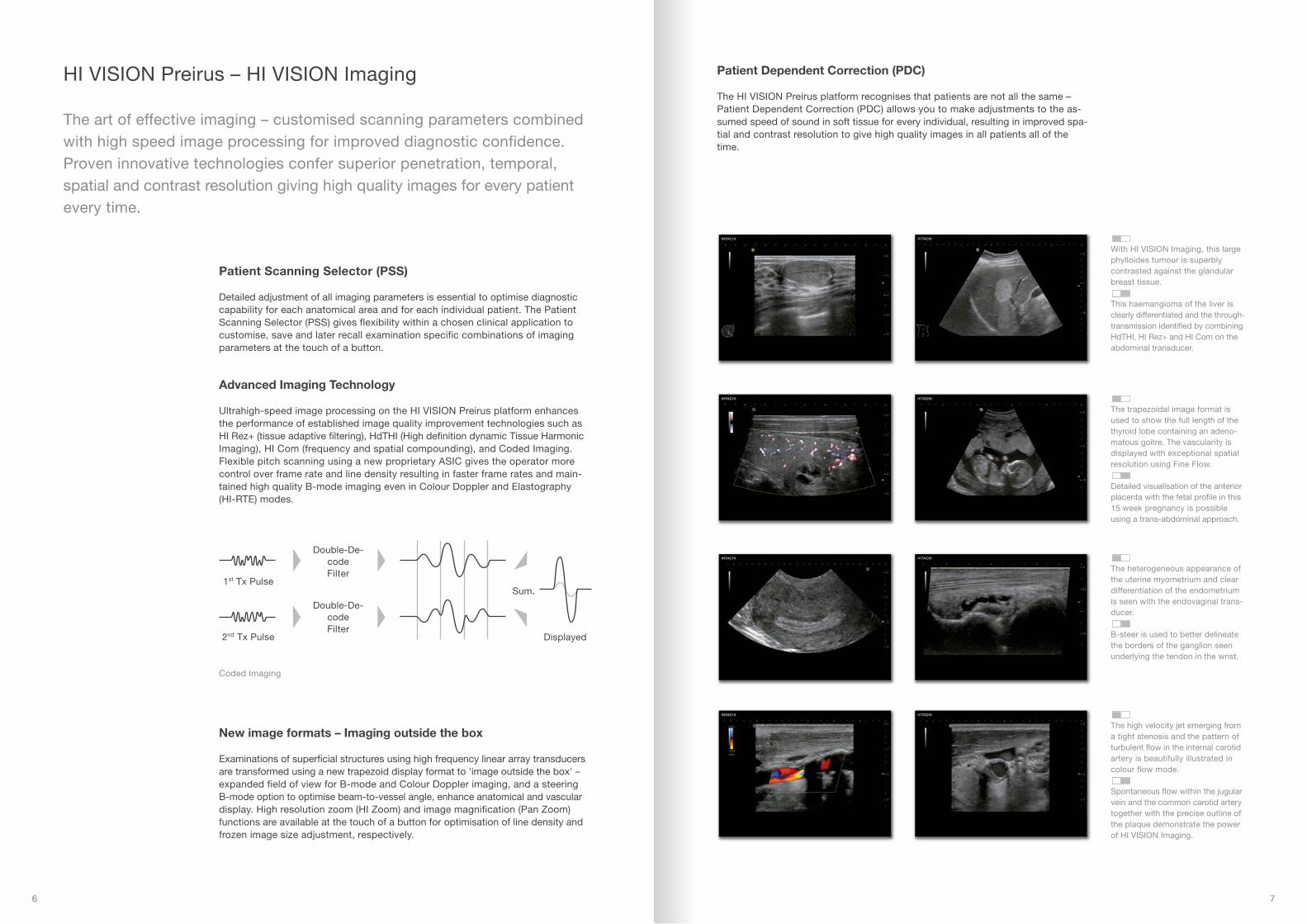
The trapezoidal image format isused to show the full length of thethyroid lobe containing an adeno-matous goitre. The vascularity isdisplayed with exceptional spatialresolution using Fine Flow.
Detailed visualisation of the anteriorplacenta with the fetal profile in this15 week pregnancy is possibleusing a trans-abdominal approach.
With HI VISION Imaging, this largephylloides tumour is superbly contrasted against the glandularbreast tissue.
This haemangioma of the liver isclearly differentiated and the through-transmission identified by combiningHdTHI, HI Rez+ and HI Com on theabdominal transducer.
The high velocity jet emerging froma tight stenosis and the pattern ofturbulent flow in the internal carotidartery is beautifully illustrated incolour flow mode.
Spontaneous flow within the jugularvein and the common carotid arterytogether with the precise outline ofthe plaque demonstrate the powerof HI VISION Imaging.
The heterogeneous appearance ofthe uterine myometrium and cleardifferentiation of the endometriumis seen with the endovaginal trans-ducer.
B-steer is used to better delineatethe borders of the ganglion seenunderlying the tendon in the wrist.
Coded Imaging
Patient Dependent Correction (PDC)
The HI VISION Preirus platform recognises that patients are not all the same –Patient Dependent Correction (PDC) allows you to make adjustments to the as-sumed speed of sound in soft tissue for every individual, resulting in improved spa-tial and contrast resolution to give high quality images in all patients all of thetime.
Hitachi Real-time Tissue Elastography (HI-RTE)*
HI-RTE has proven clinical benefits in a variety of different applications – breast,prostate, pancreas & lymph nodes, thyroid, musculoskeletal, liver and many more.With the ability to improve focal lesion visualisation and refine a differential diag-nosis in real-time using any one of 25 transducers, clinical studies evidence thatthe technique is accurate, reproducible and easy to perform. HI-RTE is rapidlybecoming an essential part of the routine clinical ultrasound examination.
Dynamic Contrast Harmonic Imaging (dCHI)*
Dynamic Contrast Harmonic Imaging (dCHI) is a wideband pulse-inversion (WPI)technology developed by Hitachi for use with ultrasound contrast agents. Wegive you increased agent-to-tissue specificity by modulation of both pulse phaseand the transmit frequency between pulses – you notice significant improvementin lateral and contrast resolution and greater sensitivity at depth with no compro-mise in axial resolution. Using the Microbubble Trace Imaging (MTI) accumulativeenhancement mode you benefit from an improved ability to interrogate micro-vessel morphology and by generating Time Intensity Curves (TIC) from storeddata you can display contrast agent enhancement over time within multiple se-lectable regions of interest (ROI).
8 9
HI VISION Preirus – Advanced Modalities
Incorporating the latest technological advances the HI VISION Preirus de-livers state-of-the-art ultrasound capability giving you improved exa-mina-tion value from raised diagnostic confidence and increased potential fortherapeutic intervention.
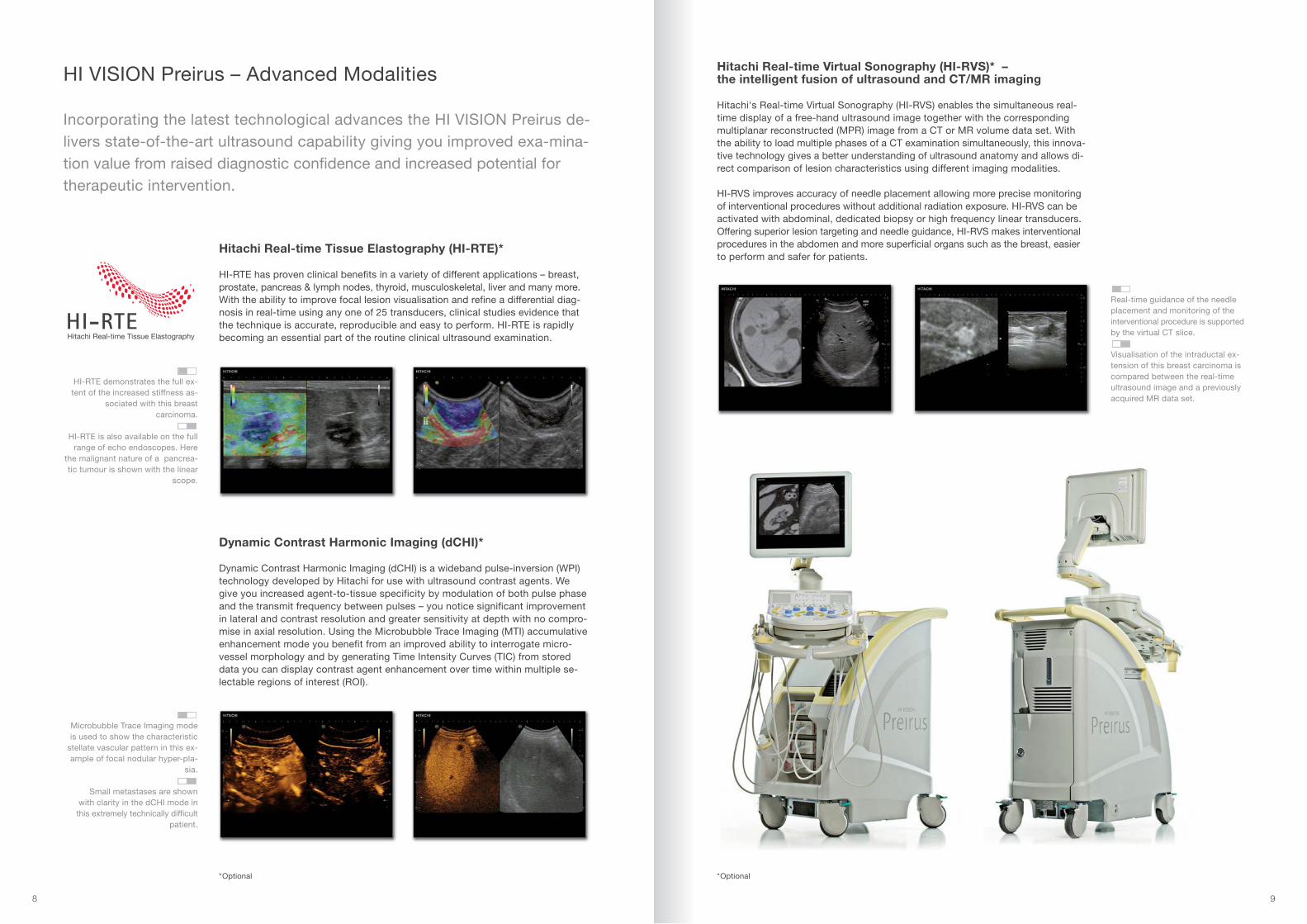
HI-RTE demonstrates the full ex-tent of the increased stiffness as-
sociated with this breast carcinoma.
HI-RTE is also available on the fullrange of echo endoscopes. Here
the malignant nature of a pancrea-tic tumour is shown with the linear
scope.
Hitachi Real-time Virtual Sonography (HI-RVS)* –the intelligent fusion of ultrasound and CT/MR imaging
Hitachi's Real-time Virtual Sonography (HI-RVS) enables the simultaneous real-time display of a free-hand ultrasound image together with the correspondingmultiplanar reconstructed (MPR) image from a CT or MR volume data set. Withthe ability to load multiple phases of a CT examination simultaneously, this innova-tive technology gives a better understanding of ultrasound anatomy and allows di-rect comparison of lesion characteristics using different imaging modalities.
HI-RVS improves accuracy of needle placement allowing more precise monitoringof interventional procedures without additional radiation exposure. HI-RVS can beactivated with abdominal, dedicated biopsy or high frequency linear transducers.Offering superior lesion targeting and needle guidance, HI-RVS makes interventionalprocedures in the abdomen and more superficial organs such as the breast, easierto perform and safer for patients.
Real-time guidance of the needleplacement and monitoring of theinterventional procedure is supportedby the virtual CT slice.
Visualisation of the intraductal ex-tension of this breast carcinoma iscompared between the real-timeultrasound image and a previouslyacquired MR data set.
*Optional
Microbubble Trace Imaging modeis used to show the characteristic
stellate vascular pattern in this ex-ample of focal nodular hyper-pla-
sia.
Small metastases are shown with clarity in the dCHI mode inthis extremely technically difficult
patient.
*Optional

10 11
An apical 4-chamber view showingautomatic calculation of the leftatrial volume.
An apical long axis view where thedegree of aortic stenosis has beenestimated from analysis of the CWspectrum.
Hitachi's 'in-house manufacture' expertise has allowed us to customise and optimisetransducer performance for each clinical application by using the most appropriatedesign features. For example, our super multi-layer technology gives you high si-gnal-to-noise ratios at depth in the abdomen whilst micro piezo-composite tech-nology is used to reduce interference and improve signal-to-noise ratio when imagingsuperficial structures with high frequency linear transducers.
The HI VISION imaging achieved with dedicated 4D transducers matches that of thestandard 2D transducers and the lightweight ergonomic design features minimiseoperator fatigue.
Biopsy Guidance
Hitachi offers outstanding technological support for interventional diagnostic andtherapeutic procedures with a choice of dedicated transducers for biopsy, attach-ments for performing biopsy with standard transducers and integrated workingchannels for endoscopes. Advanced image acquisition and display features suchas Real-time BiPlane Imaging (RTBi) and Hitachi Real-time Virtual Sonography(HI-RVS) enable you to realise the potential of ultrasound for safe, accurate, non-operative diagnosis and minimally invasive therapeutic interventions.
Advanced Transducer Technology across a Range of Applications
Hitachi earned a place in history by introducing the first curved linear arrayin 1980. Today, Hitachi still manufactures the majority of its transducersin-house and is at the leading edge of transducer materials research andcable design.
Increased frequency bandwidthsand a short ring down translatesinto improved axial resolution.
Single Crystal Transducer Technology
With the HI VISION Preirus ultrasound platform, Hitachi introduces state-of-the-artsingle crystal technology in its new EUP-S70 phased array transducer. Precisionelement slicing delivers markedly improved stability and energy efficiency givingyou higher quality B-mode images and increased Doppler sensitivity for improveddiagnostic confidence.
conventional S70
Sens
itivi
ty [d
B]
Frequency [MHz]
A decrease in cross-talk improvessignal to noise.
The EUP-S70 phased array trans-ducer with lightweight, low-capaci-tance cable.dB
ch

12 13
Network connectivity
Full DICOM connectivity allows you to interface with PACS and other image and in-formation management systems, providing integrated worklist, storage, query/retrieve and print functionality. Structured report options expedite examinationcompletion and encourage standardised reporting practice.
Intelligent patient administration – gives you smart access to patient data as and when required
At the start of each 'new patient' previous examination entries can be reviewed forcorresponding records and matching patient information automatically populatedinto relevant data fields.
Prospective worklist entry capability allows rapid patient identification and se-lection at the start of each examination and ensures accurate and consistent pa-tient records. Flexible interrogation software enables you to search the imagedatabase using patient name, date of study or keywords. 'Image Viewer' softwareallows you to retrieve stored images and measurements for offline review to faci-litate image interpretation and examination reporting.
Data Transfer
Versatile image software enables external transfer of images to USB memory de-vices (multiple ports), USB hard disk drives, DVD-R or DVD-RAM in DICOM, BMP,TIFF or AVI format. The ability to mask patient identification on transfer ensurescompliance with data protection protocols and assures patient confidentialitywhen using images for research, training and education.
HI VISION Preirus – Safe and Efficient Patient Data Inter-facing
Intelligent software solutions and universal connectivity for improved workflow – maximising patient throughputs: minimising operator effort.
DICOM Server
DICOM Viewer
DICOM Printer
Module Integration
HIS/RIS
DICOM network / PACS
14 15
Our values and services
We combine high technology with the Japanese tradition of long-term thinking, a high level of consciousness for quality aspects and the subsequent understanding of service.
In building valuable, long-term relationships with our customers, we have achievedan understanding of their different needs and expectations. This has strengthenedour commitment to deliver high-quality products which fulfil the requirements ofeach unique clinical speciality.
We provide a one-to-one service to secure first class customer satisfaction. Theclose working relationships among sales, applications and many other key membersguarantee appropriate reactions and fast responses.
We always endeavour to 'go the extra mile'. We succeed because we welcomenew ideas, products and services.
Services such as our 360° educational programme, the Hitachi Medical SystemsTechnology Academy, offering tailor-made, added-value services and solutions forprofessionals in all fields of medicine and other interested groups.
We abide by our corporate philosophy believing that we have a social responsibilityto protect our environment, so that the next generation has a firm grounding onwhich to build a secure future.
HI VISION PreirusA compact premium ultrasound platform that adapts to your environment