Hüftgelenk – MR-Anatomie, Normvarianten und …...Partial\ഠseparation of labrum from adjacent acetabulum \ 愀爀爀漀眀栀攀愀搀尩is seen on transverse oblique and sagittal
Hüftgelenk – MR-Anatomie, Normvarianten und Pitfalls
Dt.Rö-Kongress, Hamburg, 30.05., 8.00
Labrum
Kapselbänder
Lig. teres
Plicae
Knorpel
Labrum-Normalanatomie
Radiology 2012; 263:189–198
Labrum-Varianten Asymptomatische Varianten: -Formvarianten -Absenz (ca 10%)
Übergänge in Pathologie fliessend, Klinik!
Vorführender
Präsentationsnotizen
Several pitfalls have to be avoided in daily routine when evaluating the acetabular labrum for tears. The anterior portion of the labrum is absent in 10%–14% of the population (31,32). A normal posteroinferior acetabular sublabral sulcus or groove (also called a posterior labrocartilaginous cleft) is present in up to 23% of hips (33). Studler et al (34) performed a study with 57 patients who underwent subsequent hip surgery or arthroscopy and showed that a sublabral recess is found in 18% of patients, is present in the anterior aspect of the hip joint, mostly at the 8 o’clock position, and is almost always linear in shape on MR images 3(Fig 2). While a linear labral abnormality at the 9–10 o’clock position could be either a sublabral recess or a tear, a sublabral recess in the anterosuperior quadrant (10–12 o’clock), which is the typical location of labral tears, is found in only 2% of patients. Further criteria that help identify a tear versus a sublabral recess are the greater depth of a tear, a possible nonlinear morphology of the signal intensity abnormality in the labrum, and the possible presence of an associated paralabral ganglion (34). Finally, a bifid posterior labrum has been described as a rare anatomic variant at arthroscopy, and this finding might simulate a posterior labral tear at MR imaging (35).
Several labral variations and clinically insignificant findings have been described in asymptomatic patients.The posterior inferior sublabral sulcus should not be misinterpreted as posterior labral tear on axial images.axial oblique images superior to the transition between the transverse ligament and the posterior inferior labrum. A normal posteroinferior acetabular sublabral sulcus or groove (also called a posterior labrocartilaginous cleft) is present in up to 23% of hips (33). Studler et al (34) performed a study with 57 patients who underwent subsequent hip surgery or arthroscopy and showed that a sublabral recess is found in 18% of patients, is present in the anterior aspect of the hipjoint, mostly at the 8 o’clock position, and is almost always linear in shape on MR images: Sublabral recess in 9-o’clock position at arthroscopy of right hip in 35-year-old woman. Partial separation of labrum from adjacent acetabulum (arrowhead)is seen on transverse oblique and sagittal water-excitation true fast imaging with steady-state precession MRarthrograms. Contrast material interposition corresponding to recess has a linear form with regular margins. Adjacent labrum shows normal shape and signal intensity. Corresponding arthroscopic image shows sublabral recess (arrowheads) at base of normal labrum (L). Labrum was stable at probing. Normal acetabular cartilage (A) was present. The perilabral sulcus represents a normal space between the acetabular labrum and capsule visualized on coronal images.
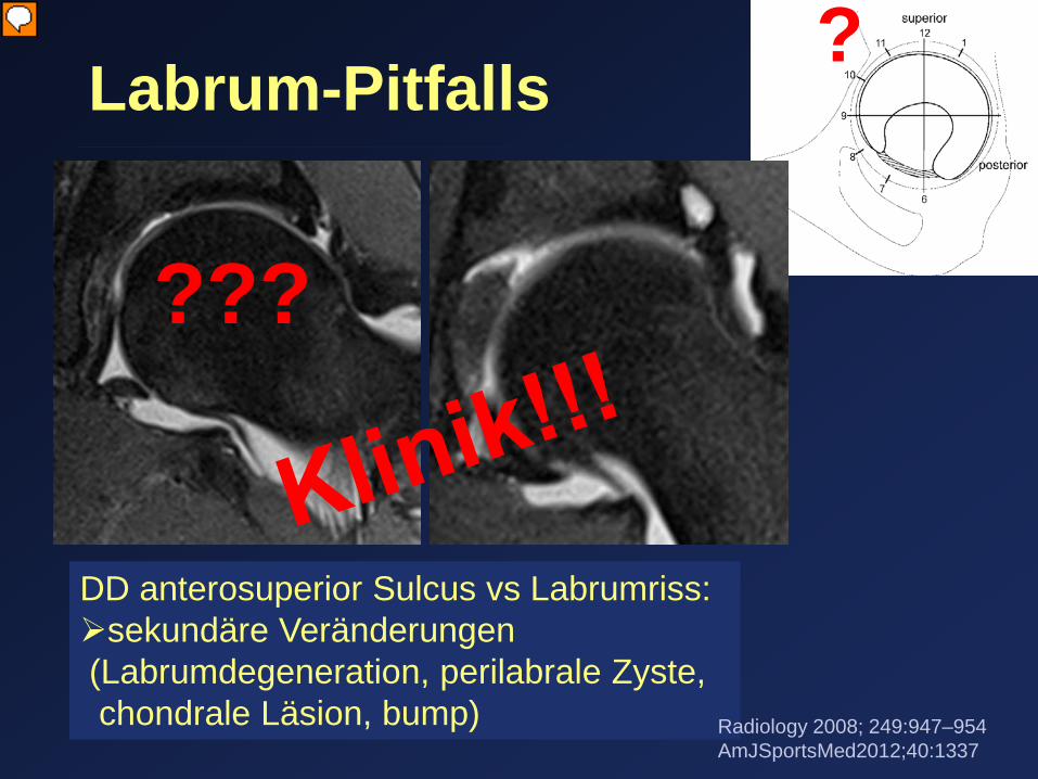
Labrum-Pitfalls
Radiology 2008; 249:947–954
?
DD anterosuperior Sulcus vs Labrumriss: Ausdehnung
Vorführender
Präsentationsnotizen
Studler et al (34) performed a study with 57 patients who underwent subsequent hip surgery or arthroscopy and showed that an anterosuperior cleft may be seen as a normal variant in the presence of a normal lateral acetabular labrum. On anterior coronal or sagittal images, this cleft is seen as a partial undercutting of the labrum on a single image. As with recesses,the extension was less than the entire diameter of the labral base in 51% (22 of 43) of tears, and 49% (21 of 43) of tears also were linear in shape. In addition, 49% (21 of 43) of tears showed no extension into the labrum, and in 26% (11 of 43) of all tears, the adjacent labrum showed no signal abnormalities. Cartilage damage was not present in 47% (20 of 43) of all labral tears. Labral tear at arthroscopy of left hip in 68-year-old woman. (a) Transverse oblique water-excitation true fast imaging with steady-state precession MR image demonstrates anterosuperior interposition of contrast material at 11-o’clock position (arrowhead). (b) Sagittal T1-weighted MRarthrogram shows partially separated labrum from acetabulum (arrows). (c) Corresponding arthroscopic image reveals extensive sublabral cleft (arrowheads) at base of mildly degenerated labrum (L). Labrum was unstable at probing. Acetabular cartilage (A) damage was not present. FHfemoral head.
Acetabular paralabral cysts were identified in 11 of 42 (26.2%) and 9 of 42 (21.4%) hips by the 2 respective radiologists, with an interobserver reliability of 90.5% (κ = .74) and intraobserver reliability of 95.2% (κ = .87). In addition, acetabular labral tears were identified in 36 of 42 (85.7%) and 34 of 42 (80.9%) hips, with an interobserver reliability of 90.5% (κ = .70) and intraobserver reliability of 95.2% (κ = .83).
Assoziation mit Labrumpathologie Os acetabuli
Clin Sports Med 30 (2011) 239–283
Herniation pit
Vorführender
Präsentationsnotizen
The exact association of fibrocystic changes of the femoral neck and FAI is not entirely clear. It is possible that these cysts are more common in patients with pincer-type impingement than cam. An association between an os acetabuli and hip impingement has been suggested. Some believe this condition is heterotopic bone formation on the acetabulum rim related to abnormal contact with the femur. The mineralization may be in the soft tissue or in the labrum.
Knorpel
Varianten
Radiology 2012; 264:651–667
Vorführender
Präsentationsnotizen
Coronal T1-weighted MR arthrographic image of right hip in a 48-year-old womanshows normal cartilage thickness gradients. Examination was performed with 7.5 kg of traction on the leg. Thickness of femoral head cartilage increases from periphery to center (white arrowheads; arrowhead size indicates thickness of cartilage), with maximal thickness next to attachment of ligamentum teres at fovea capitis (arrow). Conversely, thickness of acetabular cartilage (black arrowheads) increases from medial portion to lateral edge of the acetabulum. No cartilage is present at the acetabular fossa (∗). The stellate lesion or crease is a bare area deficient in hyaline cartilage above the anterosuperior margin of the acetabular fossa within the articular area of the acetabulum. This lesion should not be confused with an osteochondral lesion. The tubular acetabular intraosseous contrast tracking is a common magnetic resonance (MR) arthrographic finding that seems to have little or no clinical significance. Although the exact pathophysiologic mechanism is unknown, it is thought to represent dilatation of nutrient foramina along the anterior and posterior margin of the acetabular fossa.
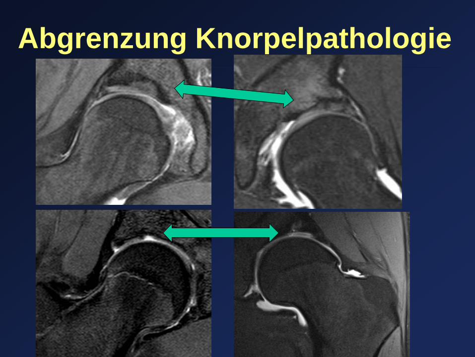
Abgrenzung Knorpelpathologie
Abgrenzung Knorpelpathologie oder diskret...
Vorführender
Präsentationsnotizen
Coronal intermediate-weighted fatsaturated MR arthrographic image of left hip in a 40-year-old woman with mixed-type FAI shows delamination of acetabular cartilage that was confirmed at subsequent arthroscopic surgery. The acetabular cartilage adjacent to acetabular rim shows linear hypointensity (arrow). This finding has 90% specificity for cartilage delamination.
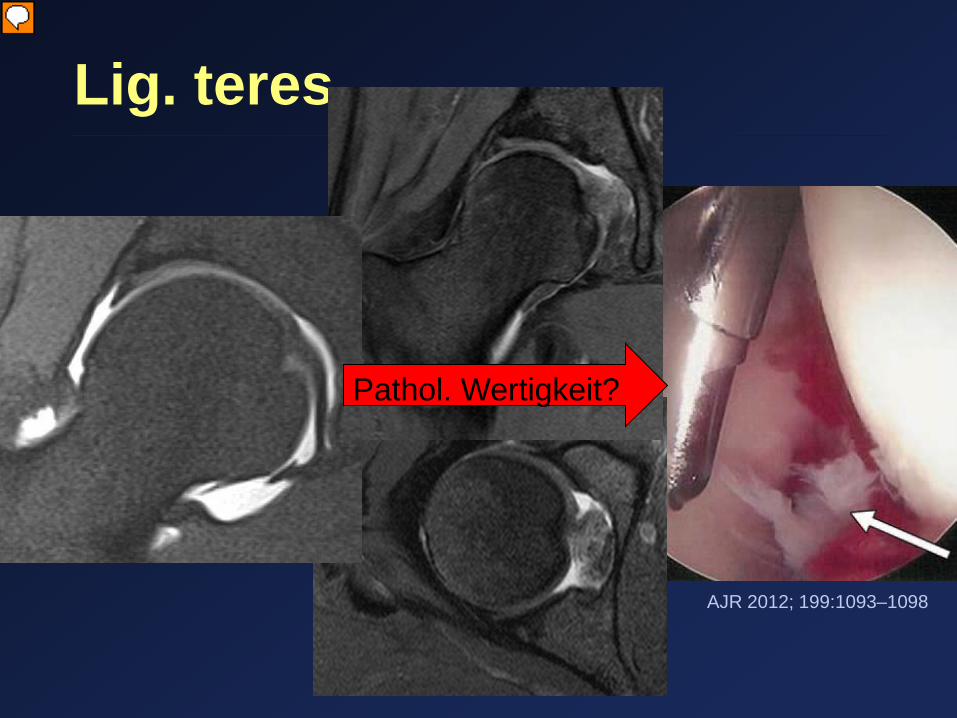
Lig. teres
Pathol. Wertigkeit?
AJR 2012; 199:1093–1098
Vorführender
Präsentationsnotizen
The intact and partially torn ligamentum teres can have similar imaging findings on MR arthrography, making the diagnosis of partial ligamentum teres tears difficult. High SI within the substance of the fibers and irregularity suggest partial tearing. Arthroscopic image shows acute tear of central band (arrow) of ligamentum teres in a patient.
Morphologic details of hip joint on coronal T1-weighted MR arthrographic images of right hip in two patients. Pectinofoveal fold arising from the medial aspect of the femoral neck, extending inferiorly to attach on to the proximal femur. The relationship of this fold to internal impingement is not known. The contour can be either smooth, as was seen in 52% of the patients in our study, or irregular. The irregular appearance should not make one think that the pectinofoveal fold is pathologically abnormal. The pectinofoveal fold more commonly inserts onto the hip capsule (75%) rather than onto the femur.(arrow).
Arthroscopic images from Case 1. a Initial appearance of the plica, the ligamentum teres, and the femoral head. b The plica is retracted away from the ligamentum teres prior to resection. c Following resection of the plica. Black arrow—hip plica, white arrow—ligamentum teres, F—femoral head
Kapselbandapparat Radiology 2012; 263:189–198
Vorführender
Präsentationsnotizen
IBILFL = inferior band of the iliofemoral ligament ILFL = iliofemoral ligament ISFL = ischiofemoral ligament PFL = pubofemoral ligament SBILFL = superior band of the iliofemoral ligament Images of the ILFL. (a) Transverse tissue slice of a cadaveric specimen and (b) corresponding T1-weighted spin-echo (600/17) MR arthrographic image show SBILFL (black arrow) inserting in superior aspect of intertrochanteric line (arrowhead). Medially, IBILFL (white arrow) is seen as distinct thickening on the whole superior-inferior extension of anterior capsule. (c) Transverse tissue slice of a cadaveric specimen and (d) corresponding T1-weighted spin-echo (600/17) MR arthrographic image at a level inferior to a and b show IBILFL (white arrow) inserting in inferior aspect of intertrochanteric line (arrowhead). Inferior portion of ZO is seen curving around inferior circumference of femoral neck (black arrow). Images of ILFL. (a) Sagittal tissue slice of cadaveric specimen and (b) corresponding T1- weighted spin-echo (600/17) MR arthrographic image show vertical orientation of IBILFL (white arrows) longitudinally sliced and visible in its whole extension. Posterior segment of ZO is seen embracing posterior circumference of femoral neck (black arrow). (c) Coronal tissue slice of a cadaveric specimen in anterior aspect of articular capsule and (d) corresponding T1-weighted spin-echo (600/17) MR arthrographic image show SBILFL (black arrows) longitudinally sliced and coursing in lateral and inferior direction to attach in upper part of intertrochanteric line. The IBILFL is partially seen medially in the articular capsule (white arrows). Images of the PFL. (a) Sagittal tissue slice of a cadaveric specimen at the level of the transverse acetabular ligament, (b) corresponding T1-weighted spin-echo (600/17) MR arthrographic image, and (c) histologic sample of the central area in a show extracapsular segment of PFL (arrow) transversely sliced with a characteristic stellate appearance, seen as a hypointense structure surrounded by fat in b, coursing inferiorly and perpendicularly to the transverse acetabular ligament (arrowheads). (Hematoxylin-eosin stain). (d) Sagittal tissue slice of a cadaveric specimen at the level of the medial portion of the articular capsule (lateral to the level in a) and (e) corresponding T1-weighted spin-echo (600/17) MR arthrographic image show capsular segment of PFL (white arrow) as subtle localized thickening of inferior capsule. The iliopsoas tendon and muscle are seen in anterior aspect of the joint (arrowhead). (f) Histologic sample of central area of the tissue slice in d at the level where PFL joins inferior portion of articular capsule shows three well-defined layers of fibers: a superficial layer of longitudinal fibers, consisting of fibers of the PFL (solid black arrow); a middle layer of transversely oriented fibers mainly derived from the ZO (white arrow); and a deep layer of longitudinal capsular fibers (dashed black arrow) that arise from the proximal insertion of the inferior capsule at the transverse acetabular ligament. F = femoral head. (Hematoxylin-eosin stain.) Images of the ISFL. (a) Axial oblique tissue slice of cadaveric specimen in middle portion of articular capsule, (b) high-spatial-resolution radiograph of this slice, and (c) corresponding T1-weighted spin-echo (600/17) MR arthrographic image show ISFL (solid white arrow) as a diffuse thickening of the posterior capsule with longitudinally oriented fibers. ZO (dashed white arrow) is seen as a distinct focal thickening forming distal free border of posterior capsule. A small synovial articular recess (arrowhead), the same as shown in Figure 3d, protrudes laterally to distal border of capsule. ILFL (black arrow) is seen as a thickening in the anterior capsule inserting distally in the intertrochanteric line.
Kapselbandapparat Wann symptomatisch ??? -Capsulitis (adhaesiva) -Mikroinstabilität
Clin Sports Med 30 (2011) 239–283
Vorführender
Präsentationsnotizen
b.Coronal T1 FS-weighted image shows a proximal iliofemoral ligament tear (arrow) and extraarticular contrast leak.
Akzessorischer Muskel
M. iliocapsularis -Stabilisator bei Hüftdysplasie?
Radiology 2008; 249:947–954
?
Vorführender
Präsentationsnotizen
Transverse T1-weighted MR image shows normal iliocapsularis muscle at level of left femoral head in a 35-year old man who had prolonged hip pain after falling on the hip. Iliocapsularis muscle (dashed line) is in proximity to iliacus (I) and rectus femoris (R) muscles. It originates at the hip capsule and anterior inferior iliac spine and inserts distal to the lesser trochanter. While its function in healthy individuals is not completely clear, it is an important stabilizer of the hip joint in patients with dysplastic hips.
Kommunizierende Bursen
Bursa iliopsoas Paralabrale Zyste
Vorführender
Präsentationsnotizen
Another potential pitfall when analyzing MR arthrographic images of the hip is the presence of bursae that can have a normal communication with the hip joint. The iliopsoas bursa lies between the musculotendinous portion of the iliopsoas muscle and the hip joint capsule, and it can have a bilobate appearance. It is the largest bursa around the hip joint, with an average size of 6 × 3 cm when filled with fluid, and it communicates with the hip joint in 15% of the population (133,134). The obturator externus bursa lies between the tendon of the obturator externus muscle and the posterior hip capsule. In a study with 200 hip MR arthrograms (135), images in 5.5% of patients showed an obturator externus bursa that communicated with the hip joint; notably, all of those patients had labral tears. When present, bursae around the hip should be identified and assessed carefully. They may be a secondary finding when intraarticular or periarticular abnormalities are present. Loose bodies can be found in the bursae around the hip (136), and bursae can be involved in neoplastic processes such as pigmented villonodular bursitis or in patients with arthritis (137–139).
a. Illustration of a dissected hip joint (posterior view) shows hip synovium (curved arrows) protruding from the margin of the ischiofemoral capsular ligament (straight arrow). (Reprinted, with permission, from reference 11.) b. T2-weighted fat-suppressed fast SE MR images (4,166/104) obtained in a patient with synovial osteochondromatosis of the left hip. Transverse MR image shows the bursa extending along the plane of the obturator externus muscle and separate from the inferior hip capsule.
Zusammenfassung
DD Labrumvarianten vs Labrumpathologie Abgrenzung Knorpelvariationen vs Knorpelpathologie Normales vs suspektes Lig. teres Plicae und ihre Variationen Anatomie und Wertigkeit Kapselbandapparat Kenntnis kommunizierender Bursen