herd health management equine science ii. what is normal? each horse is unique, but in general every...
TRANSCRIPT

Herd Health Management
Equine Science II

What is normal?
Each horse is unique, but in general every healthy horse should:Be alert and curious about surroundingsBe bright eyed, ears movingBreath easily and regularly
Both at rest and during exerciseShould eat everything given (or nearly)
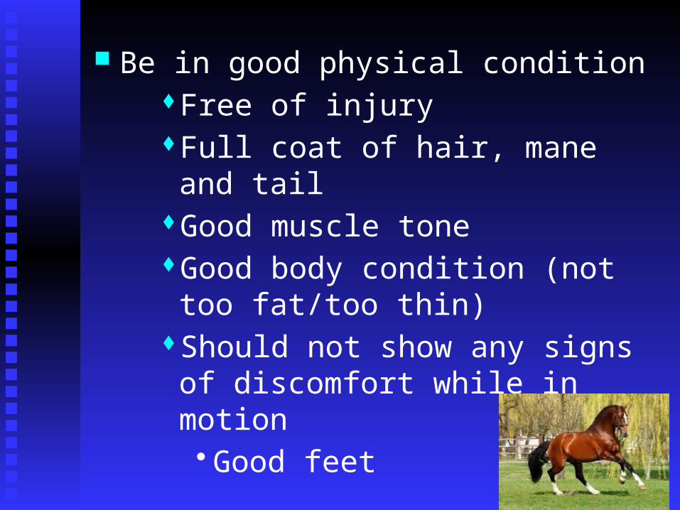
Be in good physical conditionFree of injuryFull coat of hair, mane and tailGood muscle toneGood body condition (not too fat/too
thin)Should not show any signs of
discomfort while in motion• Good feet

Know Your Horse
Each horse is unique and displays consistent behaviorsKnow what is normal for your horse
Including defecation, urination, drinking, eating, stall/pasture behaviors, social behavior
Always be looking for signs that something is not normal

Know your horse It is always good to know what is normal
for your horse BEFORE an emergency strikesTake temperature, pulse, respiration and
keep recordNote any scars, deformities,
conformational issuesKeep a record of previous health issues,
vaccination dates, travel Know the history of your horse

Recognize changesa. When something is wrong, signs are often
subtle
a. Loss of appetite is one of the first warnings that something is wrong.
b. Observe changes in the color, texture, amount, etc. of manure.
b. Dullness of eyes and coat, a runny nose or a persistent cough can all be indications that something is wrong.
c. Changes in behavior
a. Depressed, slow moving


If I suspect something is wrong… 1. General observation
How alert?Eyes, ears, posture, movementShould be continued through observation
Content horses generally display a certain degree of calmness and are alert

2. Check Vital Signs
Temperature Pulse Respiration Rate Capillary Refill Time Mucous Membranes Skin Pliability

3. Other Things to Consider
Body Fluids Movement Recent Travel or new horse brought in Hair Coat Hoof Condition Feeding Habits Body Condition and Weight Behavior Disorders

Conducting the Physical Exam
1. General ObservationsEyes, ears, mouth, legs, body
Muscle tone, exterior injuries, etc 2. T,P, R (Temp, pulse, respiration) 3. CRT (capillary refill time) 4. Hydration test (skin tent) 5. Other

Temperature
1. The normal temperature range is 99.5-101.5 degrees F. 100.5 Normal
1. Foals will have higher temp than adults Factors that May Cause Variations
Include: Time of Day, Age, Sex, Ambient Temperature, Wind,
Precipitation, Activity, and Disease

2. 102 degrees F is a mild fever, 104 degrees F is moderate and 106 degrees F is a high fever
2. The chance for recovery by an equine with a high temperature is low.
3. Rest equine with a 102 degree F temperature and call a veterinarian when the temperature rises to 103 degrees.

3. Use a veterinary thermometer to check a equine’s temperature.
a. Always take the equine’s temperature rectally.
b. Allow three minutes for accurate reading
c. Use the string attached to the thermometer to secure and retrieve.
d. Inserting the thermometer full length helps prevent breaking

4. Procedure for taking equine’s temperature: Shake the mercury down to the 95-97
degree range.

Dip the bulb of the thermometer, bulb first, full length into the rectum
Fasten the thermometer to the equine’s tail using the clip-on string.
Remove after 3 minutes, read and then wash the thermometer with soap and cool water
Finally, dip the thermometer in a disinfectant solution and rinse it again

Respiration
1. The normal rate an equine breathes while at rest is 8-16 breaths per minute.
a. Any kind of distress or activity increases a equine’s respiration rate.
b. Respiration rate should always be lower than heart rate.
c. Fitness determines how quickly RR and heart rate returns to normal

2. Respiration rate can be determined without special equipment by:
a. Counting the number of times the flanks move in and out per minute. (Flanks move in and out with each breath).

b. Counting the number of times the nostrils flare and contract per minute. (the nostrils flare and contract with each breath.)
c. Holding the hand in front of the nostrils to feel the breaths that the equine takes may also help.

Heart Rate
1. Normal heart rate varies Adult: 28-40 beats per minute
Newborn foal: 80-120 Older foals: 60-80 Yearlings: 40-60
A horse in heavy exercise can have a HR of 200 BPM!!

2. Establish the normal rate for the equine by checking the rate when the horse is calm, cool, and relaxed.
The heart rate may have to be checked several times to identify a comfortable range for the normal rate
An ill equine may have a heart rate from 80-120 beats per minute for long periods

3. Determine a equine’s heart rate by counting the pulse for 30 seconds and multiplying by 2

a. Locate an artery at one of the following points
Lower jaw The cheek 4” below eye Under the tail close to the
body Inside foreleg Inside left elbow Against chest wall Behind the knee Inside or outside of the
pastern

b. Press the fingers against an artery and count each throb.
c. Use a stethoscope just behind the equine’s left elbow

Purpose? Determine dehydration & proper blood
flow
How do we determine? Firmly press thumb against upper gum for
a couple of seconds Area should appear white
Should return to normal color in ~2 seconds >2 sec = shock, blood loss
Capillary Refill Time

Includes: Inner eyelid & nostril Inner lips & gums Vulva of the mare
Membranes should be Bright and moist and have a clear pink color Should be aware of unusual odors
Breath should not be Sweet-smelling or foul and pungent in odor
Mucous Membranes
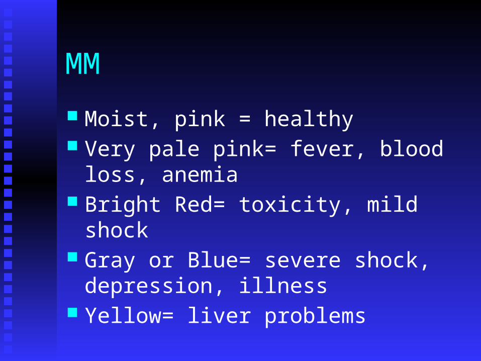
MM
Moist, pink = healthy Very pale pink= fever, blood loss, anemia Bright Red= toxicity, mild shock Gray or Blue= severe shock, depression,
illness Yellow= liver problems
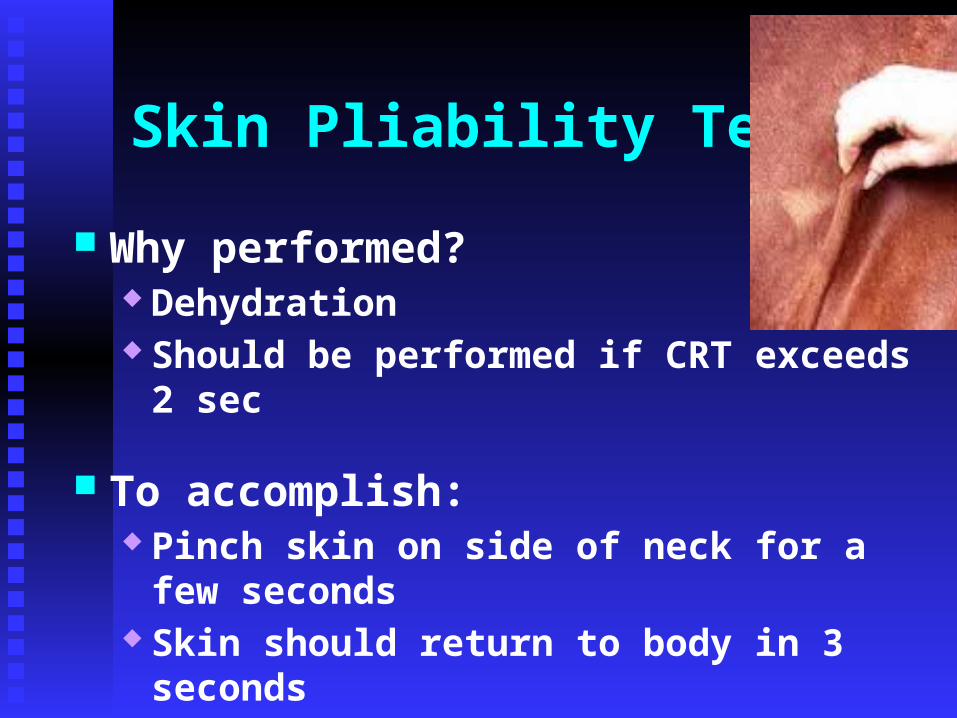
Why performed? Dehydration Should be performed if CRT exceeds 2 sec
To accomplish: Pinch skin on side of neck for a few seconds Skin should return to body in 3 seconds
Skin Pliability Test
Gut Sounds
Indicate if the horses digestive system is working
Listen at the barrel just behind the last rib, check on both sides
Should hear gut noises either with ear or stethoscopeExcessive gut noise is badNo gut noise is terrible***

Feces:Consistency and color reflect health
status
Consistency normally depends on Feed ingested
Loose bowels may indicate Disease of G.I. Tract
Other Parameters

How much will an adult horse produce in 24 hrs? 28-50 lbs Occurs 8 to 10 times/d
Blood is an indication of Severe inflammation
Feces

Normal is Turbid and Rarely Clear Color is usually pale Can be reddish-yellow and still be normal
Frequency and Amount? Urinate 5 to7 times/ 24 hr period Amount varies from 4 to 7 quarts
Mares may produce very thick Oily urine while in estrus
Urine

Important Function for the Healthy Eye: Maintained by the Lachrymal Apparatus Distributes the tears on the inner surface of the eyeball Tears are collected in the
Lower inside corner of the eye & directed in the nasal duct
Obstruction of passage will cause tears to spill over
Tears

Purpose: Thermoregulation Excitement
Unhealthy conditions: May sweat profusely from extended exercise Some loose the ability to sweat
Anhydrosis
Sweat

What is it?1 to 9
1 = Poor9 = Extremely Fat
What is the best BCS?Depends on situation
Body Condition Score

Body Condition Scoring

Body Condition Scoring

What Score Would this Be?

What Score Would this Be?

Should move in a cadenced manner
Lame vs. Sound
How do we determine lameness
Degrees of lameness Grades 1 – 4
What causes lameness
Treatment?
Movement

Hair Coat Time of year Housing Parasite control Age Etc.
Hoof Condition Should be hard, slick shiny surface
Other Parameters

Typically are content if they can Eat and drink at will
What if a horse stops eating?May be first sign of a problemMay not be a problem at all Here again, important to know your
horse
Feeding Habits
Important to understandEating, drinking, content, disturbed,
aggressive, etc.
Abnormal behavior:Stall walking Weaving Cribbing
Behavior

Travel and New Barn Mates
Anytime a horse leaves the property or a new horse arrives brings a chance for disease If your horse shows signs of illness after
either event, check with local authorities or people that attended to determine if other animals are illRecent outbreaks of WEV in Oregon,
Texas

Why First Aid
The nature of equine makes them accident-prone and they are subject to:Kicks or bites from other horses, falls and
injuries sustained by running through or over obstructions when frightened

Equines are naturally curious and may be injured as a result of pawing objects or sticking their heads through holes in fences or stall walls.

Equines may develop unsoundness from injuries because of stress and strain to tendons, ligaments, muscles, bones, etc. and are caused by:Overworking or over exercise from
trainers, riders, etc. Improper exercise (too little followed by
too much)

First aid measure are needed for open wounds so that:Excessive bleeding which can result in
death is controlled. (Some controlled bleeding is good because it helps flush the wound of contaminated material)

The contaminated wound becomes a clean wound.
The wound heals rapidly

Other reasons for first aid treatment include reducing pain, calming the animal until a veterinarian can arrive, and preventing further injury.

Common First Aid Treatments

a. Abrasions (skin scrapes) are superficial wounds caused by falls or tack and are treated by gently and thorough washing of the area with lukewarm water or a saline solution and when possible applying a light bandage.
Wounds

b. Equine who suffer wounds more serious than abrasions or superficial cuts must be treated by a veterinarian, but first aid used for all open wound should be used until a vet arrives.
1. Move the equine to a quiet area and calm it down.

2. Hose the wounded area to remove dirt, clay and other contaminants. (Excessive pressure may force foreign materials deeper into the wound.
3. Clean the wounded area with either saline solution (1 tsp of salt in 1 pint boiled water is .84% salt solution) or diluted solution of mild skin antiseptic in warm water (1% iodine-based washes such as Povidone Iodine or Chorhexidine)

4. Remove foreign objects from puncture wounds and apply a poultice to draw out contaminating materials and keep the wound open
5. Applying firm, direct pressure with a sterile pad to the wound and holding it in place to control arterial bleeding (spurts our of the wound and is bright red). Reconstituted cellulose and absorbable gelatin sponges help blood clot and may be left in the wound since they are absorbed by the body.

Lameness
a. Check each hoof for any foreign object lodged in the sole or frog, remove the object and observe the equine for signs of lameness and rest the equine.

b. First aid treatment for closed wounds, injuries or swelling include:
1. Application of cold is a common first aid treatment because it reduces pain, swelling, bleeding and inflammation
2. Cold treatment should not continue past the first 24-48 hours
3. Apply cold treatment for 20-30 minutes and then wait one hour before starting another 30 minute treatment

Bruises and Fractures
a. Call a veterinarian
b. If leg fracture is suspected, immobilize the leg with a pillow held in place as tightly as possible by wrapping bandages tightly around the pillow and leg.

Strains and Sprains
a. Muscle strains/sprains to tendons and ligaments may be treated with an alcohol rub or liniment
b. After the application of liniment, the owner or caretaker should wrap the horse’s legs in rest bandages.

Bandages used for wounds, sprains and support1. Bandages may protect a wound from
dirt, decrease movement of the wounded or affected area, allow faster healing, cover medications, minimize swelling and provided support.
a. Bandages consist of padding material and an adhesive, elastic or not-elastic wrap
b. Vet rap is a self-adhering, elastic bandage with contouring qualities which can make it useful for bandaging difficult areas, pressure bandages and ice packs

2. A stable bandage extends from below the knee or hock to the fetlock and is used to support the lower leg: support the leg opposite an injured leg: or for wounds. Apply a stable bandage by:
a. Wrapping padding snugly around the leg from just below the joint to below the fetlock and cover with wrapping flannel or knit wrap starting near the middle of the cannon bone.

b. Tuck the end of the wrap under the edge of the padding, and wrap once around the leg to just below the fetlock joint.
c. The bandage should wrap below the back of the joint but rise higher in front, causing an upside down “V” on the front of the joint.

3. Cold water bandages are used to apply cold pressure to cool a warm strained leg. Apply a cold-water bandage by applying cold water to the leg or soaking padding in ice water and applying padding directly to the leg without wringing it out.

a. Wrap the padding snugly with a knit leg wrap.
b. Run cold water over the bandage frequently and do not let the bandage dry out

4. Bandages should be changed when they are no longer functional; the leg is swollen above or below the bandage; the equine shows signs of pain; the bandage slips out of position or the bandage becomes dirty.
Hoof Anatomy
1. Parts of a horse’s foot
a. The hoof wall is a horny substance made of parallel fibers protected by a varnish-like coating called periople that also holds moisture in the hoof. The hoof wall functions to: provide a
weight bearing surface, protect the internal structure of the foot and maintain moisture in the foot.

b. The coronet, or coronary band is an area directly above the hoof wall that serves as the source of growth for the hoof wall.
c. The pastern- The part of the horse’s leg between the fetlock and the coronet that affects the stride of the horse.
d. The sole of the foot is a horny substance that protects the sensitive inner portions.
e. The frog is a triangular shaped formation in the sole of an equine’s foot. The frog of a healthy hoof must remain elastic as it acts like a shock absorber.

2. The hoof is designed to change shape when weight is applied. As weight is applied the sole flattens and the hoof expands laterally at the heel.

Growth of the Hoof1. The growth rate of the hoof is about 3/8”
per month depending on exercise and general health of the equine.
a. Hind hooves grow faster than front hooves because they have less weight to rise.

b. Unshod hooves grow faster than shod because the nails and shoe of a shod hoof limits movement.
c. The hooves of mares and geldings grow faster than stallions because they get more exercise than stallions.

2. The hoof grows at a 45-55 degree angle with the ground.

Important Points in Foot Care
1. Foot care is often neglected as a horse management practice. Foot care includes
a. Routine cleaning with a pick. Pick from the heel to the toe of the foot to prevent injury.
b. Trimming the hooves every 4-6 weeks so they retain proper shape and length.

c. Correcting minor imperfections by trimming such as splayfoot, toed in and toed out.
d. Treatment of foot diseases and injuries. Thrush is a bacterial infection that penetrates the frog, making it soft and mushy. Thrush is related to lack of cleaning. Wet conditions cause rapid drying out of the horse’s foot.

Reasons for Shoeing
1. Protect the hooves from excessive wear.
2. Provide better traction.
3. Help correct defects of stance or gait such as forging.
4. Help cure diseased or defected hooves such as inflamed tendons.

5. Shoes can provide relief from the pain of injured parts such as bruised soles and hoof wall cracks.

6. Shoes do not make walking easier; shoes do not improve agility; shoes do increase shock and road concussion and nail holes made in attaching shoes weaken the hoof wall, may cause separation and may provide entry for infection
7. A proper fitting shoe should follow closely the outline of the trimmed hoof at the toe and around the wall to the bend of the quarter. Then it should widen gradually until it extends laterally 1/8” beyond the hoof wall at the heel. This provides support for the expanded hoof when the horse places weight on the shod foot.
8. The branch of a properly fitted shoe should not project beyond the upper part of the hoof at the heel.
9. The last nail should be placed on the widest part of the hoof. Placing the nail too far to the rear hinders the lateral expansion of the foot at the heel.

Common Dental Problems
1. A common problem is painful sores in the equine’s mouth as a result of sharp edges of hooks on the molars caused when the equine’s molars do not meet evenly.
2. Wolf teeth (one to four small teeth that may develop in front of the molars) can cause “bit” problems.

3. Canine teeth on older equine can get too long, hit the opposite gum and cause sores.

4. Temporary teeth (caps) that fail to fall out may stick to the equine’s gums while the permanent teeth are coming in and can make chewing difficult.

5. Parrot mouth is a common problem that results when the lower jaw is too short and affects the equine’s ability to graze.

6. Chipped or broken incisors may result from the bad habit of cribbing.
a. Cribbing a bad habit of equines living in stalls that results from boredom.
b. Cribbing occurs when an equine grabs objects with their teeth, arch their necks and swallow air.

Recognizing Dental Problems
1. Observe the equine while it is eating to learn if it has problems chewing. Signs are:
The equine moves food around it its mouth a lot prior to swallowing and food falls from the horse’s mouth as it eats.
The equine refuses to eat

2. Make an examination of the equine's mouth by:
Grasping the equine’s lower jaw with one hand to open the mouth.
At the same time use the other hand to pull and hold the equine’s tongue to the side of the mouth so that visual inspection can be made.

Correcting Dental Problems1. A veterinarian uses a special rasp called a
float to file and remove sharp edges from an equine’s teeth.
2. A veterinarian can pull wolf teeth.

3. Equine with parrot mouth should not be bred since parrot mouth is an inherited defect.
4. Equine may be prevented from cribbing by placing:
Cribbing strap around the equine’s throatlatch.
The strap presses on the equine’s trachea when it arches its neck.

Internal Parasites
An internal parasite lives at least part of its life cycle inside the host.
There are more than 150 types of internal parasites that can infect equine.

No individual equine is ever completely free of internal parasites but relatively few internal parasites cause serious damage to the equine.
Most internal parasites live in the digestive tract, lungs, bloodstream or body cavity of the equine.
The extent of injury from internal parasites depends on:The kind of parasite.The number of parasites involved, and The length of time the parasite lives in
the host.
The general life cycle of internal parasites includes five stages:

Stage 1- Eggs from the internal parasite pass out of the equine in the feces and is deposited on the pasture.
Stage 2- Eggs hatch on the pasture and become infective larvae.
Stage 3- The equine ingest the larvae on the grass stem.

Stage 4- The immature parasites migrate through the equine’s tissues.
Stage 5- The mature parasites live in the digestive tract and lay eggs.

The MOST abundant and harmful internal parasites affecting equine are:

Large strongyles (bloodworms) pose the most serious threat to the equine’s health out of all internal parasites.Migrate within artery walls from the
digestive tract toward the heart.May cause damage to arteries so the
equine bleeds internally and dies.

Small strongyles spend their entire life cycle in the intestinal walls.Cause ulcerations in the intestinal
walls that may interfere with digestion.
They do not attach to the lining of the intestine and they do not suck blood.

Ascarids (large roundworms) are the largest parasite that infect equine.May grow to 15” long and be the size
of a pencil.Affect young equine less than two
years of age.
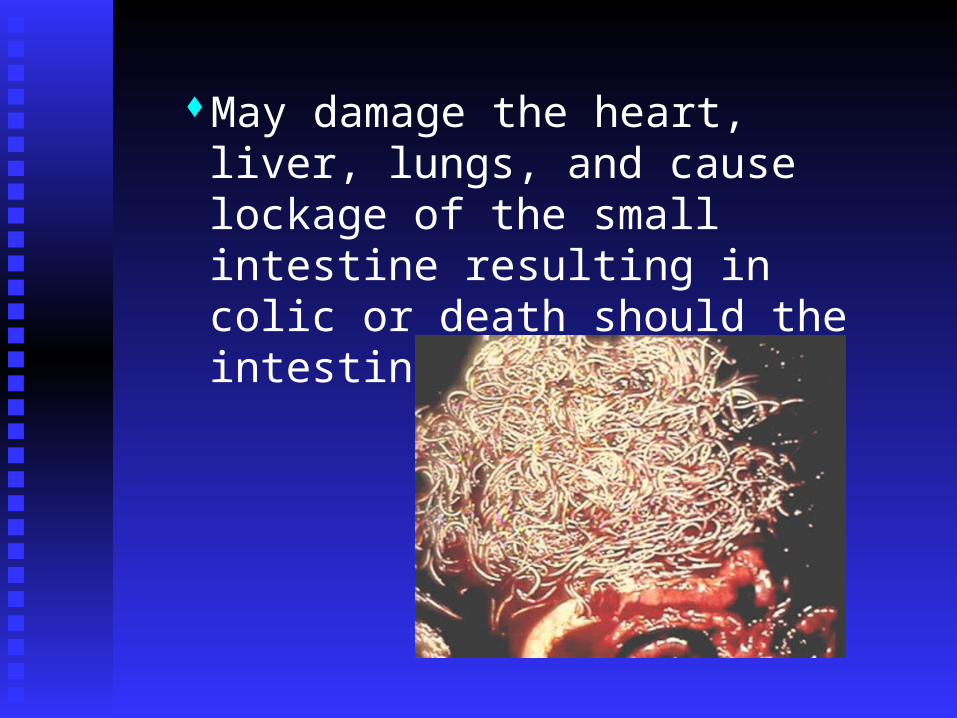
May damage the heart, liver, lungs, and cause lockage of the small intestine resulting in colic or death should the intestine rupture.

Pinworms are more of a nuisance causing digestive problems but resulting in little damage.

Bots are flies that lay eggs on various parts of the equine.Eggs hatch into larvae and are
ingested by the equine when the equine licks the area where eggs are laid.
Other eggs hatch and larvae crawl into the mouth, from the nostrils and lips where they mature in the gums and membranes of the inner lips.

When horsemen refer to bots, they usually mean the mature larvae that attach to the lining of the stomach.
Bots cause stomach problems and can block the entrance to the small intestine causing the stomach to rupture.

Management practices and treatment include:Proper manure disposal which includes:
Timely removal on a weekly basisComposting prior to spreading on
pasture grazed by animals other than equine or spread on cropland or ungrazed areas.

Proper pasture management that includesThe use of temporary pastures where
possible.Frequent mowing and grain harrowing.Rotational grazing when possible.Separate pasture for young and old
equine.Avoiding overstocking.
Never feed equine from the ground. Always use troughs and mangers.

Regular use of dewormer under the supervision of a veterinarian.Ivermectin, a form of avermectin,
controls al common internal parasites.Dewormers may be administered
easily with little chance of injury by a paste.

The most effective way to administer a dewormer is by a stomach tube but usually a veterinarian is required for that procedure.
Feed additives are effective as long as the equine will eat the materials.

External Parasites
External parasites annoy equine and may infect equine with deadly diseases.
External parasites may leave a equine weak, lower feed efficiency and produce raw sores.

Some common external parasites that attach equine include:Ticks can cause damage and transmit
disease such as African equine fever.

Lice are most often found on neglected equine.Two types of lice live on equine: The
biting louse and the sucking louse.Lice spread quickly from equine to
equine.

Mites cause the condition mange or scabies in equine. Mange is very contagiousSeparate equine with mange from
healthy equine and use different grooming equipment

Gnats are bloodsuckers and cause extreme itching after they bite.
Mosquitoes carry viruses and bacteria which cause diseases such as equine infectious anemia (sleeping sickness).
Flies are annoying to equine and carry stomach worms from equine to equine.Female screworm flies lay eggs in
fresh wounds on animals that hatch into larva that feed on the tissue resulting in large sores.

Management practices and treatment for external parasites include;Regular removal of manure, dirty stall
bedding and materials which encourage the breeding of parasites.
Maintaining fresh water supplies to discourage breeding by external parasites.

Use chemicals according to label instructions as repellants and control methods.
Use regular insecticide application on infected animals according to label instructions.

Use biological controls such as predator wasp to reduce the need for chemical controls.
Use mechanical controls such as a face mask made from scrap leather or commercial vinyl masks to keep face flies away.

Equine Infectious Anemia (EIA)
1. A viral diseases that affects the equine’s immune system resulting in recurrent fever, weight loss and anemia.
2. Once an equine is infects, it remains infected for the rest of its life.

3. Chronically infected equine may go for years without showing signs of anemia only to have the sign recur when stress, environmental conditions or other disease affects the equine.

4. Horseflies and deerflies are the major natural transmitters of the virus from one equine to another.
5. Detection- A blood test called the “Coggins test” is used to detect the presence of EIA.
6. Equine must have a negative Coggins test before they can compete or be placed in events in North Carolina
7. Prevention involves isolation from equine that are not infected and protection from biting, flying insects.
8. Treatment There is no effective treatment or
vaccination for the disease In some cases, equine with EIA are
required to be euthanized.

Equine Influenza
1. There are two common features of this viral infection
Extremely rapid spread of infection Frequent, dry cough

2. Equine with influenza have an elevated temperature of 102.5-105 degrees F which persist up to 5 days
3. The death rate from influenza is very low

4. The virus is spread to other equine when the equine exhales or coughs and may be carried by handlers on equipment that have been in contact with an infected equine.
5. Prevention Use two intramuscular injections of
influenza vaccine scheduled 2-4 weeks apart followed by a booster shot at 3-4 month intervals for horses at risk.
Isolation of infected equine and screening animals prior to transportation will reduce the spread of the disease.

6. Treatment One week of stall rest is recommended
for each day the equine has an elevated temperature due to influenza
Maintain a dust free environment during the illness and recovery by wetting hay and providing clean bedding.

Equine Rhinopneumonitis
1. This is a respiratory disease caused by EHV virus.
2. The infection is accompanied by a gold colored nasal discharge among foals.

3. The virus associated with this disease also may cause abortions in pregnant mares and occasional paralysis.
4. Equine may develop a temperature of 102-106 degrees F which lasts for 12-48 hours.

5. Equine become infected when they inhale the virus exhaled by sick or even apparently healthy equine that are infected.
6. Prevention requires a combination of: Sensible management practices
Mares should be isolated and separated from other equine
Thorough cleaning and sanitation in the event of dead or aborted fetus

Vaccination Vaccinate pregnant mares in the 5th 7th
and 9th month of pregnancy. Young equine should receive two
injections followed by a booster according to manufacturer’s recommendations

7. Treatment is limited as there is not specific anti-viral therapy available.

Strangles
1. A highly contagious bacterial infection most commonly found in young equine from one to five years of age.
2. Early signs of strangles include fever, depression and loss of appetite due to difficulty with swallowing.

3. As strangles progresses, the lower jaw and throatlatch region may become hot, swollen and painful with abscesses forming and rupturing onto the skin.
4. Pus from ruptured abscesses may contaminate water buckets and communal feeders for months.

5. When strangles develop and abscesses from on internal organs, mortality may be as high as 10%.
6. Prevention and treatment are used in conjunction.
Treat ruptured abscesses with a mild antiseptic solution to hasten healing

Administer Procaine penicillin G as an antibiotic treatment until clinical signs no longer exist for five days.
Isolate affected animals from all other equine
Vaccinate with 2 or 3 injections one month apart and apply a booster annually.

Equine Viral Arteritis (EVA)
1. EVA is a viral infection that causes respiratory illness with nasal and ocular discharges, swelling (stocking up) of the hind limbs and other areas of the body and sometimes abortion.
2. EVA may be passed by respiratory transmission when equine come in close contact.

3. Equine invariable make uneventful clinical recoveries even without treatment
4. Perhaps EVA is most pronounced as a breeding disease through the venereal spread of the disease by acutely affected stallions.
Viral persistence in the stallion can range from several weeks to the entire lifetime of the equine
Mares can be infected either at time of natural breeding or artificial breeding.

5. Prevention Immunize the breeding stallion
population with MLV vaccine. Immunize all mares if they are
inseminated with EVA positive semen.

6. Treatment There is no specific anti-viral treatment
for equine with EVA Infected stallions should receive forced
rest if they show symptoms of the disease.

Eastern, Western and Venzuelan Equine Encephalomyelitis (EEE, WEE, and VEE)
1. EEE, WEE, and VEE are viral infections that result in the inflammation of the brain.

2. The viral infections are spread by insect contact.
The virus may live in reservoir hosts such as birds and rodents for long periods of time without harming the host.

The virus is transmitted and spread by mosquitoes (vectors) to equine and humans.
The viral infection is not transmitted between equine and humans.

3. The disease has high mortality rates: EEE is 75-100% WEE is 20-50% VEE is 40-80%

4. Complete recovery is rare with equine frequently continuing to exhibit clumsiness, depression and abnormal behavior.
5. Signs of EEE, WEE, and VEE include aggression, propulsive walking, excitability, and a number of things that indicate the equine is confused.

6. Prevention focuses on mosquito control. Use insecticides and repellants when
possible and practical Eliminate standing water Screen stalls, use fans to move air, and
limit the use of incandescent lights in stall areas

Brings equine inside prior to dusk Vaccinate with 3 injections at 3, 4, and 6
months of age and give a booster at the beginning of insect season

7. Treatment There is not specific treatment available
Equine Colic
1. Colic is the behavioral signs of abdominal pain in equine characterized by various activities including, but not limited to: tail twitching; head tossing; kicking toward the belly with one of the hind limbs; pawing the ground; grinding the cheek teeth and frequent attempts to lie down and roll on the back, etc.

2. True colic is due to intestinal obstruction or disease which causes pain
3. 95% of all colic cases are from two causes Spasmodic causes where their is intestinal
spasm caused by numerous things such as stress, anxiety, diet change, parasite damage, dewormers, etc.
Large intestinal impaction (constipation) caused most often by improper diet and exercise (Human management is often culprit)

4. Prevention Use a parasite management program to
avoid problems Careful rotation of pasture helps
reduce levels of parasites Avoid overstocking pastures Use “Ivermectin” as a deworming
compound Pay attention to the equine’s diet

5. Treatment Place the equine in a safe environment Treat with analgesia for pain relief Treat with mineral oil as a laxative

Remove feed until the equine has improved and then reintroduce feed gradually
Do not allow the equine to eat too much hay or roughage (lightly grazing the pasture is recommended)

Founder
1. Characterized by the hoof collapsing as a result of laminitis (inflammation of the supporting tissue between the hoof wall and pedal bone)
2. Causes lameness in the horse and if left unchecked can cause permanent lameness or even require euthanization.

3. The leading cause of founder is gastrointestinal disturbance from any number of factors such as colic, grain overload, lush grass, excessive cold water, etc.
4. Other factors that cause founder are exhaustion, excessive concussion (road founder) contact with black walnut shavings, etc.

5. Prevention Control diet Avoid overworking the equine

6. Treatment Identify cause and direct treatment
toward alleviating the problem Increase blood flow to the laminae
through the use of drugs and IV fluids Use aspirin or heparin to thin blood

Use anti-inflammatory non-steroidal drugs (Ketaproten) to reduce laminar swelling
Pack the frog and sole to increase support Use specialized shoes to relieve pressure
to the hoof area.

Tetanus (Lockjaw)
1. Caused by a neurotoxin that allows uncontrolled muscle contraction and muscle spasms
2. Affected horses most often have sustained a wound from 2 days to a month prior to the onset of tetanus; however, tetanus bacteria also live in the equine’s digestive tract

3. Tetanus has become less common due to vaccination, but is still highly fatal
4. Equine usually die from suffocation, cardiac arrest or starvation since their muscular system does not function

5. Prevention Vaccination with two doses of toxoid
vaccine given one month apart followed by a booster shot annually
Equine that are injured should receive the toxoid booster if there is no record of prior shots

6. Treatment Administer penicilln Aggressively clean the wound Administer tetanus anitoxin Use tranquilizers and muscle relaxes to
relieve spasms and muscle pain Provide IV fluids and nutritional
support

Potamac Horse Fever (PHF)
1. PHF is a disease which in its extreme form results in profuse, watery diarrhea, fever, shock and laminitis
2. The occurrence of PHF disease is consistently within 5 miles of a river

3. PHF is caused by an organism that can survive within living cells and must be transmitted through blood cells by insects such as ticks
4. Equine with PHF are not considered contagious to other equine

5. Prevention Vaccinate with two injections one
month apart Give a booster injection annually in
May or June

6. Treatment A veterinarian should use tetracycline
(anti-microbial drug) for 4-5 days IV fluids, frog pads, sole support, and
anti-inflammatory drugs also help

Borreliosis (Lyme Disease)
1. Lyme disease is a bacterial infection transmitted primarily by certain ticks
2. Rare among equine and only a problem where Lyme disease is found among human populations
3. Arthritis is the most commonly reported sign of Lyme disease in equine

4. Prevention Careful grooming to remove ticks Avoiding tick-infested areas

5. Treatment Use antibiotics such as tetracycline and
ampicillin Prolonged treatment of 10-30 days
usual

West Nile Virus (WNV)
1. Viral infection that can cause inflammation of the brain and often mimics EEE in equine
First introduced in western hemisphere in 1999
Equine are more often affected by WNV than any other domesticated animal
2. WNV is spread by mosquitoes when they bite an infected bird and then bite a human or an equine
Equine are not contagious and do not pose a health risk to other animals or humans
About 30% of horses who show clinical signs either die or have to be euthanized


3. Clinical sign of WNV may include circling, aimless wandering, head pressing, impaired vision, hyper excitability, etc.

4. Prevention See mosquito and control techniques
listed for EEE, WEE and VEE A veterinarian should vaccinate with
“protocol” and follow with a booster injection three weeks later
5. Treatment is still developing

THE END!!!