her2 expression and gene amplification is rarely
TRANSCRIPT

AUS DER KLINIK UND POLIKLINIK FÜR MUND-, KIEFER- UND GESICHTSCHIRURGIE
(NORDWESTDEUTSCHE KIEFERKLINIK)
DES KOPF- UND NEUROZENTRUMS
DES UNIVERSITÄTSKLINIKUMS HAMBURG-EPPENDORF
DIREKTOR: PROF. DR. MED. DR. MED. DENT. M. HEILAND
HER2 EXPRESSION AND GENE AMPLIFICATION IS RARELY DETECTABLE IN PATIENTS WITH ORAL SQUAMOUS CELL CARCINOMAS
DISSERTATION
ZUR ERLANGUNG DES GRADES EINES DOKTORS DER MEDIZIN AN DER MEDIZINISCHEN FAKULTÄT DER UNIVERSITÄT HAMBURG
VORGELEGT VON:
ROBERT ANDRÉ GAUDIN
AUS BERLIN
HAMBURG 2014

2
Angenommen von der Medizinischen Fakultät der Universität Hamburg am: Veröffentlicht mit Genehmigung der Medizinischen Fakultät der Universität Hamburg. Prüfungsausschuss, der/die Vorsitzende: Prüfungsausschuss, zweite/r Gutachter/in: Prüfungsausschuss, dritte/r Gutachter/in:

3
„Wirkliches Neuland in einer Wissenschaft kann wohl nur gewonnen werden, wenn man
an einer entscheidenden Stelle bereit ist, den Grund zu verlassen, auf dem die bisherige
Wissenschaft ruht, und gewissermaßen ins Leere zu springen.“
Werner Heisenberg (1901-76)

4
Table of Contents
I. Publication ............................................................................................................... 5
II. Introduction ........................................................................................................... 10
III. History and state of knowledge of Her2 ................................................................ 10
IV. Head and neck squamous cell carcinoma/Her2 .................................................... 11
V. Aims of our study .................................................................................................. 12
VI. Brief summary of methods and materials .............................................................. 12
a. Tissue-Micro Array (TMA) ..................................................................................... 12
b. Immunohistochemistry (IHC) ................................................................................. 13
c. Fluorescence in situ hybridization (FISH) .............................................................. 15
d. Statistical analysis ................................................................................................. 16
VII. Summary of results with discussion ...................................................................... 16
VIII. References ............................................................................................................ 18
IX. Erklärung des Eigenanteils an der Publikation ...................................................... 20
X. Danksagung .......................................................................................................... 21
XI. Lebenslauf ............................................................................................................. 22
XII. Eidesstattliche Versicherung ................................................................................. 24

5
I. Publication

6

7
8

9

10
Her2 expression and gene amplification is rarely detectable in patients with oral
squamous cell carcinomas
II. Introduction
The Her2 oncogene seems to be important for the growth of many different tumours. It is most
strongly associated with the important therapeutic target in breast cancer. This gene product
which shows a significant association to cell proliferation and cell cycle control, is of high
scientific interest not only for breast cancer but also for other cancers like in our case for
squamous cell carcinomas of the head and neck region, because it could be a possible target for
a specific therapeutic approach and eventually be a prognostic marker. Since there is
inconsistent data to this topic our objectives were to clarify the significance of HER-2 expression
and HER-2 gene amplification for the squamous cell carcinoma of the head and neck region.
III. History and state of knowledge of Her2
Her2 (human epidermal growth factor receptor 2, also known as erbB2 (standing for its origin in
the Erb-b gene which is responsible for avian erythroblastosis virus)), is an oncogene which has
been localised to chromosome 17q21 and encodes for one of the epithelial growth factor
receptors on the cell. It has a molecular weight of 185.000 dalton (Stern et al. 1986) and
belongs to the HER family of four transmembrane receptor tyrosine kinases involved in signal
transduction pathways that regulate cell growth and differentiation. It was discovered at the
Massachusetts Institute of Technology by the Weinberg scientists Group in 1982-1984. From
then on it was in the focus of science and research. It was shown that it was amplified in a range
of tumor types including breast, ovarian, bladder, salivary gland, endometrial, pancreatic and
non-small-cell lung cancer (NSCLC)(Scholl et al. 2001). Her2 is involved in disease initiation and
progression, associated with poor prognosis, and may also predict the response to
chemotherapy and hormonal therapy(Scholl et al. 2001). Based on this data anti-Her2
monoclonal antibodies have been designed to specifically antagonise the function of the HER2
receptor in Her2-positive tumours (Scholl et al. 2001). The therapy with these agents such as
Trastuzumab „targeted therapy of HER-2“ is well established for the metastasized HER2 positive

11
breast cancer (Tripathy et al. 2004). Although there is a positivity of HER2 reported for many
human tumor types the data of positivity varies. This applies to immunohistochemical
examination procedures (IHC), which result in a wide span of positive HER2 test results through
the use of several different reagents and records of the examination criteria. It is reported for
example that there is a HER2 overexpression of 5.7-88.8% of the non-small cell lung cancer
cases (Ugocsai et al. 2005) and in 3-54% of the colon cancer cases (Ooi et al. 2004). On the
other hand there is a variability of HER2 positivity in the clinical analysis of amplification.
Different detection methods for example Southern blot or fluorescent in situ hybridization (FISH)
and different evaluation criteria seem to be responsible for the different HER-2 amplification
results given in the literature. Reports on tumour examinations at the head and neck region state
also a different extent of HER2 overexpression in 11%-60% of the cases and a HER2
amplification in 18%-46% of the cases (Scheer 2003).
IV. Head and neck squamous cell carcinoma/Her2
Head and neck cancer is a broad term used to describe malignancies that arise in the nasal and
oral cavities, pharynx and larynx, as well as the paranasal sinuses. Most of these epithelial
cancers are squamous cell carcinomas. Head and neck squamous cell carcinoma (HNSCC) is a
common, morbid, and frequently lethal malignancy (Hanken* et al. 2014). HNSCC is the sixth
most common non-skin cancer in the world, with an incidence of about 600,000 cases per year
and mortality rate of about 50% (Ferlay et al. 2010). Besides alcohol and tobacco abuse, it is
becoming evident that human papilloma virus (HPV) infection is a more and more important risk
factor for the development of HNSCC (Jain et al. 2013). Despite recent advances in research,
the survival rates for many types of HNSCC have improved little over the past forty years (Gupta
et al. 2009). A crucial step to improve the prognosis is the identification of tumour specific
proteins like Her2 that could be used as therapeutic targets. As mentioned above, reports on
tumour examinations at the head and neck region state a different extent of HER2
overexpression in 11%-60% of the cases and a Her2 amplification in 18%-46% of the cases
(Scheer 2003). Her2 could be a possible target for a specific therapeutic approach and
eventually be a prognostic marker. Therefore it is important to clarify the significance of HER-2
expression and HER-2 gene amplification for the squamous cell carcinoma of the oral cavity.

12
V. Aims of our study
We conducted these studies in order to learn more on the rate of HER2 gene amplification and
overexpression of the receptor in head and neck squamous cell carcinoma (HNSCC) and to find
out more about the subanatomical entity of oral squamous cell carcinomas (OSCC) and the
significance of HER2 gene amplification and expression in HNSCC and the subgroup of OSCC.
VI. Brief summary of methods and materials
Two tissue microarrays (TMA) were used for the immunohistochemical (IHC) and fluorescence
in situ hybridization (FISH) examinations to reach maximum standardization of investigation for
the tumour collective. This technique for the assessment of Her2 status is well established.
Several authors have validated the use of TMA technology in this setting in comparative studies
using archived material. Over the years of use it has been shown that the TMA technology is a
useful tool for the validation of different HER-2 FISH protocols and for assessment of
interlaboratory reproducibility (Graham et al. 2008). In the presented studies, in order to clarify
the significance of HER-2 expression and HER-2 gene amplification for the squamous cell
carcinoma of the oral cavity, the method authorised by the FDA (US Food and Drug
Administration) has been used together with the associated evaluation scoring of the
immunohistochemical examination (Hercep TestTM; DAKO, Glostrup, Denmark) and the
fluorescent in situ hybridization (PathVysionTM; Vysis). The studies were conducted at a Tissue-
Micro-Array (TMA) in order to reach maximum standardisation of investigation for the tumour
collective.
a. Tissue-Micro Array (TMA)
Tissue Micro Arrays consist of paraffin blocks in which up to 1000 separate tissue cores are
brought into an array format and can be analysed simultaneously. Based on paraffin-embedded
tissue blocks, core needle biopsies are taken from specific locations and are re-embedded into
one arrayed block from which the analysation is taking place. The TMA technology is a fast,
cost-effective, and statistically powerful method that will substantially facilitate translational
research (Simon & Sauter 2004).
The first TMA in this study contained 222 oral squamous cell carcinomas (OSCC) from 157 male
and 67 female patients treated in the department for oral- and maxillofacial surgery of the
University Medical Center Hamburg-Eppendorf, Germany, between 1988 and 2007. Among
these oral carcinomas, 33 were at the tongue’s margin, 122 at the floor of the mouth, 35 in the

13
alveolar process, 10 located oropharynegeal and 22 in the upper jaw. The second well-
established TMA from Basel was a comparative analysis consisted of 427 carcinomas of the
head- and neck region (HNSCC) including 92 laryngeal carcinomas, 215 pharyngeal carcinomas
and 120 OSCC`s.
b. Immunohistochemistry (IHC)
Immunohistochemistry refers to the process of detecting antigens (e.g. proteins) in cells of a
tissue section by exploiting the principle of antibodies binding specifically to antigens in
biological tissues. The IHC technique is a combination of immunologic and chemical reactions
visualised with a photonic microscope. It starts with deparaffination of tissue sections, then
includes preincubation steps (e.g. antigen retrieval, blocking of nonspecific activities), incubation
with the primary antibody, and labeling of the antigen-antibody reaction and ends with slide
counterstaining and coverslipping (Ramos-Vara & Miller 2014). In our TMA sections we used the
Food and Drug Administration–approved HercepTest kit (polyclonal rabbit antibody, undiluted,
DAKO, Glostrup, Denmark) to detect the Her2 expression. For the visualisation of the bound
antibody we used the EnVision Kit (DAKO, Glostrup, Denmark). A hematoxylin counterstain was
done. Two pathologist examined all spots of the TMA in a blind manner and rated the extent and
intensity of background staining as well as nuclear and non-nuclear staining of tumour cells.
Only spots with more than 20 tumour cells were evaluated. HER2 immunohistochemical
expression was scored exactly as described for the Food and Drug Administration–approved
HercepTest in four categories: 0, 1+, 2+, and 3+ (Wolff et al. 2013) by the producer (DAKO
(Hercep-Test®, Hamburg DAKO; table 1) (M. Blessmann 2008).

14
Tab.1: Evaluation criteria for Hercep-Score (HER-2).
Figure 1: Representative pictures of (A) negative and (B) positive Her2 immunostaining in oral
squamous cell carcinomas (Hanken* et al. 2014).
Staining pattern HER-2
Overexpression Score
Absence of flourescence or membrane coloring in less
than 10 % of tumour cells. Negative 0
In more than 10 % of tumour cells weakly/hardly
visible membrane coloring. Only part of membrane. Negative 1+
In more than 10 % of tumour cells weakly to moderately
visible coloring of entire cell membrane.
weakly positive 2+
In more than 10 % of tumour cells strongly
visible coloring of entire cell membrane.
strongly positive 3+
15
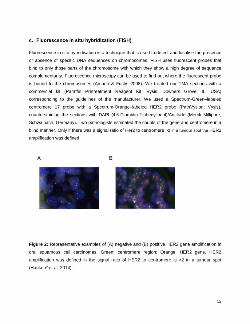
c. Fluorescence in situ hybridization (FISH)
Fluorescence in situ hybridisation is a technique that is used to detect and localise the presence
or absence of specific DNA sequences on chromosomes. FISH uses fluorescent probes that
bind to only those parts of the chromosome with which they show a high degree of sequence
complementarity. Fluorescence microscopy can be used to find out where the fluorescent probe
is bound to the chromosomes (Amann & Fuchs 2008). We treated our TMA sections with a
commercial kit (Paraffin Pretreatment Reagent Kit, Vysis, Downers Grove, IL, USA)
corresponding to the guidelines of the manufacturer. We used a Spectrum-Green–labeled
centromere 17 probe with a Spectrum-Orange–labeled HER2 probe (PathVysion; Vysis),
counterstaining the sections with DAPI (4′6-Diamidin-2-phenylindol)/Antifade (Merck Millipore,
Schwalbach, Germany). Two pathologists estimated the counts of the gene and centromere in a
blind manner. Only if there was a signal ratio of Her2 to centromere >2 in a tumour spot the HER2
amplification was defined.
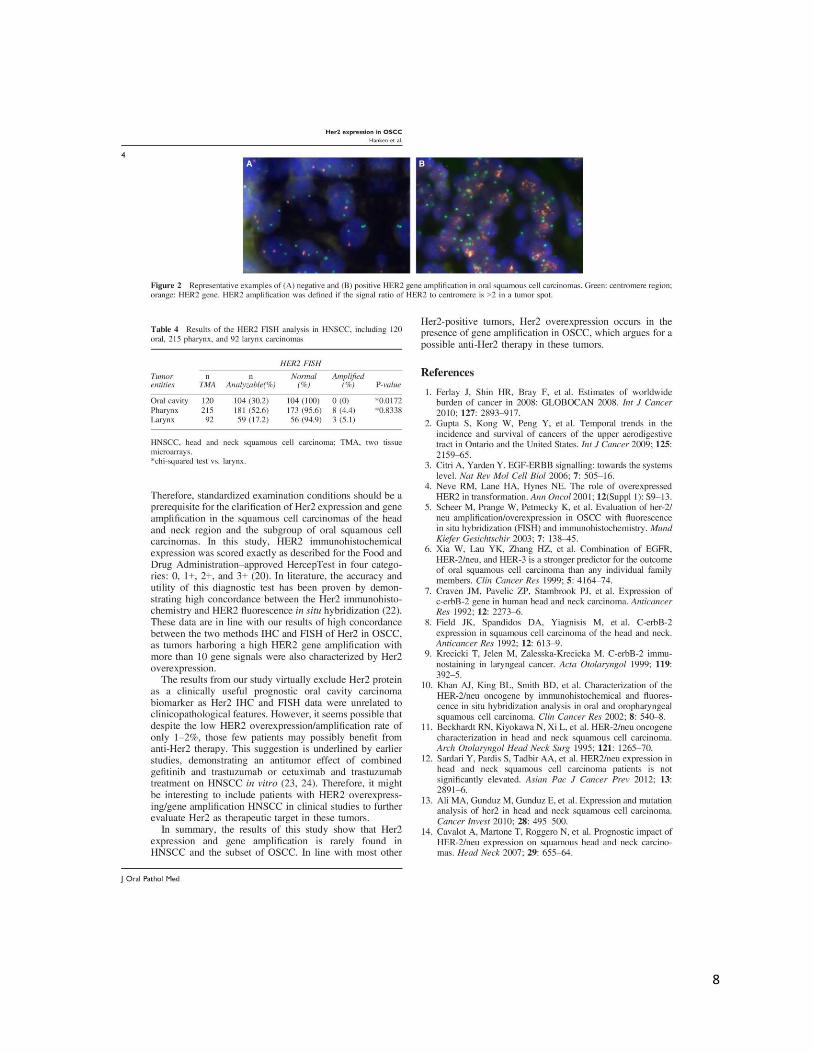
Figure 2: Representative examples of (A) negative and (B) positive HER2 gene amplification in
oral squamous cell carcinomas. Green: centromere region; Orange: HER2 gene. HER2
amplification was defined in the signal ratio of HER2 to centromere is >2 in a tumour spot
(Hanken* et al. 2014).

16
d. Statistical analysis
We analysed the clinical data on tumour size, grading, lymph node involvement, tumour relapse,
distant metastasis, and survival for a total of 216 patients. For statistical analysis JMP 5.0.1.2
(SAS Institute Inc., Böblingen, Germany) was used. To search for associations between
molecular parameters and tumor phenotype, contingency tables and chi²-test were performed.
Survival curves were calculated according to Kaplan-Meier, setting raw survival times as an end
point. In order to detect survival differences that are significant between the groups a Log-Rank
test was applied.
VII. Summary of results with discussion
As mentioned above, we conducted these studies in order to learn more on the rate of HER2
gene amplification and overexpression of the receptor in head and neck squamous cell
carcinoma (HNSCC) and to clarify the subanatomical entity of oral squamous cell carcinomas
(OSCC) as well as to clarify the importance of HER2 gene amplification and expression in
HNSCC and the subgroup of OSCC. The clinical data on tumor size, grading, lymph node
involvement, tumour relapse, distant metastasis, and survival were available for 216 patients
with a median follow-up of 46 months (range: 1–306 months) (Hanken* et al. 2014) . Our study
revealed that the Her2 expression in the immunohistochemical stains was rarely detectable.
Furthermore, gene amplification in the TMA of Her2 was absent in the majority and unrelated to
tumour phenotype or survival of the patients suffering from oral squamous cell carcinomas as a
sub anatomical region of the HNSCC. In the immunohistochemical stains 26 (11.7%) tissue
spots of the 222 OSCC had to be excluded due to the complete lack of tissue or absence of
unequivocal cancer cells on the respective TMA spots. The Her2 expression could be detected
in 4 of 196 (2%) analysable tumours including a score of 1+ in 0.5%, 2+ in 1%, and 3+ in 0.5%
of cases and was unrelated to gender (p=0.4558), tumour stage (p=0.4513), lymph node status
(p=0.4514) and tumour grading in OSCC (p=0.9810) as shown in table 1 in the attached paper
above. Within the TMA containing squamous cell carcinomas of 92 larynx, 120 oral and 215
pharynx carcinomas, Her2 expression was absent in the majority of HNSCC and detectable in 6
of 177 (3.2%) pharynx including a score of 1+ in 2.3% and 2+ in 1.1% and in 1 of 64 (1.6%)
larynx carcinomas with a score of 2+ as shown in table 2 in the attached paper (Hanken* et al.
2014). We could demonstrate a high concordance between the Her2 immunohistochemistry and
HER2 fluorescence in-situ hybridisation. Also in the HER2 FISH analysis a total of 15 (6.8%)
tissue spots of the 222 OSCC were excluded due to the complete lack of tissue or absence of

17
unequivocal cancer cells on the respective TMA spots. HER2 gene amplification with at least 2
copies per cell was found in 6 of 207 (3%) analysable oral squamous cell carcinomas including 3
tumours (1.5%) with 4-6 HER2 gene signals and 3 tumours (1.5%) with more than 10 gene
signals. These findings were unrelated to tumour phenotype shown in table 3 of the attached
paper. As shown in table four of the attached paper, HER2 gene amplification was only found in
8 of 181 (4.4%) interpretable pharynx and in 3 of 59 (5.1%) larynx carcinomas within the head
and neck cancer TMA (Hanken* et al. 2014). To summarise the results with the selected
experimental conditions, detectable HER2 gene amplification and the expression of the receptor
was found in a minority of HNSCC and the subset of OSCC. In earlier studies it was either
reported that the Her2 expression and gene amplification in HNSCC and OSCC was high or not
feasible. Making it on the one hand a promising target for adjuvant treatment or on the other
hand not relevant. By analysing these studies it sticks out that there was not a uniform
methodically approach such as preparation of antibodies, the tissue, staining or evaluation. We
suggest using standardised examination condition would make the study of Her2 expression and
gene amplification in the squamous cell carcinomas of the head and neck region with the
subgroup of oral squamous cell carcinomas more transparent and more meaningful. Looking at
our data of IHC and FISH three tumours harboring a high gene amplification with more than 10
gene signals were also characterised by Her2 protein overexpression. Therefore we can
conclude that there is a meaningful linkage between expression and amplification. Looking at the
data from our study one is likely to exclude Her2 protein as a clinically useful prognostic oral
cavity carcinoma biomarker as Her2 IHC and FISH data were unrelated to clinic-pathological
features. But it is underlined in the literature that the minority of 1-2% patients of our study
possibly benefits from anti-Her2 therapy demonstrating an antitumour effect of combined
gefitinib and trastuzumab or cetuximab and trastuzumab treatment on HNSCC in vitro. For that
reason, it makes sense to clarify in further studies the best treatment for HER2 highly amplified
and simultaneously HER2 positive oral cavity carcinomas. This study should also give an
impulse for further studies for the importance of oncogenes as tumour markers in Head and
neck squamous cell carcinomas (HNSCCs).

18
VIII. References
Amann, R. & Fuchs, B.M., 2008. Single-cell identification in microbial communities by improved fluorescence in situ hybridization techniques. Nature reviews. Microbiology, 6(5), pp.339–48. Available at: http://www.ncbi.nlm.nih.gov/pubmed/18414500 [Accessed January 21, 2014].
Ferlay, J. et al., 2010. Estimates of worldwide burden of cancer in 2008: GLOBOCAN 2008. International journal of cancer. Journal international du cancer, 127(12), pp.2893–917. Available at: http://www.ncbi.nlm.nih.gov/pubmed/21351269 [Accessed January 21, 2014].
Graham, a D. et al., 2008. Tissue microarray technology in the routine assessment of HER-2 status in invasive breast cancer: a prospective study of the use of immunohistochemistry and fluorescence in situ hybridization. Histopathology, 52(7), pp.847–55. Available at: http://www.ncbi.nlm.nih.gov/pubmed/18494613 [Accessed February 12, 2014].
Gupta, S. et al., 2009. Temporal trends in the incidence and survival of cancers of the upper aerodigestive tract in Ontario and the United States. International journal of cancer. Journal international du cancer, 125(9), pp.2159–65. Available at: http://www.ncbi.nlm.nih.gov/pubmed/19569190 [Accessed February 12, 2014].
Hanken*, H. et al., 2014. Her2 expression and gene amplification is rarely detectable in patients with oral squamous cell carcinomas. J Oral Pathol Med.
Jain, K.S. et al., 2013. Synchronous cancers in patients with head and neck cancer: risks in the era of human papillomavirus-associated oropharyngeal cancer. Cancer, 119(10), pp.1832–7. Available at: http://www.ncbi.nlm.nih.gov/pubmed/23423883 [Accessed October 19, 2013].
M. Blessmann, 2008. Molekulargenetische Veränderungen des Plattenepithelkarzinoms der Mundhöhle.
Ooi, A. et al., 2004. Protein overexpression and gene amplification of HER-2 and EGFR in colorectal cancers : an immunohistochemical and fluorescent in situ hybridization study. Modern Pathology, 2, pp.895–904.
Ramos-Vara, J. a & Miller, M. a, 2014. When tissue antigens and antibodies get along: revisiting the technical aspects of immunohistochemistry--the red, brown, and blue technique. Veterinary pathology, 51(1), pp.42–87. Available at: http://www.ncbi.nlm.nih.gov/pubmed/24129895 [Accessed January 29, 2014].
Scheer, M., 2003. Überexpression / Amplifikation oralen Plattenepithelkarzinomen. Mund Kiefer Gesichts Chirurgie, 7, pp.138–145.
Scholl, S., Beuzeboc, P. & Pouillart, P., 2001. Targeting HER2 in other tumor types. Annals of Oncology, 12(suppl 1), pp.S81–S87. Available at: http://annonc.oxfordjournals.org/cgi/content/long/12/suppl_1/S81 [Accessed February 12, 2014].
19
Simon, R. & Sauter, G., 2004. Tissue microarray (TMA) applications: implications for molecular medicine. Expert Reviews in Molecular Medicine, 5(26), pp.1–12. Available at: http://www.journals.cambridge.org/abstract_S1462399403006781 [Accessed March 14, 2014].
Stern, D.F., Heffernan, P.A. & Weinberg, R.A., 1986. p185 , a Product of the neu Proto-Oncogene , Is a Receptorlike Protein Associated with Tyrosine Kinase Activity. Molecular and Cellular Biology, 6(5).
Tripathy, D. et al., 2004. Safety of Treatment of Metastatic Breast Cancer With Trastuzumab Beyond Disease Progression. Journal of clinical oncology : official journal of the American Society of Clinical Oncology, 22(6), pp.1063–1070.
Ugocsai, K. et al., 2005. Investigation of HER2 Overexpression in Non-small Cell Lung Cancer. Anticancer Research, 3066, pp.3061–3066.
Wolff, A.C. et al., 2013. Recommendations for human epidermal growth factor receptor 2 testing in breast cancer: American Society of Clinical Oncology/College of American Pathologists clinical practice guideline update. Journal of clinical oncology : official journal of the American Society of Clinical Oncology, 31(31), pp.3997–4013. Available at: http://www.ncbi.nlm.nih.gov/pubmed/24101045 [Accessed January 21, 2014].

20
IX. Erklärung des Eigenanteils an der Publikation
Im Rahmen meiner Doktorarbeit am Kopf- und Neurozentrum / Klinik und Poliklinik für Mund-,
Kiefer- und Gesichtschirurgie ist folgende Publikation entstanden: "Her2 expression and gene
amplification is rarely detectable in patients with oral squamous cell carcinomas". Die Arbeit
wurde im Januar 2014 im Journal of Oral Pathology and Medicine veröffentlicht.
In Zusammenarbeit mit Herrn PD Dr. Dr. Blessmann und Herrn Dr. Dr. Hanken habe ich das
Follow-up und Die Dantenanalyse der Gewebeproben und klinischen Daten von Patienten der
Klinik und Poliklinik für Zahn-, Mund-, Kiefer- und Gesichtschirurgie des Universitätsklinikum
Hamburg-Eppendorf, die aufgrund eines Mundhöhlenkarzinoms in einem Zeitraum von 19
Jahren (1988 bis 2007) behandelt wurden, ausgewertet. Die entsprechenden 222 Tumorblöcke
wurden im Institut für Pathologie zusammengesammelt und die Amplifikationen von HER2
mittels Fluoreszenz in situ Hybridisierung (FISH) und die Expression von Her-2
immunhistochemisch (IHC) dargestellt. In Zusammenarbeit mit Herrn PD Dr. Dr. Blessmann und
Herrn Dr. Dr. Hanken habe ich die von der Pathologie erstellten IHC und FISH Analysen in
Bezug auf die Her2 Expression und Amplifikation ausgewertet und zur Erstellung des
Manuscripts beigetragen.

21
X. Danksagung
Mein besonderer Dank gilt Herrn Prof. Dr. Dr. Max Heiland, Direktor der Klinik und Poliklinik
für Mund-, Kiefer- und Gesichtschirurgie am Universitätsklinikum Hamburg-Eppendorf, für die
Bereitstellung der Doktorarbeit.
Mein Großer Dank richtet sich an Herrn PD Dr. Dr. Marco Blessmann für die Bereitstellung des
spannenden Themas, die Möglichkeit zur Durchführung der Arbeit und die engagierte und
strukturierte Betreuung. Insbesondere danke ich ihm für die große Unterstützung und Förderung
seit Beginn meiner Zeit als sein Doktorand bis heute.
Ebenfalls möchte ich Dr. Dr. Henning Hanken meinen Dank aussprechen. Auch er hat meine
Arbeit maßgeblich mitbetreut. Ich konnte in fachlichen Diskussionen mit ihm viel lernen und
freue mich, dass ich mich mit all meinen Fragen immer an ihn wenden konnte. Ich danke ihm
sehr für die Unterstützung und die wertvolle Mitbetreuung meiner Arbeit.
Vielen Dank ebenfalls der Abteilung Pathologie unter Professor Dr. Guido Sauter und PD Dr.
Ronald Simon am UKE für die Aufarbeitung unseres Tumorgewebes und die Bereitstellung der
Daten aus der biomedizinischen Forschung.
Dieses Projekt war ebenfalls nur möglich durch die Bereitstellung der TMAs des
Universitätsklinikums Basel. Ich danke allen Mitwirkenden dafür sehr!
Herrn Professor Dr. Dr. Ralf Smeets spreche ich meinen Dank aus, da er mir als Mentor und
Vorbild immer mit Rat und Tat zur Seite stand und mich für dieses Projekt vorgeschlagen hat.
Meine Eltern, Dr. med. Bernd Peter Gaudin und Eciel Gaudin, und meine Freunde, standen
auch in dieser Zeit unverbrüchlich zu mir. Vielen herzlichen Dank!

22
XI. Lebenslauf
Name: Robert Andrè Gaudin
Geburtsdatum: 12. September 1988
Geburtsort: Berlin
Staatsangehörigkeiten: Deutsch
Familienstand: ledig
Schulausbildung
27.08.2005 - 20.06.2007 Windsor High School, Windsor, Virginia
(Advanced High School Diploma GPA 3.99)
30.08.2007 Privates Schiller Gymnasium Potsdam (Abitur 1,6)
Studium:
Seit Oktober 2008 Medizin an der Universität Hamburg, seit 2010 mit abgeschlossenem
1. Abschnitt der Ärztlichen Prüfung, Note 3.5
Seit September 2012 bestandenes USMLE Step 1
Geplant Oktober 2014, 2. Abschnitt der Ärztlichen Prüfung
Forschung und Publikationen:
06.2011- 10.2013:
Im Tierlabor „Evaluation einer neuartigen mikrochirurgischen Anastomose - Technik mit einem
resorbierbaren ST-Stent im Tiermodell im Bereich „Tissue Engineering“ bei Univ.-Prof. Dr. med.
Dr. med. dent. Max Heiland in der MKG am UKE
01.04.2013 - 30.05.2013
Forschung im Rocco Lab im Massachusetts General Hospital der Harvard Medical School,
Erstellung einer Datenbank zu HPV-Tumoren im Oropharynx
10.2013 – 01.2014 Datenanalyse und –akquise zur Auswertung und Erstellung des Manuscripts
"Her2 expression and gene amplification is rarely detectable in patients with oral squamous cell
carcinomas" bei PD Dr. Dr. Marco Blessmann in der MKG am UKE

23
19.09.2013 – 09.12.2013
Forschung bei Professor Gieri Cathomas, Leiter der Pathologie, Kantonspitaal Liestal, Schweiz,
Bearbeitung und Auswertung einer Datenbank zu HPV-Tumoren im Oropharynx
- HER2 in head and neck Squamous cell carcinoma (Doktorarbeit, MKG UKE PD Dr. Dr.
Blessmann), Veröffentlicht 2014 im: Journal of Oral Pathology and Medicine
- Case Report: Henoch- Schönlein Purpura in adults (Institut Innere Medizin, Universitätsspital
Liestal, Schweiz, Professor Leuppi), Veröffentlicht 2014 im: Die PRAXIS Journal Schweiz
-> Posterpräsentation auf dem European and Swiss Congress of Internal Medicine in
Genf am 15.05.2014
- Ausstehend: Paper zu HPV orophanryngeal cancer (Harvard Medical School, Professor
Rocco)
- Ausstehend: Paper: “ Bilateral tonsilar manifestation of Her2 positive oropharyngeal cancers”
Professor Gieri Cathomas, Pathologie, Kantonspitaal Liestal, Schweiz
- Ausstehend: Evaluation einer neuartigen mikrochirurgischen Anastomose - Technik mit einem
resorbierbaren ST-Stent im Tiermodell (MKG UKE Professor Heiland)

24
XII. Eidesstattliche Versicherung
Ich versichere ausdrücklich, dass ich die Arbeit selbständig und ohne fremde Hilfe
verfasst, andere als die von mir angegebenen Quellen und Hilfsmittel nicht benutzt
und die aus den benutzten Werken wörtlich oder inhaltlich entnommenen Stellen
einzeln nach Ausgabe (Auflage und Jahr des Erscheinens), Band und Seite des
benutzten Werkes kenntlich gemacht habe.
Ferner versichere ich, dass ich die Dissertation bisher nicht einem Fachvertreter an
einer anderen Hochschule zur Überprüfung vorgelegt oder mich anderweitig um
Zulassung zur Promotion beworben habe.
Ich erkläre mich einverstanden, dass meine Dissertation vom Dekanat der
Medizinischen Fakultät mit einer gängigen Software zur Erkennung von Plagiaten
überprüft werden kann.
Unterschrift: ......................................................................