hepatitis b x-interacting protein induces hepg2 cell proliferation through activation of the...
TRANSCRIPT

http://ebm.sagepub.com/Experimental Biology and Medicine
http://ebm.sagepub.com/content/236/1/62The online version of this article can be found at:
DOI: 10.1258/ebm.2010.010179
2011 236: 62Exp Biol Med (Maywood)Feng-ze Wang, Hong-rong Fei, Li-hui Lian, Jian-mei Wang and Yu-yu Qiu
phosphatidylinositol 3-kinase/Akt pathwayHepatitis B x-interacting protein induces HepG2 cell proliferation through activation of the
Published by:
http://www.sagepublications.com
On behalf of:
Society for Experimental Biology and Medicine
can be found at:Experimental Biology and MedicineAdditional services and information for
http://ebm.sagepub.com/cgi/alertsEmail Alerts:
http://ebm.sagepub.com/subscriptionsSubscriptions:
http://www.sagepub.com/journalsReprints.navReprints:
http://www.sagepub.com/journalsPermissions.navPermissions:
What is This?
- Jan 1, 2011Version of Record >>
at University of Central Florida Libraries on January 27, 2014ebm.sagepub.comDownloaded from at University of Central Florida Libraries on January 27, 2014ebm.sagepub.comDownloaded from
Original Research
Hepatitis B x-interacting protein induces HepG2 cell
proliferation through activation of the phosphatidylinositol
3-kinase/Akt pathway
Feng-ze Wang1, Hong-rong Fei2, Li-hui Lian1, Jian-mei Wang1 and Yu-yu Qiu1
1Department of Biology; 2College of Pharmacy, Taishan Medical University, Chang Cheng Road, Taian 271016, PR China
Corresponding author: Professor Feng-ze Wang. Email: [email protected]
AbstractHepatitis B x-interacting protein (HBXIP), a co-factor of survivin, was originally identified by its binding with the C-terminus of
the hepatitis B virus x protein (HBx). We have recently shown that HBXIP promotes the growth of both normal liver cells and
hepatoma cells in vitro, but the molecular mechanisms of this have not been documented. In this study, we investigated the
potential effects of HBXIP on the proliferation of HepG2 cells and the intracellular signaling pathway mediating these changes.
Over-expression of the HBXIP gene promoted the proliferation of HepG2 cells, as shown by the MTT assay. We also showed
that HBXIP induced cellular accumulation in the S phase concomitantly with up-regulation of cyclinD1 and down-regulation of
p21 and p53 levels. Moreover, HBXIP over-expression cells showed activation of the PI3K/Akt pathway; this activation was
accompanied by an increase in phosphorylation of glycogen synthase kinase 3b. LY294002, a specific inhibitor of PI3K,
blocked HBXIP-stimulated Akt phosphorylation and suppressed the cell cycle promotion induced by HBXIP in HepG2
cells. The increase in cyclinD1 protein levels induced by HBXIP was inhibited when cells were incubated with LY294002. In
conclusion, our data suggest that the proliferation of HepG2 cells promoted by HBXIP is associated with activation of the
PI3K/Akt signaling pathway.
Keywords: HBXIP, hepatoma cell, cell proliferation, PI3K/Akt
Experimental Biology and Medicine 2011; 236: 62–69. DOI: 10.1258/ebm.2010.010179
Introduction
Hepatocellular carcinoma (HCC) is one of the most commoncancers in the world, and it has been estimated that morethan 50% of HCC cases worldwide are associated withhepatitis B virus (HBV). Moreover, research has shownthat individuals carrying HBV have a greater than100-fold increased relative risk of developing HCC.1
Among the four overlapping genes of HBV, such as S/preS, C/preC, P and X, hepatitis B virus X gene, whichencodes a 17-kDa protein known as HBV x protein (HBx),is strongly linked to the development of HCC.2,3
Hepatitis B x-interacting protein (HBXIP) is a cellularprotein which was originally identified by its interactionwith the C-terminus of HBx.4 The sequences of the HBXIPgene, containing a putative leucine zipper motif and twoconsensus phosphorylation sites for protein kinase C andcasein kinase II, are well conserved among mammalianspecies. HBXIP can reduce the replication of wild-typeHBV and suppress the transactivity of activating protein-1enhanced by HBx expression. HBXIP also forms a complexwith survivin, an antiapoptotic protein, and selectively
inhibits apoptosis via the mitochondrial/cytochrome cpathway.5 Recently, another HBXIP interactor namedhSuv3p, a human ATP-dependent RNA/DNA helicase,has been identified. The HBXIP-binding domain in thehSuv3p protein is important for its stability and transportinto the mitochondria.6 As a regulator of centrosome dupli-cation, HBXIP is required for bipolar spindle formation inHeLa cells and primary mouse embryonic fibroblast cells.7
Knockdown of endogenous HBXIP expression arrests thecells in prometaphase with monopolar spindles, and over-expression of HBXIP causes tripolar or multipolar spindlesdue to excessive centrosome replication. Our previousstudies have suggested that HBXIP promotes cellular pro-liferation in both cancer cells and normal liver cells,8 butthe molecular mechanisms of this are still unclear.
The phosphatidylinositol (PI) 3-kinase (PI3K)/Akt signal-ing pathway is a central regulator of cell proliferation andcell survival.9,10 PI3Ks are dual-specificity enzymes thatplay a pivotal role in the regulation of many cellular pro-cesses.11,12 Activated PI3K phosphorylates PI substrates toproduce PI(3)P, PI(3,4)P2 and PI(3,4,5)P3. These molecules
ISSN: 1535-3702
Copyright # 2011 by the Society for Experimental Biology and Medicine
Experimental Biology and Medicine 2011; 236: 62–69
at University of Central Florida Libraries on January 27, 2014ebm.sagepub.comDownloaded from

act as secondary messengers, recruiting the PI3K-dependentserine/threonine kinases (PDK1) and Akt from the cyto-plasm to the plasma membrane.13 Akt, also known asprotein kinase B, is an important target of PI3K. Akt canfunction to regulate cell proliferation, cell cycle and cell sur-vival by interacting with numerous other regulatory pro-teins, such as p53, cyclinD1, p21Cip1, glycogen synthasekinase-3 (GSK3) and mammalian target of rapamycin.14,15
To be fully activated, Akt is phosphorylated on two criticalresidues: Thr308 (T308-Akt) and Ser473 (S473-Akt), andalterations of the Akt pathway have been detected in avariety of cancerous human malignancies.16,17
Here, we present evidence that HBXIP promotes hepa-toma cell proliferation, partly by activating the PI3K/Aktsignaling pathway. Up-regulation of the expression ofcyclinD1 by HBXIP over-expression is involved in promot-ing a G0/G1 to S phase transition in HCC cells. The PI3Kinhibitor, LY294002, blocks HBXIP-stimulated Akt phos-phorylation and suppresses the cell cycle promotioninduced by HBXIP in HepG2 cells.
Materials and methods
Cancer cell culture
The HepG2 cell line was obtained from the Type CultureCollection of the Chinese Academy of Sciences (Shanghai,China) and cultured in RPMI 1640 medium supplementedwith 10% fetal calf serum (Gibco BRL, Gaithersburg, MD,USA), 2 mmol/L glutamine, 100 U/mL penicillin and100 mg/mL streptomycin at 378C in humidified 5% CO2.
Stable transfection
HepG2 cells were transfected with pCMV-hbxip8 orpCMV-tag2B (empty vector) with Lipofectamine 2000 accord-ing to the manufacturer’s instructions. Twenty-four hours aftertransfection, the cells were diluted 1:10 and cultured in growthmedium containing G418 (700 mg/mL) for three weeks. Stabletransfected clones were picked and maintained in a mediumcontaining 350 mg/mL G418 for additional studies.
MTT assay
Cell proliferation was measured using the MTT assay every24 h. Briefly, 200 mL of cell suspensions (5 � 103 cells/mL)was added to each well of a 96-well plate and incubated at378C for six days. A total of 20 mL of MTT (5 mg/mL;Genview, Houston, TX, USA) was added and incubated at378C for 4 h. After removing the supernatant, 150 mL ofdimethyl sulfoxide was added to dissolve formazan crystals,and the value of the optical density was detected at 570 nm.To explore the role of the PI3K/Akt pathway, HepG2-hbxipcells and control cells were treated with 10 and 40 mmol/LLY294002 (Cell Signaling, Dallas, TX, USA), respectively.
Flow cytometry analysis
Cells (1 � 106) were harvested by trypsinization and washedtwice with phosphate-buffered saline (PBS), then fixed in
75% ethanol at 48C for 18 h. The fixed cells were rinsedtwice with PBS and resuspended in propidium iodine (PI)solution containing 50 mg/mL PI and 50 mg/mL RNaseA(Sigma, St Louis, MO, USA) in PBS without calcium andmagnesium and incubated at 378C for 30 min in the dark.Stained cells were passed through a nylon-mesh sieve toremove cell clumps and analyzed by a FACScan flow cyt-ometer and Cell Quest analysis software (BectonDickinson, San Jose, CA, USA). Flow cytometry analysiswas repeated three times.
Western blot analysis
After washing twice with cold PBS, hepatoma cells were lysedwith ice-cold lysis buffer (150 mmol/L NaCl, 20 mmol/LTris-HCl [pH 7.4], 0.1% SDS, 1.0% Nonidet P-40, 0.5%Na-deoxycholate [Na-DOC], 0.2 mmol/L phenylmethylsulfo-nyl fluoride, protease inhibitor cocktails and phosphataseinhibitors). Lysates were centrifuged at 12,000g for 20 min,and the supernatants were used as total cell lysates. Theprotein concentrations were determined by Bradford proteinassay (Bio-Rad, Hercules, CA, USA). A quantity of 30–40 mgtotal protein per lane was separated by sodium dodecylsulfate-polyacrylamide gel electrophoresis (SDS-PAGE) andtransferred onto polyvinylidene fluoride membranes(Millipore, Bedford, MA, USA). Membranes were blockedwith 5% milk powder in 0.05% Tween-TBS and incubatedwith one of the following primary antibodies: mouseanti-b-Actin (Sigma, 1:10,000 dilution), goat anti-HBXIP(Santa Cruz Biotechnology, Santa Cruz, CA, USA, 1:300dilution), rabbit anti-cyclinB1 (Santa Cruz Biotechnology,1:600), rabbit anti-cyclinD1 (Bioworld, St Louis Park, MN,USA, 1:600 dilution), mouse anti-p21 (NeoMarkers, Fremont,CA, USA, 1:500 dilution), mouse anti-p53 (Santa CruzBiotechnology, 1:500 dilution), rabbit anti-Akt (CellSignaling, 1:1000 dilution), mouse anti-p-Akt (Cell Signaling,1:200 dilution), rabbit anti-p-GSK3b (Cell Signaling, 1:400dilution), rabbit anti-ERK (Cell Signaling, 1:800 dilution),mouse anti-p-ERK (Cell Signaling, 1:600 dilution); the second-ary antibody was diluted in 3% BSA/Tween-TBS. Detection ofthe target proteins on the membranes was performed usingthe ECL Western Blot Detection Reagents (Thermo ScientificPierce, Rockford, IL, USA). Western blot analysis was repeatedthree times.
Akt kinase assay
Akt kinase activity was measured using the Akt kinaseassay kit from Cell Signaling Technology. Briefly, cells trans-fected with the pCMV-hbxip or pCMV-tag2B plasmid werewashed twice with cold PBS and lysed in ice with 600 mL oflysis buffer containing 1% Triton X-100, 1 mmol/L EDTA,1 mmol/L EGTA, 2.5 mmol/L sodium pyrophosphate,150 mmol/L NaCl, 20 mmol/L Tris-HCl (pH 7.5), 1 mg/mLaprotinin, 1 mg/mL leupeptin, 1 mmol/L phenylmethylsulfo-nyl fluoride, 1 mmol/L b-glycerophosphate and 1 mmol/LNa3VO4 according to the protocol. Resuspended, immobi-lized Akt antibody slurry (20 mL) was added to 200 mL ofcell lysate supernatant to selectively immunoprecipitate Aktfrom the lysate with gentle rocking for 12 h at 48C.
................................................................................................................................................Wang et al. HBXIP induces cell proliferation through Akt activation 63
at University of Central Florida Libraries on January 27, 2014ebm.sagepub.comDownloaded from

Immunoprecipitations were washed three times with lysisbuffer and twice with Akt kinase buffer (25 mmol/L Tris[pH 7.5], 5 mmol/L b-glycerophosphate, 2 mmol/L dithio-threitol, 0.1 mmol/L Na3VO4 and 10 mmol/L MgCl2). Theimmunoprecipitated pellet was then incubated with kinasebuffer containing 200 mmol/L ATP and 1 mg of GSK-3fusion protein according to the manufacturer’s instructionsfor the non-radioactive Akt kinase assay. Samples were ana-lyzed by Western blot analysis using 15% SDS-PAGE.Akt-induced phosphorylation of GSK-3 was detected byWestern blot using the phospho-GSK-3a/b(Ser21/9) anti-body. Akt kinase assay was repeated three times.
Statistical analysis
All data are expressed as mean+SD. Statistical analysiswas performed using the Student’s t-test and P , 0.05 indi-cated statistical significance.
Results
Up-regulation of cyclinD1 and down-regulation ofp53 and p21 expression are involved in the G0/G1 to Sphase transition induced by HBXIP in HepG2 cells
To evaluate the biological function of HBXIP in the prolifer-ation of hepatoma cells, HepG2 cells were engineered tostable express high levels of HBXIP gene and were namedHepG2-hbxip. The expression levels of HBXIP in HepG2-hbxip cells were assessed by Western blot assay. Comparedwith the control (pCMV-tag2B), HepG2-hbxip cells showedhigh expression of HBXIP (Figure 1a). The results of theMTT assay in Figure 2 indicated that HBXIP can promoteHepG2 cells proliferation in vitro. Furthermore, flow cytome-try analysis was used to measure the cell cycle distributionin HepG2-hbxip cells. As predicted, HBXIP significantly accel-erated the cell cycle progression. The percentage of cellsre-entering into the S phase in HepG2-hbxip cells was39.2+1.32, which was higher than that of pCMV-tag2B cells(32.06+1.46) (Figure 3, P , 0.05), suggesting that HBXIPmight promote hepatoma cell growth by accelerating theG0/G1 to S phase transition in the cell cycle.
To identify molecules responsible for the cell proliferationeffect induced by HBXIP, the expression of several activatorsand inhibitors of the cell cycle was detected by Western blotassay. As shown in Figure 4a, HepG2-hbxip cells containeda greater amount of cyclinD1 and a lower amount of p21and p53 protein when compared with control cells. Theexpression level of cyclinB1 was not significantly differentbetween HepG2-hbxip and pCMV-tag2B cells. These dataindicated that up-regulation of cyclinD1 and down-regulation of p21 and p53 might contribute to theHBXIP-induced G0/G1 to S phase transition.
PI3K/Akt signaling pathway is involved inHBXIP-induced human HCC cell proliferation
To investigate the possible molecular mechanism ofHBXIP-induced cell proliferation, the PI3K/Akt signalingpathway and the MAPK/ERK pathway were examined.
Western blot analysis of the phosphorylated Akt (p-Akt)level indirectly showed that Akt activity was increased inHepG2-hbxip cells when compared with the pCMV-tag2Bcontrol cells (Figure 5a). No obvious changes for total Aktprotein levels were observed, demonstrating that HBXIPhas no effect on protein stability. In agreement with theincreased phosphorylation of Akt in HepG2-hbxip cells,over-expression of HBXIP in HepG2 cells led to anup-regulation in the phosphorylation of GSK-3b. We also
Figure 1 Examination of the levels of HBXIP in HepG2 cells stably trans-
fected with the HBXIP gene or control vector. (a) Western blot analysis of
the expression of HBXIP. Detection of b-actin with an anti-b-actin antibody
was used as a loading control. (b) Bands were analyzed using Glyco
Band-Scan software. Each bar corresponds to the mean+SD for three inde-
pendent experiments. �P , 0.05 versus control cells, Student’s t-test. HBXIP,
hepatitis B x-interacting protein
Figure 2 HBXIP induces hepatoma cell proliferation. Cell proliferation was
measured using an MTT assay in HepG2-hbxip cells and pCMV-tag2B cells.
The y-axis represents the optical density (OD). Mean value and standard devi-
ation are shown (n ¼ 3). �P , 0.05 versus control cells, Student’s t-test.
HBXIP, hepatitis B x-interacting protein (A color version of this figure is avail-
able in the online journal)
................................................................................................................................................64 Experimental Biology and Medicine Volume 236 January 2011
at University of Central Florida Libraries on January 27, 2014ebm.sagepub.comDownloaded from

examined the effect of HBXIP on the MAPK/ERK pathway,and no significant changes of phosphorylated ERK wereobserved in the Western blot assay results.
Phosphorylation of both Thr308 and Ser473 is requiredfor full activation of Akt, and Akt activity can be measuredusing an in vitro kinase assay that detects phosphorylationof GSK-3 induced by immunoprecipitated Akt. To evaluatewhether Akt enzymatic activity was directly induced byHBXIP over-expression, Akt activity was measured inHepG2 cells after being transiently transfected with eitherplasmids containing the HBXIP gene or empty plasmid con-trols. As shown in Figure 6a, HBXIP enhanced Akt enzy-matic activity in HepG2 cells.
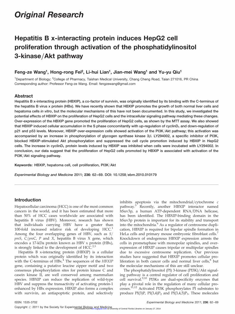
To examine whether the Akt pathway was directlyinvolved in the HBXIP-induced proliferation of HepG2cancer cells, HepG2-hbxip cells were treated withLY294002, a specific inhibitor of the PI3K/Akt pathway,18
followed by the MTT assay, cell cycle distribution andWestern blot analyses. As expected, MTT results showedthat LY294002 treatment reduced HBXIP-enhanced prolifer-ation of HepG2 cells (Figure 7). In comparison with thedimethyl sulfoxide-treated cells, the percentage ofHepG2-hbxip cells in the S phase decreased by 10.86+1.42% at 12 h after LY294002 treatment, while that ofpCMV-tag2B cells was unaffected (Figure 8). Western blotresults showed that the expression of p-Akt in theHepG2-hbxip cells was decreased after treatment ofLY294002, and the total levels of Akt were unchanged(Figure 9a). Furthermore, LY294002 potently inhibited thecyclinD1 induction by HBXIP over-expression, but p21 inhi-bition by HBXIP was unaffected. These data indicated thatthe PI3K/Akt pathway was involved in the promotion ofHBXIP-induced cell proliferation, and that the HBXIPup-regulation of cyclinD1 required PI3K/Akt activation.
Discussion
HBXIP, a co-factor of survivin, was originally identified byits interaction with viral HBx. Although some binding pro-teins of HBXIP, such as HBx, survivin and hSuv3p, havebeen identified in the past few years, these interactions are
Figure 3 Flow cytometry analysis for cell cycle in HepG2 cells stably transfected with HBXIP gene or control vector. Data shown are from representative exper-
iments repeated three times with similar results. HBXIP, hepatitis B x-interacting protein (A color version of this figure is available in the online journal)
Figure 4 Up-regulation of cyclinD1 and down-regulation of p53 and p21
expression by HBXIP. (a) Examination of expression levels of p21, p53,
cyclinB1 and cyclinD1 by Western blot analysis. (b) Bands were analyzed
using Glyco Band-Scan software. Each bar corresponds to the mean+SD
for three independent experiments. �Indicates that P , 0.05 versus control
using the Student’s t-test. HBXIP, hepatitis B x-interacting protein
................................................................................................................................................Wang et al. HBXIP induces cell proliferation through Akt activation 65
at University of Central Florida Libraries on January 27, 2014ebm.sagepub.comDownloaded from

almost irrelevant to cellular proliferation. In hepatoma cells,HBXIP bridges HBx and survivin together to suppresscaspase activation, inhibit apoptosis and regulate centro-some dynamics. hSuv3p is another interactor of HBXIPthat is mainly involved in the processes of RNA turnoverand apoptosis. Previously, we found that HBXIP promotedthe growth of both normal liver cells and hepatoma cells invitro, but the mechanisms were not clearly expatiated; there-fore, we speculate that other signaling pathways areinvolved in HBXIP-regulated cellular proliferation.
In this study, we first provide evidence that HBXIP pro-motes HepG2 cell proliferation by facilitating the G1 to Sphase transition (Figure 3) that is commonly seen in
cancer cells. Many kinases and kinase inhibitors in the cellcycle are involved in mediating the G1/S transition.19,20
p21 is central to cell cycle arrest, and down-regulation orloss of p21 expression has been found in a variety ofhuman cancers.21,22 In HepG2 cells, high expression ofHBXIP decreased the levels of p21 (Figure 4a). Becausep21 protein levels are directly stimulated by the p53protein,23 we then examined the effect of HBXIP on p53expression. Western blot results indicated that HBXIPdown-regulated the levels of p53. A recent study showedthat HBXIP was a substrate of ATM involved in the cellularresponse to DNA damage,24 and that activation of p53 wasmainly dependent on signaling by ATM kinase when DNAdamage occurred; thus, regulation of p53 expression byHBXIP may be involved in the cellular response to DNAdamage. We also determined the expression levels ofcyclinB1 and cyclinD1 by Western blot. As shown in
Figure 5 HBXIP activates the PI3K/Akt pathway in HepG2 cells. (a) Western blot analysis of phosphorylation of Akt, GSK3b and ERK. (b) Bands were analyzed
using Glyco Band-Scan software. Each bar corresponds to the mean+SD for three independent experiments. �Indicates that P , 0.05 and �� indicates that P ,
0.01 versus control cells using the Student’s t-test. HBXIP, hepatitis B x-interacting protein; GSK3b, glycogen synthase kinase 3b
Figure 6 Kinase reactions and Western blot analysis were performed in
anti-Akt immunoprecipitates from the corresponding lysates. (a) HepG2
cells were transiently transfected with either pCMV-hbxip or pCMV-tag2B
plasmids. Forty hours after transfection, cells were washed with ice-cold
phosphate-buffered saline and ice-cold lysis buffer was added. Akt activity
was assessed using GSK-3a/b as a substrate for phosphorylation
(p-GSK-3a/b). Phosphorylation of GSK-3 was measured by Western blot
using a p-GSK-3a/b (Ser21/9) antibody. (b) Bands were analyzed using
Glyco Band-Scan software. Each bar corresponds to the mean+SD for
three independent experiments. ��Indicates that P , 0.01 versus control
cells using the Student’s t-test. HBXIP, hepatitis B x-interacting protein;
GSK3b, glycogen synthase kinase 3b
Figure 7 Effects of PI3K inhibitor, LY294002, on HBXIP-induced HepG2 cell
proliferation assessed by MTT assay. HepG2 cells stably transfected with
HBXIP were treated with LY294002 at 10 and 40 mmol/L and then analyzed
by the MTT assay. Values are expressed as percentage of untreated cells,
and each bar represents the mean+SD of triplicate determinations (n ¼ 3).�Indicates that P , 0.05 versus control cells. HBXIP, hepatitis B x-interacting
protein
................................................................................................................................................66 Experimental Biology and Medicine Volume 236 January 2011
at University of Central Florida Libraries on January 27, 2014ebm.sagepub.comDownloaded from
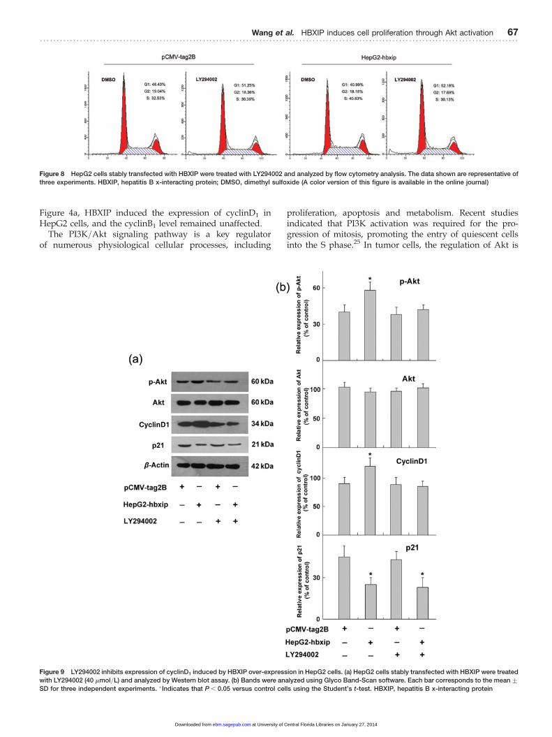
Figure 4a, HBXIP induced the expression of cyclinD1 inHepG2 cells, and the cyclinB1 level remained unaffected.
The PI3K/Akt signaling pathway is a key regulatorof numerous physiological cellular processes, including
proliferation, apoptosis and metabolism. Recent studiesindicated that PI3K activation was required for the pro-gression of mitosis, promoting the entry of quiescent cellsinto the S phase.25 In tumor cells, the regulation of Akt is
Figure 8 HepG2 cells stably transfected with HBXIP were treated with LY294002 and analyzed by flow cytometry analysis. The data shown are representative of
three experiments. HBXIP, hepatitis B x-interacting protein; DMSO, dimethyl sulfoxide (A color version of this figure is available in the online journal)
Figure 9 LY294002 inhibits expression of cyclinD1 induced by HBXIP over-expression in HepG2 cells. (a) HepG2 cells stably transfected with HBXIP were treated
with LY294002 (40 mmol/L) and analyzed by Western blot assay. (b) Bands were analyzed using Glyco Band-Scan software. Each bar corresponds to the mean+SD for three independent experiments. �Indicates that P , 0.05 versus control cells using the Student’s t-test. HBXIP, hepatitis B x-interacting protein
................................................................................................................................................Wang et al. HBXIP induces cell proliferation through Akt activation 67
at University of Central Florida Libraries on January 27, 2014ebm.sagepub.comDownloaded from
complex, and phosphorylation of Akt at Ser473 is still anunclear mechanism. Here, we have shown that PI3K/Aktis induced by HBXIP over-expression. GSK-3b, a down-stream target of PI3K/Akt, has been implicated in diversebiological processes including cell proliferation and cell sur-vival signaling in various cell types. Therefore, we alsoinvestigated whether HBXIP enhanced the phosphorylationof GSK-3b in HepG2 cells. As shown in Figure 5a, HBXIPover-expression stimulated a significant increase in thephosphorylation of GSK-3b. It has been previously reportedthat activation of MAPK/ERK occurs in human HCC,26 sowe also examined the effect of HBXIP on the activation ofERK. Western blot results showed that HBXIP did nothave a significant impact on the phosphorylation of ERK(Figure 5a).
This study further investigated whether the activation ofthe PI3K/Akt pathway induced by HBXIP was specific.HepG2-hbxip cells and control cells were treated withLY294002, a widely used PI3K inhibitor. The MTT assayshowed that pretreatment with LY294002 blocked the cellproliferation promoted by HBXIP (Figure 7). Inhibition ofthe PI3K/Akt signaling pathway partially reversedHBXIP-induced cell cycle progression (Figure 8). Both p21and cyclinD1 are downstream factors of the PI3K/Akt sig-naling pathway, and are involved in the regulation of cellcycle progression via the Akt pathway.27,28 Moreover, weobserved that HBXIP inhibited the level of p21 andinduced the expression of cyclinD1, so Western blot assaywas performed to detect whether or not p21 and cyclinD1
were involved in Akt activation. As shown in Figure 9a,the PI3K chemical inhibitor LY294002 suppressed the induc-tion of cyclinD1 but not p21 in HepG2 cells, suggesting thatthe PI3K/Akt pathway is involved in HBXIP-induced cellproliferation, possibly through the induction of cyclinD1.
In summary, our findings suggest that one of the func-tions of HBXIP is associated with cell proliferationregulation. Over-expression of HBXIP targets cyclinD1, pro-moting HCC cell proliferation through activation of thePI3K/Akt pathway. Blocking the PI3K/Akt pathway usingLY294002 decreases the p-Akt protein level as well as thefraction of cells in the S phase. Up-regulating the level ofcyclinD1 is involved in the Akt activation, and the down-regulation of p21 and p53 by HBXIP may be relevant toother signaling pathways in HepG2 cells.
Author contributions: F-zW conceived the experiment andconducted the experiment together with H-rF, J-mW andL-hL; Y-yQ performed the data analyses; F-zW and H-rFco-wrote the paper.
ACKNOWLEDGEMENTS
This work was supported by grants from the NationalNatural Science Foundation of China (No. 30800422).
REFERENCES
1 Block TM, Mehta AS, Fimmel CJ, Jordan R. Molecular viral oncology ofhepatocellular carcinoma. Oncogene 2003;22:5093–107
2 Benhenda S, Cougot D, Buendia MA, Neuveut C. Hepatitis B virus Xprotein molecular functions and its role in virus life cycle andpathogenesis. Adv Cancer Res 2009;103:75–109
3 Lupberger J, Hildt E. Hepatitis B virus-induced oncogenesis. World JGastroenterol 2007;13:74–81
4 Melegari M, Scaglioni PP, Wands JR. Cloning and characterization of anovel hepatitis B virus x binding protein that inhibits viral replication. JVirol 1998;72:1737–43
5 Marusawa H, Matsuzawa S, Welsh K, Zou H, Armstrong R, Tamm I,Reed JC. HBXIP functions as a cofactor of survivin in apoptosissuppression. EMBO J 2003;22:2729–40
6 Minczuk M, Mroczek S, Pawlak SD, Stepien PP. Human ATP-dependentRNA/DNA helicase hSuv3p interacts with the cofactor of survivinHBXIP. FEBS J 2005;272:5008–19
7 Fujii R, Zhu C, Wen Y, Marusawa H, Bailly-Maitre B, Matsuzawa S,Zhang H, Kim Y, Bennett CF, Jiang W, Reed JC. HBXIP, cellular target ofhepatitis B virus oncoprotein, is a regulator of centrosome dynamics andcytokinesis. Cancer Res 2006;66:9099–107
8 Wang FZ, Sha L, Zhang WY, Wu LY, Qiao L, Li N, Zhang XD, Ye LH.Involvement of hepatitis B X-interacting protein (HBXIP) in proliferationregulation of cells. Acta Pharmacol Sin 2007;28:431–8
9 Gayer CP, Chaturvedi LS, Wang S, Craig DH, Flanigan T, Basson MD.Strain-induced proliferation requires the phosphatidylinositol 3-kinase/
AKT/glycogen synthase kinase pathway. J Biol Chem 2009;284:2001–1110 Okada M, Jang SW, Ye K. Akt phosphorylation and nuclear
phosphoinositide association mediate mRNA export and cellproliferation activities by ALY. Proc Natl Acad Sci USA 2008;105:8649–54
11 Zhao L, Vogt PK. Class I PI3K in oncogenic cellular transformation.Oncogene 2008;27:5486–96
12 Liu P, Cheng H, Roberts TM, Zhao JJ. Targeting the phosphoinositide3-kinase pathway in cancer. Nat Rev Drug Discov 2009;8:627–44
13 Carracedo A, Pandolfi PP. The PTEN-PI3K pathway: of feedbacks andcross-talks. Oncogene 2008;27:5527–41
14 Franke TF. PI3K/Akt: getting it right matters. Oncogene 2008;27:6473–8815 Manning BD, Cantley LC. AKT/PKB signaling: navigating downstream.
Cell 2007;129:1261–7416 Chen M, Cassidy A, Gu J, Delclos GL, Zhen F, Yang H, Hildebrandt MA,
Lin J, Ye Y, Chamberlain RM, Dinney CP, Wu X. Genetic variations inPI3K-AKT-mTOR pathway and bladder cancer risk. Carcinogenesis2009;30:2047–52
17 Sarker D, Reid AH, Yap TA, de Bono JS. Targeting the PI3K/AKTpathway for the treatment of prostate cancer. Clin Cancer Res2009;15:4799–805
18 Moon DO, Kim MO, Choi YH, Kim GY. Beta-sitosterol induces G2/Marrest, endoreduplication, and apoptosis through the Bcl-2 and PI3K/Aktsignaling pathways. Cancer Lett 2008;264:181–91
19 Stein GS, van Wijnen AJ, Stein JL, Lian JB, Montecino M, Zaidi SK,Braastad C. An architectural perspective of cell-cycle control at the G1/Sphase cell-cycle transition. J Cell Physiol 2006;209:706–10
20 Blain SW. Switching cyclin D-Cdk4 kinase activity on and off. Cell Cycle2008;7:892–8
21 Abbas T, Dutta A. p21 in cancer: intricate networks and multipleactivities. Nat Rev Cancer 2009;9:400–14
22 Mottet D, Pirotte S, Lamour V, Hagedorn M, Javerzat S, Bikfalvi A,Bellahcene A, Verdin E, Castronovo V. HDAC4 represses p21(WAF1/
Cip1) expression in human cancer cells through a Sp1-dependent,p53-independent mechanism. Oncogene 2009;28:243–56
23 Kang JY, Kim JJ, Jang SY, Bae YS. The p53-p21(Cip1/WAF1) pathwayis necessary for cellular senescence induced by the inhibition ofprotein kinase CKII in human colon cancer cells. Mol Cells2009;28:489–94
24 Matsuoka S, Ballif BA, Smogorzewska A, McDonald ER, Hurov KE, LuoJ, Bakalarski CE, Zhao Z, Solimini N, Lerenthal Y, Shiloh Y, Gygi SP,Elledge SJ. ATM and ATR substrate analysis reveals extensive proteinnetworks responsive to DNA damage. Science 2007;316:1160–6
25 Akiyama H, Furukawa S, Wakisaka S, Maeda T. Cartducin stimulatesmesenchymal chondroprogenitor cell proliferation through bothextracellular signal-regulated kinase and phosphatidylinositol 3-kinase/
Akt pathways. FEBS J 2006;273:2257–6326 Ito Y, Sasaki Y, Horimoto M, Wada S, Tanaka Y, Kasahara A, Ueki T,
Hirano T, Yamamoto H, Fujimoto J, Okamoto E, Hayashi N, Hori M.Activation of mitogen-activated protein kinases/extracellular
................................................................................................................................................68 Experimental Biology and Medicine Volume 236 January 2011
at University of Central Florida Libraries on January 27, 2014ebm.sagepub.comDownloaded from

signal-regulated kinases in human hepatocellular carcinoma. Hepatology1998;7:951–8
27 Lu S, Ren C, Liu Y, Epner DE. PI3K-Akt signaling is involved in theregulation of p21(WAF/CIP) expression and androgen-independentgrowth in prostate cancer cells. Int J Oncol 2006;28:245–51
28 Fatrai S, Elghazi L, Balcazar N, Cras-Meneur C, Krits I, Kiyokawa H,Bernal-Mizrachi E. Akt induces beta-cell proliferation by regulating
cyclin D1, cyclin D2, and p21 levels and cyclin-dependent kinase-4activity. Diabetes 2006;55:318–25
(Received June 6, 2010, Accepted October 6, 2010)
................................................................................................................................................Wang et al. HBXIP induces cell proliferation through Akt activation 69
at University of Central Florida Libraries on January 27, 2014ebm.sagepub.comDownloaded from