hemodynamic monitoring in the critically patient
TRANSCRIPT

Med Intensiva. 2014;38(3):154---169
www.elsevier.es/medintensiva
CONSENSUS STATEMENT
Hemodynamic monitoring in the critically patient.
Recommendations of the Cardiological Intensive Care
and CPR Working Group of the Spanish Society
of Intensive Care and Coronary Units�
A. Ochagavíaa,∗, F. Baigorria, J. Mesquidaa, J.M. Ayuelab, A. Ferrándizc, X. Garcíaa,M.I. Monged, L. Mateuc, C. Sabatiera, F. Clau-Terrée, R. Vichof, L. Zapatag, J. Maynarh,A. Gild, Working Group Intensive Care and Cardiology CPR SEMICYUC
a Servicio de Medicina Intensiva, Hospital de Sabadell, CIBER Enfermedades Respiratorias, Corporació, Sanitària Parc Taulí,Institut Universitari Parc Taulí, Universitat Autònoma de Barcelona, Sabadell, Barcelona, Spainb Servicio de Medicina Intensiva, Hospital de Burgos, Burgos, Spainc Servicio de Medicina Intensiva, Hospital Universitario General de Castellón, Castellón, Spaind Servicio de Cuidados Críticos y Urgencias, Hospital del SAS Jerez, Jerez de la Frontera, Cádiz, Spaine Institut Recerca, Hospital de Vall d’Hebron y Consorci Sanitàri Terrasa, Barcelona, Spainf Servicio de Medicina Intensiva, Clínica USP-Palmaplanas, Palma de Mallorca, Spaing Servicio de Medicina Intensiva, Hospital de la Santa Creu i Sant Pau, Barcelona, Spainh Servicio de Medicina Intensiva, Hospital Universitario Araba, Vitoria, Álava, Spain
Received 8 October 2013; accepted 20 October 2013
Available online 5 May 2014
KEYWORDSHemodynamicmonitoring;Critically ill patient;Shock
Abstract Hemodynamic monitoring offers valuable information on cardiovascular perfor-
mance in the critically ill, and has become a fundamental tool in the diagnostic approach
and in the therapy guidance of those patients presenting with tissue hypoperfusion. From
introduction of the pulmonary artery catheter to the latest less invasive technologies,
hemodynamic monitoring has been surrounded by many questions regarding its usefulness and
its ultimate impact on patient prognosis. The Cardiological Intensive Care and CPR Working
Group (GTCIC-RCP) of the Spanish Society of Intensive Care and Coronary Units (SEMICYUC)
has recently impulsed the development of an updating series in hemodynamic monitoring.
Now, a final series of recommendations are presented in order to analyze essential issues in
hemodynamics, with the purpose of becoming a useful tool for residents and critical care
practitioners involved in the daily management of critically ill patients.
© 2013 Elsevier España, S.L. and SEMICYUC. All rights reserved.
� Please cite this article as: Ochagavía A, Baigorri F, Mesquida J, Ayuela JM, Ferrándiz A, García X, et al. Monitorización hemodinámica enel paciente crítico. Recomendaciones del Grupo de Trabajo de Cuidados Intensivos Cardiológicos y RCP de la Sociedad Espanola de MedicinaIntensiva, Crítica y Unidades Coronarias. Med Intensiva. 2014;38:154---169.
∗ Corresponding author.E-mail address: [email protected] (A. Ochagavía).
2173-5727/$ – see front matter © 2013 Elsevier España, S.L. and SEMICYUC. All rights reserved.

Hemodynamic monitoring in the critically patient 155
PALABRAS CLAVEMonitorizaciónhemodinámica;Paciente crítico;Hipoperfusión tisular
Monitorización hemodinámica en el paciente crítico. Recomendaciones del Grupo
de Trabajo de Cuidados Intensivos Cardiológicos y RCP de la Sociedad Espanola de
Medicina Intensiva, Crítica y Unidades Coronarias
Resumen La monitorización hemodinámica nos permite obtener información sobre el fun-
cionalismo cardiovascular del paciente crítico, por lo que constituye una pieza fundamental
en la aproximación diagnóstica y en la guía terapéutica del paciente con hipoperfusión tisular.
Desde la aparición del catéter de arteria pulmonar hasta el desarrollo reciente de tecnologías
mínimamente invasivas, la monitorización hemodinámica se ha rodeado de interrogantes en
cuanto a su utilidad y su impacto final sobre el pronóstico de nuestros pacientes. El Grupo de
Trabajo de Cuidados Intensivos Cardiológicos y RCP (GTCIC y RCP) de la SEMICYUC ha impulsado
recientemente la realización de la serie de «Puesta al día en monitorización hemodinámica» y
ha querido además desarrollar unas recomendaciones que pretenden analizar cuestiones fun-
damentales en la valoración cardiovascular del paciente crítico, con la intención final de ser
una herramienta útil para residentes, intensivistas y otros profesionales que afrontan el manejo
diario de estos pacientes.
© 2013 Elsevier España, S.L. y SEMICYUC. Todos los derechos reservados.
Introduction
The study of cardiovascular function is a fundamentalaspect in critical patient care. Hemodynamic monitoringallows us to obtain information on cardiocirculatory phys-iopathology that will help in establishing a diagnosis andin orienting patient management in situations of hemody-namic instability. The pulmonary artery catheter (PAC) hasbeen the most widely used technique since its introductionover 40 years ago. Although its contribution to the in-depth knowledge of cardiovascular function is undeniable,the use of the PAC has decreased because of controversyregarding its indications and its limitations. This in turnhas intensified the search for new monitoring methods. Atpresent, a range of technological advances offer us manysystems that can be used to explore the most importantaspects of hemodynamics (preload, ventricular function,hemodynamic resuscitation targets or goals, etc.). In thesame way as PAC, these systems have advantages andlimitations that must be known before they are used inclinical practice.1,2 Echocardiography, while not a continu-ous monitoring system in the strict sense, offers anatomicaland functional information that can be enormously use-ful for the hemodynamic assessment of the critically illpatient.3,4
The Cardiological Intensive Care and CPR Working Group(GTCIC-RCP) of the Spanish Society of Intensive Care andCoronary Units (SEMICYUC) has impulsed an ‘‘Update inhemodynamic monitoring’’ series,5 composed of differentchapters that offer a review of the most relevant aspectsin the field. The series has recently been published in thisjournal. On the other hand, it has been the aim of theWorking Group to develop and publish a series of recom-mendations on specific issues in hemodynamic monitoringand resuscitation, essentially based on the contents of thementioned chapters and on the respective supporting lit-erature searches. The objective of these recommendationsis to afford a guide that is useful in clinical practice.
The concrete issues or questions raised are the following:(1) What are the objectives of hemodynamic resuscita-tion? (2) How do we evaluate the factors determiningcardiac yield? (3) Initial basic hemodynamic monitoring.Continuous hemodynamic monitoring. When and with what?(4) What role does echocardiography play in hemodynamicresuscitation? (5) What evidence is there of the useful-ness of hemodynamic monitoring in the critical patient?Each of these five issues was addressed by a group com-posed of several of the professionals who participated inthe drafting of the recommendations---all of them experts inhemodynamic monitoring and/or echocardiography in thecritical patient. The final document was discussed, andconsensus was established among all the participants. Inaddition, the document was forwarded to the members ofthe Working Group for due assessment and approval. Thedocument has received the scientific endorsement of theSEMICYUC.
The level of recommendation and the quality of the evi-dence have been defined according to the criteria of theGRADE system,6 which scores evidence as high (grade A),moderate (grade B), low (grade C) or very low (grade D),according to factors that include the methodology of thestudies and the consistency and precision of the results,among other aspects. The GRADE system classifies the rec-ommendations as strong (L1) or weak (L2), on the basis offactors such as the balance between benefit and risk, thequality of the evidence, the costs, and resource utiliza-tion.
Definition of the scenario. Type of patients andprofessionals to whom the recommendations areaddressed
The recommendations refer to patients with systemichypoperfusion, independently of the underlying cause, andrun parallel to the specific measures applicable to each

156 A. Ochagavía et al.
disease condition (e.g., septic drainage, coronary vesselopening, and fibrinolysis in cases of pulmonary throm-boembolism). The recommendations are addressed to allintensivists, residents in training and other professionalswho care for critically ill patients in their daily prac-tice.
Question 1. What are the objectivesof hemodynamic resuscitation?
An obligate first step in the initial evaluation of thecritical patient is to determine tissue perfusion status.The presence and/or persistence of cellular oxygena-tion problems is a key factor in the development oforganic damage, multiorgan failure and, eventually, patientdeath. What we commonly call hemodynamic instabilityusually refers to the presence of clinical signs sugges-tive of hypoperfusion (sensory alterations, poor capillaryfilling, etc.), and especially to arterial hypotension. Inthis regard, in recent years the evidence that the pres-ence of hypoperfusion even in the absence of hypotensionand/or of the abovementioned clinical signs---a conditionknown as occult or compensated shock---is also associatedto significantly increased morbidity-mortality7 has led tointensified effort to detect such situations of hypoperfu-sion.
In the critical patient, we speak of shock or car-diovascular insufficiency when there is evidence oftissue hypoperfusion. The incapacity to maintain ade-quate tissue perfusion causes an increase in peripheral(microcirculatory) oxygen extraction, together with theintervention of anaerobic pathways in order to maintaincellular respiration. In clinical practice, we refer to shockwhen the venous oxygen levels are seen to decrease and/orthe serum lactate concentrations rise, beyond the simplepresence or not of arterial hypotension.8
The main determinants of oxygen supply to the tissuesare (a) perfusion pressure and (b) global oxygen transport.Hemodynamic resuscitation, based on the manipulation ofthese pressure and flow variables, seeks to restore thebalance between oxygen transport (DO2) and consumption(VO2) by the tissues, with the consequent reversion of anaer-obiosis. The correction of dysoxia should be achieved asquickly as possible, since its duration conditions organ fail-ure, with direct consequences in relation to the patientprognosis.
Arterial pressure
We use mean arterial pressure (MAP) as an estimationof tissue perfusion pressure. Since from the physiologicalperspective the vascular system loses its autoregulatingcapacity at MAP values of less than 60---65 mmHg, moststudies have proposed a target MAP of 65 mmHg. In sep-tic patients, for example, a MAP cutoff value of 65 mmHgduring the first 48 h of admission was found to be thevalue that best discriminated between survivors and non-survivors.9 Furthermore, the achievement of higher MAPvalues did not result in better outcomes. However, it shouldbe mentioned that recent capillary videomicroscopic stud-ies have shown that the microcirculation of certain patients
may indeed benefit from higher MAP values,10 thus suggest-ing the possibility of individualizing MAP according to itseffect upon the microcirculation. However, we lack prospec-tive studies on the prognostic impact of the individualizedoptimization of MAP according to microcirculatory parame-ters.
Two special situations require mention in the manage-ment of MAP in acute critical illness: (a) uncontrollablebleeding in trauma patients and (b) patients with severetraumatic brain injury in the absence of systemic bleeding.11
In the first situation, it is advisable to maintain a MAP level of40 mmHg until surgical control of the bleeding is achieved.In situations of severe traumatic brain injury with neurolog-ical impairment but no evidence of systemic bleeding, sincewe do not know the brain perfusion pressure, it is advisableto maintain a MAP level of 90 mmHg. Once intracranial pres-sure is monitored, we adjust the MAP level to ensure brainperfusion.12
Oxygen transport variables
In addition, to ensure correct tissue perfusion, we mustadapt DO2 to the metabolic needs. Our aim in this regardis to increase DO2. Although hemodynamic resuscitation isfundamentally based on optimization of the flow variables(ensuring arterial O2 content), as a general rule we do notseek any predetermined values but rather the adaptation ofthese variables to secure normalization of the parametersthat reflect the global oxygen supply-consumption balance.Accordingly, after discarding or correcting arterial hypox-emia, it does not seem logical to seek concrete valuesreferred to DO2, cardiac output (CO) or hemoglobin, butto adjust them according to the global condition of tissueoxygenation.
Special mention should be made of the resuscitation ofhigh surgical risk patients---a population in which there isenough evidence to recommend the use of certain levelsof DO2 (>600 ml O2/min/m2) as a target for hemodynamicoptimization before, during and after surgery.13 However, itseems reasonable to assume that the use of variables whichinform us about the balance between DO2 and VO2 offersadvantages compared with the isolated use of DO2 variables,with a view to avoiding both over- and under-resuscitation(with the problems this entails).
Global hypoperfusion markers
As we have mentioned, beyond the achievement of pre-determined DO2 or flow values, hemodynamic resuscitationseeks to normalize the global perfusion markers. In clini-cal practice we fundamentally have two extremely usefulvariables for this purpose: venous oxygen saturations andthe lactate levels.
Venous oxygen saturations
Mixed venous oxygen saturation (SvO2), recorded in the pul-monary artery, is probably the best indicator of the adequacyof DO2. In different critical situations, central venous oxygensaturation (SvcO2), recorded in the right atrium, has showngood correlation to SvO2 (though with over-estimations ofabout 5%), as well as consistent parallelism with changes

Hemodynamic monitoring in the critically patient 157
in the latter parameter.14 A decrease in CO and/or anincrease in the metabolic needs leads to a compensatoryincrease in oxygen extraction, with the consequent lower-ing of the venous saturations. This decrease occurs early, andcan even precede the rise in serum lactate concentration.The incorporation of venous saturation as an end metabolictarget in the resuscitation process has been shown to bene-fit the prognosis of different critical patient populations.15
However, in certain situations of distributive shock, thepresence of SvcO2 elevation has also been associated withincreased mortality.16 This could be attributed to differentmechanisms such as shunt phenomena, heterogeneous flow,or alterations in oxygen extraction. It is therefore essen-tial to know the limitations of this variable and, within anadequate clinical context, to make use of other parame-ters that inform us about the tissue perfusion status of thepatient.
LactateIn general, elevation of the blood lactate concentrationindicates the presence of tissue hypoxia and anaerobicmetabolism. The magnitude of this increase in lactate lev-els has been directly correlated to the prognosis of thepatient with acute critical disease.17 Regarding its useful-ness in guiding the resuscitation process, the monitoringof lactate clearance in response to treatment interventionshas not been found to be inferior to resuscitation guided bySvcO2.18
In situations of tissue hypoxia, and in addition to theproduction of lactate, anion elevations secondary to anaer-obiosis are observed, as well as defects in CO2 clearancefrom the body. In this respect, the determination of stan-dard base excess and the CO2 arteriovenous difference(P(v − a)CO2) may be of help in evaluating the global tis-sue oxygenation status. Although the prognostic usefulnessof alterations in the initial standard base excess values hasbeen shown to be similar to that of lactate, their evolu-tion over time is influenced by a range of factors otherthan cellular hypoxia. The use of standard base excess istherefore not recommended as an independent parameterin guiding resuscitation.19 Regarding P(v − a)CO2 (whethercentral or mixed), different studies have found its valueto be inversely correlated to the cardiac index. Levelsof P(v − a)CO2 > 6 mmHg have been shown to be useful indetecting persistent hypoperfusion despite the normaliza-tion of SvcO2
20 --- though the incorporation of this variableto resuscitation algorithms has not been evaluated todate.
Regional circulation and/or microcirculationmarkers
Although in recent years there has been growing evidenceof the prognostic value of different microcirculatory param-eters, no studies have evaluated their usefulness as targetsor goals in the hemodynamic resuscitation process.8 Thus,although very promising, it is presently not possible torecommend the incorporation of these markers to clin-ical practice as guiding parameters in the reversion ofshock.
Detection of shock
1. Shock is a life-threatening situation characterizedby an alteration in DO2 and/or in the capacity touse oxygen, giving rise to tissue dysoxia.
2. The presence of arterial hypotension (MAP< 65 mmHg) is not a prerequisite for shock.
3. In the presence of a suggestive clinical condition,the alteration of a tissue perfusion marker (lactateand/or venous oxygen saturation) serves to defineshock, whether accompanied by arterial hypoten-sion or not.
Recommendations: hemodynamicresuscitation goals
1. Hemodynamic resuscitation measures should beadopted immediately, and the established manage-ment goals should be reached as quickly as possible(ideally in the first 6 h). (L1; B)
2. The first step in hemodynamic resuscitationis to quickly reach and maintain minimumacceptable tissue perfusion pressure, defined asMAP ≥ 65 mmHg. (L1; B)
3. Once perfusion pressure has been secured, we mustcorrect the tissue dysoxia, defined as the restora-tion of normal global hypoxia tissue marker values:SvcO2 ≥ 70% (or SvO2 ≥ 65%), and/or normalize thelactate levels. (L1; A)
4. The guiding of hemodynamic resuscitation based onthe monitoring of lactate clearance has not beenshown to be inferior to the monitoring of SvcO2.(L1; B)
5. In high-risk surgical patients we can aim to optimizeDO2 to values ≥600 ml O2/min/m2 in order to avoidtissue hypoperfusion. (L1; A)
6. In situations of SvcO2 ≥ 70%, a high arteriove-nous CO2 gradient may indicate the persistence ofhypoperfusion in certain territories; we thus couldaim to optimize DO2 to P(v − a)CO2 values <6 mmHg.(L2; B)
7. At present, the use of technologies for evalu-ating the microcirculation or regional circulationhas not been explored in the context of theresuscitation process; its routine incorporation toclinical practice is therefore not recommended.(L1; B)
8. Integration of the different objectives or goalsin early resuscitation algorithms or bundles willafford improved patient prognosis. (L1; A)

158 A. Ochagavía et al.
Question 2. How do we evaluate the factorsdetermining cardiac yield?
When should we monitor cardiac output in theshock? Available techniques
Cardiac output (CO) is considered a marker or evalua-tor of global cardiac function. It moreover also offersdata on the cause of shock and of organ failure. CO istherefore a fundamental parameter in the hemodynamicevaluation of the critical patient. Nevertheless, the valueof CO must be integrated with other hemodynamic variables(measures of preload, contractility, postload), biologicalsigns and tissue oxygenation parameters, in order to obtainfull information with which to guide our treatment deci-sions.
There is little evidence in support of the systematicmonitoring of CO in critical patients.1,2,11,21,22 In manycases the situation of hemodynamic instability can beresolved through more simple assessment and monitor-ing (physical examination, diuresis, blood pressure [BP],estimation of preload and volume response parameters,etc.), without having to resort to additional measuresor procedures. However, some patients continue to showsigns of hypoperfusion 3---6 h after the start of treatment.In these cases it may be useful to use more exhaus-tive monitoring producing more detailed information oncardiovascular function, and which may allow us to under-stand the reason for initial management failure and thusmore adequately guide the resuscitation measures. Suchmonitoring, which must include CO, should be introducedearly once the patient is found to be refractory to initialtreatment.2,23
In patients with severe initial hypoxemia and suspectedheart failure, or in patients with complex cardiopul-monary problems, it seems reasonable to monitor COfrom earlier stages, since the initial resuscitation mea-sures (volume expansion, mechanical ventilation, etc.)can worsen cardiac and respiratory function. On theother hand, in cardiogenic shock, correct and earlymonitoring of CO is particularly important not only interms of the diagnosis but also in guiding posteriortreatment.1,2,21
Lastly, and as commented above, when dealing with high-risk surgical patients, the optimization of CO during theoperation and in the hours immediately after surgery hasa direct impact upon the patient prognosis.13
Many methods are currently available for monitoringCO, with important differences among them. Informationcan be found regarding the existing devices in the chap-ter dedicated to the estimation of CO of the ‘‘Update inhemodynamic monitoring’’ series.23 These monitoring sys-tems can be classified according to their invasiveness. Inthis regard we have invasive devices (PAC), semi-invasivesystems (transpulmonary thermodilution, lithium dilution,pulse wave analysis, esophageal Doppler ultrasound, etc.)and noninvasive techniques (ultrasound, bioreactance,Doppler technology, etc.).
Cardiac output obtained by thermodilution with thePAC has been regarded as the gold standard for the mea-surement of CO since its introduction in 1970.24,25 Most
of the methods for estimating CO have been evaluatedby comparison with the data afforded by thermodilutionwith the PAC, despite the fact that this technique haslimitations and might not be the best comparator in thissense. The PAC moreover allows us to obtain relevanthemodynamic parameters such as pulmonary artery pres-sure (PAP), pulmonary artery occlusion (wedge) pressure(PAOP), and parameters referred to DO2 and VO2. However,the use of PAC has decreased because of its invasivenessand controversy regarding its possible complications and itsindications.25,26
The semi-invasive methods for estimating CO in turninclude transpulmonary thermodilution, lithium dilution andpulse wave analysis.
Transpulmonary thermodilution is a variant of the ther-modilution method in which injection of the saline bolus ismade through a central venous catheter, and the temper-ature change is detected by a sensor placed in an artery(femoral or axillary)---the CO value being calculated froma modification of the Stewart-Hamilton equation. Its useis subject to debate in situations characterized by impor-tant body temperature variations, the use of extracorporealfiltration systems, and intracardiac shunts. A number ofstudies have validated this technique in different criticalpatient populations.2,23,27,28
Lithium dilution is based on the use of lithium chlorideas a tracer for calculating CO. Calibration is performed byinjecting a lithium chloride bolus into a central or peripheralvenous line, and an electrode placed in an artery detectsthe lithium concentration in arterial blood. Cardiac outputis calculated using the area under the concentration---timecurve (AUC). The technique is contraindicated in patientsreceiving treatment with lithium or non-depolarizing musclerelaxants, and in patients with intracardiac shunts. Lithiumdilution has also been found to be useful in different studiesconducted in Intensive Care Units (ICUs) and in the surgicalsetting.29
Many devices offer us the possibility of obtaining CO ona continuous basis using arterial pulse wave analysis. Thesystems currently found on the market are PiCCO® (Pul-sion), PulseCO® (LiDCO), Modelflow® (TNO/BMI), Most Care®
(Vygon) and FloTrac®/Vigileo (Edwards Lifesciences). Theydiffer in the way of transforming the information providedby the blood pressure wave profile in beat-to-beat strokevolume (SV), in the algorithms used in each case, in theform of calibration (since some require manual calibrationwhile others do not), the arterial catheterization site, theparameters analyzed, and the accuracy with which theyare able to determine CO.30---33 Most of the techniques atthe same time generate continuous information referred topreload, postload and contractility, and also allow calcula-tion of percentage pulse pressure variation (PPV) or strokevolume variation (SVV), used to guide fluid therapy and ana-lyze its effects. The main importance of these devices is thatthey allow us to calculate CO in a continuous and scantlyinvasive manner. On the other hand, there are resuscita-tion protocols based on hemodynamic parameters obtainedwith these systems that have been shown to reduce mor-bidity among postsurgical and critical patients.32,34 Theirmain disadvantage is that they lose precision and reli-ability in situations characterized by important changes involemia and changes in vascular tone --- these aspects being

Hemodynamic monitoring in the critically patient 159
particularly important in the case of systems without exter-nal calibration.
Esophageal Doppler ultrasound estimates blood flowin the descending aorta and allows us to calculateSV. The limitations of this technique include opera-tor dependency and the fact that the probe is poorlytolerated in non-ventilated patients.2,23,35 It should benoted that this system has been shown to be usefulfor fluid optimization in high-risk surgical patients, resul-ting in shorter hospital stay and fewer postoperativecomplications.
Among the noninvasive methods used to moni-tor CO, mention should be made of bioreactance,transthoracic Doppler ultrasound and echocardiogra-phy.
Bioreactance, used by the NICOM® system (CheetahMedical), is based on analysis of the phase shift thattakes place in the high-frequency electric impulses tar-geted to the thorax, produced by changes in blood volume.Promising results have been obtained in heart surgerypatients, though there are still not enough studies onits usefulness and reliability in broader critical patientpopulations.2,23,36 Transthoracic Doppler ultrasound in turnconsists of applying a blind Doppler probe over differentthoracic areas to measure flow at different levels of thecardiovascular system. This technique has a fast learn-ing curve, with no need for calibration, but is operatordependent. The most widely used system, and which issupported by the largest number of studies in the lit-erature, is the USCOM® monitor (Pty Ltd.). Despite itspurported advantages, however, the literature comparingits utilization with the PAC in the intensive care setting isscarce.2,23,37
Echocardiography allows us to determine CO in a nonin-vasive (transthoracic echocardiography, TTE) or minimallyinvasive manner (transesophageal echocardiography, TEE),and moreover affords abundant hemodynamic information.Despite its multiple applications and the rapid spread ofits use in the ICU, the technique requires adequate train-ing in order to guarantee the quality and reliability of themeasurements.3,4
The choice of one technique or the other in estimatingCO is influenced by a number of factors, some of whichare related to the device itself (e.g., its advantages andlimitations), while others may be of an institutional natureor related to the patient. On the other hand, it should betaken into account that the use of a less invasive systemmay be preferable if it is able to quickly and easily offerus information, even if such information is slightly lessexact---particularly in situations in which rapid assessmentof the patient condition is required. In turn, the compa-rability of the techniques for the monitoring of changesand trends in CO may be more relevant in clinical practicethan the degree of concordance or agreement among theabsolute values. In many cases, the choice of hemodynamicmonitoring device depends not only on the technique forestimating CO but also on additional parameters providedby the system, the severity of the patient condition, andthe etiology underlying shock.1,21,22 The indication andtype of continuous monitoring will be addressed in anothersection of these recommendations.
Estimation of preload and cardiovascular responseto volume expansion
Volume expansion constitutes the first line therapy in mostsituations of hemodynamic instability, despite the fact thatonly 50% of the patients admitted to the ICU respond tofluid administration by increasing SV and CO.38---40 Recentdata suggest that resuscitation with early and ‘‘aggressive’’volume administration can limit or revert tissue hypoxia,progression to organ failure, and improve the prognosis.15,41
On the other hand, a clear association has been foundbetween the accumulated water balance and mortalityamong critical patients.42 These findings underscore howimportant it is for the hemodynamic parameters proposedin deciding volume administration to be able to identifythose patients who will benefit from volume expansion byincreasing SV (responders) and those who will not (non-responders)---with a view to avoiding useless and potentiallyharmful treatments.
The parameters traditionally used to decide fluid admin-istration are the preload estimators known as staticparameters: filling pressures (central venous pressure [CVP]and PAOP), and the volumes and areas (global end-diastolicvolume, right ventricle end-diastolic volume and left ven-tricle end-diastolic area). Central venous pressure, inparticular, and also PAOP are still used in daily practiceas routine tools for deciding when to administer volume.In this respect, a recently published German registry hasshown that over 92% of all intensivists and anesthetists useCVP for monitoring patient resuscitation following heartsurgery.43 Likewise, the guidelines of the Surviving SepsisCampaign44 recommend the use of CVP within an early resus-citation protocol, based on the results of the study of Riverset al.15 However, failure of these parameters to predict vol-ume response has been evidenced by many studies.40,45---47
No significant differences have been found in the base-line CVP or PAOP values between patients who respond andpatients who do not respond to volume expansion. Otherpreload estimators such as the end-diastolic area obtainedby echocardiography or the volumes obtained with PAC orPiCCO® likewise have not been identified as good predictorsof cardiovascular response to volume administration. Nev-ertheless, it is accepted that very low CVP or PAOP values(<5 mmHg) could be predictive of a favorable response.47
The reasons why the static parameters are not reliableestimators of volume response include technical measure-ment aspects and the possibility of inadequate estimationof transmural pressures secondary to the effect of positiveend-expiratory pressure (PEEP), abdominal hypertension, oralterations of ventricular distensibility. Moreover, from thepurely physiological point of view, the knowledge of a con-crete preload value does not allow us to know the preloadreserve of a ventricle, due on one hand to the fact thatthe Frank---Starling curve or ventricular function curve iscurvilinear (ascending segment: dependence on preload;descending segment: independence of preload), and on theother hand to the fact that there is no single ventricularfunction curve but rather a whole family of curves thatrelate preload and SV according to the changes in postloador contractility. For this reason, and for a given preloadvalue, the increase in SV will depend on the part of the

160 A. Ochagavía et al.
Frank---Starling curve on which both ventricles are operat-ing.
The use of dynamic parameters based on a ‘‘functional’’hemodynamic evaluation has recently been proposed. Thisapproach consists of introducing a reversible and transientchange in cardiac preload, followed by observation of theeffects upon SV or CO. The most widely studied dynamicparameters in patients subjected to mechanical ventilationare those obtained from the analysis of the changes in SVand blood pressure during a mechanical respiratory cycle,based on heart---lung interaction.38,39,47---51 The main hemo-dynamic effect of an increase in intrathoracic pressure is adecrease in venous return and in right ventricle ejection,which may give rise to a drop in preload and in SV of theleft ventricle. The magnitude of the respiratory changesof SV depends on the position of both ventricles on theventricular function curve. Thus, when both ventricles areoperating on the ascending part of the curve (the zoneof preload dependence), a change in preload induced bypositive intrathoracic pressure gives rise to a significantchange in SV and in pulse pressure (since the latter isdirectly proportional to SV). Many studies have found thatPPV values ≥13% [(maximum pulse pressure − minimumpulse pressure)/(maximum pulse pressure + minimum pulsepressure)/2 × 100] and SVV ≥ 10% [(SV maximum − SV min-imum)/(SV maximum + SV minimum)/2 × 100] predict theresponse to volume expansion with high sensitivity andspecificity in different critical patient populations. Fur-thermore, it repeatedly has been shown that the dynamicparameters are better predictors than the static param-eters. In this sense, a recent systematic review of 29studies involving 685 patients referred to the predic-tive capacity of dynamic parameters derived from theblood pressure curve showed the areas under the curve(ROC) for PPV and SVV to be 0.94 and 0.84, respectively,while in comparison the areas of the static parame-ters were 0.55 for CVP; 0.56 for the indexed globalend-diastolic volume; and 0.64 for the left ventricleend-diastolic area.50 Other methods based on the samephysiological concept, such as the variation in aortic flowvelocity as determined by esophageal Doppler and thevariation in peak velocity or the velocity---time integralof aortic flow determined by echocardiography, are alsouseful for predicting the response to volume expansion.Analysis of the amplitude of the plethysmographic sig-nal in pulsoxymetry and the plethysmographic variabilityindex has even been proposed.52---55 Likewise, other indicesbased on heart---lung interaction, such as variations of thesuperior and inferior vena cava, have been validated asreliable and precise predictors in patients subjected tomechanical ventilation.56,57
Despite the undeniable usefulness of these parametersin the evaluation of patient response to volume expan-sion, it is important to know their limitations. Firstly,the patient must be subjected to controlled mechanicalventilation, without spontaneous respiratory activity. Onthe other hand, these parameters have not been validatedin the presence of cardiac arrhythmias, and their predictivevalue is lower in patients ventilated with tidal volumesbelow 8 ml/kg.48,58
Raising the legs of the patient can also be useful inpredicting the response to volume administration. Raising
the legs to an angle of 45◦ with respect to the bed for atleast 1 min reversibly produces the cardiovascular effectsof the administration of a volume of 300 ml. The hemody-namic response to this maneuver---regarded as a test ratherthan a treatment---can be used in patients subjected tomechanical ventilation and with spontaneous breathing. Itspredictive capacity has also been demonstrated in patientswith arrhythmias. An increase in SV > 10---12% estimated byechocardiography, PiCCO® system, esophageal Doppler orother devices during the maneuver is able to predict anincrease in SV > 15% following the administration of volume,with high sensitivity and specificity. A recent metaanalysisincluding the results of 8 studies confirms the excellent valueof the leg-raising maneuver in predicting volume responsein critical patients, with an area under the curve (ROC) of0.95.59,60
In patients with spontaneous breathing, some studieshave examined different maneuvers that could be usefulin clinical practice, though confirmatory studies are stillneeded: the variation in arterial pulse pressure during theValsalva maneuver, the variation in blood pressure with end-expiratory occlusion maneuvering, and the variation in rightatrial pressure.40
Lastly, the evaluation of CO response to the admin-istration of a certain volume of CO (fluid challenge)has been used for many years in clinical practice toassess the efficacy and safety of volume expansiontherapy. This maneuver should be used in cases inwhich none of the above described parameters can beevaluated.61
How do we evaluate contractility and postloadin critical patients?
Contractility can be defined as the capacity of the heartto generate external work independently of the loadingconditions. Cardiac dysfunction is mainly caused by fail-ure of the ventricular pump, which is unable to affordsufficient hydraulic energy to maintain effective circula-tion. Most of the indices used to evaluate contractilityat experimental or clinical level are partially dependentupon preload or postload---a fact that may complicatethe evaluations. The slope of the end-systolic ventricu-lar pressure---volume ratio, known as end-systolic elastance(Ees), is regarded as the reference contractility index,due to its relative independence of the loading condi-tions and its sensitivity to changes in inotropism. Thegeneration of complete systolic and diastolic data requiresinvasive left ventricle instrumentation for the simulta-neous estimation of measures of pressure and volume,which complicates determination,62---65 though echocardi-ography could partially facilitate the recording of Ees atthe patient bedside in a noninvasive manner. However,it is not possible to determine Ees in unstable patientsthrough occlusion of the inferior vena cava and the admin-istration of vasodilator or vasoactive drugs, since the pre-and postload alterations could trigger a life-threateningsituation. A number of methods have been proposed forrecording Ees through the estimation of a single heartbeat, without the need to modify the loading condi-tions, though there is not enough clinical evidence in

Hemodynamic monitoring in the critically patient 161
critical patient s to apply such methods on a systematicbasis.66
Traditionally, the parameter most widely used in the ICUfor assessing ventricular function has been CO. However,it must be remembered that this hemodynamic parameterdepends not only on contractility but also on preload andpostload. The joint utilization of CO and filling pressures(CVP, PAOP) yields a series of hemodynamic patterns orreferences that can be very useful in clinical practice. Inthis sense, severe left heart failure is characterized bylow CO and elevated PAOP. The most characteristic exam-ple of the usefulness of these patterns is the Forresterclassification of acute myocardial infarction.67 However,the data obtained from the combination of CO and fill-ing pressures does not afford sufficient information toknow the mechanisms (alteration of ventricular disten-sibility, decreased contractility, increased postload, etc.)underlying the clinical and hemodynamic findings of thepatient. Another parameter classically used in the ICUfor the evaluation of contractility at the patient bedsideis stroke work (SW), defined as the product of SV andthe difference between MAP and PAOP. This index alsodepends on preload, but is independent of postload; asa result, the observation of low SW could serve to iden-tify diminished contractile function in situations in whichvolume expansion has been adequate. Many studies in thefields of heart failure, ischemic heart disease and sep-sis have used this parameter as an indicator of alteredcontractility, and it has even been related to patientprognosis.64,68
The heart may be regarded as a mechanical pumpcapable of generating hydraulic energy. Accordingly, thepumping capacity of the heart can be expressed as car-diac power (CP), defined as the product of the flow andpressure generated by the heart. Thus, CP is the prod-uct of CO and MAP determined simultaneously. Cardiacpower is not significantly influenced by postload, thoughit can change with variations in preload. For this rea-son, we must make sure that the heart of the patient isnot dependent upon preload before affirming that CP---andtherefore heart pumping function---is diminished. In recentyears, the maximum CP recorded after pharmacologicalstimulation or exercise has been used to evaluate patientswith acute or chronic heart failure. In this regard, differ-ent studies have shown that the maximum CP reached is amore powerful predictor of the evolution of patients withchronic heart failure than VO2, CO or left ventricular ejec-tion fraction (LVEF).69 These results have been confirmedin critical patients in situations of cardiogenic shock. Tanand Littler70 found the hemodynamic evaluation of cardiacreserve (difference between maximum CP maxima and basalCP) through dobutamine stimulation to clearly distinguishbetween survivors and non-survivors. Patients with maxi-mum CP < 1 W (normal basal value for the healthy adult)or SW index <0.25 j/m2 died, while all those with highervalues survived for over one year. A posterior confirmationof the prognostic value of CP has recently been publishedin the same population of patients.71 For the time being,no data have been published referred to broader criticalpatient populations.
Echocardiography plays a key role in the evalu-ation of contractility, since it offers us a range ofparameters that can be useful in the ICU for assessingcontractility. The left ventricular ejection fraction is theparameter most widely used to evaluate contractility inpatients with heart disease.3,72,73 In this regard, LVEF isconsidered fundamental in patients with heart failure notonly because of its prognostic importance (the lower theLVEF, the poorer the patient prognosis) but also becausemost studies select patients based on their LVEF.72 At thetime of its interpretation and evaluation in critical patients,we must take into account that LVEF is also dependentupon preload, heart rate and, fundamentally, postload. Thisdependence would be particularly significant in situationscharacterized by an important increase or decrease in post-load, as occurs in sepsis. Furthermore, echocardiographyoffers us other parameters of known usefulness in the eval-uation of contractility, such as the Tei index, dP/dt max,systolic tricuspid annular displacement, and the maximumvelocity of the S-wave in mitral or tricuspid tissue Dopplerimaging (TDI), which show greater or lesser sensitivity tochanges in inotropism and loading conditions.74
At present, the new monitoring systems offer othercontractility-estimating parameters such as the cardiacfunction index and global ejection fraction, obtained bytranspulmonary thermodilution, though there are still notenough studies to allow us to recommend their use on aroutine basis. Even the measurement of natriuretic pep-tides could play a role in the noninvasive assessment ofventricular dysfunction and in the evaluation of left ven-tricle filling pressures---though the existing data are not yetconclusive.64
Postload, defined as the ‘‘load’’ or opposition which theheart must overcome in order to expel its stroke volume,is an important determinant of CO for a given set of con-tractility and preload conditions. Although aortic pressureis one of the main components of ventricular postload,the parameter best able to estimate the latter remainsthe subject of debate. A form of assessment could be theestimation of systolic ventricular wall stress according tothe equation of Laplace. Echocardiography can facilitatethe determination of this parameter.
The estimation of postload in the ICU has been a tra-ditional practice, despite its limitations, based on thedetermination of systemic and pulmonary vascular resis-tances using PAC. The current technological advances allowus to use other devices, including ultrasound, to obtain thissame parameter in a less invasive manner.
It has been suggested that the ratio between PPV andSVV, called dynamic elastance by Monge Garcia et al.,75
could be of help in estimating vascular tone and, con-sequently, might be useful in hemodynamic resuscitationalgorithms---though there is still not enough literature sup-port in this respect.63,64,75,76
In conclusion, the new technological resources allow thecombination of different hemodynamic monitoring tech-niques, affording information or a different approach to thesame problems, with more precise evaluation of the contrac-tility and postload alterations that may be found in criticalpatients.

162 A. Ochagavía et al.
Recommendations: monitoring of cardiacoutput
1. Routine and systematic monitoring of CO is notrecommended in patients with hemodynamic insta-bility. (L1; B)
Recommendations: preload and estimationof the response to volume expansion
1. The measures of preload, such as the intravascu-lar pressures (CVP or PAOP), volumes or areas, areunable to offer a reliable prediction of the responseto volume expansion (L1; B), though low preloadvalues (CVP, PAOP < 5 mmHg) can be associated to apositive response to the administration of volume.(L2; C)
2. SVV or its related parameters (pulse pressure varia-tion, aortic flow variation, variation of peak aorticflow velocity, etc.) have been found to be goodpredictors of volume response in critical patientssubjected to controlled mechanical ventilation,without spontaneous breathing effort, and withsinus node rhythm (L1; A). The respiratory varia-tion of the diameter of the inferior vena cava andthe collapsibility index of the superior vena cavaallow us to predict volume response in the samepopulation of patients. (L1; B)
3. The passive leg raising maneuver (monitored bypulse wave profile, echocardiography or esophagealDoppler) allows very reliable identification of thosepatients who respond to the administration of flu-ids. The predictive capacity of this maneuver is notaffected in cases of atrial fibrillation, low tidal vol-umes or spontaneous breathing. (L1; B)
4. A fluid challenge test is recommended in clinicalsituations in which the static and dynamic predic-tive parameters cannot be used. (L1; C)
Recommendations: evaluation ofcontractility and postload
1. Despite its relative dependence upon the loadingconditions, LVEF estimated by echocardiography isthe fundamental parameter for estimating contrac-tility in clinical practice. (L1; C)
2. The parameters obtained by PAC (SW, vascular resis-tance, CO and filling pressures, among others) andother echocardiographic parameters are useful forestimating contractility/postload at the patientbedside. (L2; B)
3. The information obtained with the new monitoringsystems and with the determination of natriureticpeptides could allow the assessment of contractility,though confirmatory studies are needed. (L2; C)
Question 3. Initial basic hemodynamicmonitoring. Continuous hemodynamicmonitoring. When and with what?
Hemodynamic monitoring aims to be the support and guideto the global tissue O2 supply optimization process, based onthe premise that detection, knowledge and understandingof the physiopathological disorders of critical illness mustresult in better patient treatment and recovery. It is impor-tant to stress that no hemodynamic monitoring system initself is able to improve the prognosis. In order to secureclinical benefit, (1) the data obtained from the monitoringsystem must be sufficiently precise to be able to influencetherapeutic decision making; (2) the data must be clinicallyrelevant to the patient; and (3) the treatment provided,guided by interpretation of the data obtained, must have afavorable impact upon the prognosis of the patient.
It should be mentioned that the hemodynamic monitor-ing process can yield two fundamental types of variables:(a) those which could be defined as targets, objectives orgoals of the resuscitation process and (b) those which weregard as hemodynamic evaluation tools of potential use-fulness in decision making. The former type of variablescomprises those parameters already mentioned in these rec-ommendations, such as MAP, lactate level and venous oxygensaturation.8 Examples of the second type of variables wouldbe all those parameters that explore preload dependency,such as PPV and SVV, and which are also analyzed in detailin these recommendations.40 The combination of these twotypes of variables allows us to create algorithms or to sys-tematize the hemodynamic resuscitation process, with theultimate aim of securing faster and better recovery, asdemonstrated by Rivers in his early goal-directed therapy(EGDT) protocol.15 Lastly, and as occurs with all algorithms,good understanding of the underlying physiopathologicalbases, as well as of the limitations and inconveniences ofthe variables used, will allow better management of thehemodynamic resuscitation process.
Initial basic monitoring
Initial management of shock patients comprises adequateanamnesis and physical examination, together with elec-trocardiographic (heart rate and ECG) and pulsoxymetricmonitoring (SpO2)---not only to assess possible cardiovascu-lar failure, but also as necessary complements in diagnosingthe clinical condition.
Initial basic hemodynamic monitoring of patients withpotentially critical acute disease is determined by thedefinition of shock itself.8 Thus, the measurement ofblood pressure and of at least one variable informing ofglobal tissue oxygenation, such as serum lactate or SvcO2,is essential. The importance of monitoring MAP as an

Hemodynamic monitoring in the critically patient 163
indicator of tissue perfusion pressure has been consideredin detail in another section. Different studies have shownthat the duration of hypotension in the first hours of shockexerts an accumulative effect upon the development oforgan failure9---thus pointing to the need to establish fre-quent and precise blood pressure measurements. On theother hand, the monitoring of blood pressure using non-invasive systems loses precision in situations of shock. Ittherefore seems reasonable to indicate invasive and con-tinuous blood pressure monitoring in the critical patient.In any case, in the early stages, the monitoring of bloodpressure could be carried out on a noninvasive basis dur-ing admission to the hospital ward or in the emergencyservice.
The clinical detection of tissue hypoperfusion (with orwithout arterial hypotension) implies the need to adoptmeasures to improve DO2. In this dysoxia reversion or hemo-dynamic resuscitation process, the first step always includesthe need to optimize volemia. Consequently, and in additionto the abovementioned goals of the resuscitation process,it is advisable from the start to monitor some parame-ter estimating preload dependency and which can be ofhelp in making initial resuscitation decisions. However, thepossibilities at the time of initial monitoring are limited,fundamentally when the patient has not been admitted tothe ICU. In addition to an anamnesis and clinical explo-ration suggesting hypovolemia, the measurement of CVP byinserting a venous catheter with the distal tip positionedin the right atrium or superior vena cava will be the mainparameter offering information on volemia, despite its lim-ited capacity to predict the response to volume expansion.Rather than a specific parameter of volemia, or of preloaddependency, CVP could be viewed as a marker of mini-mum circulating volemia. In this respect, extremely lowCVP values are probably associated to insufficient volemia,though elevated CVP values are entirely nonspecific in pre-dicting the response to volume expansion.39 Despite all itslimitations, different multicenter studies have found ini-tial volume expansion with CVP safety limits of 8 mmHg(12 mmHg in the case of patients subjected to mechanicalventilation), within the setting of a bundle of measures, tobe associated with improved patient prognosis.15,44 Further-more, if an invasive arterial catheter is available, we can usePPV as an estimator of the response to volume expansion inpatients subjected to mechanical ventilation and with sinusrhythm.
Echocardiography currently plays a relevant role in theearly diagnosis and management of patients with hemody-namic instability, as will be commented in the followingsection of these recommendations.3,77
Continuous hemodynamic monitoring. Whenand with what?
In general, adequate initial management based on theinformation obtained from the clinical history, the physi-cal examination and basic monitoring may suffice to securea favorable outcome. However, in some patients, despiteadequate initial resuscitation, persistent shock may beobserved, or new complications related to the initial dis-ease process or with the adopted treatments may develop.
As has already been mentioned in the section referred to theestimation of CO, in patients with an insufficient responseto the measures applied in the first 3---6 h, or in patients withorgan failure and/or comorbidities capable of causing inter-ferences or of worsening during the resuscitation process,we should consider a greater degree of continuous hemo-dynamic monitoring to help optimize our interventions,quantify their effects and avoid complications resulting fromthe applied treatments.1,2,21---23
Continuous hemodynamic monitoring should provideinformation on CO and its determinants: preload/preloaddependency, contractility and postload. Thus, in addi-tion to monitoring of the variables used as goals ortargets---whether MAP and venous O2 saturation and/orlactate clearance---an intensive resuscitation process willrequire the incorporation of technologies that allow thecontinuous evaluation of these parameters, with a viewto correcting them. Monitoring of the mentioned targetvariables will remain essential, since they will mark theend of the resuscitation process. It will be necessary tomeasure these variables repeatedly after the treatmentinterventions to confirm that they remain normalized overtime.
When choosing a monitoring system, and in additionto factors inherent to the patient, we also must takeother elements into account, such as the technologi-cal resources available in the center, the experience ofthe team with each system, the place where monitoringand resuscitation will be carried out (emergency ser-vice, ICU, operating room, etc.), and cost-effectivenesscriteria. The ideal hemodynamic monitoring system issimple, safe, relatively versatile, easy to use, operator-independent, cost-effective, reliable and precise, andshould yield variables and information capable of guidingpatient treatment.21,22 No system is presently able to satisfyall these conditions. Nevertheless, the following principlescan help us choose a continuous hemodynamic monitoringsystem:
- Monitoring has been shown to be particularly useful in theearly phases of hemodynamic resuscitation, and provesless useful once organ failure has become established.
- Although less invasive systems are preferable, they can-not always be used, since in complex situations we needfull, precise and reliable information that is not alwaysobtainable with less invasive techniques.
- Noninvasive systems could be used in the hospital wardor emergency service to confirm a preliminary diagno-sis, to assess the course in lesser risk patients, or formonitoring prior to admission to the ICU. For the timebeing, the use of such systems is not recommended inapplication to more critically ill patients. In general, thegreater the severity and complexity of the disease condi-tion, the greater the need for intensive treatments andincreased precision in the measures obtained---and suchconditions are currently associated with increased inva-siveness of the systems used.21
- Monitoring of hemodynamic changes in short periods oftime is important (e.g., volume administration or the useof inotropic drugs). Moreover, continuous monitoring of
164 A. Ochagavía et al.
the hemodynamic variables and the beat-to-beat estima-tion of CO can be particularly useful.1
- The PAC can be useful for the management of complexcirculatory conditions in which it is particularly impor-tant to know PAP, PAOP and tissue oxygenation parameters(e.g., acute left- and right-side failure, pulmonary hyper-tension, difficult weaning, patients subjected to heartsurgery or pending heart transplantation).1,21,78
- The transpulmonary dilution techniques that determineintrathoracic volumes and extravascular lung water canbe regarded as the methods of choice in guiding hemo-dynamic management and the application of mechanicalventilation in patients with acute lung injury or acuterespiratory distress syndrome. In these cases, PAC couldalso be used.2
- The systems that determine CO through pulse wave anal-ysis would be indicated in surgical areas, in situations ofseptic shock, or in other clinical scenarios without seri-ous respiratory complications. Devices without externalcalibration lose reliability in the presence of importantalterations in vascular tone.2,21
Recommendations: initial basic monitoring
1. Complete physical examination, including ECG andSpO2. (L1; C)
2. Initial and frequent measurement of blood pressure(preferably on an invasive basis) in patients withconditions suggestive of cardiovascular failure. (L1;C)
3. Initial measurement of a DO2/VO2 equilibriummetabolic marker, fundamentally serum lactate.(L1; A)
4. Repeated measurement of a DO2/VO2 equilibriummetabolic marker, serum lactate or venous O2 sat-uration, during the resuscitation process. (L1; A)
5. In the initial management of shock, and inthe absence of other variables assessing preloaddependency and/or CO, the monitoring of CVP mayprove useful in the decision-making process. (L1; B)
Recommendations: continuoushemodynamic monitoring
1. Repeated measurement is recommended of aDO2/VO2 equilibrium metabolic marker, serumlactate or venous O2 saturation, during the resus-citation process. (L1; A)
2. Continuous hemodynamic monitoring is recom-mended in patients who continue to show signsof tissue hypoperfusion 3---6 h after the start oftreatment and/or in those patients in whom morein-depth assessment of the physiopathology of thedisease process is required. (L1; B)
3. The PAC would be indicated in complex circulatoryconditions in which the knowledge of PAP, PAOP andtissue oxygenation parameters is considered to beparticularly important. (L1; C)
4. Noninvasive systems are presently not recommendedfor the hemodynamic monitoring of critically illpatients in the ICU. Such systems should be reservedfor patients with less serious conditions and/or forpatients admitted to the hospital ward or emergencyservice. (L2; C)
Question 4. What role does echocardiographyplay in hemodynamic resuscitation?
Echocardiography is a useful tool for evaluating cardio-vascular function in the critical patient, since it offersreal-time imaging at the patient bedside, and in a nonin-vasive (transthoracic echocardiography [TTE]) or minimallyinvasive manner (transesophageal echocardiography [TEE]).The information obtained is interpreted and immediatelyintegrated in the global assessment of the patient. Themain indication of echocardiography in the ICU is thestudy of cardiocirculatory function in shock, since itallows us to obtain information on the underlying cause,and can be of great help as a guide and for moni-toring the treatment provided.3,77 The latest Americancardiological guides on the use of echocardiography con-sider the technique to have grade A recommendation(appropriate use: the test is generally acceptable andis a reasonable procedure for that indication) in situa-tions of hypotension or hemodynamic instability.79 Onthe other hand, the guides on acute and chronic heartfailure recommend TTE for the evaluation of cardiac struc-ture and function, for establishing the diagnosis, and inplanning and monitoring the treatment, as well as forobtaining prognostic information (recommendation grade IC).73
Although no randomized studies have analyzed theimpact of echocardiography upon the treatment and progno-sis of the critical patient, several studies in the ICU settinghave found that the information obtained by TEE or TTEleads to changes in therapeutic and diagnostic manage-ment in 30---60% of the patients.80 This technique moreoverallows us to discard structural anomalies such as valve dis-ease and left ventricle outflow tract obstruction that cannotbe detected by any other hemodynamic monitoring sys-tem. Most critical patients can be adequately studied withTTE, thanks to technological advances that have served toimprove ultrasound quality (second harmonic, digital acqui-sition, etc.). However, TEE is preferable in situations inwhich we may have difficulties in acquiring optimum images,such as for example when applying mechanical ventilationwith positive pressure, in the presence of edemas or obesity,etc. Furthermore, TEE should be regarded as the techniqueof choice in the following situations: aortic dissection, endo-carditis, intracavitary thrombi, study of the thoracic aorta,and the assessment of valve prostheses.

Hemodynamic monitoring in the critically patient 165
Initial basic monitoring
(Physical exam, pulsioxymetry, ECG, RF)
“Goal-directed”
basic echocardiography
Insufficient response to treatment and/or
need for further insight to physiopathology
Advanced echocardiography
Continuous advanced monitoring
Target variables:
Target variables:
• MAP (NIP vs invasive)
• Tissue perfusion: lactate and/or SvcO2
• MAP (invasive BP)• Tissue perfusion: lactate and/or SvcO2
• Noninvasive
• Semi-invasive
Pulse wave analysis without calibration
(very limited utility in critical patients)
(not recommended in case of altered vascular tone)Esophageal Doppler
PAC
Pulse wave analysis with calibration
(transpulmonary thermodilution, lithium dilution)– Invasive
Advanced hemodynamic monitoring systems
• CVP/PPV
Assessment of preload–dependency
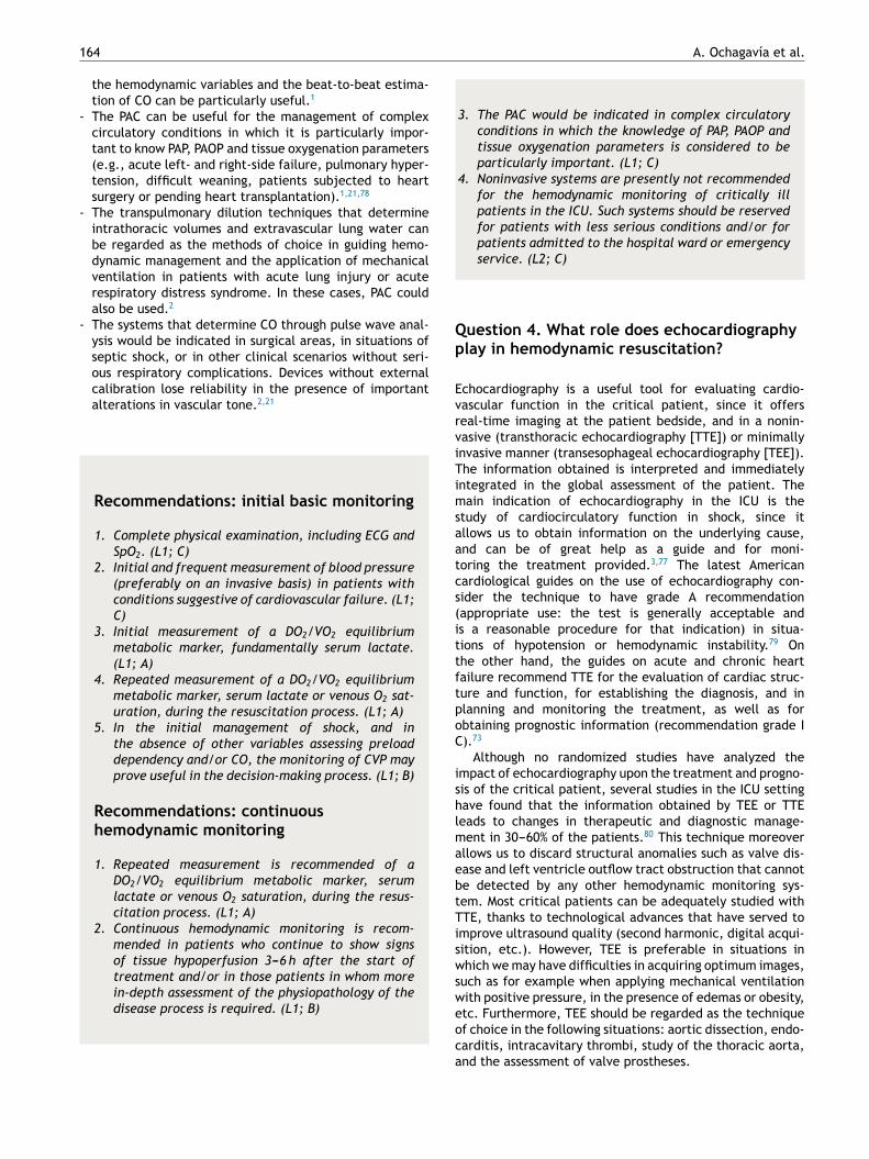
Figure 1 Algorithm for the evaluation of cardiovascular function and hemodynamic monitoring in situations of shock. PAC:
pulmonary artery catheter; ECG: electrocardiogram; RF: respiratory frequency; BP: blood pressure; MAP: mean arterial pres-
sure; NIP: noninvasive pressure; CVP: central venous pressure; SvcO2: central venous oxygen saturation; PPV: pulse pressure
variation.
The growing incorporation of ultrasound to the ICUhas caused many national Intensive Care societiesthroughout the world to promote the learning of thistechnique. On one hand, basic training in echocar-diography is advocated for intensivists, adopting agoal-directed approach, i.e., focused on solving spe-cific issues in Critical Care, while on the other handadvanced training is advised only for intensivists witha specific interest in furthering their knowledge ofechocardiography. Joint statements have been pub-lished by the American and French societies regardingthe competencies in ultrasound in the ICU.4 Theserecommendations define the skills and competen-cies required for the different levels of knowledge inechocardiography and in other ultrasound techniquesin the ICU, such as thoracic, abdominal and vascularultrasound. Likewise, the European Society of IntensiveCare Medicine, together with representatives of other soci-eties, has recently proposed a series of standard trainingrequirements in ultrasound.81
In relation to the basic level, emphasis is placed onthe need for echocardiographic examination to be quali-tative, dynamic and goal oriented. It should include basicassessment of left and right ventricular function, as wellas the assessment of cardiac tamponade, the estimation ofvolume response, and evaluation of massive valve regur-gitation. Evaluation should aim to answer a limited seriesof specific questions regarding situations of hemodynamicinstability (Is left ventricle function normal? Is the rightventricle dilated? What about right ventricle function?Are there echocardiographic signs of cardiac tamponade?Are there signs of severe hypovolemia? Is there seriousvalve disease?). More complete and detailed information
in turn would form part of an advanced level of trainingin echocardiography. The skills at this advanced level inthe ICU emphasize specific knowledge of aspects relatedto hemodynamic assessment of the critical patient, such asvolume response indices, filling pressures, CO, the impactof mechanical ventilation upon right ventricle function,etc.
Basic echocardiography should be applied in the initialassessment of shock, since it allows us to quickly detectthe characteristic causal conditions of shock: severe leftventricle failure, right ventricle failure generally secondaryto pulmonary thromboembolism, cardiac tamponade, mas-sive valve insufficiency, and hypovolemia. At this pointof the evaluation, before the adoption of any treatmentmeasure (pharmacological or surgical), the diagnostic yieldof the technique is maximum. Posteriorly, a more com-plete echocardiographic evaluation should be made in theevent of insufficient treatment response or if further insightinto the physiopathology of the process is required. Thismore exhaustive exploration requires training in advancedechocardiography, affording reliable, detailed and morein-depth information about relevant aspects of cardiovas-cular function in the hemodynamic management of thecritical patient. Discontinuous but repeated echocardi-ographic evaluations contribute to further hemodynamicassessment and to evaluate and guide treatment. On theother hand, general ultrasound is also very useful for theglobal evaluation of shock patients, and can help iden-tify a non-cardiogenic origin of hemodynamic instability.Clearly, the information afforded by ultrasound explorationmust always be accompanied by other clinical assessmentelements in critical patients such as the case history andinitial examination, imaging and laboratory tests, and the

166 A. Ochagavía et al.
data obtained with other hemodynamic monitoring systems(Fig. 1).3,4,77
Recommendations: echocardiography
1. In the initial phase of the evaluation of shock, basicechocardiography is extremely useful for obtain-ing information on the underlying cause, and canbe of great help as a guide and for monitoring thetreatment provided. (L1; B)
2. In situations of shock characterized by insuffi-cient treatment response, or if further insight intothe physiopathology of the process is required,advanced level echocardiography is indicated. (L1;B)
Question 5. What evidence is there of theusefulness of hemodynamic monitoring in thecritical patient?
As we have commented above, hemodynamic monitoring isbased on the premise that the detection and treatment ofthe physiopathological alterations of critical disease shouldresult in improved patient outcomes. Despite the logic ofthis affirmation, the concept of ‘‘hemodynamic monitor-ing’’ has been subject to endless discussion due to thelack of studies demonstrating that monitoring in itself isable to improve the patient prognosis. This attitude maybe wrong, however, since the use of certain variables (suchas venous oxygen saturation) in cardiovascular resuscitationalgorithms has indeed shown a beneficial impact upon theprognosis of different disease conditions. The most evidentexample of this can be found in recent studies in septicpatients, such as the work of Rivers et al.,15 in which resusci-tation guided by previously defined hemodynamic objectivesor goals had a strong impact upon the survival of thesepatients.
On the other hand, demonstrating the isolated progno-stic value of the many existing hemodynamic variables willbe very complicated. This would be the case, for exam-ple, of PPV and/or SVV as predictors of the responseto volume expansion in certain patients. Despite theirundeniable physiological meaning and the fact that theyhave been shown to be clearly superior to the cardiacfilling pressures, the lack of randomized studies specifi-cally demonstrating that the isolated use of such dynamicvariables is directly correlated to improved prognosis hascaused them to be excluded from the different rec-ommendations and management guides---while in contrastthe mentioned cardiac filling pressures have been main-tained.
Thus, the impact that hemodynamic monitoring willhave upon our patients depends not only on the reliabil-ity of the systems used, but also on our knowledge oftheir limitations, as well as on adequate understanding ofthe physiological principles and correct interpretation of the
variables obtained. The adequate use of these variables, asa goal or tool in decision making, is the factor ultimatelydeciding the beneficial impact upon patient outcome. Thereasoning, therefore, is that hemodynamic monitoring willonly result in an improved patient prognosis when it isassociated to a treatment that has been shown to affordbenefits.82
Recommendations: evidence of theusefulness of hemodynamic monitoring
No hemodynamic monitoring system will have a posi-tive impact upon the prognosis of the critical patientunless it is associated to treatments of establishedefficacy. (L1; C)
Conflicts of interest
Ana Ochagavia and Javier Maynar: payment for conferences,consultants (Pulsion): Ignacio Monge: payment for confer-ences, consultants (Edwards).
References
1. Vincent JL, Rhodes A, Perel A, Martin GS, Della RocaG, Vallet B, et al. Clinical review: update on hemo-dynamic monitoring-a consensus of 16. Crit Care. 2011;15:229.
2. Mateu Campos M, Ferrándiz Sellés A, Gruartmoner de VeraG, Mesquida Febrer J, Sabatier Cloarec C, Poveda HernándezY, et al. Técnicas disponibles de monitorización hemod-inámica. Ventajas y limitaciones. Med Intensiva. 2012;36:434---44.
3. Ayuela Azcárate JM, Clau Terré F, Ochagavía A, Vicho PereiraR. Papel de la ecocardiografía en la monitorización hemod-inámica de los pacientes críticos. Med Intensiva. 2012;36:220---32.
4. Mayo PH, Beaulieu Y, Doelken P, Feller-Kopman D, HarrodC, Kaplan A, et al. American College of Chest Physician/LaSociété de Réanimation de Langue Francaise statement oncompetence in critical care ultrasonography. Chest. 2009;135:1050---60.
5. Ochagavía A, Baigorri F. Introducción de la serie «Puesta aldía: monitorización hemodinámica en el paciente crítico». MedIntensiva. 2011;35:497---8.
6. GRADE Working Group. Grading quality of evidenceand strength of recommendations. BMJ. 2004;328:1490---8.
7. Howell MD, Donnino M, Clardy P, Talmor D, ShapiroNI. Occult hypoperfusion and mortality in patientswith suspected infection. Intensive Care Med. 2007;33:1892---9.
8. Mesquida J, Borrat X, Lorente JA, Masip J, Baigorri F.Objetivos de la reanimación hemodinámica. Med Intensiva.2011;35:499---508.
9. Varpula M, Tallgren M, Saukkonen K, Voipio-Pulkki LM. Hemody-namic variables related to outcome in septic shock. Crit CareMed. 2005;31:1066---71.

Hemodynamic monitoring in the critically patient 167
10. Dubin A, Pozo MO, Casabella CA, Pálizas Jr F, MuriasG, Moseinco MC, et al. Increasing arterial blood pres-sure with norepinephrine does not improve microcircula-tory blood flow: a prospective study. Crit Care. 2009;13:R92.
11. Antonelli M, Levy M, Andrews PJD, Chastre J, HudsonLD, Manthous C, et al. Hemodynamic monitoring inshock and implications for management. InternationalConsensus Conference. Intensive Care Med. 2007;33:575---90.
12. Feyen BFE, Sener S, Jorens PG, Menovsky T, Maas AI. Neu-romonitoring in traumatic brain injury. Minerva Anestesiol.2012;78:949---58.
13. Kern JW, Shoemaker WC. Meta-analysis of hemodynamicoptimization in high risk surgical patients. Crit Care Med.2002;30:1686---92.
14. Reinhart K, Kuhn HJ, Hartog C, Bredle DL. Continuouscentral venous and pulmonary artery oxygen saturation mon-itoring in the critically ill. Intensive Care Med. 2004;30:1572---8.
15. Rivers E, Nguyen B, Havstad S, Ressler J, Muzzin A, KnoblichB, et al. Early goal-directed therapy in the treatment ofsevere sepsis and septic shock. N Engl J Med. 2001;345:1368---77.
16. Textoris J, Fouche L, Wiramus S, Antonini F, Tho S, Martin C,et al. High central venous oxygen saturation in the latter stagesof septic shock is associated with increased mortality. Crit Care.2011;15:R176.
17. Shapiro NI, Howell MD, Talmor D, Nathanson LA, Lisbon A, WolfeRE, et al. Serum lactate as a predictor of mortality in emer-gency department patients with infection. Ann Emerg Med.2005;45:524---8.
18. Jones AE, Shapiro NI, Trzeciak S, Arnold RC, Claremont HA, KlineJA. Lactate clearance vs central venous oxygen saturation asgoals of early sepsis therapy: a randomized clinical trial. JAMA.2010;303:739---46.
19. Smith I, Kumar P, Molloy S, Rhodes A, Newman PJ, GroundsRM, et al. Base excess and lactate as prognostic indicatorsfor patients admitted to intensive care. Intensive Care Med.2001;27:74---83.
20. Vallee F, Vallet B, Mathe O, Parraguette J, Mari A, Silva S, et al.Central venous-to-arterial carbon dioxide difference: an addi-tional target for goal-directed therapy in septic shock. IntensiveCare Med. 2008;34:2218---25.
21. Alhashemi J, Cecconi M, Hofer CH. Cardiac output monitoring:an integrative perspective. Crit Care. 2011;15:214.
22. Slagt C, Breuker RM, Groeneveld J. Choosing patient-tailored hemodynamic monitoring. Crit Care. 2010;14:208.
23. García X, Mateu L, Maynar J, Mercadal J, Ochagavía A, FerrandizA, et al. Estimación del gasto cardíaco. Utilidad en la prácticaclínica. Monitorización disponible invasiva y no invasiva. MedIntensiva. 2011;35:5552---61.
24. Swan HJC, Ganz W, Forrester JS, Marcus H, Diamond G,Chonette D. Catheterization of the heart in man with theuse of a flow-directed balloon tipped catheter. N Engl J Med.1970;283:447---51.
25. Carrillo López A, Fiol Sala M, Rodríguez Salgado A. El papeldel Swan-Ganz en la actualidad. Med Intensiva. 2010;34:203---14.
26. Connors AF, Speroff T, Dawson NV, Thomas C, Harrell Jr FE, Wag-ner D, et al., for the SUPPORT Investigators. The effectivenessof right heart catheterization in the initial care of critically illpatients. JAMA. 1996;276:889---97.
27. Goedje O, Hoeke K, Lichtwarck-Aschoff M, Faltchauser A,Lamm P, Reichart B. Continous cardiac output by femoralarterial thermodilution calibrated pulse contour analysis:
comparison with pulmonary arterial thermodilution. Crit CareMed. 1999;27:2407---12.
28. Friesechke F, Heinrich A, Abel P, Felix SB. Comparison ofpulmonary artery and aortic transpulmonary thermodilutionfor monitoring of cardiac output in patients with severeheart failure: validation of a novel method. Crit Care Med.2009;37:119---23.
29. Linton R, Band D, Haire K. A new method of measuringcardiac output in man using lithium dilution. Br J Anaesth.1993;71:262---6.
30. Sakka SG, Kozieras J, Thuemer O, van Hout N. Measure-ment of cardiac output: a comparison between transpulmonarythermodilution and uncalibrated pulse contour analysis. Br JAnaesth. 2007;99:337---42.
31. Hofer CK, Senn A, Weibel L, Zollinger A. Assessment ofstroke volume variation for prediction of fluid responsivenessusing the modified FloTrac and PiCCO plus. Crit Care. 2008;12:282.
32. Mayer J, Boldt J, Mengistu AM, Röhm KD, Suttner S.Goal-directed intraoperative therapy based on autocalibratedarterial pressure reduces hospital stay in high-risk surgicalpatients: a randomized controlled trial. Crit Care. 2010;14:R18.
33. Mathews L, Singh K. Cardiac output monitoring. Ann CardiacAnaesth. 2008;11:56---68.
34. Benes J, Chytra I, Altmann P, Hluchy M, Kasal E, Svitak R, et al.Intraoperative fluid optimization using stroke volume variationin high surgical patients: results of prospective randomizedstudy. Crit Care. 2010;14:R118.
35. Monge MI, Estella A, Díaz JC, Gil A. Monitorización hemod-inámica mínimamente invasiva con eco-Doppler esofágico. MedIntensiva. 2008;32:33---44.
36. Squara P, Denjean D, Estagnasie P, Brusset A, Dib JC,Dubois C, et al. Noninvasive cardiac output monitoring(NICOM): a clinical validation. Intensive Care Med. 2007;33:1191---4.
37. Thom O, Taylor DM, Wolf RE, Cade J, Myles P, Krum H, et al.Comparison of a supra-sternal cardiac output monitor (USCOM)with the pulmonary artery catheter. Br J Anaesth. 2009;103:800---4.
38. Bendjelid K, Romand JA. Fluid responsiveness inmechanically ventilated patients: a review of indicesused in intensive care. Intensive Care Med. 2003;29:352---60.
39. Michard F, Teboul JL. Predicting fluid responsiveness inICU patients: a critical analysis of the evidence. Chest.2002;121:2000---8.
40. Sabatier C, Monge I, Maynar J, Ochagavía A. Valoración de laprecarga y la respuesta cardiovascular al aporte de volumen.Med Intensiva. 2012;36:45---55.
41. Levy MM, Macias WL, Russell JA, Williams MD, Trzaskoma BL,Silva E, et al. Failure to improve during the first day of therapy ispredictive of 28-day mortality in severe sepsis. Chest. 2004;124Suppl.:120S.
42. Murphy CV, Schramm GE, Doherty JA, Reichley RM, GajicO, Afessa B, et al. The importance of fluid management inacute lung injury secondary to septic shock. Chest. 2009;136:102---9.
43. Kastrup M, Markewitz A, Spies C, Carl M, Erb J, Grosse J, et al.Current practice of hemodynamic monitoring and vasopressorand inotropic therapy in post-operative cardiac surgery patientsin Germany: results from a postal survey. Acta AnaesthesiolScand. 2007;51:347---58.
44. Dellinger RP, Levy M, Rhodes A, Annane D, Gerlach H, Opal SM,et al. Surviving Sepsis Campaign: international guidelines frommanagement of severe sepsis and septic shock: 2012. Crit CareMed. 2013;41:580---637.

168 A. Ochagavía et al.
45. Reuse C, Vincent JL, Pinsky M. Measurements of right ven-tricular volumes during fluid challenge. Chest. 1990;98:1450---4.
46. Marik PE, Baram M, Vahid B. Does central venous pres-sure predict fluid responsiveness? A systematic review of theliterature and the tale of seven mares. Chest. 2008;134:172---8.
47. Teboul JL, et le groupe d’experts de la SRLF. Recomman-dations d’experts de la SRLF «Indicateurs du remplissagevasculaire au cours de l’insuffisance circulatoire». Réanimation.2004;13:255---63.
48. Michard F, Teboul JL. Using heart---lung interactions to assessfluid responsiveness during mechanical ventilation. Crit Care.2000;4:282---9.
49. Michard F, Boussat S, Chemla D, Anguel N, Mercat A, Lecarpen-tier Y, et al. Relation between respiratory changes in arterialpulse pressure and fluid responsiveness in septic patientswith acute circulatory failure. Am J Respir Crit Care Med.2000;162:134---8.
50. Marik PE, Cavallazzi R, Vasu T, Hirani A. Dynamic changes inarterial waveform derived variables and fluid responsiveness inmechanically ventilated patients: a systematic review of theliterature. Crit Care Med. 2009;37:2642---7.
51. Berkenstadt H, Margalit N, Hadani M, Friedman Z, Segal E, VillaY, et al. Stroke volume variation as a predictor of fluid respon-siveness in patients undergoing brain surgery. Anesth Analg.2001;92:984---9.
52. Monnet X, Rienzo M, Osman D, Anguel N, Richard C, Pinsky MR,et al. Esophageal Doppler monitoring predicts fluid responsive-ness in critically ill ventilated patients. Intensive Care Med.2005;31:1195---201.
53. Feissel M, Michard F, Mangin I, Ruyer O, Faller JP, Teboul JL.Respiratory changes in aortic blood velocity as an indicator offluid responsiveness in ventilated patients with septic shock.Chest. 2001;119:867---73.
54. Natalini G, Rosano A, Taranto M, Faggian B, Vittorielli E,Bernardini A. Arterial versus plethysmographic dynamic indicesto test responsiveness for testing fluid administration inhypotensive patients: a clinical trial. Anesth Analg. 2006;103:1478---84.
55. Loupec Th, Nanadoumgar H, Frasca D. Pleth variability indexpredicts fluid responsiveness in critically ill patients. Crit CareMed. 2011;39:294---9.
56. Feissel M, Richard F, Faller JP, Teboul JL. The respi-ratory variation in inferior vena cava diameter as aguide to fluid therapy. Intensive Care Med. 2004;30:1834---7.
57. Vieillard-Baron A, Chergui K, Rabiller A. Superior venacava collapsibility as a gauge of volume status in ven-tilated septic patients. Intensive Care Med. 2004;30:1734---9.
58. De Backer D, Heenen S, Piagnerelli M, Koch M, VincentJL. Pulse pressure variations to predict fluid responsive-ness: influence of tidal volume. Intensive Care Med. 2005;31:517---23.
59. Monnet X, Rienzo M, Osman D, Anguel N, Richard C,Pinsky MR, et al. Passive leg raising predicts fluidresponsiveness in the critically ill. Crit Care Med. 2006;2006:5.
60. Cavallaro F, Sandroni C, Marano C, La Torre G, Mannocci A,De Waure C, et al. Diagnostic accuracy of passive leg rais-ing for prediction of fluid responsiveness in adults: systematicreview and meta-analysis of clinical studies. Intensive CareMed. 2010;36:1475---83.
61. Vincent JL, Weil MH. Fluid challenge revisited. Crit Care Med.2006;34:1333---7.
62. Poelaert J. Haemodynamic monitoring. Curr Opin Anaesthesiol.2001;14:27---32.
63. Pinsky MR. Functional hemodynamic monitoring: applied phys-iology at the bedside. In: Vincent JL, editor. Yearbook ofintensive care med. Berlin: Springer-Verlag; 2002. p. 537---51.
64. Ochagavía A, Zapata L, Carrillo A, Rodríguez A, GuerreroM, Ayuela JM. Evaluación de la contractilidad y la poscargaen la unidad de cuidados intensivos. Med Intensiva. 2012;36:365---74.
65. Grossman W, Braunwald E, Mann T, McLaurin LP, Green LH.Contractile state of the left ventricle in man as evalu-ated from end-systolic pressure---volume relations. Circulation.1977;56:845---52.
66. Chen CH, Fetics B, Nevo E, Rochitte CE, Chiou KR, Ding PA,et al. Noninvasive single-beat determination of left ventri-cular end-systolic elastance in humans. J Am Coll Cardiol.2001;38:2028---34.
67. Crexells C, Chatterjee K, Forrester JS, Dikshit K, Swan HJ.Optimal level of filling pressure in the left side of the heartin acute myocardial infarction. N Engl J Med. 1973;289:1263---6.
68. La Vechia L, Varotto L, Spadaro GL, Zanolla L, FontanelliA. Left ventricular stroke work reserve as prognostic markerin severe heart failure. Minerva Cardioangiol. 2006;54:249---55.
69. Cotter G, Williams SG, Vered Z, Tan LB. Role of car-diac power in heart failure. Curr Opin Cardiol. 2003;18:215---22.
70. Tan LB, Littler WA. Measurement of cardiac reserve in cardio-genic shock: implications for prognosis and management. BrHeart J. 1990;64:121---8.
71. Fincke R, Hochman JS, Lowe AM, Menon V, Slater JN,Webb JG, et al. Cardiac power is the strongest hemody-namic correlate of mortality in cardiogenic shock: a reportfrom the shock trial registry. J Am Coll Cardiol. 2004;44:340---8.
72. O’Gara PT, Kushner FG, Ascheim DD, Casey Jr DE, ChungMK, de Lemos JA, et al. 2013 ACCF/AHA guidelines for themanagement of ST-elevation myocardial infarction. Circulation.2013;127:e362---425.
73. McMurray J, Adamopoulus S, Anker SD, Auricchio A, Böhm M,Dickstein K, et al., ESC Committee for Practice Guidelines.ESC Guidelines for the diagnosis and treatment of acute andchronic heart failure 2012: the Task Force for the Diagnosisand Treatment of Acute and Chronic Heart Failure 2012 of theEuropean Society of Cardiology. Developed in collaboration withthe Heart Failure Association (HFA) of the ESC. Eur Heart J.2012;33:1787---847.
74. Slama M, Maizel J. Echocardiographic measurementof ventricular function. Curr Opin Crit Care. 2006;12:241---8.
75. Monge Garcia MI, Gil Cano A, Gracia Romero M. Dynamic arte-rial elastance to predict arterial pressure response to volumeloading in preload-dependent patients. Crit Care. 2011;15:R15,http://dx.doi.org/10.1186/cc9420.
76. Pinsky MR. Protocolized cardiovascular management based onventricular-arterial coupling. In: Pinsky MR, Payen D, editors.Functional hemodynamic monitoring. Berlin: Springer-Verlag;2005. p. 381---95.
77. Schmidt GA, Koenig S, Mayo PH. Ultrasound to guide diagnosisand therapy. Chest. 2012;142:1042---8.
78. Richard Ch, Monnet X, Teboul JL. Pulmonary arterycatheter monitoring in 2011. Curr Opin Crit Care. 2011;17:296---302.
79. American College of Cardiology Foundation AppropriateUse Criteria Task Force; American Society of Echocar-diography; American Heart Association; American Societyof Nuclear Cardiology; Heart Failure Society of America;Heart Rhythm Society; Society for Cardiovascular Angiogra-phy and Interventions; Society of Critical Care Medicine;

Hemodynamic monitoring in the critically patient 169
Society of Cardiovascular Computed Tomography; Societyfor Cardiovascular Magnetic ResonanceDouglas PS, GarcíaMJ, Haines DE, Lai WW, Manning WJ, Patel AR, et al.ACCF/ASE/AHA/ASNC/HFSA/HRS/SCAI/SCCM/SCCT/SCMR 2011Appropriate Use Criteria for Echocardiography. A Report ofthe American College of Cardiology Foundation AppropriateUse Criteria Task Force, American Society of Echocardi-ography, American Heart Association, American Society ofNuclear Cardiology, Heart Failure Society of America, HeartRhythm Society, Society for Cardiovascular Angiography andInterventions, Society of Critical Care Medicine Societyof Cardiovascular Computed Tomography, and Society for
Cardiovascular Magnetic Resonance Endorsed by the Ameri-can College of Chest Physicians. J Am Coll Cardiol. 2011;57:1126---66.
80. Beaulieu Y. Bedside echocardiography in the assessment of thecritically ill. Crit Care Med. 2007;35 Suppl.:S235---49.
81. Expert Round Table on Ultrasounds in ICU. Interna-tional expert statement on training standards for criticalcare ultrasonography. Intensive Care Med. 2011;37:1077---83.
82. Gil Cano A, Monge García IM, Baigorri González F. Evidencia dela utilidad de la monitorización hemodinámica en el pacientecrítico. Med Intensiva. 2012;36:650---5.