heme-regulated eif2 kinase activated atf4 signaling pathway in
TRANSCRIPT

RED CELLS, IRON, AND ERYTHROPOISESIS
Heme-regulated eIF2� kinase activated Atf4 signaling pathway in oxidative stressand erythropoiesisRajasekhar N. V. S. Suragani,1 Roshini S. Zachariah,1 Jason G. Velazquez,1 Sijin Liu,1,2 Chiao-Wang Sun,3 Tim M. Townes,3
and Jane-Jane Chen1
1Harvard–MIT Division of Health Sciences and Technology, Massachusetts Institute of Technology, Cambridge, MA; 2State Key Laboratory of EnvironmentalChemistry and Ecotoxicology, Research Center for Eco-Environmental Science, Chinese Academy of Sciences, Beijing, China; and 3Department ofBiochemistry and Molecular Genetics, University of Alabama, Birmingham, AL
Heme-regulated eIF2� kinase (Hri) is nec-essary for balanced synthesis of hemeand globin. In addition, Hri deficiencyexacerbates the phenotypic severity of�-thalassemia intermedia in mice. Activa-tion of Hri during heme deficiency and in�-thalassemia increases eIF2� phosphor-ylation and inhibits globin translation.Under endoplasmic reticulum stress andnutrient starvation, eIF2� phosphoryla-tion also induces the Atf4 signaling path-way to mitigate stress. Although the func-tion of Hri in regulating globin translation
is well established, its role in Atf4 signal-ing in erythroid precursors is not known.Here, we report the role of the Hri-activated Atf4 signaling pathway in reduc-ing oxidative stress and in promotingerythroid differentiation during erythro-poiesis. On acute oxidative stress, Hri�/�
erythroblasts suffered from increased lev-els of reactive oxygen species (ROS) andapoptosis. During chronic iron deficiencyin vivo, Hri is necessary both to reduceoxidative stress and to promote erythroiddifferentiation. Hri�/� mice developed inef-
fective erythropoiesis during iron defi-ciency with inhibition of differentiation atthe basophilic erythroblast stage. Thisinhibition is recapitulated during ex vivodifferentiation of Hri�/� fetal liver ery-throid progenitors. Importantly, the Hri-eIF2�P-Atf4 pathway was activated andrequired for erythroid differentiation. Wefurther demonstrate the potential of modu-lating Hri-eIF2�P-Atf4 signaling withchemical compounds as pharmaceuticaltherapies for �-thalassemia. (Blood. 2012;119(22):5276-5284)
Introduction
Heme-regulated eIF2� kinase (Hri) was the first discoveredmember of the family of eIF2� kinases, which control translationalinitiation under diverse stress conditions by phosphorylation of the�-subunit of eIF2 (eIF2�P).1,2 Hri is necessary to coordinatetranslation of globin mRNAs with the availability of heme for theproduction of large amounts of hemoglobin during erythroidmaturation.3 In Hri deficiency, excessive globins synthesizedduring heme deficiency precipitate and form inclusions causingproteotoxicity.3,4 Beyond heme deficiency, Hri is also activated byoxidative stress and denatured proteins,5 both of which occur inthalassemia.6 Indeed, Hri is required to reduce the phenotypicseverity of the Hbb�/� mouse model of �-thalassemia intermedialacking both copies of �-globin major.4
Besides Hri, there are 3 additional mammalian eIF2� kinases:the double-stranded RNA-dependent eIF2� kinase (Pkr), the Gcn2protein kinase, and the Pkr-like ER resident kinase (Perk).1 Eachkinase elicits a major physiologic response in vivo: Pkr responds toviral infection; Gcn2 senses nutrient starvations; Perk is activatedby endoplasmic reticulum (ER) stress; and Hri is regulated byheme. In addition to inhibition of global protein synthesis, thesecond function of eIF2� phosphorylation is to reprogram transla-tion and transcription necessary for adaptation to stress7 asillustrated in Figure 7A. In mammalian cells, translation ofactivating transcriptional factor 4 (Atf4) is selectively increased byeIF2�P amid general inhibition of protein synthesis during ER stressand amino acid starvation via upstream open reading frames in the
5�-UTR (untranslated region).8,9 A major target gene of Atf4 is thetranscription factor, C/EBP Homologous Protein-10 (Chop), whichis up-regulated on stress.10 In ER stress, induction of Chop leads toexpression of Gadd34, which recruits eIF2�P for dephosphoryla-tion by PPase1.11,12 This feedback mechanism of Gadd34 inregenerating active eIF2 is necessary for the recovery of proteinsynthesis during the late stage of the stress response.13,14
Although we demonstrated the necessity of Hri in inhibiting globintranslation during iron/heme deficiency and in �-thalassemia, therole of Hri under these stress conditions is more than just inhibitionof translation. Hri is also necessary to reduce ineffective erythropoi-esis.3,4 To further understand the molecular mechanism by whichHri mitigates stress and reduces ineffective erythropoiesis, weinvestigated the eIF2�P-Atf4 signaling pathway in the stressresponse of the erythroid lineage.
In this article we demonstrate that Hri activates the Atf4 stressresponse pathway in nucleated erythroid precursors for adaptationto oxidative stress. Significantly, this Hri-dependent eIF2�P-Atf4pathway is also operative and necessary for erythroid differentia-tion, especially under stress conditions. Our study reveals thefunction of Hri in translational activation of Atf4 mRNA and thesubsequent regulation of gene expression necessary for stressadaptation. We also demonstrate the feasibility of targeting theHri-eIF2�P-Atf4 pathway for the treatment of �-thalassemia withthe small chemical salubrinal, which inhibits dephosphorylation ofeIF2�P and promotes the Atf4 signaling pathway.15
Submitted October 25, 2011; accepted April 2, 2012. Prepublished online asBlood First Edition paper, April 12, 2012; DOI 10.1182/blood-2011-10-388132.
The online version of this article contains a data supplement.
The publication costs of this article were defrayed in part by page chargepayment. Therefore, and solely to indicate this fact, this article is herebymarked ‘‘advertisement’’ in accordance with 18 USC section 1734.
© 2012 by The American Society of Hematology
5276 BLOOD, 31 MAY 2012 � VOLUME 119, NUMBER 22
For personal use only.on April 1, 2018. by guest www.bloodjournal.orgFrom

Methods
Animals
Mice were maintained at the Massachusetts Institute of Technology (MIT)animal facility, and all experiments were carried out using the protocolsapproved by the Division of Comparative Medicine at MIT. Hri�/� micewere generated in our laboratory.3 The generations of Atf4�/� and Hri�/
�Hbb�/� mice were as previously described.4,16 All mice used for this studywere of C57BL/6J genetic background. Mice were maintained in aniron-deficient diet (Harlan Teklad) on weaning for 2 months before analysis.
Fetal liver cell isolation and ex vivo differentiation
Ter119� cells were isolated from E14.5 fetal liver (FL) and cultured for exvivo differentiation as described.17 Morphologic examination of cells wascarried out on cytospin slides stained with Giemsa and benzidine.17
Sodium arsenite and H2O2 treatments
E14.5 FL cells from Hri �/� and �/� mice were recovered for 1 hour inIscove medium supplemented with cytokines17 before sodium arsenitetreatment as indicated. Peripheral blood (2 �L) was resuspended in 1 mL ofHanks balanced salt solution (HBSS). Red blood cells (RBCs) were washedonce with HBBS, resuspended in 200 �L of HBSS, and recovered for20 minutes at 37°C incubator. The RBCs were then preincubated with5�M of 5 and 6-chloromethyl-2�,7�-dichlorodihydrofluorescein diacetate(CM-H2DCFDA; Invitrogen) for 30 minutes followed by treatment withH2O2 for 20 minutes.
Analyses of ROS, erythroid differentiation, and apoptosis byflow cytometry
All the flow cytometric analyses were performed at the MIT Core Facility,which provides excellent technical support for gating, analyzing and sortingcell populations. For cellular reactive oxygen species (ROS) measurements,cells were resuspended in prewarmed phosphate-buffered saline (PBS)supplemented with 5% fetal bovine serum (FBS) and incubated with5�M 2�, 7�-dichlorodihydrofluorescein diacetate (DCF; Invitrogen) in thedark for 30 minutes at room temperature, and then were analyzedimmediately by flow cytometry.18 Erythroid differentiation was analyzed aspreviously described.17 Reticulocyte formation during ex vivo culture wasmeasured by enucleation.19 Apoptosis was measured by annexin V (AnV)binding.3 Propidium iodide (BD Pharmingen) positive cells that contain latestage AnV� apoptotic and necrotic cells were excluded from these analyses.
Western blot analysis
Cell extracts were prepared as previously described.18 Proteins (5-20 �g)were separated by 7% (for Hri) and 10% (for other proteins) SDS-PAGE.Blots were incubated overnight at 4°C with primary antibodies againstHri (1:1000),18 eIF2� (1:1000; BioSource), phosphorylated eIF2� (1:1000;BioSource), Chop (1:500; Santa Cruz Biotechnology), Ho-1 (1:1000;Stressgen), or Atf4 (1:500; Abnova).
Quantitative real time PCR
RNAs were isolated using the Promega SV Total RNA Isolation kit. Thereverse transcription of mRNA and polymerase chain reaction (PCR) wereperformed using Quantitect kits (QIAGEN RT:205311, PCR:204143).Primer sequences for quantitative PCR are listed in supplemental Table 1(available on the Blood Web site; see the Supplemental Materials link at the top ofthe online article). eIF2� was used as internal control for normalization. Raw datawere processed through LinRegPCR for baseline correction to determine C(t)values. Relative expression of mRNA was determined by the �[�C(t)] method.
Knockdown of Atf4 expression
Knockdown of Atf4 expression in differentiating mouse erythroid leukemic(MEL) cells was performed with Sigma-Aldrich Mission small interfering
RNAs (siRNAs) using the protocol described by Paradkar et al.20
Atf4 knockdown siRNA sequences were shown in supplemental Table2. Mission siRNA Universal Negative Control #1 (Sigma-Aldrich) wasused as a negative control.
Salubrinal treatment and measurement of protein synthesis
Blood samples from adult Hri�/�Hbb�/� mice, which contained approxi-mately 30% reticulocytes, were recovered in complete DMEM medium asdescribed.3 Cells were then treated with salubrinal as indicated. E14.5FL cell were pretreated with 5�M of arsenite for 2 hours to enhance eIF2�Plevels. Cells were washed with PBS to remove arsenite and then treatedwith various concentrations of salubrinal as indicated. Splenic Ter119�
cells from Hbb�/� mice were isolated using magnetic-activated cell sortingMACS column purification (Miltenyi Biotec). Cells were then recoveredfor 30 minutes before salubrinal treatment at concentrations indicated for3.5 hours. The rate of globin synthesis was determined by S35-methionine/cysteine incorporation into globin proteins.3
Data analysis
The statistical analyses among the various groups were performed by the2-tailed Student t test with a P value of � .05 for statistical significance.
Results
Activation of Hri-eIF2�P-Atf4 pathway in erythroid precursorson arsenite-induced oxidative stress
We have shown earlier that Hri is the eIF2� kinase activated bytreatment with sodium arsenite in erythroid precursors and that theactivation of Hri by arsenite is prevented by pretreatment with theROS scavenger N-acetylcysteine.5 To gain further insight intomechanistic details of ROS production and Hri activation, ROSlevels in E14.5 FL cells, which are highly enriched in nucleatederythroid precursors, were measured after arsenite treatment. Asshown in Figure 1A, ROS levels increased rapidly within 15 minutesand peaked at 30 minutes on arsenite treatment of FL cells.Concomitantly, activation of Hri also peaked at 30 minutes afterarsenite treatment. Hri remained activated for at least 60 minutes asshown by the hyperphosphorylation of Hri and the increased levelsof eIF2�P (Figure 1B).
Arsenite treatments also resulted in the Hri-dependent inductionof the eIF2�P-Atf4 signaling pathway as shown by increased levelsof eIF2�P, Atf4 and Chop proteins in Hri�/� FL cells at 1 to 6 hoursof exposure to arsenite (Figure 1C). There was no increase ineIF2�P, Atf4, or Chop protein levels in Hri�/� cells on arsenitestress. Importantly, the Atf4 mRNA level did not change signifi-cantly on arsenite stress in either Hri�/� or Hri�/� cells (Figure1D), consistent with the immediate translational regulation ofAtf4 by increased eIF2�P.9 Both Chop and Gadd34 mRNA levelswere increased in Hri�/� cells on arsenite stress, consistent withtheir transcriptional induction by Atf4. In contrast to Gadd34, theconstitutively expressed regulator of eIF2�P dephosphorylationCrep21 was not affected by arsenite stress as would be expected(Figure 1D). These results establish that activation of Hri byarsenite stress elicits the eIF2�P signaling pathway of up-regulating Atf4, Chop, and Gadd34 in primary erythroid precursors.
Increased oxidative stress in Hri�/� erythroid precursors onarsenite stress
To investigate whether this up-regulation of the Hri-Atf4 signalingpathway might contribute to adaptation to oxidative stress, levels ofROS in Hri�/� and Hri�/� FL cells were measured at different time
HRI-ATF4 SIGNALING IN ERYTHROPOIESIS 5277BLOOD, 31 MAY 2012 � VOLUME 119, NUMBER 22
For personal use only.on April 1, 2018. by guest www.bloodjournal.orgFrom

points after arsenite treatment. ROS levels in Hri�/� cells weresignificantly higher than those in Hri�/� cells at 3, 6, and 9 hoursafter treatment with 25�M arsenite (supplemental Figure 1A).Furthermore, ROS levels declined to baseline in Hri�/� cells at9 hours of sodium arsenite treatments whereas ROS levels inHri�/� cells continued to rise (Figure 1E, supplemental Figure 1A).These results demonstrate that Hri is required to mitigate acuteoxidative stress induced by arsenite. In addition, Hri is necessaryfor recovery from oxidative stress after exposure to arsenite for3 hours (supplemental Figure 1B-C). Hri�/� cells not only failed torecover from oxidative stress induced by arsenite treatment, butalso suffered further increase in oxidative stress during therecovery phase. This increased oxidative stress of Hri�/� FL cellson arsenite exposure resulted in increased apoptosis accompaniedwith elevated activation of caspases 3 and 9 (supplemental Figure 2).
Hri and Atf4 dependent expression of Ho-1 and antioxidantgenes
To further investigate the mechanism by which Hri protects againstoxidative stress, we examined the expression of heme oxygenase1 (Ho-1), which catabolizes heme to biliverdin, CO, and Fe, andplays a pivotal role in reducing oxidative stress.22 We found thatHo-1 protein in Hri�/� erythroblasts was increased by treatmentwith arsenite from 0.1 to 3�M (Figure 2A). Interestingly, theincrease of Ho-1 expression by arsenite in Hri�/� cells was less
prominent and required higher concentrations compared withHri�/� cells (Figure 2A). Furthermore, the level of Ho-1 mRNAwas significantly increased on arsenite treatment in Hri�/� cells,but not in Hri�/� cells (Figure 2C).
We then examined whether the induction of Ho-1 by arseniterequired Atf4, a downstream target of Hri signaling pathway(Figure 1C). As shown in Figure 2B, there is less Ho-1 protein inAtf4�/� FL cells compared with Atf4�/�or Atf4�/� cells at thebaseline without arsenite treatment. On arsenite treatment, Ho-1protein expression was increased in FL cells from all the 3 genotypes.However, there was still less Ho-1 protein in Atf4�/� cells (Figure2B). These results demonstrate that Ho-1 expression in primaryerythroid precursors is dependent on both Hri and Atf4.
Besides Ho-1, arsenite treatment also induced expression of otherantioxidant genes (Figure 2C), such as glutathione S-tranferase� (Gst�), NAD(P)H quinone oxidoreductase 1 (Nqo1) and super-oxide dismutase 2 (Sod2) in Hri�/�, but not in Hri�/� FL cells.Similarly, induction of Ho-1 and Nqo1 mRNA was observed inAtf4�/�, but not in Atf4�/� cells (Figure 2D). These resultsdemonstrate that both Hri and Atf4 are necessary to induce someantioxidant genes to mitigate oxidative stress incurred by arsenitetreatment. Consistent with decreased expression of Ho-1 and Nqo1,RBCs from Hri�/� and Atf4�/� mice were more sensitive toH2O2-induced oxidative stress and exhibited increased ROS levelscompared with RBCs from wild-type (WT) and Atf4�/� mice(Figure 2E; quantitative data of ROS levels are shown in supplemen-tal Figure 3A). These results indicate that Hri-Atf4 signaling may
Figure 2. Hri and Atf4 dependent induction of antioxidant genes. (A) Hri-dependent Ho-1 protein expression. Hri�/� and Hri�/� FL cells were treated for5 hours with increasing concentrations of arsenite as indicated. (B) Atf4-dependentHo-1 protein expression. Atf4 �/�, �/�, and �/� FL cells were treated with 5�Marsenite for 3 hours. (C-D) Relative expression of mRNA of antioxidant genes. Hri�/�,Hri�/�, Atf4�/�, and Atf4�/� FL cells were treated with 5�M arsenite for 3 hours.mRNA levels of treated and untreated cells were analyzed by qPCR. Data arepresented as mean SD (n 3). P values denote the comparison between arsenite-treated Hri�/� and Hri�/� samples; P � .01 for Ho-1 and Nqo1, P � .05 for Gst�, andP .06 for Sod2; or between the comparison of arsenite-treated Atf4�/� and Atf4�/�
samples; P � .005 for Chop, P � .05 for Ho-1, P � .001 for Nqo1. (E) Elevated ROSlevels in Hri�/� and Atf4�/� RBCs upon H2O2 treatment. Blood samples were treated with50�M H2O2. (F) ROS levels in RBCs of Hri�/� (wt) and Hri�/� (Ko) mice in iron deficiency.
Figure 1. Induction of the Hri-eIF2�P-Atf4 signaling pathway and protectionagainst oxidative stress in FL erythroblasts. (A) ROS levels. Hri�/� FL cells weretreated with 25�M sodium arsenite. At times indicated, cellular ROS levels weremeasured by DCF fluorescence. (B) Hyperphosphorylation and activation of Hri.Hyperphosphorylation of Hri was examined by the slower migration in SDS-PAGE.eIF2� was used as a loading control. (C) Induction of the Hri-dependent Atf4 signalingpathway. Cells were treated with 5�M As. At times indicated, samples were analyzed forexpression of eIF2�P, Atf4, Chop, and eIF2� proteins. (D) Relative expression ofmRNAs of the Hri-Atf4 signaling pathway components. Atf4, Chop, Gadd34, andCrep mRNA levels from FL cells treated with 5�M As for 3 hours were analyzed byqPCR. Expression of each of the mRNA in untreated Hri�/� cells was defined as 1.Data are presented as mean SD (n 3). P values denote the comparison betweenarsenite-treated Hri�/� and Hri�/� samples; P � .001 for Atf4 and Gadd34; P � .005for Chop. (E) ROS levels. Hri�/� and Hri�/� FL cells treated with 5 or 25�M arsenitefor 9 hours before ROS measurement.
5278 SURAGANI et al BLOOD, 31 MAY 2012 � VOLUME 119, NUMBER 22
For personal use only.on April 1, 2018. by guest www.bloodjournal.orgFrom
also be required during erythroid development. Impairment of thispathway may generate RBCs that are more sensitive to oxidative insults.
Increased oxidative stress in Hri�/� erythroid cells duringchronic iron deficiency in vivo
As shown above, Hri is required for reducing acute oxidative stressin vitro (Figure 1E). We investigated whether Hri also participatedin reducing oxidative stress of erythroid cells in vivo duringchronic iron deficiency. Levels of ROS were measured in RBCsfrom WT and Hri�/� (KO) mice maintained on either a normal oriron-deficient diet. Representative histograms of ROS levels byFACS analysis are shown in Figure 2F. The quantitative data ofROS levels are shown in supplemental Figure 3B. It is important tonote that iron deficiency alone (WT-Fe) did not significantlyincrease ROS levels in RBCs. However, during iron deficiency, theabsence of Hri (KO-Fe) resulted in dramatically elevated ROSlevels in RBCs compared with those of WT-Fe cells (Figure 2F).These results demonstrate that Hri is necessary to mitigate oxida-tive stress in erythroid cells under chronic stress of iron deficiencyin vivo. These findings are also consistent with our gene profilingdata indicating that expression of many redox genes, such ascatalase, Sod2, Gsts, thioredoxin reductase 2, peroxiredoxins,ferritins, and Ho-1 is significantly lower in KO-Fe FL cellscompared with WT-Fe cells.23 Together, these results indicate thatboth Hri-mediated inhibition of globin translation and up-regulation of the Atf4 signaling pathway contribute in mitigatingoxidative stress during iron deficiency.
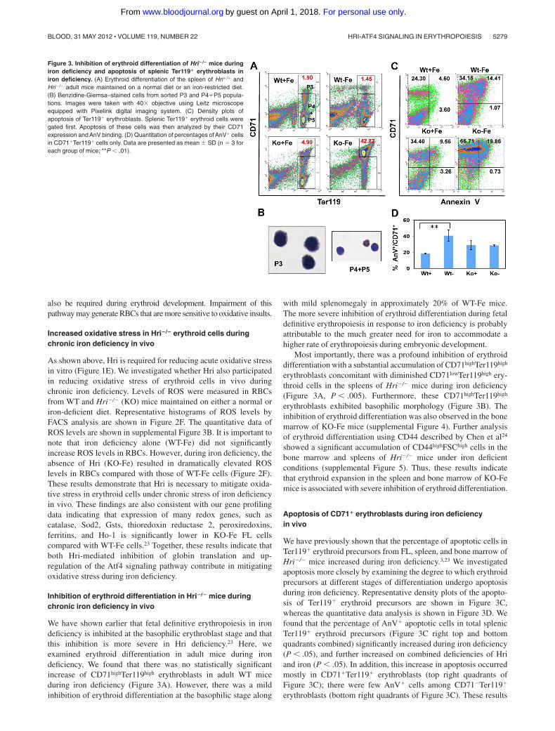
Inhibition of erythroid differentiation in Hri�/� mice duringchronic iron deficiency in vivo
We have shown earlier that fetal definitive erythropoiesis in irondeficiency is inhibited at the basophilic erythroblast stage and thatthis inhibition is more severe in Hri deficiency.23 Here, weexamined erythroid differentiation in adult mice during irondeficiency. We found that there was no statistically significantincrease of CD71highTer119high erythroblasts in adult WT miceduring iron deficiency (Figure 3A). However, there was a mildinhibition of erythroid differentiation at the basophilic stage along
with mild splenomegaly in approximately 20% of WT-Fe mice.The more severe inhibition of erythroid differentiation during fetaldefinitive erythropoiesis in response to iron deficiency is probablyattributable to the much greater need for iron to accommodate ahigher rate of erythropoiesis during embryonic development.
Most importantly, there was a profound inhibition of erythroiddifferentiation with a substantial accumulation of CD71highTer119high
erythroblasts concomitant with diminished CD71lowTer119high ery-throid cells in the spleens of Hri�/� mice during iron deficiency(Figure 3A, P � .005). Furthermore, these CD71highTer119high
erythroblasts exhibited basophilic morphology (Figure 3B). Theinhibition of erythroid differentiation was also observed in the bonemarrow of KO-Fe mice (supplemental Figure 4). Further analysisof erythroid differentiation using CD44 described by Chen et al24
showed a significant accumulation of CD44highFSChigh cells in thebone marrow and spleens of Hri�/� mice under iron deficientconditions (supplemental Figure 5). Thus, these results indicatethat erythroid expansion in the spleen and bone marrow of KO-Femice is associated with severe inhibition of erythroid differentiation.
Apoptosis of CD71� erythroblasts during iron deficiencyin vivo
We have previously shown that the percentage of apoptotic cells inTer119� erythroid precursors from FL, spleen, and bone marrow ofHri�/� mice increased during iron deficiency.3,23 We investigatedapoptosis more closely by examining the degree to which erythroidprecursors at different stages of differentiation undergo apoptosisduring iron deficiency. Representative density plots of the apopto-sis of Ter119� erythroid precursors are shown in Figure 3C,whereas the quantitative data analysis is shown in Figure 3D. Wefound that the percentage of AnV� apoptotic cells in total splenicTer119� erythroid precursors (Figure 3C right top and bottomquadrants combined) significantly increased during iron deficiency(P � .05), and further increased on combined deficiencies of Hriand iron (P � .05). In addition, this increase in apoptosis occurredmostly in CD71�Ter119� erythroblasts (top right quadrants ofFigure 3C); there were few AnV� cells among CD71�Ter119�
erythroblasts (bottom right quadrants of Figure 3C). These results
Figure 3. Inhibition of erythroid differentiation of Hri�/� mice duringiron deficiency and apoptosis of splenic Ter119� erythroblasts iniron deficiency. (A) Erythroid differentiation of the spleen of Hri�/� andHri�/� adult mice maintained on a normal diet or an iron-restricted diet.(B) Benzidine-Giemsa–stained cells from sorted P3 and P4�P5 popula-tions. Images were taken with 40� objective using Leitz microscopeequipped with Pixelink digital imaging system. (C) Density plots ofapoptosis of Ter119� erythroblasts. Splenic Ter119� erythroid cells weregated first. Apoptosis of these cells was then analyzed by their CD71expression and AnV binding. (D) Quantitation of percentages of AnV� cellsin CD71�Ter119� cells only. Data are presented as mean SD (n 3 foreach group of mice; **P � .01).
HRI-ATF4 SIGNALING IN ERYTHROPOIESIS 5279BLOOD, 31 MAY 2012 � VOLUME 119, NUMBER 22
For personal use only.on April 1, 2018. by guest www.bloodjournal.orgFrom

also confirm an expansion of splenic CD71� erythroblasts amongTer119� erythroid precursors from KO-Fe mice (top left and rightquadrants of Figure 3C).
It is possible that the increase in percentage of AnV� apoptoticcells in KO-Fe splenic Ter119� erythroid precursors might simplybe because of the expansion of CD71�Ter119� erythroblasts in thespleens of KO-Fe mice. We therefore analyzed the percentage ofAnV� apoptotic cells in CD71�Ter119� erythroblasts (Figure 3D).The percentage of apoptotic cells in CD71�Ter119� erythroblastswas increased in WT-Fe spleens, but was not further increased inKO-Fe spleens (Figure 3D). Similar observations were made withbone marrow samples (data not shown).
Collectively, these results demonstrate that ineffective erythro-poiesis of KO-Fe mice is primarily because of the profoundinhibition of erythroid differentiation at the CD71�Ter119� eryth-roblast stage. The increase of apoptotic cells in total Ter119�
erythroid precursors observed in KO-Fe mice is the consequence ofthe expansion and inhibition of erythroid differentiation ofCD71�Ter119� erythroblasts, which undergo apoptosis normally.25
Hri is necessary for differentiation of erythroid progenitorsex vivo
Ter119� cells in E14.5 FLs are primarily erythroid progenitors,which undergo proliferation and differentiation in ex vivo culture.17
We used this system to further investigate the role of Hri inerythroid differentiation. Differentiation of Hri�/� Ter119� ery-throid progenitors progressed more slowly than that of Hri�/� cells(Figure 4). Compared with Hri�/� cells, Hri�/� cells had a greatlyreduced population in the P3 basophilic erythroblast stage at 20 hoursof culture (top panels, P � .001, supplemental Figure 6A). Whereasthe percentage of cells in the P3 stage appeared similar at 40 hours,Hri�/� cells had higher CD71 and lower Ter119 expression,indicative of retarded erythroid differentiation (middle panels). At60 hours of differentiation (bottom panels), there was a signifi-cantly lower percentage of Hri�/� cells in the P4 polychromato-
philic and orthochromatophilic erythroblast stage compared withHri�/� cells (P � .005, supplemental Figure 6A).
To further substantiate the inhibition of erythroid differentiationof Hri�/� erythroblasts, the formation of reticulocytes duringex vivo differentiation was measured. Reticulocytes, which aredevoid of nuclei and are characterized as the Hoechst33 342lowTer119high population, are shown in magenta in Figure 4B.Reticulocyte formation was greatly reduced in the differentiation ofHri�/� Ter119� erythroid progenitors compared with Hri�/� cellsat 20, 40 and 60 hours (Figure 4B). This inhibition of differentia-tion was further corroborated by observations of cell morphologiesand hemoglobinization (supplemental Figure 6B). Taken together,these results demonstrate that Hri is required for differentiation oferythroid progenitors ex vivo, similar to the requirement of Hri inerythroid differentiation of adult spleens in vivo during irondeficiency (Figure 3A). Thus, this ex vivo erythroid differentiationsystem recapitulates the stress erythropoiesis observed in vivo, andprovides an excellent in vitro system to investigate the molecularmechanism by which Hri regulates erythroid differentiation.
Activation of the Hri signaling pathway during erythroiddifferentiation
We then examined the activation of the Hri-eIF2�P-Atf4 signalingpathway during ex vivo differentiation of Hri�/� and Hri�/� cells.Hri protein expression was increased at 36 and 48 hours. Further-more, Hri was activated by hyperphosphorylation (Hri-P; Figure4C). Most notably, there was Hri-dependent eIF2� phosphorylationand induction of Atf4, Chop, and Ho-1 protein expression duringerythroid differentiation of Hri�/�, but not Hri�/� Ter119� ery-throid progenitors (Figure 4C). Furthermore, we found that at36 hours of ex vivo differentiation, Hri�/� cells also had signifi-cantly lower levels of Atf4, Chop, Ho-1, Gst�, and Sod2 mRNAs(Figure 4D). Together, these results demonstrate the activation ofthe Hri signaling pathway during erythroid differentiation ex vivo.
Figure 4. Differentiation of Ter119� FL erythroid progenitors ex vivoand the activation of the Hri signaling pathway. (A) Erythroid differen-tiation, and (B) reticulocyte production of Hri �/� and �/� FL Ter119�
erythroid progenitors at 20, 40, and 60 hours of ex vivo culture.(C) Activation of Hri and protein expression of its downstream targets.Vertical lines have been inserted to indicate a repositioned gel lane. Themiddle 3 lanes, which contains Hbb�/� samples, were removed of thisWestern blot because these results is not necessary for this figure.(D) qPCR analysis of the mRNA expression at 36 hours of ex vivo culture.Data are presented as relative expression normalized to eIF2� control withmean SD (n 3; ***P � .005; **P � .01). Triplicate of ex vivo differen-tiation were carried out using Hri �/� or �/� FL erythroid progenitorsisolated from embryos of the same mother. This set of experiment wasrepeated 3 times with similar results.
5280 SURAGANI et al BLOOD, 31 MAY 2012 � VOLUME 119, NUMBER 22
For personal use only.on April 1, 2018. by guest www.bloodjournal.orgFrom

MEL cell is an established cell line, which undergoes erythroiddifferentiation on treatment with dimethylsulfoxide (DMSO). Wehave shown that Hri expression is up-regulated on induction oferythroid differentiation of MEL cells,26 and that overexpression ofthe dominant-negative Hri mutant inhibits erythroid differentia-tion.27 We found that the Hri-eIF2�P-Atf4 pathway was alsoactivated during DMSO-induced erythroid differentiation of MELcells (supplemental Figure 7).
Requirement of Atf4 for erythroid differentiation
To determine whether Atf4 is necessary for erythroid differentia-tion, siRNA knockdown of Atf4 expression was performed in MELcells. Three Atf4 siRNAs, S1, S2, and S3 were used individuallyand in combination. Both S1 and S2 siRNA significantly reducedAtf4 mRNA, wheras S3 was less effective (Figure 5A). Thecombination of all 3 siRNAs (S123) worked most effectively andreduced the Atf4 mRNA level to 34.5% of the control siRNA(Figure 5A). Importantly, S123 knockdown samples also had alower mRNA level of �-globin major compared with the controlsiRNA (Figure 5B), indicating an inhibition of differentiation byknocking down Atf4 expression. Erythroid differentiation of MELcells after treatment with control and S123 siRNAs was furtherexamined by benzidine-Giemsa staining. Cells at different stages ofdifferentiation were shown in Figure 5C and were scored morpho-
logically (Figure 5D). These results demonstrated that knockdownof Atf4 expression in MEL cells resulted in inhibition of erythroiddifferentiation at the basophilic stage and in reduction of benzidinepositive cells. Furthermore, S123-treated cells were less hemoglo-binized as assessed by benzidine staining (Figure 5C-D), consistentwith decreased �-globin major mRNA (Figure 5B). Together, theseresults support the requirement of Atf4 for erythroid differentiation.
It was reported that ablation of the Atf4 gene in mice results ingrowth retardation and transient fetal anemia, which is attributed toa proliferation deficit in erythroid burst-forming unit (BFU-E) anderythrocyte colony-forming unit (CFU-E) erythroid progenitors.16
The effect of Atf4 knockout on erythroid differentiation has notbeen investigated. We found that the in vivo erythroid differentia-tion of E14.5 Atf4�/� FLs was modestly inhibited at the P1 stagewith an increased percentage of cells at P1 and a decreasedpercentage of cells at the P3 stage (supplemental Figure 8B). Thisdifference in erythroid differentiation between the Atf4�/� andHri�/� FLs23 suggests the involvement of other eIF2� kinases aterythroid progenitor and proerythroblast stages because Atf4expression is regulated by all 4 eIF2 � kinases.2 In addition, wefound that Hri activation and eIF2�P were not altered in Atf4deficiency (supplemental Figure 8C), consistent with Atf4 beingdownstream of the Hri signaling pathway (Figure 1B-C).
Modulation of Hri signaling pathway in �-thalassemic erythroidprecursors by salubrinal
Increased oxidative stress and decreased erythroid differentiationexacerbate severity of �-thalassemia.28 We have shown earlier thatHri�/�Hbb�/� embryos died of severe anemia at E18.5 andHri�/�Hbb�/� mice had more severe adult �-thalassemic pheno-type.4 We investigated here the proof of concept for Hri and itssignaling pathway as possible potential novel pharmaceuticaltargets for treatment of �-thalassemia by reducing ineffectiveerythropoiesis.
We used salubrinal, a small chemical that selectively inhibitsthe dephosphorylation of eIF2�P, to test its capability to enhancethe Hri signaling pathway in Hri�/�Hbb�/� �-thalassemic ery-throid precursors. As shown in Figure 6A, salubrinal treatment ofHri�/�Hbb�/� reticulocytes resulted in increased eIF2�P as well asin a significant decrease in the rate of globin protein synthesis(Figure 6B). It is important to note that phosphorylation of only20%–30% of eIF2� is sufficient to completely inhibit proteinsynthesis.29 Furthermore, there was less 35S-globin in the insolublepellet fractions of salubrinal-treated reticulocytes compared withthe DMSO-treated control (Figure 6B). Salubrinal treatment alsoincreased the levels of eIF2�P and Chop in Hri�/�Hbb�/� FL cells(Figure 6C).
Similarly, salubrinal treatment of Hri�/�Hbb�/� Ter119� ery-throid precursors increased eIF2�P and inhibited protein synthesisin a concentration-dependent manner (Figure 6D). It is to be notedthat Atf4 protein was synthesized in Hri�/�Hbb�/� Ter119�
erythroid precursors, consistent with activation of Hri in �-thalasse-mia.4 Importantly, at 10�M salubrinal, Atf4 mRNA translation (asdetermined by immunoprecipitation of newly synthesized 35S-Atf4protein) was increased by 21%, whereas globin mRNA translationwas decreased by 17%. At 25�M salubrinal, eIF2�P was furtherincreased resulting in the shut-off of protein synthesis includingAtf4. This is expected, as no eIF2 can be recycled when eIF2�Psequesters all eIF2B.
Together, these results demonstrate that salubrinal is effective inincreasing eIF2�P and reducing denatured globins in �-thalassemic
Figure 5. Inhibition of erythroid differentiation of MEL cells by knockdown ofthe Atf4 expression. (A) Knockdown of Atf4 expression in MEL cells by siRNAs.(B) Decreased expression of �-globin major mRNA in Atf4 knockdown cells. Data arepresented as mean SD (n 3, 3 separate transfections were performed).(C-D) Inhibition of erythroid differentiation in Atf4 knockdown cells. At 5 days aftersiRNA transfection, differentiating cells were stained with Giemsa and benzidine.Representative cells at different stages of differentiation stained are shown in panelC. Cell images were obtained with 40� objectives as describe for Figure 3B.(D) Percentages of cells at the basophilic erythroblast (Baso), polychromaticerythroblast (Poly), and orthochromatic erythroblast (Ortho) stages as well asbenzidine-positive cells were determined by scoring the Giemsa-benzidine stainedcells.
HRI-ATF4 SIGNALING IN ERYTHROPOIESIS 5281BLOOD, 31 MAY 2012 � VOLUME 119, NUMBER 22
For personal use only.on April 1, 2018. by guest www.bloodjournal.orgFrom

reticulocytes. Furthermore, salubrinal also enhances Atf4 transla-tion and its subsequent signaling pathway in �-thalassemic ery-throid precursors. These observations provide the foundation forexploiting the Hri-eIF2�P signaling pathway for treatment ofthalassemia.
Discussion
Regulation of ROS levels and oxidative stress is extremelyimportant in erythropoiesis. Starting at the basophilic erythroblaststage, erythroid precursors synthesize large amounts of hemoglo-bin, which requires heme as a prosthetic group. Thus, iron uptakefor heme biosynthesis also increases, potentially generating ROSthrough the iron-catalyzed Fenton reaction.30 To date, 2 nonredun-dant Foxo3 and Nrf2-mediated pathways to combat oxidative stresshave been identified in erythroid cells.31-33 We show here for thefirst time that the Hri-eIF2�P-Atf4 pathway is also required in theerythroid lineage for the adaptation to oxidative stress.
As illustrated in Figure 7A, phosphorylation of eIF2� byactivated Hri not only leads to inhibition of globin translation butalso leads to the selective enhanced translation of Atf4 and
subsequent expression of redox genes (ie, Ho-1, Gst�, and Nqo1)for adaptation to acute and chronic oxidative stress. During chroniciron deficiency of Hri�/� mice, both reduced antioxidant geneexpression (Figure 2 and Liu et al23) and heme-free globinprecipitates3 probably contribute to the increased ROS levels inHri�/� erythroid cells. Although the exact mechanisms by whichmisfolded proteins in the cytosol generate oxidative stress have yetto be defined, there are 2 possibilities. Denatured heme-free globinaggregates may overwhelm and compromise capacities of molecu-lar chaperons and proteosomal degradation. Both of these pro-cesses have been shown to mitigate ROS during ER stress.Reduction of improper disulfide bonds in misfolded proteins byg lutathione (GSH) can deplete GSH level. In addition, degradationof misfolded proteins is necessary to prevent ROS accumulationduring ER stress.34
Furthermore, this Hri-eIF2�P-Atf4 pathway is also necessaryfor erythroid differentiation (Figure 7B). In nucleated erythro-blasts, Hri not only inhibits globin translation, but also increasesAtf4 translation to mitigate oxidative stress and to promoteerythroid differentiation. At the enucleated reticulocyte stage, therole of Hri is only to regulate globin translation to prevent
Figure 6. Modulation of Hri signaling by salubrinal in �-thalassemic erythroid precursors. (A) eIF2�P levels in Hri�/�Hbb�/� reticulocytes. Cells were treated for 6 hourswith concentrations of salubrinal indicated. (B) Globin protein synthesis in Hri�/�Hbb�/� reticulocytes. After treatment with 100�M salubrinal for 2 hours, globin proteinsynthesis at times indicated was measured in the supernatant and pellet fractions. Vertical lines have been inserted to indicate a repositioned gel lane. The middle lanebetween Control and Sal in the bottom rows (pellet), which was the salubrinal treated sample at time zero before 35S-Met/Cys labeling, was removed. (C) eIF2�P and Chop inHri�/�Hbb�/� FL erythroid precursors. Cells were treated for 12 hours with salubrinal at concentrations indicated. Numbers in panels A and C denote the ratio of eIF2�P/ eIF2�or Chop/eIF2�. Numbers in panel B denote the ratio of 35S-globin/total globin. (D) eIF2�P levels and protein synthesis in Ter119� cells from Hri�/�Hbb�/� spleen. Cells weretreated for 3.5 hours with salubrinal as indicated, and labeled with 35S-Met/Cys for the last 3 hours. Total protein syntheses in the cell lysates are shown in the middle panel.Newly synthesized 35S-Atf4 immunoprecipitated with antibody (Abnova) is shown in bottom panel. Numbers indicate ratio of eIF2�P/eIF2� or globin and Atf4 syntheses relativeto 0 �M controls.
5282 SURAGANI et al BLOOD, 31 MAY 2012 � VOLUME 119, NUMBER 22
For personal use only.on April 1, 2018. by guest www.bloodjournal.orgFrom
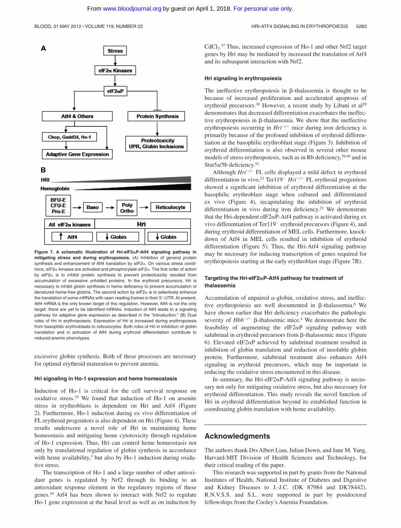
excessive globin synthesis. Both of these processes are necessaryfor optimal erythroid maturation to prevent anemia.
Hri signaling in Ho-1 expression and heme homeostasis
Induction of Ho-1 is critical for the cell survival response onoxidative stress.35 We found that induction of Ho-1 on arsenitestress in erythroblasts is dependent on Hri and Atf4 (Figure2). Furthermore, Ho-1 induction during ex vivo differentiation ofFL erythroid progenitors is also dependent on Hri (Figure 4). Theseresults underscore a novel role of Hri in maintaining hemehomeostasis and mitigating heme cytotoxicity through regulationof Ho-1 expression. Thus, Hri can control heme homeostasis notonly by translational regulation of globin synthesis in accordancewith heme availability,3 but also by Ho-1 induction during oxida-tive stress.
The transcription of Ho-1 and a large number of other antioxi-dant genes is regulated by Nrf2 through its binding to anantioxidant response element in the regulatory regions of thesegenes.36 Atf4 has been shown to interact with Nrf2 to regulateHo-1 gene expression at the basal level as well as on induction by
CdCl2.37 Thus, increased expression of Ho-1 and other Nrf2 targetgenes by Hri may be mediated by increased the translation of Atf4and its subsequent interaction with Nrf2.
Hri signaling in erythropoiesis
The ineffective erythropoiesis in �-thalassemia is thought to bebecause of increased proliferation and accelerated apoptosis oferythroid precursors.38 However, a recent study by Libani et al28
demonstrates that decreased differentiation exacerbates the ineffec-tive erythropoiesis in �-thalassemia. We show that the ineffectiveerythropoiesis occurring in Hri�/� mice during iron deficiency isprimarily because of the profound inhibition of erythroid differen-tiation at the basophilic erythroblast stage (Figure 3). Inhibition oferythroid differentiation is also observed in several other mousemodels of stress erythropoiesis, such as in Rb deficiency,39,40 and inStat5a/5b deficiency.41
Although Hri�/� FL cells displayed a mild defect in erythroiddifferentiation in vivo,23 Ter119� Hri�/� FL erythroid progenitorsshowed a significant inhibition of erythroid differentiation at thebasophilic erythroblast stage when cultured and differentiatedex vivo (Figure 4), recapitulating the inhibition of erythroiddifferentiation in vivo during iron deficiency.23 We demonstratethat the Hri-dependent eIF2�P-Atf4 pathway is activated during exvivo differentiation of Ter119� erythroid precursors (Figure 4), andduring erythroid differentiation of MEL cells. Furthermore, knock-down of Atf4 in MEL cells resulted in inhibition of erythroiddifferentiation (Figure 5). Thus, the Hri-Atf4 signaling pathwaymay be necessary for inducing transcription of genes required forerythropoiesis starting at the early erythroblast stage (Figure 7B).
Targeting the Hri-eIF2�P-Atf4 pathway for treatment ofthalassemia
Accumulation of unpaired �-globin, oxidative stress, and ineffec-tive erythropoiesis are well documented in �-thalassemia.6 Wehave shown earlier that Hri deficiency exacerbates the pathologicseverity of Hbb�/� �-thalssemic mice.4 We demonstrate here thefeasibility of augmenting the eIF2�P signaling pathway withsalubrinal in erythroid precursors from �-thalassemic mice (Figure6). Elevated eIF2�P achieved by salubrinal treatment resulted ininhibition of globin translation and reduction of insoluble globinprotein. Furthermore, salubrinal treatment also enhances Atf4signaling in erythroid precursors, which may be important inreducing the oxidative stress encountered in this disease.
In summary, the Hri-eIF2�P-Atf4 signaling pathway is neces-sary not only for mitigating oxidative stress, but also necessary forerythroid differentiation. This study reveals the novel function ofHri in erythroid differentiation beyond its established function incoordinating globin translation with heme availability.
Acknowledgments
The authors thank Drs Albert Liau, Julian Down, and Jane M. Yang,Harvard-MIT Division of Health Sciences and Technology, fortheir critical reading of the paper.
This research was supported in part by grants from the NationalInstitutes of Health, National Institute of Diabetes and Digestiveand Kidney Diseases to J.-J.C. (DK 87984 and DK78442).R.N.V.S.S. and S.L. were supported in part by postdoctoralfellowships from the Cooley’s Anemia Foundation.
Figure 7. A schematic illustration of Hri-eIF2�P-Atf4 signaling pathway inmitigating stress and during erythropoiesis. (A) Inhibition of general proteinsynthesis and enhancement of Atf4 translation by eIF2�. On various stress condi-tions, eIF2� kinases are activated and phosphorylate eIF2�. The first order of actionby eIF2� is to inhibit protein synthesis to prevent proteotoxicity resulted fromaccumulation of excessive unfolded proteins. In the erythroid precursors, Hri isnecessary to inhibit globin synthesis in heme deficiency to prevent accumulation ofdenatured heme-free globins. The second action by eIF2� is to selectively enhancethe translation of some mRNAs with open reading frames in their 5�-UTR. At present,Atf4 mRNA is the only known target of this regulation. However, Atf4 is not the onlytarget; there are yet to be identified mRNAs. Induction of Atf4 leads to a signalingpathway for adaptive gene expression as described in the “Introduction.” (B) Dualroles of Hri in erythropoiesis. Expression of Hri is increased during erythropoiesisfrom basophilic erythroblasts to reticulocytes. Both roles of Hri in inhibition of globintranslation and in activation of Atf4 during erythroid differentiation contribute toreduced anemic phenotypes.
HRI-ATF4 SIGNALING IN ERYTHROPOIESIS 5283BLOOD, 31 MAY 2012 � VOLUME 119, NUMBER 22
For personal use only.on April 1, 2018. by guest www.bloodjournal.orgFrom

Authorship
Contribution: R.N.V.S.S., R.S.Z., S.L., and J.-J.C. designed experi-ments; R.N.V.S.S., R.S.Z., and J.G.V. performed experiments;C.-W.S. and T.M.T. provided the Atf4�/� mice; R.N.V.S.S., R.S.Z.,and J.-J.C. wrote the paper; and all authors participated in theediting of the paper.
Conflict-of-interest disclosure: The authors declare no compet-ing financial interests.
The current affiliation for R.N.V.S.S. is Acceleron Pharma Inc,Cambridge, MA.
Correspondence: Jane-Jane Chen, Harvard-MIT Division ofHealth Sciences and Technology, Massachusetts Institute of Tech-nology, E25-421A, 77 Massachusetts Ave, Cambridge, MA 02139;e-mail: [email protected].
References
1. Chen JJ. Regulation of protein synthesis by theheme-regulated eIF2alpha kinase: relevance toanemias. Blood. 2007;109(7):2693-2699.
2. Sonenberg N, Hinnebusch AG. Regulation oftranslation initiation in eukaryotes: mechanismsand biological targets. Cell. 2009;136(4):731-745.
3. Han AP, Yu C, Lu L, et al. Heme-regulatedeIF2alpha kinase (HRI) is required for transla-tional regulation and survival of erythroid precur-sors in iron deficiency. EMBO J. 2001;20(23):6909-6918.
4. Han AP, Fleming MD, Chen JJ. Heme-regulatedeIF2alpha kinase modifies the phenotypic sever-ity of murine models of erythropoietic protopor-phyria and beta-thalassemia. J Clin Invest. 2005;115(6):1562-1570.
5. Lu L, Han AP, Chen JJ. Translation initiation con-trol by heme-regulated eukaryotic initiation factor2alpha kinase in erythroid cells under cytoplasmicstresses. Mol Cell Biol. 2001;21(23):7971-7980.
6. Rivella S. Ineffective erythropoiesis and thalasse-mias. Curr Opin Hematol. 2009;16(3):187-194.
7. Harding HP, Zhang Y, Zeng H, et al. An integratedstress response regulates amino acid metabolismand resistance to oxidative stress. Mol Cell. 2003;11(3):619-633.
8. Harding HP, Zhang Y, Ron D. Protein translationand folding are coupled by an endoplasmic-reticulum-resident kinase. Nature. 1999;397:271-274.
9. Harding HP, Novoa II, Zhang Y, et al. Regulatedtranslation initiation controls stress-induced geneexpression in mammalian cells. Mol Cell. 2000;6(5):1099-1108.
10. Wang XZ, Lawson B, Brewer JW, et al. Signalsfrom the stressed endoplasmic reticulum induceC/EBP-homologous protein (CHOP/GADD153).Mol Cell Biol. 1996;16(8):4273-4280.
11. Novoa I, Zeng H, Harding HP, Ron D. Feedbackinhibition of the unfolded protein response byGADD34-mediated dephosphorylation ofeIF2alpha. J Cell Biol. 2001;153(5):1011-1022.
12. Connor JH, Weiser DC, Li S, Hallenbeck JM,Shenolikar S. Growth arrest and DNA damage-inducible protein GADD34 assembles a novelsignaling complex containing protein phospha-tase 1 and inhibitor 1. Mol Cell Biol. 2001;21(20):6841-6850.
13. Novoa I, Zhang Y, Zeng H, Jungreis R, Harding HP,Ron D. Stress-induced gene expression requiresprogrammed recovery from translational repres-sion. EMBO J. 2003;22(5):1180-1187.
14. Kojima E, Takeuchi A, Haneda M, et al. The func-tion of GADD34 is a recovery from a shutoff ofprotein synthesis induced by ER stress: elucida-tion by GADD34-deficient mice. Faseb J. 2003;17(11):1573-1575.
15. Boyce M, Bryant KF, Jousse C, et al. A selectiveinhibitor of eIF2alpha dephosphorylation protectscells from ER stress. Science. 2005;307(5711):935-939.
16. Masuoka HC, Townes TM. Targeted disruption ofthe activating transcription factor 4 gene results insevere fetal anemia in mice. Blood. 2002;99(3):736-745.
17. Zhang J, Socolovsky M, Gross AW, Lodish HF.Role of Ras signaling in erythroid differentiation ofmouse fetal liver cells: functional analysis by aflow cytometry-based novel culture system.Blood. 2003;102(12):3938-3946.
18. Liu S, Suragani RN, Wang F, et al. The function ofheme-regulated eIF2alpha kinase in murine ironhomeostasis and macrophage maturation. J ClinInvest. 2007;117(11):3296-3305.
19. Ji P, Jayapal SR, Lodish HF. Enucleation of cul-tured mouse fetal erythroblasts requires RacGTPases and mDia2. Nat Cell Biol. 2008;10(3):314-321.
20. Paradkar PN, Zumbrennen KB, Paw BH, Ward DM,Kaplan J. Regulation of mitochondrial iron importthrough differential turnover of mitoferrin 1 andmitoferrin 2. Mol Cell Biol. 2009;29(4):1007-1016.
21. Jousse C, Oyadomari S, Novoa I, et al. Inhibitionof a constitutive translation initiation factor 2alphaphosphatase, CReP, promotes survival ofstressed cells. J Cell Biol. 2003;163(4):767-775.
22. Nath KA. Heme oxygenase-1: a provenance forcytoprotective pathways in the kidney and othertissues. Kidney Int. 2006;70(3):432-443.
23. Liu S, Bhattacharya S, Han A, et al. Haem-regulated eIF2alpha kinase is necessary foradaptive gene expression in erythroid precursorsunder the stress of iron deficiency. Br J Haematol.2008;143(1):129-137.
24. Chen K, Liu J, Heck S, Chasis JA, An X,Mohandas N. Resolving the distinct stages inerythroid differentiation based on dynamicchanges in membrane protein expression duringerythropoiesis. Proc Natl Acad Sci U S A. 2009;106(41):17413-17418.
25. Liu Y, Pop R, Sadegh C, Brugnara C, Haase VH,Socolovsky M. Suppression of Fas-FasL coex-pression by erythropoietin mediates erythroblastexpansion during the erythropoietic stress re-sponse in vivo. Blood. 2006;108(1):123-133.
26. Crosby JS, Lee K, London IM, Chen J-J. Ery-throid expression of the heme-regulated eIF-2alpha kinase. Mol Cell Biol. 1994;14:3906-3914.
27. Crosby JS, Chefalo PJ, Yeh I, et al. Regulation ofhemoglobin synthesis and proliferation of differ-entiating erythroid cells by heme-regulatedeIF-2alpha kinase. Blood. 2000;96(9):3241-3247.
28. Libani IV, Guy EC, Melchiori L, et al. Decreaseddifferentiation of erythroid cells exacerbates inef-
fective erythropoiesis in beta-thalassemia. Blood.2008;112(3):875-885.
29. Hinnebusch AG. Mechanism and regulation ofinitiator methionyl-tRNA binding to ribosomes. In:Sonenberg N, Hershey JWB, and Mathews MB,eds. Translational Control of Gene Expression.Cold Spring Harbor, New York: Cold SpringHarbor Laboratory Press; 2000:185-243.
30. Ghaffari S. Oxidative stress in the regulation ofnormal and neoplastic hematopoiesis. AntioxidRedox Signal. 2008;10(11):1923-1940.
31. Kawatani Y, Suzuki T, Shimizu R, Kelly VP,Yamamoto M. Nrf2 and selenoproteins are es-sential for maintaining oxidative homeostasis inerythrocytes and protecting against hemolyticanemia. Blood. 2011;117(3):986-996.
32. Marinkovic D, Zhang X, Yalcin S, et al. Foxo3 isrequired for the regulation of oxidative stress inerythropoiesis. J Clin Invest. 2007;117(8):2133-2144.
33. Yu D, dos Santos CO, Zhao G, et al. miR-451protects against erythroid oxidant stress by re-pressing 14-3-3zeta. Genes Dev. 2010;24(15):1620-1633.
34. Haynes CM, Titus EA, Cooper AA. Degradation ofmisfolded proteins prevents ER-derived oxidativestress and cell death. Mol Cell. 2004;15(5):767-776.
35. Alam J, Cook JL. How many transcription factorsdoes it take to turn on the heme oxygenase-1gene? Am J Respir Cell Mol Biol. 2007;36(2):166-174.
36. Itoh K, Mimura J, Yamamoto M. Discovery of thenegative regulator of Nrf2, Keap1: a historicaloverview. Antioxid Redox Signal. 2010;13(11):1665-1678.
37. He CH, Gong P, Hu B, et al. Identification of acti-vating transcription factor 4 (ATF4) as an Nrf2-interacting protein. Implication for heme oxygen-ase-1 gene regulation. J Biol Chem. 2001;276(24):20858-20865.
38. Schrier SL. Pathophysiology of thalassemia. CurrOpin Hematol. 2002;9(2):123-126.
39. Sankaran VG, Orkin SH, Walkley CR. Rb intrinsi-cally promotes erythropoiesis by coupling cellcycle exit with mitochondrial biogenesis. GenesDev. 2008;22(4):463-475.
40. Spike BT, Dirlam A, Dibling BC, et al. The Rbtumor suppressor is required for stress erythro-poiesis. EMBO J. 2004;23(21):4319-4329.
41. Socolovsky M, Nam H, Fleming MD, Haase VH,Brugnara C, Lodish HF. Ineffective erythropoiesisin Stat5a(�/�)5b(�/�) mice due to decreasedsurvival of early erythroblasts. Blood. 2001;98(12):3261-3273.
5284 SURAGANI et al BLOOD, 31 MAY 2012 � VOLUME 119, NUMBER 22
For personal use only.on April 1, 2018. by guest www.bloodjournal.orgFrom

online April 12, 2012 originally publisheddoi:10.1182/blood-2011-10-388132
2012 119: 5276-5284
Tim M. Townes and Jane-Jane ChenRajasekhar N. V. S. Suragani, Roshini S. Zachariah, Jason G. Velazquez, Sijin Liu, Chiao-Wang Sun, oxidative stress and erythropoiesis
kinase activated Atf4 signaling pathway inαHeme-regulated eIF2
http://www.bloodjournal.org/content/119/22/5276.full.htmlUpdated information and services can be found at:
(865 articles)Red Cells, Iron, and Erythropoiesis Articles on similar topics can be found in the following Blood collections
http://www.bloodjournal.org/site/misc/rights.xhtml#repub_requestsInformation about reproducing this article in parts or in its entirety may be found online at:
http://www.bloodjournal.org/site/misc/rights.xhtml#reprintsInformation about ordering reprints may be found online at:
http://www.bloodjournal.org/site/subscriptions/index.xhtmlInformation about subscriptions and ASH membership may be found online at:
Copyright 2011 by The American Society of Hematology; all rights reserved.of Hematology, 2021 L St, NW, Suite 900, Washington DC 20036.Blood (print ISSN 0006-4971, online ISSN 1528-0020), is published weekly by the American Society
For personal use only.on April 1, 2018. by guest www.bloodjournal.orgFrom