hematopoietic and lymphoid neoplasm project 1. acknowledgments american college of surgeons (acos)...
TRANSCRIPT

Hematopoieticand Lymphoid NeoplasmProject
1

Acknowledgments
• American College of Surgeons (ACOS) Commission on Cancer (COC)
• Canadian Cancer Registries (CCR)• National Cancer Registrars Association (NCRA)• National Program of Cancer Registries (NPCR) of
the Centers for Disease Control (CDC)• North American Association of Central Cancer
Registries (NAACCR)
2

With Special Thanks to
• Graca Dores, MD• Charles Platz, MD• Amy Blum, RHIT, CTR
3

Disease Presentations andDiagnostic Process
Carol Hahn Johnson, BS, CTRNCI-SEEROctober 2009
4
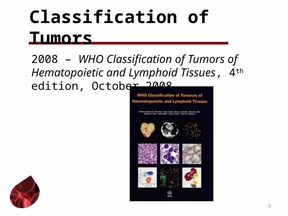
Classification of Tumors
2008 – WHO Classification of Tumors of Hematopoietic and Lymphoid Tissues, 4th edition, October 2008
5
Objectives
Understand the basis of the WHO Classification
Understand the presentation and workup for hematopoietic and lymphoid neoplasms
Recognize provisional diagnoses
6

Objectives
Recognize the significance of immunophenotyping and genetic testing
Understand the terminology used in immunophenotyping and genetic testing
7
New Classifications of Hematopoietic and Lymphoid Neoplasms
8

Regenerative Medicine, 2006. 9

2008 WHO Classification of Tumors of Haematopoietic and Lymphoid Tissues
Basic principle: Classification for all neoplasms based on:
• Morphology and biologic features• Genetic• Immunophenotype• Clinical features
10

Disease Definitions and Symptoms
11

Tumors Primary in Tissue
Lymphoma: Malignant tumor in lymph nodes or lymphoid tissue
Myeloid sarcoma: Solid tumor of immature white blood cells
Plasma cell tumor (MM, extraosseous, osseous): Tumors comprised of plasma cells
12

Lymphoma Presentation
Not specific to diseaseSwollen lymph nodesChest pain/breathing problemsUnexplained weight lostRecurring fevers/night sweatsRashesLower back painSore LN after alcohol consumption
13
Leukemia Presentation/SymptomsLeukemia limited to BM involvement
Chronic leukemiaUsually asymptomatic
Acute leukemiaSymptomaticSymptoms vary with type of leukemia
14

Acute Leukemia Symptoms
AnemiaShortage of red blood cellsSymptoms: SOB, tiredness, pallor
LeukopeniaShortage of normal white blood cells; too few
mature granulocytesWhite blood cells do not protect against infection
15

Acute Leukemia Symptoms
ThrombocytopeniaLow blood plateletsPlatelets control blood clotting by closing “holes”
in damaged blood vesselsSymptoms: excessive bruising, bleeding,
nosebleeds, and bleeding from gums
16

Initial Diagnostic Procedures
17

Lymphoma, Myeloid Sarcoma, Plasma Cell Tumor
Tissue biopsyLymph nodeOrganSkinBoneBone marrow
18

Leukemia
1. Blood counts (CBC; peripheral smear)2. Bone marrow aspiration/biopsy
19

Provisional Diagnoses
20

Types of Diagnoses
NOS histology onlyNOS with a “possible/probable” specific
histology
21

Provisional Diagnoses
NOS histology onlyNOS with a “possible/probable” specific
histology
22

NOS Diagnosis
NOS histologyProvisional –awaiting test resultsOnly diagnosis available now
Use Appendix E to identify NOS
23

Example: NOS DX Only Option Available Chronic myeloproliferative neoplasm (MPN),
NOSClinical, lab, and morphologic features +Does not meet criteria for specific MPN ORFeatures overlap two or more MPD categories
Initial stageLate stage
24

Provisional Diagnoses
NOS histology onlyNOS with a “possible/probable” specific
histology
25

NOS with Probable Specific
1. MPN (9960/3), probably PV (9950/3)
26

Tests That Identify Specific Hematopoietic and Lymphoid Histologies
27

2008 WHO Classification of Tumors of Haematopoietic and Lymphoid Tissues
Basic principle: Classification for all neoplasms based on:
• Morphology and biologic features• Genetic• Immunophenotype• Clinical features
28

Genetic Testing
Laboratory studies of blood, bone marrow, or tissue to analyze DNA to identify chromosome abnormalities which diagnose specific neoplasms
29

Normal Chromosomes
46 in each cellEach chromosome has a specific number
Example: (1;2) Short arm “p” and a long arm “q”
Example: (p13;q22)
30

Genetic Abnormalities
1. Translocation: t(1;2) 2. Inversion: inv163. Deletion: -7 or 7-4. Addition: +8 or 8+
31

Gene Translocation
32Courtesy: National Human Genome Research Institute

Gene Inversion
33Diego Diez, Ph, Bioinformatics Center, Institute for Chemical Research, Kyoto University.Gokasho, Uji, Kyoto 611-0011 JAPAN [email protected]

Gene Deletion
34
Courtesy: National Human Genome Research Institute

Gene Addition
35
Walters L, Palmer JG. “The Ethics of Human Gene Therapy.” Oxford University Press. 1997.
Genetic Testing
FISH: Identifies genetic changes and translocations.
Polymerase chain reaction (PCR): Measures cancer cells that cannot be detected by FISH.
Karyotyping: To arrange and classify chromosomes based on number, size, shape, and other characteristics.
36

FISH to Identify NPM/ALK Fusion Gene
37http://www.pathologyoutlines.com

Karyotype
38http://www.pathologyoutlines.com

Immunophenotyping
Cells from blood, BM, tissue used to determine types of antigens or markers on surface of cell. Referred to as CD
CD; cluster of differentiation: Used to define the findings in immunophenotyping .
39

Additional Immunophenotyping
Flow cytometry: Cells from blood, BM, tissue are treated with antibodies and passed in front of a laser beam.
Immunocytochemistry (IHC): Shows specific antigens in cells from blood, BM, by using either fluorescent dyes or enzymes as markers
40

Immunohistochemistry
41
http://www.pathologystudent.com/?tag=acute-myeloid-leukemia

Genetic Studies and Immunophenotyping
Cytogenetics: The study of the DNA to identify antigen receptors and translocations.
42

Genetic Testing/Cytogenetics
43Appelbaum, MD, Frederick R. Leukemia [Internet]. Version 5. Knol. 2008 Jul 28. Available from: http://knol.google.com/k/frederick-r-appelbaum-md/leukemia/pOIC0j0O/gRxHJw

Identifying Definitive Diagnosis
44

Required to Identify Specific HistologyUse Hematopoietic DB to identify definitive
diagnostic method(s)
45

46

An Additional Diagnostic Method
47

Types of Diagnoses
NOS histology onlyNOS with a “possible/probable” specific
histologyDiagnosis of exclusion (clinical)
48

Diagnosis of Exclusion (Clinical)
Tests are equivocalDiagnosis based on equivocal tests and
clinical presentation
Examples: myelodysplastic syndrome, unclassifiable ; refractory thrombocytopenia
49

50

Get Information on Tests
Check with laboratory to get samples of testsAsk HIM dept
Where tests are filedHow tests that arrive after MR is complete are filed
Follow-back with physician if tests have been ordered
51

Major Points
• Diagnostic/work-up process different• Genetic data and immunophenotyping
• Do NOT use ambiguous terminology• Do NOT code to higher ICD-O-3 code• Histology code updated to more specific• Use Hematopoietic DB to identify Definitive
Diagnostic Procedures
52

Conclusion
• The new hematopoietic and lymphoid neoplasm rules go into effect for cases diagnosed January 1, 2010, and after
• Email address for [email protected]
53