hematological manifestations of human t lymphotropic virus
TRANSCRIPT

C
Hla
AAa
b
c
a
A
R
A
A
I
I1woeMCstt
aT
H
h1r
rev bras hematol hemoter. 2 0 1 6;3 8(1):75–78
www.rbhh.org
Revista Brasileira de Hematologia e HemoterapiaBrazilian Journal of Hematology and Hemotherapy
ase Report
ematological manifestations of human Tymphotropic virus type 1 infection: a possiblessociation with autoimmune myelofibrosis
na Ciminelli a, Frederico Meloa, Marie-Christine Copinb,nna Bárbara Carneiro-Proietti c, Suely Meireles Rezendea,∗
Universidade Federal de Minas Gerais (UFMG), Belo Horizonte, MG, BrazilUniversité de Lille, Lille, FranceFundacão HEMOMINAS, Belo Horizonte, MG, Brazil
r t i c l e i n f o
rticle history:
eceived 14 August 2015
ccepted 30 August 2015
vailable online 9 October 2015
With an insidious manifestation and chronic evolution,
ntroduction
t is estimated that the human T lymphotropic virus type (HTLV-1) infects approximately 20 million people world-ide. Associated diseases, however, manifest only in 5–10%f infected individuals.1 Studies have shown that HTLV-1 isndemic in Southern Japan, the Caribbean, South America,elanesian islands, Papua New Guinea, the Middle East andentral and Southern Africa. In Brazil, HTLV-1 infection is con-idered a public health concern as the country figures amonghe endemic areas in the world with prevalence rates from 1%o 5%.1
Since the discovery of HTLV-1 in 1979, it has been clearlyssociated with hematologic disorders, specifically with adult-cell leukemia/lymphoma (ATL), an aggressive neoplasm
∗ Corresponding author at: Universidade Federal de Minas Gerais (UFMGorizonte, MG, Brazil.
E-mail addresses: [email protected], [email protected]://dx.doi.org/10.1016/j.bjhh.2015.08.006516-8484/© 2015 Associacão Brasileira de Hematologia, Hemoterapiaeserved.
with poor prognosis.1 Although ATL has clinical forms withdifferent manifestations, it is generally characterized as aclonal proliferation of CD4+ T cells containing randomly dis-tributed HTLV-1 proviral integration sites. The onset of thedisease usually occurs 20–30 years after viral infection andis primarily associated with vertical transmission, mainlythrough breastfeeding by a seropositive woman.1
Apart from ATL, other diseases have been consistentlylinked to HTLV-1 infection over the years. Tropical spasticparaparesis/HTLV-1-associated myelopathy (TSP/HAM) morefrequently affects women, with onset ranging from yearsto decades after infection, at an average age of 40 years.1
), Avenida Alfredo Balena, 190, 2◦ andar, sala 243, 30130-100, Belo
om.br (S.M. Rezende).
HAM/TSP takes years to progress from the onset of symp-toms, such as weakness and spasticity of one or both legsand minor sensory changes, to wheelchair confinement and
e Terapia Celular. Published by Elsevier Editora Ltda. All rights
mote
76 rev bras hematol hebowel/bladder incontinence.1 Other diseases such as uveitis,infective dermatitis, pulmonary and rheumatic disorders suchas rheumatoid arthritis and Sjögren’s syndrome, as well asother autoimmune conditions such as bronco-alveolar pneu-monitis, autoimmune thyroiditis and endemic polymyositis,have also been associated with HTLV-1 infection.1,2 In addi-tion, in the last two decades, HTLV-1 has been reportedin association with other blood diseases, such as acutemyeloid leukemia,3 idiopathic thrombocytopenic purpura4
and myelodysplastic syndrome.5
Case report
A 27-year-old woman was referred for a hematology consulta-tion to investigate pancytopenia. At admission, she had spas-tic paraparesis, and bowel and bladder dysfunction, clinicalsigns consistent with the initial presentation of HAM/TSP. Hersymptoms started as a progressive weakness of the legs whenshe was 22 years old. The patient was born in a known endemicarea for HTLV-1 infection, located in the Northern Region ofMinas Gerais State, Brazil. Although there was no history ofblood transfusion, she reported that she was breastfed by awet nurse. HTLV-1 testing was performed and the positiveenzyme immunoassay results were confirmed by western blot.No alterations were found in cerebrospinal fluid analysis or acomputed tomography (CT) scan of the lumbosacral spine. Atadmission, she presented with pancytopenia that had beeninvestigated in another hospital in 1996 by bone marrow aspi-rate and biopsy; at that time, no abnormalities were found.These results could not be checked due to the lack of material.
Later in the same year, the patient was hospitalized foranother investigation of the pancytopenia and metrorrha-gia. During her hospitalization, the patient received 600 mL ofpacked red cells and daily platelet transfusions. The absoluteneutrophil count ranged from 1.310 × 10⁹/L to 2.060 × 10⁹/L(reference range [RR] = 4.0 × 10⁹/L to 11.0 × 10⁹/L); plateletcount varied from 3.74 × 10⁹/L to 64.00 × 10⁹/L (RR = 150 × 109/Lto 450 × 109/L) and hemoglobin levels were between 6.0 g/dLand 8.4 g/dL (RR = 12 g/dL to 14 g/dL). The reticulocyte countwas 3.7% (RR = 0.5% to 1.5%) with the absolute red cell countof 2.35 × 10³/�L. Abdominal ultrasound revealed liver at 4 cmbelow the lower edge of the right costal margin and the spleenwas classified as Boyd grade III.
Several tests were carried out to investigate the cause ofthe pancytopenia, including leishmaniasis, HIV, hepatitis Band C, syphilis and antinuclear antibody test, all with neg-ative results. Tests for vitamin B12, folate, iron serum levelsand other routine tests were also performed, with resultswithin normal reference ranges. A bone marrow aspirate andbiopsy showed increased cellularity, abnormal distribution oferythropoiesis, atypical megakaryocytes, immature granulo-cytes and evidence of reactive marrow fibrosis. Marrow ironstores were normal. The metrorrhagia was controlled with theadministration of combined oral contraceptives.
Throughout 11 years, a non-progressive pancytopenia was
the only persistent sign of a hematological disorder. A sig-nificant hepatosplenomegaly was noticed and confirmed byimaging in 2002. The liver and spleen were no longer pal-pable in the patient’s first consultation in our outpatients’r. 2 0 1 6;3 8(1):75–78
clinic in 2009. An abdominal CT scan, performed in 2009,revealed mild splenomegaly with no other abdominal abnor-malities. Petechiae were present in her consultation, mainlyin the oral cavity. Platelet counts were consistently below40.0 × 109/�L and absolute neutrophil counts varied from1.5 × 109/L to 2.0 × 10⁹/L, with relative lymphocytosis (58% to65%). A discrete normocytic normochromic anemia could beobserved in most of the blood tests, with hemoglobin varyingfrom 10.5 g/dL to 12 g/dL. In addition, the patient presentedincreased lactate dehydrogenase (LDH) levels, with valuesranging from 902 U/L to 1020 U/L (RR: 313–618 U/L). Hepaticenzymes and calcium levels were unremarkable. A high doseof prednisone (60 mg per day) was prescribed to increaseplatelet counts but was stopped as it was not effective.
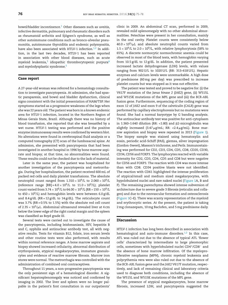
The patient was tested and proved to be negative for: (i) theV617F mutation of the Janus kinase 2 (JAK2) gene, (ii) W515Land W515K mutations of the MPL gene and (iii) the BCR-ABLfusion gene. Furthermore, sequencing of the coding region ofexon 12 of JAK2 and exon 9 of the calreticulin (CALR) gene wasperformed by capillary electrophoresis but no mutations werefound. She had a normal karyotype by G banding analysis.The antinuclear antibody test was positive for anti-cytoplasmin 1:360–1:640 dilution (RR: <1:80) and �2-microglobulin wasslightly increased (3.47 �g/mL; RR: <2.4 �g/mL). Bone mar-row aspiration and biopsy were repeated in 2013 (Figure 1).The biopsy sample was stained using hematoxylin andeosin, periodic acid-Schiff (PAS), giemsa, silver impregnation(Gordon-Sweet), Masson’s trichrome, and Perls. Immunostain-ing was performed for CD2, CD3, CD4, CD5, CD8, CD20, CD30,CD34, CD56 and FOXP3. The lymphocytes reacted with variableintensity for CD2, CD3, CD4, CD5 and CD8 but were negativefor CD56 and FOXP3. The reaction with CD4 was more intensethan with CD8. CD34 positive blasts were not identified.The reaction with CD61 highlighted the intense proliferationof atypical/small and medium sized megakaryocytes, withhypolobulated nuclei and rare naked nuclei (Figure 1A, B andF). The remaining parenchyma showed intense subversion ofarchitecture due to severe grade 3 fibrosis (reticulin and colla-gen) and due to the neovascular formation and osteosclerosis(Figure 1C–E). There was scanty representation of the myeloidand erythrocytic series. At the present, the patient is taking2 mg clonazepam, 10 mg Baclofen, and 5 mg prednisone daily.
Discussion
HTLV-1 infection has long been described in association withhematological and auto-immune disorders.1,2 In this case,ATL was ruled out due to the absence of typical ATL ‘flowercells’ characterized by intermediate to large pleomorphiccells, sometimes with hyperlobulated nuclei CD4+/CD8− andthe absence of bone marrow infiltration. Of the myelopro-liferative neoplasms (MPN), chronic myeloid leukemia andpolycythemia vera were also ruled out due to the absence ofthe BCR-ABL fusion gene and the JAK2 V617F mutation, respec-tively, and lack of remaining clinical and laboratory criteria
used to diagnose both conditions, including the absence ofthe W515L and W515K mutations of the MPL gene.The presence of atypical megakaryocytes, bone marrowfibrosis, increased LDH, and pancytopenia suggested the

rev bras hematol hemoter. 2 0 1 6;3 8(1):75–78 77
Figure 1 – Bone marrow biopsy. (A and B) Bone marrow biopsy stained using hematoxylin and eosin showing clusters ofabnormal megakaryocytes (arrow). (C and D) Bone marrow biopsy stained using Gordon Sweet stain showing a markedincrease in coarse reticulin fibers (arrow). (E) Perivascular collagen fibrosis stained using Masson’s trichrome (arrow). (F)Immunostaining for CD61 with megakaryocyte hyperplasia (arrow).
dcmtaMmactettvca
h4odtoastc
nea
iagnosis of myelofibrosis. Myelofibrosis, characterized byhronic and persistent pancytopenia associated with bonearrow fibrosis and proliferation of megakaryocytes, is one of
he most common types of MPN. It can be primary or develops an end-stage bone marrow failure, secondary to other MPN.yelofibrosis can also be present as a reaction of the bonearrow against other neoplasms or inflammatory processes,
nd can emerge as an autoimmune phenomenon.6,7 Classi-al findings of primary myelofibrosis are hepatosplenomegaly,eardrop erythrocytes and leukoerythroblastosis in the periph-ral blood, which were not present in this patient. Moreover,he benign, non-progressive course of the disease disfavoredhe diagnosis of primary myelofibrosis as the median sur-ival of patients with this condition is about 69 months.8 Thelinical picture and laboratory tests suggest the diagnosis ofutoimmune myelofibrosis (AIMF).
AIMF is an under-recognized cause of marrow fibrosis, andas been defined using the following criteria: (1) Grade 3 or
reticulin fibrosis of the bone marrow; (2) lack of clusteredr atypical megakaryocytes; (3) lack of myeloid or erythroidysplasia, eosinophilia, or basophilia; (4) lymphocyte infiltra-ion of the bone marrow; (5) lack of osteosclerosis; (6) absentr mild splenomegaly; (7) presence of autoantibodies; and (8)bsence of a disorder known to cause myelofibrosis.6 A recenttudy reported that mild atypias including occasional clus-ered, left-shifted, small, and hypolobated megakaryocytes,an be observed in AIMF.7
AIMF is classified as primary or secondary, the latteramed when associated with autoimmune disorders. Dis-ases such as systemic lupus erythematous (SLE), sclerodermand Sjögren’s syndrome are the main disorders described
in association with secondary AIMF.6,7 AIMF has also beendescribed in association with HIV infection9 but not with HTLVinfection. The differentiation between AIMF and the MPN-related myelofibrosis is imperative once these disorders havedifferent management and prognoses. AIMF tends to respondwell to steroids and/or other immunosuppressive agents andhas a better prognosis.
An additional unusual aspect of this case is the rare over-lap of HTLV-1-related diseases: HAM/TSP and a hematologicaldisorder. ATL and HAM/TSP seem to differ in their routeof transmission (ATL being mainly via breast-feeding, andHAM/TSP via blood transfusion), pathogenesis and immuno-logical response. After infection, both cellular and humoralimmune responses are formed against HTLV. The humoralreaction contributes as delayed protection by producing anti-bodies against viral proteins, including Tax. However, recently,Tax-antibodies have been associated with the developmentof HAM/TSP, suggesting an autoimmune component to thedisease. This antibody could also be associated with AIMF. Fur-thermore, HAM/TSP apparently relates to patients with highproviral loads, which is supposedly determined by host fac-tors such as polymorphisms in the major histocompatibilitycomplex class I (MHC-I) molecules and its influence on antigenpresentation to CD8+ T-cells.
In conclusion, this is the case of a young woman with HTLV-1 infection and HAM/TSP, who evolved with pancytopenia andbone marrow fibrosis. To our knowledge, except for a shortreport by Engels,10 who does not provide detailed information,
this is the first report of AIMF associated with HTLV-1 infec-tion. This case report strengthens the described associationbetween HTLV-1 and autoimmune/hematological disorders.
mote
r
1A. Persistent human herpesvirus 8 viremia associated with
78 rev bras hematol he
Conflicts of interest
The authors declare no conflicts of interest.
Acknowledgements
The authors wish to thank Laboratorio Fleury, São Paulo,Brazil, in particular Dr. Maria Carolina Tostes Pintão, for per-forming some of the molecular tests for the diagnosis ofmyeloproliferative neoplasms.
e f e r e n c e s
1. Goncalves DU, Proietti FA, Ribas JG, Araújo MG, Pinheiro SR,Guedes AC, et al. Epidemiology, treatment, and prevention ofhuman T-cell leukemia virus type 1-associated diseases. ClinMicrobiol Rev. 2010;23(3):577–89.
2. Pinheiro SR, Lana-Peixoto MA, Proietti AB, Oréfice F,Lima-Martins MV, Proietti FA. HTLV-I associated uveitis,myelopathy, rheumatoid arthritis and Sjögren’s syndrome.Arq Neuropsiquiatr. 1995;53(4):777–81.
3. Tsukasaki K, Koba T, Iwanaga M, Murata K, Maeda T, AtogamiS, et al. Possible association between adult T-cellleukemia/lymphoma and acute myeloid leukemia. Cancer.1998;82(3):488–94.
r. 2 0 1 6;3 8(1):75–78
4. Matsushita K, Ozaki A, Arima N, Tei C. HumanT-lymphotropic virus type I infection and idiopathicthrombocytopenic purpura. Hematology. 2005;10(2):95–9.
5. Karlic H, Möstl M, Mucke H, Pavlova B, Pfeilstöcker M, Heinz R.Association of human T-cell leukemia virus andmyelodysplastic syndrome in a central European population.Cancer Res. 1997;57(21):4718–21.
6. Pullarkat V, Bass RD, Gong JZ, Feinstein DI, Brynes RK. Primaryautoimmune myelofibrosis: definition of a distinctclinicopathologic syndrome. Am J Hematol. 2003;72(1):8–12.
7. Vergara-Lluri ME, Piatek CI, Pullarkat V, Siddiqi IN, O’ConnellC, Feinstein DI, et al. Autoimmune myelofibrosis: an updateon morphologic features in 29 cases and review of theliterature. Hum Pathol. 2014;45(11):2183–91.
8. Cervantes F, Dupriez B, Pereira A, Passamonti F, Reilly JT,Morra E, et al. New prognostic scoring system for primarymyelofibrosis based on a study of the International WorkingGroup for Myelofibrosis Research and Treatment. Blood.2009;113(3):2895–901.
9. Lee AC, Fong CM. Autoimmune myelofibrosis as the firstmanifestation of human immunodeficiency virus infection inan infant. Ann Hematol. 2012;91(5):809–10.
0. Engels EA, Eastman H, Ablashi DV, Wilks RJ, Braham J, Manns
coinfection with human T-cell lymphotropic virus type I andmyelofibrosis. J Acquir Immune Defic Syndr. 2000;23(3):283–6.