helper and killer t cells do not express b cell
TRANSCRIPT

H E L P E R A N D K I L L E R T C E L L S D O N O T E X P R E S S
B C E L L I M M U N O G L O B U L I N J O I N I N G
A N D C O N S T A N T R E G I O N G E N E S E G M E N T S *
BY MITCHELL KRONENBERG, MARK M. DAVIS, PHILIP W. EARLY, LEROY E. HOOD, AND JAMES D. WATSON
From The Division of Biology, California Institute of Technology, Pasadena, California 91125; and The Department of Microbiology, University of California at Irvine, Irvine, California 92717
The immune system is characterized by both the specificity and breadth of its response. The generation of an immune response depends upon the specific recognition of antigens by lymphocytes. There are two classes of lymphocytes: B cells capable of secreting large quantities of immunoglobul in and T cells that can carry out facilitation or help of lymphocyte responses (1, 2), suppression of lymphocyte responses (3), or cytotoxicity directed against appropriate target cells (4). A T cell is apparently committed to a single functional subclass prior to any exposure to antigen (5, 6).
The antigen-binding receptor on the surface of B lymphocytes is an immunoglob- ulin molecule composed of two identical light chain and two identical heavy chain polypeptides. Both light and heavy chains have variable regions responsible for antigen binding and constant regions responsible for various effector functions such as complement fixation. The immunoglobulins are encoded by three unlinked gene families: two light chain families, t¢ and )% and a heavy chain family. In mice, there are one or more constant region genes for each light chain family, and at least eight heavy chain constant region genes (#, ~, y3, yl, yZb, y2a, a, and E). Each mature B cell and its progeny can respond to only one or a few antigenic determinants because it can synthesize only one light chain and one heavy chain variable region. However, during their development, B cells switch from the expression of IgM molecules containing # heavy chains to the expression of other immunoglobulin classes contain- ing different heavy chain constant regions, while continuing to express the same VH region and light chain (7, 8).
Recent experiments employing recombinant DNA techniques have clarified some of the molecular events necessary for B-cell antibody expression. Both the commitment to express single VH and VL regions, and the switch in expression of heavy chain constant region are characterized by DNA rearrangements. In the mouse, the light chain is encoded by three separate gene segments--V, j1 (joining), and C (constant) (9). The V and J gene segments together code for the variable region. For mouse K chains, there are multiple germline V~ gene segments (2200) (10) and four J~ gene segments (11, 12). The heavy chain is encoded by four gene segments--VH, D (diversity), J n and CH, and in some cases an M (membrane) exon (13-16). The V
* Supported by grant AI 10781 from the U. S. Public Health Service. Abbreviations used in this paper: C, constant; CT, constant region; D, diversity; J, j'oining; JT, J gene
segment; kb, kilobase; V, variable.
J. Exv, M ED. © The Rockefeller University Press • 0022-1007/80/12/1745/17 $1.00 1 745 Vo[tlln(" 152 December 1980 1745-1761
Dow
nloaded from http://rupress.org/jem
/article-pdf/152/6/1745/1091261/1745.pdf by guest on 02 Decem
ber 2021

1746 T CELLS DO NOT EXPRESS C GENES
gene segment (and presumably the D gene segment) expressed in a given B cell is rearranged from its germ-line context and becomes contiguous with a J gene segment that is located a few kilobases 5' from a C gene (I 1-13, 17). DNA sequences between the V and J gene segments are deleted during this rearrangement (11, 18, 19). Heavy chain constant-region switching takes place through replacement of the/z constant- region gene and some of its flanking sequence by another CH gene (20).z
Individual T cells can also respond specifically to one or a few antigenic determi- nants (21-23). However, the molecular properties of the T-cell antigen-binding receptor are a subject of great controversy (24). Most attempts to define the receptor have relied on antisera made to either B-cell derived immunoglobulin (25-31) or to responding T cells (32, 33) in conjunction with immunological or genetic experiments. There is considerable evidence that T cells synthesize VH regions without light chains or any of the Cn regions expressed in B cells (29-31), but there are a number of reports of the expression of light chains and Cn regions in T cells (25-28, 34, 35).
The development of two relatively new techniques has persuaded us to reexamine the controversy regarding immunoglobulin synthesis by T cells. First, cell lines grown in T-cell growth factor or Interleukin II (36) provided us with large numbers of mature, functional T cells that are entirely free of contaminating B cells and have a well-defined antigen specificity. Second, using the methods of recombinant DNA research, we have obtained cloned DNA probes for immunoglobulin heavy and light chain J and C gene segments. We have used our DNA probes to ask whether these J and C gene segments are rearranged and transcribed into RNA by T cells. Because both the T cells and the DNA probes are homogeneous and well characterized, we hoped to avoid the ambiguities of some of the earlier experiments concerning T-cell expression of immunoglobulin constant regions. In addition, by using probes for the heavy and light chain J gene segments, we could obtain information on T-cell expression of the 3' part of the variable region. We have obtained convincing data that the B-cell immunoglobulin light and heavy chain J and C gene segments are neither rearranged nor expressed in the monoclonal helper and killer T cells that we have analyzed.
Materials and Methods T Cells. BALB/c Cum mice were originally obtained from Cumberland View Farms,
Clinton, Tenn., and have since then been bred in our animal colony at the California Institute of Technology. C57BL/6J mice were obtained from The Jackson Laboratory, Bar Harbor, Maine. Thymus tissue was dissected from 2- to 4-wk-old BALB/c mice killed by inhalation of ether. Mice were injected with 0.1 ml of India ink prior to sacrifice to facilitate visualization and removal of parathymic lymph nodes. The antigen specificity and cell-surface phenotype of our monoclonal T cells are presented in Table I. WEHI-22 is an irradiation-induced BALB/c T lymphoma (27). It has been reported to synthesize large amounts of a 68,000 mol wt protein that cross-reacts with an antiserum made against mouse immunoglobulin (28). WEHI-22 cells were a gift of Dr. Noel Warner, University of New Mexico, Albuquerque, N. Mex. and were grown in suspension culture in Dulbecco's modified Eagle's medium (Grand Island Biological Co., Grand Island, N. Y.) supplemented with 10% fetal bovine serum. The helper cell line HT- 1 (23) and the alloreactive killer cell line CTLLi6 (37) were grown in RPMI-1640 medium (Grand Island Biological Co.) supplemented with 10% heat-inactivated fetal bovine serum, 1 mM glutamine, 50 U/ml penicillin, 50 #g/ml streptomycin, and 5 × 10 -5 M 2-mercaptoethanol.
2 Davis, M. M., S. Kim, and L. Hood. DNA sequences mediating heavy chain switching in alpha immunoglobulin genes. Manuscript submitted for publication.
Dow
nloaded from http://rupress.org/jem
/article-pdf/152/6/1745/1091261/1745.pdf by guest on 02 Decem
ber 2021

KRONENBERG, DAVIS, EARLY, HOOD, AND WATSON
TABLE I
Monoclonal T Cells Analyzed with Cloned Immunoglobulin DNA Probes
1747
Cell line Strain of Function Antigen Cell surface Description Reference origin specificity phenotype
HT-I C57BI/6J Helper Sheep eryth- 0 +, Lyt-l* Growth factor- 23 rocytes dependent
Helps H-2 h B cell line cells
CTLLi6 C57BI/6J Killer H-2 d O +, Lyt-2 + Growth factor- 37 dependent cell line
WEHI-22 BALB/c ? ? 0 + T lymphoma 27
TABLE II
Cloned Immunoglobulhl Probes
Region Source Reference
C, (C region Subclone from BALB/c subclone) genomic DNA
j , ( j region Subclone from BALB/c subclone) genomic DNA
Cy (pAbyr7) cDNA from HOPC 2020 mRNA
JH Fragment from BALB/c genomic DNA
Switch (p5.1) Subclone from BALB/c myeloma DNA
Cu (p104E#6) cDNA from MI04E mRNA
C~ cDNA from S107 mRNA
C region subcione
laJ'r I (13
5' ! !,,,j I nnl I 3' Jx CK I kb
I-.-.M
d region subclone
5' ~ 3' Cx 3'UT IOObp
5'
38
38
RI RI RI RI 5'!! .... 13'
JH I -6 .2 kb-~ C~'l k.b
JR probe
RI F]I RI
VHD~H " q Ca Ikb 20 switch site
I,=-5.1 kb~l switch probe
I 3 ~ 14 I"~CH3-'f'~C H 4 ~ 3'UT tOObp
5 ' ~ 3' IOObp 20 ,-'- CH F~I-CHL:~
A. Bothwell. Personal communi- cat ion.
20; this report
A superna tan t fraction from concanaval in A-st imulated lymphocytes enriched for Inter leukin II was prepared as described (23) and added to the med ium at a concent ra t ion of 10 U / m l .
Immunoglobulin Probes. Description of the immunoglobu l in DNA probes employed in these studies is presented in Tab le II. T he C~ and J , gene segments were subcloned from a liver DNA
Dow
nloaded from http://rupress.org/jem
/article-pdf/152/6/1745/1091261/1745.pdf by guest on 02 Decem
ber 2021

1748 T CELLS DO NOT EXPRESS C GENES
genomic clone. These subclones were a gift of Dr. Michael Steinmetz, California Institute of Technology, Calif. (38). A Ca cDNA clone, pAb~.x-7, was a gift of Dr. Alfred Bothwell and Dr. David Baltimore, Massachusetts Institute of Technology, Boston, Mass. A probe containing the Jn gene segments was prepared by electroeluting a 6.2 kilobase (kb) EcoRI fragment from the ChSp/x27 clone originally isolated as a C,-containing clone from a collection or library of BALB/c sperm DNA clones (20). The probe containing heavy chain constant region switch sites was subcloned from the Ch603a6 clone. This clone was originally isolated from a library made from the DNA of the IgA-producing myeloma McPC603 (17). The original a6 clone contains the 603 VH gene segment joined to a JH gene segment, with Ca gene found 6.8 kb to the 3' side of the JH gene segment. The subclone is a 5.1 kb EcoRI fragment containing intervening DNA sequence and the 5' half of the Ca gene (17). In the embryo, 1.5 kb of this intervening sequence is found adjacent to the C~ gene and not the Ca gene (20). The point at which C~ and Ca adjacent sequences are joined is denoted the switch site. Five examples of CH switching have thus far been analyzed, including three from rearranged genes of C~-producing myeloma tumors (17, 20), 2 one from a Crx producer (39, 40), and one from a Cr2b producer (41). All the active genes from these myelomas have C~ flanking sequence extending from a Jn gene segment towards the expressed C v or Ca gene. The point of deletion of the C~ flanking sequence or switch site is not the same for every tumor, but they are all found within about 300 base pairs of one another. The 5.1 kb sub¢lone we used as a probe should hybridize with restriction fragments containing all these known switch sites as well as several kb of surrounding DNA.
Preparation of DNA and Southern Blots. High molecular weight DNA was prepared according to the method of Blin and Stafford (42). Embryo DNA was prepared from whole 12-d BALB/c embryos. DNA digested with restriction enzymes was electrophoresed in 0.7% agarose gels, transferred to nitrocellulose filters (43), and hybridized with nick-translated probes (44). Unless otherwise noted, hybridizations were performed at 68°C in 1.0 M NaCI/0.045 M Trisodium citrate/0.2% bovine serum albumin/0.2% Ficoll/0.2% polyvinylpyrrolidone/0.1% NaDodSO4/100 #g/ml denatured Salmon sperm DNA/25 #g/ml poly-rA. Subsequent to hybridization, filters were washed extensively in 0.30 M NaCI/0.03 M Trisodium citrate/0.1% NaDodSO4/0.1% sodium pyrophosphate at 68°C and autoradiographed.
Preparation of RNA and Northern Blots. Purified immunoglobulin mRNA containing the ct (MOPC 167) and/~ (M 104E) heavy chain constant regions and the ~ (S 107) light chain constant region were prepared as described (17). Total RNA was prepared from T cells homogenized in guanidinium thiocyanate and centrifuged through cesium chloride (45). RNA preparations electrophoresed on agarose gels and stained with ethidium bromide were undegraded, free of contaminating DNA, and had a 260 nm/280 nm absorbance ratio of about 2.1 : 1. all-labeled RNA from sea urachin (Strongylocentrotus purpuratus) embryos (a gift of Dr. Frank Costantini, California Institute of Technology) was added at the beginning of the T cell RNA preparation as a recovery marker. Between 43 and 64% of the added counts per minute were present in the final RNA solution. From the absorbance of the mouse T cell RNA at 260 nm, and the yield of labeled S. purpuratus RNA estimated from recovered counts per minute, we calculated that WEHI-22 has 12.7 pg of RNA/cell, HT-1 has 5.8 pg/cell, CTLLi6 has 3.7 pg/cell, and thymus has 1.6 pg/cell. Polyadenylated [poly(A) +] RNA was selected by oligo(dT)-cellulose chroma- tography (46). Poly(A) ÷ RNA was denatured and electrophoresed on agarose gels containing 2.2 M formaldehyde (47). Denatured RNA was transferred directly onto nitrocellulose filters (B. Seed and D. Goldberg. Manuscript in preparation.), and filters were hybridized at 42°C in 50% formamide (48). In control experiments, Szp-labeled RNA from Sindbis virus-infected cells (a gift of Charles Rice, California Institute of Technology) was electrophoresed on agarose gels, transferred to nitrocellulose filters, and incubated in hybridization and wash solutions. Virla RNA as large as 13.0 kb was efficiently transferred to filters. About 45%Of the RNA remained bound to the nitrocellulose as measured by densitometry of viral RNA bands present on autoradiographs of the filter exposed before and after incubation in hybridization buffers. In one experiment, diazophenyhhioether paper (a gift of Brian Seed, California Institute of Technology) rather than nitrocellulose, was used as a solid support to bind RNA.
Resu l t s
Experimental Strategy. T cell DNA was analyzed for rear rangement using the Southern blot technique (43). High molecular weight genomic DNA was digested
Dow
nloaded from http://rupress.org/jem
/article-pdf/152/6/1745/1091261/1745.pdf by guest on 02 Decem
ber 2021

KRONENBERG, DAVIS, EARLY, HOOD, AND WATSON 1749
with a restriction enzyme and the resulting fragments were separated according to molecular weight in an agarose gel. The double-stranded DNA was then denatured so that single strands were available for hybridization. The size-separated and denatured DNA was transferred from the agarose gel to a solid support, generally a nitrocellulose filter, and the filter-bound DNA was hybridized with a radioactive- cloned DNA probe. The filter was autoradiographed, and restriction fragments, having sequences complementary to the probe, appeared as bands on the film. By comparing the band pattern in T cells with the band pattern obtained from germ- line DNA, we assessed whether a particular DNA sequence was rearranged. In analyzing the DNA from WEHI-22 and BALB/c thymus, cells from 12-d BALB/c embryos and adult livers provided a source of germ-line DNA. For the HT-1 and CTLLi6 cell lines, which come from C57BL/6J mice, DNA undifferentiated with respect to immunoglobulin genes was obtained from the cells of adult liver and kidney.
We used Northern blots to detect immunoglobulin sequences in T cell RNA. The experimental strategy is very similar to that employed to detect DNA rearrangements. Poly(A) ÷ total cell RNA from T cells was size-separated on agarose gels, transferred to nitrocellulose filters, hybridized with radioactive immunoglobulin DNA probes, and the filters were then autoradiographed.
T Cell DNA Analyzed for Gene Rearrangement Using Light Chain Probes. The results obtained from hybridizing light chain DNA probes to restriction fragments of T cell DNA are presented in Table III and Fig. 1. All the bands present in the T cell DNA are also present in the germ-line DNA from the same strain, indicating that C~, J~, and Cx sequences are not rearranged in the T cells. In a few cases, however, C57BL/
TABLE III Sizes of Restriction Fragments Containing Light Chain Immunoglobulin Genes in T Cell and Nonlymphoid
DNA
Probes
Digests C, J. C~
Bam Eco Bam Eco Bam HI RI Hind III HI RI Hind III HI Eco RI Hind III
C57B16 DNA HT-1 13.0 15.0 4.3, 2.7 13.0 15.0 2.7 7.3 9.0 8.4, 3.2 CTLLi6 13.0 15.0 4.3, 2.7 13.0 15.0 2.7 7.3 9.0 8.4, 3.2 Liver or kid- 13.0 15.0 4.3, 3.5 13.0 15.0 3.5, 2.7 7.3 9.0, 6.6 8.4, 3.2
ney 2.7
BALB/e DNA WEHI-22 13.0 15.0 4.3, 2.7 ND:~ 15.0 ND ND 9.0 ND Thymus 13.0 15.0 4.3, 2.7 ND 15.0 2.7 ND 9.0 ND Liver or era- 13.0 15.0 4.3, 2.7 ND 15.0 2.7 ND 9.0 ND
bryn
* Sizes of restriction fragments containing immunoglobulin genes were estimated from the migration of molecular weight standards that included Eco RI- and Hind III-digested )k bacteriophage DNA, and pBr322 plasmid digested with both Ava I and Hind II. In every case, T cell DNA was electrophoresed in a lane adjacent to one of the nonlymphoid DNA samples. WEHI-22 and BALB/c thymus DNA were electrophoresed on the same gels; HT-1 and CTLLi6 DNA were electrophoresed on separate gels. ND, not determined.
Dow
nloaded from http://rupress.org/jem
/article-pdf/152/6/1745/1091261/1745.pdf by guest on 02 Decem
ber 2021

1750 T CELLS DO NOT EXPRESS C GENES
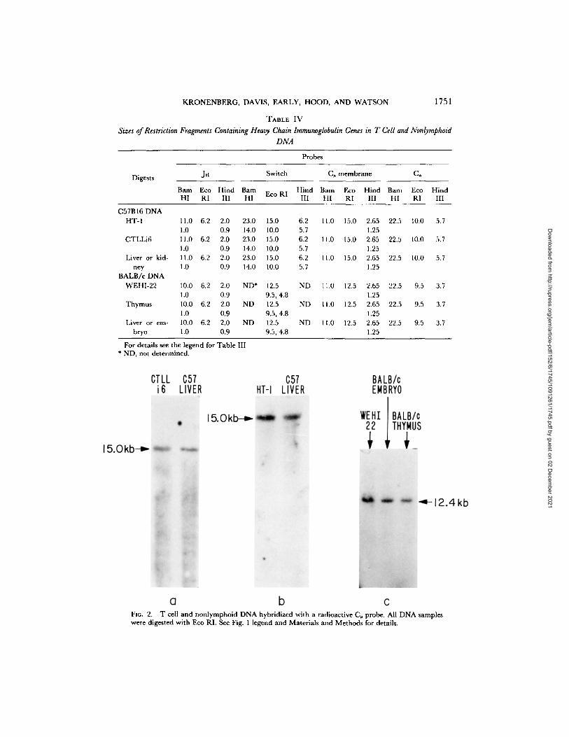
Fxc. 1. T cell and nonlymphoid DNA hybridized with a radioactive C. probe. (a-c) are autora- diographs of DNA from separate gels. In (a) DNA samples were digested with restriction enzyme Eco RI; in (b) they were digested with Barn HI; and (c) they were digested with Hind III. The sizes of the hybridizing fragments are indicated. For experimental details see Materials and Methods.
6j liver and kidney DNA had a restriction fragment hybridizing with the light chain probes that was not apparent in the two monoclonal T cell lines (Table III). The extra band in C57BL/6J liver and kidney is sometimes faint, and a technical artifact probably accounts for this result. A number of much less likely explanations are formally possible, however, including a genetic polymorphism in C57BL/6J mice, a T-cell specific sequence deletion (as opposed to rearrangement), or even an immu- noglobulin gene rearrangement specific to liver and kidney cells.
T Cell DNA Analyzed for Gene Rearrangement Using Heavy Chain Probes. The results obtained from hybridizing heavy chain probes to restriction fragments of T-cell DNA are presented in Table IV and Figs. 2 and 3.
We observed no rearrangement o f J n gene segments in the T cells tested. The 6.2 kb Jn probe contains a sequence at its 5' end that is repeated in the mouse genome (P. W. Early. Unpublished observations.). To reduce the background caused by hybridization with these repeated sequences, filters hybridized with the 6.2 kb Jn probe were washed extensively in low salt conditions (20 mM cation at 68°C), that favor melting of imperfectly matched hybrid molecules. After the low salt washes, one (EcoRI and Hind III digests) or two (Barn HI digest) dark bands remained on the filter, and the sizes of these major bands are presented in Table IV. The Eco RI fragment from BALB/c DNA that hybridizes with the probe is the same size as the EcoRI fragment used to isolate the probe from a sperm DNA clone, suggesting that this major band is not simply a large tandem array of repeated sequences or some other artifact. A 5.1 kb subclone containing sequences involved in B-cell heavy chain
Dow
nloaded from http://rupress.org/jem
/article-pdf/152/6/1745/1091261/1745.pdf by guest on 02 Decem
ber 2021
KRONENBERG, DAVIS, EARLY, HOOD, AND WATSON 1751
TABLE IV
Sizes of Restriction Fragments Containing Heavy Chain lmmunoglobulin Genes in T Cell and Nonlymphoid DNA
Probes
Digests JH Switch C¢ membrane C,,
Barn Eco Hind Bam Hind Barn Eco Hind Bam Eco Hind Eco RI
HI RI III HI III HI RI III HI RI III
C57B16 DNA HT-I 11.0 6.2 2.0 23.0 15.0 6.2 11.0 1 5 . 0 2.65 22.5 10.0 5.7
1.0 0.9 14.0 10.0 5.7 1.25 CTLLi6 11.0 6.2 2.0 23.0 15.0 6.2 11.0 1 5 . 0 2.65 22.5 10.0 5.7
1.0 0.9 14.0 10.0 5.7 1.25 Liver or kid- 11.0 6.2 2.0 23.0 15.0 6.2 11.0 1 5 . 0 2.65 22.5 10.0 5.7
hey 1.0 0.9 14.0 10.0 5.7 1.25 BALB/c DNA
WEHI-22 10.0 6.2 2.0 ND* 12.5 ND 11.0 I2.5 2.65 22.5 9.5 3.7 1.0 0.9 9.5, 4.8 1.25
Thymus 10.0 6.2 2.0 ND 12.5 ND t1.0 1 2 . 5 2.65 22.5 9.5 3.7 1.0 0.9 9.5, 4.8 1.25
Liver or em- 10.0 6.2 2.0 ND 12.5 ND 11.0 1 2 . 5 2.65 22.5 9.5 3.7 bryo 1.0 0.9 9.5, 4.8 1.25
For details see the legend for Table III. * ND, not determined.
Fro. 2. T cell and nonlymphoid DNA hybridized with a radioactive C~ probe. All DNA samples were digested with Eco RI. See Fig. 1 legend and Materials and Methods for details.
Dow
nloaded from http://rupress.org/jem
/article-pdf/152/6/1745/1091261/1745.pdf by guest on 02 Decem
ber 2021

1752 T CELLS DO N O T EXPRESS C GENES
Fla. 3. T cell and nonlymphoid DNA hybridized with a radioactive probe containing heavy chain constant region switch sites. In 3 a, DNA was digested with Bam HI, whereas in Figs. 3 b and c the DNA was digested with Eco RI. See Fig. 1 legend and Materials and Methods for details.
Fro. 4. Filter-bound poly(A) + RNA hybridized with C, and C, probes. RNA molecular weight standards included Escherichia coli 16, and 23S ribosomal RNA, and murine 18 and 28S ribosomal RNA. Purified myeloma RNA is present in lanes l, 2, and 4. When the probes are hybridized to separate filters, the C~ RNA sequence in thymus is 1.2 kb, whereas the larger C , RNA is 1.9 kb. We could detect 1.4 copies/cell o fg R N A and <1.0 copy/cell o f a RNA on this autoradiograph.
Dow
nloaded from http://rupress.org/jem
/article-pdf/152/6/1745/1091261/1745.pdf by guest on 02 Decem
ber 2021
KRONE NBE RG, DAVIS, EARLY, HOOD, AND W A T S O N 1753
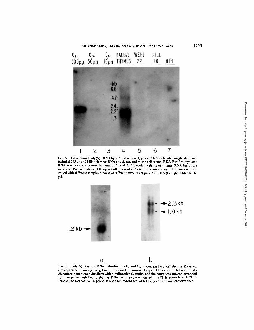
FIo. 5. Fiher-bound poly(A) + RNA hybridized with a (3~, probe. RNA molecular weight standards included 26S and 42S Sindbis virus RNA and E. coli, and murine ribosomal RNA. Purified myeloma RNA standards are present in lanes 1, 2, and 3. Molecular weights of thymus RNA bands are indicated. We could detect 1.8 copies/cell or less o f# RNA on this autoradiograph. Detection limit varied with different samples because of different amounts of poly(A) + RNA (1-10 #g) added to the gel.
FIo. 6. Poly(A) + thymus RNA hybridized to C, and C~ probes. (a) Poly(A) ÷ thymus RNA was size-separated on an agarose gel and transferred to diazotized paper. RNA covalently bound to the diazotized paper was hybridized with a radioactive C, probe, and the paper was autoradiographed. (b) The paper with bound thymus RNA, as in (a), was washed in 95% formamide at 68°(3 to remove the radioactive C, probe. It was then hybridized with a (3~ probe and autoradiographed.
Dow
nloaded from http://rupress.org/jem
/article-pdf/152/6/1745/1091261/1745.pdf by guest on 02 Decem
ber 2021

1754 T CELLS DO NOT EXPRESS C GENES
constant region switching was hybridized to the T cell DNA. No rearrangement of these sequences was observed (Fig. 2). The C, probe we used is a cDNA clone made from RNA coding for the membrane form of # heavy chain. We have detected no rearrangement of the C. gene in the T cells (Fig. 3). The C~ gene was also present in the germline or undifferentiated configuration in all the T cells analyzed.
In addition, because of reports that T cells make a polypeptide serologically cross- reactive with the ~ chain (25, 26), we hybridized the C. probe at 55°C to BALB/c and C57B1/6J liver DNA cut with various restriction enzymes. The filter was subsequently washed at 55°C. All other conditions were as described in Materials and Methods. These low temperature conditions should permit hybridization of two nucleotide sequences that are only 70% homologous (see Discussion). No bands besides those known to contain the C. gene hybridized with the probe, therefore the gene for the putative C. homologue was not detected in this experiment.
Northern Blot Analysis of T Cell RNA. Although gene rearrangement is closely associated with immunoglobulin gene expression in B cells, it is possible that T cells could express immunoglobulin genes without rearranging them. We therefore ana- lyzed T-cell RNA for the presence of immunoglobulin sequences. Size-separated poly(A) + RNA from T cells was hybridized with 32P-labeled C~ (Fig. 4), C , (Fig. 4), C~ (Fig. 5), and Ca (data not shown) probes. Purified immunoglobulin RNA were run on the same gels, and provided us with an estimate of our detection limit, which ranged from 10 to 50 pg of RNA in a series of five gels. Given the detection limit, the amount of poly(A) + RNA/cell (which is about 1% of the total cell RNA calculated in Materials and Methods) and the micrograms of RNA added per gel lane, we estimate that we could detect less than two molecules of immunoglobulin RNA/cell in every case. No immunoglobulin RNA was detected in three monoclonal T-cell lines. Whole thymus, however, contained ~150 pg of C~ RNA (7 copies/cell), 30 pg of C~ RNA (0.7 copies/cell), and 680 pg of C, RNA (16 copies/cell) as judged by densitometric comparison of thymus RNA bands to the bands obtained with purified immunoglob- ulin RNA. In addition, a 4.7 kb species that hybridized with the C, probe was present on some gels at ~0.4 copies/cell. Whole thymus was not tested for the presence of Ca RNA. The CI, and C,~ RNA in the thymus were found in the poly(A) + fraction only (data not shown), and were approximately the same molecular weight as the polysomal C~ and Ca mRNAs found in B lymphocytes. The C~ RNA was found in a diffuse band ranging from ~2.3 to 1.7 kb in size. The/~ m R N A species in B cells are 2.7 and 2.4 kb (13). The relatively low molecular weight for C~ sequences in thymus was observed in two separate RNA preparations, and a control experiment, in which both the ~¢ and # probes were hybridized to the same RNA, verified that degradation was not responsible for the diffuse band and decreased size. The thymus RNA sequences hybridizing with the x probe appeared to be a single species of about 1.2 kb (Fig. 6 a), whereas the sequences hybridizing with the C~ probe had a characteristic broad molecular weight distribution, ranging in this case from ~2.6 to 1.7 kb, with major bands visible at ~2.3 and 1.9 kb (Fig. 6b).
Discussion
After 10 years of intensive investigation, the structure of the T-cell antigen receptor remains controversial. Approaches to T-cell antigen recognition molecules based on the use of antisera to immunoglobulins have failed to yield definitive results. In this
Dow
nloaded from http://rupress.org/jem
/article-pdf/152/6/1745/1091261/1745.pdf by guest on 02 Decem
ber 2021

KRONENBERG, DAVIS, EARLY, HOOD, AND WATSON 1755
study we have employed cloned probes containing immunogiobulin coding and flanking genomic DNA sequences to test for rearrangement and expression of immu- noglobulin genes in T cells. The experiments depend upon the ability of our labeled probes to hybridize with genomic DNA or poly(A) + RNA containing immunoglobulin sequences. This approach has two distinct advantages: (a) The physical and chemical basis of nucleic acid hybridization is well understood and we can estimate the degree of sequence homology required for hybridization. (b) We can detect events, such as the synthesis of RNA molecules, which occur at two copies/cell or less. Most of our experiments were performed on three cloned cell lines, eliminating the possibility of any B cell contamination of our DNA and RNA preparations. In addition, HT-1 and CTLLi6 are bona fide mature T cells with a defined antigen specificity and functional subclass.
There is No Rearrangement of Immunoglobulin J and C Gene Segments in the Monoclonal T Cells. We used the Southern blot technique to determine whether T cells rearrange immunoglobulin J and C gene segments. Such rearrangements are clearly involved in V gene expression and heavy chain constant region switching in B cells. A rearranged DNA sequence could, however, give a band pattern identical to embryo DNA on Southern blots for two reasons: (a) The sequence is fortuitously rearranged to a new restriction fragment the same size as the embryonic fragment. (b) The restriction enzyme employed cuts a fragment that does not span the part of the DNA sequence rearranged. For example, the restriction enzyme might cut a fragment from the middle of a CH gene stretching several kb to the 3' side of this gene. This cleavage would not detect DNA rearrangement in B cells because when the V and J gene segments are joined in these cells, all the sequences 3' to the constant region remain in the germ-line configuration. To reduce the possibility of missing a rearrangement, we generally hybridized each of our probes to separate T cell DNA samples cut with three different restriction enzymes. This is particularly important for the C57BL/6J- derived T cells because the position of restriction enzyme sites with respect to the immunoglobulin genes is not as well characterized in this inbred strain as it is in BALB/c mice. In total, we have carried out 45 separate comparisons of the T-cell DNA with liver, kidney, and embryo DNA using the heavy chain probes, and another 27 with the light chain probes (Table III, Table IV, M. Kronenberg. Unpublished observations.). All the restriction fragments that hybridize with immunoglobulin probes in T cells also are present in the nonlymphoid DNA. The J~, C~, Cx, Jn, C~, and C~ gene segments, as well as the defined heavy chain constant region switch sites, are therefore not rearranged in the T ceils analyzed. Although we have not directly tested for the rearrangement of the C8 or Cv heavy chain genes, it seems unlikely that T cells express these sequences because the JH gene segments and heavy chain constant region switch sequences are not rearranged. Because the Jx gene segment should be on the same EeoRI restriction fragment as the Ca gene in BALB/c, we can also infer that the Jx gene segment is not rearranged in WEHI-22.
It is difficult to draw definitive conclusions from Southern blots of thymus DNA. If some thymus cells rearrange immunoglobulin genes, we would expect each clone of cells to generate a new restriction fragment when it joins a J gene segment to a particular V gene segment. The detection limit on our Southern blots was between 0.1 and 0.5 copies/cell. The concentration in thymus DNA of any rearranged fragment particular to a given clone of cells would therefore be too low to detect on Southern
Dow
nloaded from http://rupress.org/jem
/article-pdf/152/6/1745/1091261/1745.pdf by guest on 02 Decem
ber 2021
1756 T CELLS DO NOT EXPRESS C GENES
blots. Blots on thymus DNA then can only rule out a rearrangement common to most thymus cells that might, for example, be involved in the inactivation of the immu- noglobulin gene loci. There is no evidence for joining of variable gene segments to Jrl and J , gene segments in thymus, and our data clearly indicate that monoclonal, mature T cells, which certainly developed in the thymus, do not rearrange these gene segments.
Monoclonal T Cells Do Not Synthesize RNA Containing Immunoglobulin Sequences. In addition to testing for gene rearrangement, we analyzed T cells for transcription of immunoglobulin genes. We could not detect any C,, Cx, C,, or C~ RNA in the monoclonal T ceils. Because of the susceptibility of RNA molecules to enzymatic degradation, conclusions based upon negative data from Nothern blots must be viewed with caution. However, poly(A) ÷ T-cell RNA electrophoresed on agarose gels and stained with ethidium bromide appeared intact as judged by general size distribution and the staining of some residual 18 and 28S ribosomal RNA bands. In addition, spleen and S117 myeloma RNA, prepared and handled exactly as the T- cell RN~t were, and run in parallel on the same gels, gave strong hybridization with the radioactive immunoglobulin probes (data not shown). Finally, Southern blot analysis has indicated that the C~, Ca, C~, and C~ gene segments are found in the germ-line configuration. Thus, if we did miss a small amount of RNA synthesis, the transcribed constant region gene was not close to a V gene segment, and this transcription is therefore unlikely to be involved in antigen-receptor biosynthesis. It has recently been reported that WEHI-22 cells synthesize about three copies of C a RNA/ceU (49). We retested our poly(A) ÷ RNA from WEHI-22, under conditions where we should have been able to detect ~0.4 copies/cell, and found no hybridization with the C~ probe (M. Kronenberg. Unpublished observation.). This discrepancy is most probably caused by some heterogeneity in the WEHI-22 cell line.
Thymocytes May Synthesize Some RNA with C~ Sequences. In preparations of total cell RNA from thymus, we find small amounts of C~, Cu, and Ca RNA in the poly(A) ÷ fraction. We have no information as to whether the immunoglobulin RNA is in the nucleus, free in the cytoplasm, or on polysomes, nor is it entirely clear which cell type synthesizes the RNA.
The sizes of the K and a RNA are similar to that found in antibody-secreting B cells. It is therefore unlikely that this RNA originated from cells with unrearranged x and a genes. In our experiments, B-cell contamination may be a likely explanation for RNA containing K and a sequences. Given the amount of immunoglobulin RNA in plasmacytomas (50), if our thymus preparations contained ~0.01% plasma cells, we would have obtained the C~ and C~ hybridization that was observed on Northern blots. Storb et al. have previously reported the presence of C~ and C~ RNA in thymocytes, although they detected about 50-fold more RNA/cell than we did (51, 52). Conflicting data have been obtained on the question of whether or not the C~ RNA is synthesized by contaminating B cells (53, 54).
The unusual molecular weight distribution observed for C~ RNA in the thymus is similar to that previously observed for thymocytes (53), and for 7 out of 13 different T lymphomas ([49]; and D. Kemp. Personal communication.). This suggests that the C~ RNA that we have detected may originate from thymocytes rather than contam- inating B cells. Because most thymocytes are not immunologically competent (55), one could speculate that immature T cells synthesize/x heavy chain before switching
Dow
nloaded from http://rupress.org/jem
/article-pdf/152/6/1745/1091261/1745.pdf by guest on 02 Decem
ber 2021

KRONENBERG, DAVIS, EARLY, HOOD, AND WATSON 1757
to expression of T cell constant regions. T cell-derived/~ RNA is transcribed from unrearranged DNA and does not contain either VH or Jn gene segments (D. Kemp. Personal communication.). The relationship of this C~ sequence transcription to eventual expression of antigen receptors that presumably contain V regions is unclear.
The Gene for a Putative T Cell Constant Region Must Have Diverged by at Least 30%from the C, Gene Segment. There are reports in the literature that T cells synthesize a polypeptide serologically cross-reactive with/~ heavy chain (25, 26). In DNA cleaved with several restriction enzymes, we have found only one band containing a single gene (15) that hybridizes with the C~ probe. We can obtain a rough estimate of how far a homologous C~ gene must have diverged in order to have not cross-hybridized with the C~ probe. Hybridization of a C~ probe to a homologous gene will depend upon a number of conditions including salt, temperature, percent guanine and cytosine content of the DNA sequence, and the length of the hybridizing fragments (56). Under our standard hybridization conditions, we are ~30°C below the temper- ature at which 50% of hybrid molecules formed will separate (Tm) for perfectly matched sequences. We therefore estimate that a gene 25% divergent in DNA sequence from a probe can be detected, and this has been empirically verified (S. Crews. Unpublished observations.). For a 55°C hybridization, we might hope to detect a gene that is another 10% (i.e., 35%) divergent from our C~ probe. Although there are uncertainties in the estimates, given our Southern blot results, if there is a polypeptide homologous to/~ made by T cells, it is likely that the gene coding for this protein diverged by at least 30% from the B cell C~ gene sequence.
The T Cell Receptor Genes May Have J and C Gene Segments that are Distinct from Those of Their B Cell Counterparts. In Fig. 7 is given our model of the organization of the genes encoding the B and T cell antigen receptors. Evolutionary considerations indicate that multigene families can duplicate to generate new families that can acquire different functions and interact with gene products of the old family (57). This suggests that the gene families encoding the B and T cell antigen receptors could have evolved from a common ancestral gene family. If so, they should share common or homologous gene elements and mechanisms of DNA rearrangement for gene expres- sion. This has been demonstrated for the B-cell K, ~, and heavy chain gene families (9, 11-13, 57). Our model is based upon these evolutionary considerations, data drawn from the literature indicating that B and T cells express the same VH gene segments (29-33), and the data presented in this paper demonstrating that T cells do not express B-cell J and C gene segments. Several points should be emphasized. (a) We presume that T cells express VH gene segments through a mechanism similar to the mechanism that has been defined for B cells. This implies that T cells express VH gene segments in conjunction with T cell-specific constant region (CT) genes, and that there are multiple CT genes that are expressed differentially on the functional subclasses of T cells. We believe that the rearrangement of a VH gene segment, with
JH JT r-~
VHI VH2 VH3 VHn 12:54 ,,C/~ .C8 C a 1234 CTI CT2 CTn I I i I I I . . . I I i , " I I I I , , , I I / / I I I I 1 I I I . . . I I
FiG. 7. A model of the genes encoding the B and T cell antigen binding receptors. Exons and intervening DNA sequences are not drawn to scale. Subscript T denotes gene segments expressed in T cells only. The position of the postulated JT and CT gene segment cluster with respect to the other indicated gene segments is unknown.
Dow
nloaded from http://rupress.org/jem
/article-pdf/152/6/1745/1091261/1745.pdf by guest on 02 Decem
ber 2021

1758 T CELLS DO NOT EXPRESS C GENES
or without a D segment, to a T cell J gene segment (JT) will place that VR gene segment in the proper context for expression along with CT genes. (b) The JH gene segment codes for much of the third hypervariable region of B-cell derived antibody molecules (13). If T cells employ different Jn gene segments along with the B-cell Vn gene segments, then we might expect to find some differences in the antigen-binding specificities of T cells. This may partially explain experiments indicating that T cells recognize different antigenic determinants than B cells (58). In addition, some idiotypic markers that depend upon specific residues in the J region should be absent from T cell antigen binding receptors. This has implications for experiments employ- ing anti-idiotypic reagents as probes for antigen-binding receptors and for the proposed regulation of immune responses via idiotypic antiidiotypic interactions. (c) Although there is much evidence for VH expression, there is little evidence for VL gene segment expression by idiotype-positive T cells (31, 32, 59). If T cells do express VL gene segments, then the statements we make concerning the heavy chain gene family can be readily extended to include the light chain family. (d) If there is a CT gene product that cross-reacts serologically with the/~ chain, we have obtained data indicating the gene for this protein is not likely to be >70% homologous to the C~ heavy chain gene sequence. This is not surprising because CT genes probably diverged from B-cell Ca genes about the time of appearance of vertebrates with circulating immunoglobulin, and we would therefore expect only limited sequence homology between the B and T cell C gene clusters.
S u m m a r y
We have analyzed four kinds of T cells for rearrangement and expression of immunoglobulin genes. These cells include: (a) whole thymus; (b) WEHI-22, a T-cell lymphoma; (c) HT-1, an major histocompatability complex-restricted T helper line; and (d) CTLLi6, an H-2 alloreactive killer cell line. None of the B-cell joining and constant gene segments are rearranged in the T cells. The monoclonal cells do not express any C~, Cx, C~, or C~ RNA species. Small amounts of C,, Ca, and C~ sequences are present in RNA prepared from the thymus, although the significance of this RNA for T-cell antigen receptor synthesis is uncertain. The data support the hypothesis that expression of B-cell joining and C gene segments is unnecessary for T-cell helper and T-cell killer activity.
We thank Dr. David Kemp for helpful discussions, and Joanne Dugdale and Connie Katz for preparation of this manuscript.
Received for publication 12 August 1980.
References 1. Mitchell, (3. F., and J. F. A. P. Miller. 1968. Cell-to-cell interaction in the immune response.
II. The source of hemolysin-forming cells in irradiated mice given bone marrow and thymus or thoracic duct lymphocytes..]. Exp. Med. 128:821.
2. Cantor, H., and E. A. Boyse. 1975. Functional subclasses ofT lymphocytes bearing different Ly antigens. II. Cooperation between subclasses of Ly + cells in the generation of killer activity..]. Exp. Med. 141:1390.
3. Gershon, R. K. 1974. T-cell suppression. Contemp. Top. Immunobiol. 3:1. 4. Golstein, P., H. Wigzell, H. Blomgren, and E. A. J. Svedmyr. 1979. Cells mediating in vitro
Dow
nloaded from http://rupress.org/jem
/article-pdf/152/6/1745/1091261/1745.pdf by guest on 02 Decem
ber 2021

KRONENBERG, DAVIS, EARLY, HOOD, AND WATSON 1759
cytotoxicity, II. Probable autonomy of thymus-processed lymphocytes (T-cells) for the killing of allogeneic target cells.J. Exp. Med. 135:890.
5. Cantor, H., and E. A. Boyse. 1975. Functional subclasses o fT lymphocytes hearing different Ly antigens. I. The generation of functionally distinct T-cell subclasses is a differentiative process independent of antigen.J. Exp. Med. 141:1376.
6. Jandinski, J., H. Cantor, T. Tadakuma, D. L. Peavy, and C. W. Pierce. 1976. Separation of helper T-cells from suppressor T-cells expressing different Ly components. I. Polyclonal activation: suppressor and helper activities are inherent properties of distinct T-cell subsets.
J. Exp. Med. 143:1382. 7. Cooper, M. D., J. F. Kearney, P. M. Lydyard, C. E. Grossi, and A. R. Lawton. 1976.
Studies of generation of B-cell diversity in mouse, man, and chicken. Cold Spring Harbor Syrup. Quant. Biol. 41:139.
8. Pernis, B., L. Forni, and A. L. Luzzati. 1976. Synthesis of multiple immunoglobulin classes by single lymphocytes. Cold Spring Harbor Syrup. Quant. Biol. 41:175.
9. Bernard, O., N. Hozumi, and S. Tonegawa. 1978. Sequence of germline and rearranged )~ genes. Cell. 15:1133.
10. Valbuena, O., K. B. Marcu, M. Weigert, and R. P. Perry. 1978. Multiplicity of germline genes specifying a group of related mouse I¢ chains with implications for the generation of immunoglobulin diversity. Nature (Lond.). 276:780.
11. Sakano, H., K. Hiippi, G. Heinrich, and S. Tonegawa. 1979. Sequences at the somatic recombination sites of immunogiobulin light-chain genes. Nature (Lond.). 280:.288.
12. Max, E. E., J. G. Seidman, and P. Leder. 1979. Sequences of five potential recombination sites encoded close to an immunoglobulin x constant region gene. Proc. NatL Acad. Sci. U. S. A. 76:3450.
13. Early, P., H. Huang, M. Davis, K. Calame, and L. Hood. 1980. An immunoglobulin heavy chain variable region gene is generated from three segments of DNA: Vn, D and JH. Cell' 19:981.
14. Rogers, J., P. Early, C. Carter, K. Galame, M. Bond, L. Hood, and R. Wall. 1980. Two mRNAs with different 3' ends encode membrane-bound and secreted forms of immuno- globulin/a chain. Cell. 20:303.
15. Early, P., J. Rogers, M. Davis, K. Calame, M. Bond, R. Wall, and L. Hood. 1980. Two mRNAs can be produced from a single immunoglobulin kt gene by alternative RNA processing pathways. Cell. 20.313.
16. Kehry, M., S. Ewald, R. Douglas, C. Sibley, W. Raschke, D. Fambrough, and L. Hood. 1980. The immunoglobulin # chains of membrane-bound and secreted IgM molecules differ in their C-terminal segments. Cell. 21:393.
17. Early, P. W., M. M. Davis, D. B. Kaback, N. Davidson, and L. Hood. 1979. Immunoglob- ulin heavy chain gene organization in mice: analysis of a myeloma genomic clone containing variable and a constant regions. Proc. Natl. Acad. Sci. U. S. A. 76:857.
18. Rabbitts, T. H., A. Forster, W. Dunnick, and D. L. Bentley. 1980. The role ofgene deletion in the immunoglobulin heavy chain switch. Nature (Lond.). 283:351.
19. Cory, S., J. Jackson, and J. M. Adams. 1980. Deletions in the constant region locus can account for switches in immunoglobulin heavy chain expression. Nature (Lond.). 285:450.
20. Davis, M. M., K. Calame, P. W. Early, D. L. Livant, R. Joho, I. L. Weissman, and L. Hood. 1980. An immunoglobulin heavy-chain gene is formed by at least two recombina- tional events. Nature (Lond.). 283:733.
21. Kindred, B., and R. B. Corley. 1978. Specificity of helper T cells for different antigens. Eur. j . Immunol. 8:67.
22. yon Boehmer, H., H. Hengartner, M. Nabholz, W. Lernhardt, M. Schrieir, and W. Haas. 1979. Fine specificity of a continuously growing killer cell clone specific for H-Y antigen. Eur. J. Immunol. 9:592.
Dow
nloaded from http://rupress.org/jem
/article-pdf/152/6/1745/1091261/1745.pdf by guest on 02 Decem
ber 2021

1760 T CELLS DO NOT EXPRESS C GENES
23. Watson, J. 1979. Continuous proliferation of murine antigen specific helper T-cell lympho- cytes in culture.J. Exp. Med. 150:1510.
24. Loor, F. 1977. Structure and dynamics of the lymphocyte surface in relation to differentia- tion, recognition, activation. Prog. Allergy. 23:38.
25. Marehalonis, J. J. 1975. Lymphocyte surface immunoglobulins. Science (Wash. D.C.). 190: 20.
26. Hammerling, U., C. Mack, and H. G. Pickel. 1976. Immunofluorescence analysis of Ig determinants of mouse thymocytes and T cells. Immunochemistly. 13:525.
27. Harris, A. W., A. D. Bankhurst, S. Mason, and N. L. Warner. 1973. Differentiated functions expressed by cultured mouse lymphoma cells. II. 0 antigen, surface immunoglobulin and a receptor for antibody on cells of a thymoma cell line. J. Immunol. 110:431.
28. Szenberg, A., J. J. Marchalonis, and N. L. Warner. 1977. Direct demonstration of murine thymus-dependent cell surface endogenous immunoglobulin. Proc. Natl. Acad. Sci. U. S. A. 74:2113.
29. Binz, H., and H. Wigzell. 1977. Antigen-binding, idiotypic T-lymphocyte receptors. Contemp. Topics Immunobwl. 7: l 13.
30. Cosenza, H., M. H. Julius, and A. A. Augustin. 1977. Idiotypes as variable region markers: Analogies between receptors on phosphorylcholine-specific T and B lymphocytes. Immunol. Rev. 34:3.
31. Rajewsky, K., and K. Eichmann. 1977. Antigen receptors o f T helper cells. Contemp. Topics Immunobiol. 7:69.
32. Binz, H., H. Frischknecht, C. Mercolli, and H. Wigzell. 1978. Partial characterization of cell surface idiotypes on alloantigen-activated T lymphoblasts. Scand. J. Immunol. 7:481.
33. Krammer, P. H. 1978. Alloantigen receptor on activated T cells in mice. I. Binding of alloantigens and anti-idiotypic antibodies to the same receptors.J. Exp. Med. 147:25.
34. Avrameas, S., P. Hosli, M. Stanislawski, M. Rodrigot, and E. Vogt. 1979. A quantitative study at the single cell level of immunoglobulin antigenic determinants present on the surface of murine B and T lymphocytes.J. Immunol. 122:648.
35. Lahat, N., C. Moroz, and I. Ashkenazi. 1978. Immunoglobulin biosynthesis in mouse thymus cells. The expression of a and ~ chains in BALB/c, C57B 1 and intercrossed hybrid mice. Immunochemistry. 15:883.
36. Watson, J., S. Gitlis, J. Marbrook, D. Mochizuki, and K. A. Smith. 1979. Biochemical and biological characterization of lymphocyte regulatory molecules. I. Purification of a class of murine lymphokines.J. Exp. Med. 150:849.
37. Watson, J., and D. Mochizuki. 1980. Interleukin 2: a class of T cell growth factors. Immunol. Rev. 51:352.
38. Steinmetz, M., and H. G. Zachau. 1980. Two rearranged immunoglobulin kappa light chain genes in one mouse myeloma. Nucleic Acids Res. 8:1693.
39. Kataoka, T., T. Kawakami, N. Takahashi, and T. Honjo. 1980. Rearrangement of immunoglobulin Ta chain gene and mechanism for heavy-chain class switch. Proc. Natl. Acad. Sci. U. S. A. 77:919.
40. Takahashi, N., T. Kataoka, and T. Honjo. 1980. Nucleotide sequences around class switch recombination site of the immunoglobulin y2b chain gene of mouse. Gene (Amsterdam.). In press.
41. Sakano, H., R. Maki, Y. Kurosawa, W. Roeder, and S. Tonegawa. 1980. Two types of somatic recombination necessary for generation of complete immunoglobulin heavy chain genes. Nature (Lond. ). 286:676.
42. Blin, N. and D. W. Stafford. 1976. A general method for isolation of high molecular weight DNA from eukaryotes. Nucleic Acids Res. 3:2303.
43. Southern, E. M. 1975. Detection of specific sequences among DNA fragments separated by gel electrophoresis.J. Mol. BioL 98:503.
Dow
nloaded from http://rupress.org/jem
/article-pdf/152/6/1745/1091261/1745.pdf by guest on 02 Decem
ber 2021

KRONENBERG, DAVIS, EARLY, HOOD, AND WATSON 1761
44. Maniatis, T., O. A. Jeffrey, and D. G. Kleid. 1975. Nucleotide sequence of the rightward operator of phage )L Pro¢. Natl. Acad. Sci. U. S. A. 72:1184.
45. Chirgwin, J. M., A. E. Przybla, R. J. MacDonald and W. J. Rutter. 1979. Isolation of biologically active ribonucleic acid from sources enriched in ribonuclease. Biochemistry. 18: 5294.
46. Aviv, H., and P. Leder. 1972. Purification of biologically active globin messenger RNA by chromatography on oligothymidylic acid-cellulose. Proc. Natl. Acad. Sci. U. S. A. 69:1408.
47. Lehrach, H., D. Diamond, J. M. Wozney, and H. Boedtker. 1977. RNA molecular weight determinations by gel electrophoresis under denaturing conditions, a critical re-examina- tion. Biochemistry. 16.'4743.
48. Alwine, J. C., D. J. Kemp, B. A. Parker, J. Reiser, J. Renart, G. R. Stark, and G. M. Wahl. 1979. Detection of specific RNAs or specific fragments of DNA by fractionation in gels and transfer to diazobenzyloxymethyl paper. Methods Enzyrnol. 68:220.
49. Kemp, D. J., A. W. Harris, S. Cory, and J. M. Adams. 1980. Expression of the immuno- globulin C~ gene in mouse T and B lymphoid and myeloid cell lines. Proc. Natl. Acad. ScL U. S. A. 77:2876.
50. Schibler, U., K. B. Marcu and R. P. Perry. 1978. The synthesis and processing of the messenger RNAs specifying heavy and light chain immunoglobulins in MPC-11 cells. Cell 15:1495.
51. Storb, U., L. Hager, R. Wilson, and D. Putnam. 1977. Expression of immunoglobulin and globin genes in B and Ty lymphocytes and other cells. Biochemistry. 16:5432.
52. Near, R. I., and U. Storb. 1979. RNA sequences homologous to the 3' portion of immunoglobulin a-chain mRNA in thymus-derived lymphocytes. Biochemistry. 18:964.
53. Kemp, D. J., A. Wilson, A. W. Harris, and K. Shortman. 1980. The immunoglobulin p. constant region gene is expressed in mouse t hymocytes. Nature (Lond.). 286:168.
54. Storb, U. 1978. Direct demonstration of immunoglobulin ~ chain RNA in thymus T cells by in situ hybridization. Proc. Natl. dcad. Sci. U. S. A. 75:2905.
55. Cantor, H., and I. Weissman. 1976. Development and function of subpopulations of thymocytes and T lymphocytes. Prog. Allergy. 20:1.
56. Wetmur, J. G. 1976. Hybridization and renaturation kinetics of nucleic acids. Annu. Rev. Biophys. Bioeng. 5:337.
57. Hood, L., J. H. Campbell, and S. C. R. Elgin. 1975. The organization, expression, and evolution of antibody genes and other muhigene families. Annu. Rev. Genet. 9:305.
58. Janeway, C. A. 1976. The specificity of T lymphocyte responses to chemically defined antigens. Transplant. Rev. 29:164.
59. Cramer, M., U. Krawinkel, I. Melchers, T. Imanishi-Kari, Y. Ben-Neriah, D. Givol, and K. Rajewsky. 1979. Isolated hapten-binding receptors of sensitized lymphocytes. IV. Expression of immunoglobulin variable regions in (4-hydroxy-3-nitrophenyl)acetyl (NP)- specific receptors isolated from murine B and T lymphocytes. Eur. J . Immunol. 9.'332.
Dow
nloaded from http://rupress.org/jem
/article-pdf/152/6/1745/1091261/1745.pdf by guest on 02 Decem
ber 2021