heart failure 1: pathophysiology, etiology, and … failure 1: pathophysiology, ... moderate renal...
TRANSCRIPT

Carol Jacobson RN, MN www.cardionursing.com 1
Heart Failure 1: Pathophysiology, Etiology,
and Disease Progression
Carol Jacobson RN, MN Cardiovascular Nursing Education Association
Relax and Learn at The Farm
2013
Heart Failure Guidelines
Yancy et al. 2013 ACCF/AHA guideline for the management of heart failure: a report of the American College of Cardiology Foundation/American Heart Association Task Force on Practice Guidelines. Circulation. 2013;128
Lindenfeld J et al. HFSA 2010 Comprehensive Heart Failure Practice Guideline. J Card Fail. 2010;16:1-194.
McMurray et al. ESC Guidelines for the diagnosis and treatment of acute and chronic heart failure 2012: The Task Force for the Diagnosis and Treatment of Acute and Chronic Heart Failure 2012 of the European Society of Cardiology. Eur Heart J. 2012;33:1787-847.

Carol Jacobson RN, MN www.cardionursing.com 2
Classification of Recommendations and Levels of Evidence
Heart Failure Facts
5.1 million people in US have heart failure • 650,000 new cases diagnosed each year
• By 2030, >8 million people in the United States (1 in every 33) will have HF.
Mortality rate is 50% within 5 years of diagnosis SCD is 6-9 times higher in HF patients than in general
population
Primary diagnosis in >1 million hospitalizations every year
Total cost of HF care in the US in 2012 exceeded $40 billion
Why? • Aging population, improved survival after acute MI, and use of
ICDs has contributed to increased number of HF patients
Carol Jacobson RN, MN www.cardionursing.com 3
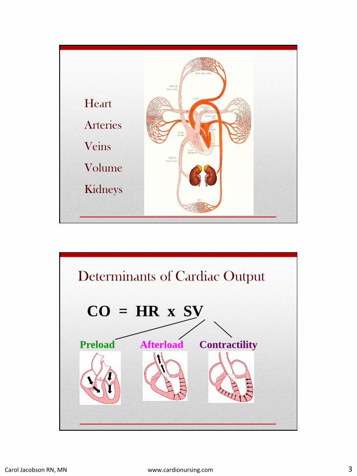
Heart
Arteries
Veins
Volume
Kidneys
Determinants of Cardiac Output
CO = HR x SV
Preload Afterload Contractility

Carol Jacobson RN, MN www.cardionursing.com 4
Venous tone Body Position Intrathoracic Intrapericardial pressure pressure Blood Volume Distribution of Atrial Kick LV Function blood volume PRELOAD Reducing preload in HF:
• Diuretics
• Venous Dilators: nitrates, ACEI, ARBs,
aldosterone blockers, nesiritide
Circulating vasodilator or
vasoconstrictor mediators
AFTERLOAD
SVR
Aortic Pressure
and Compliance
Aortic Stenosis
HOCM
Arteriolar Tone
Sympathetic NS
Reducing afterload in HF: • Arterial dilators: nitroprusside, hydralazine,
ACEI, ARBs

Carol Jacobson RN, MN www.cardionursing.com 5
CONTRACTILITY
Ventricular
Muscle Mass Catecholamines Metabolic
State
Drugs
SNS Adrenals H+ CO2 O2 Hypoxia
Ischemia
Increasing contractility in HF: • Positive inotropes: dobutamine,
milrinone, dopamine, digoxin
Carol Jacobson RN, MN www.cardionursing.com 6
Heart Failure
A complex clinical syndrome characterized by abnormal ventricular filling or ejection resulting in a low cardiac output state.
As the syndrome progresses, a variety of compensatory mechanisms including hemodynamic, renal, neurohormonal and cellular process occur
These compensatory mechanisms contribute to the progression of the disease
Heart Failure
A complex clinical syndrome that can result
from any structural or functional cardiac
disorder that impairs the ability of the ventricle
to fill with or eject blood.
Manifestations are:
• Dyspnea and fatigue which limit exercise
tolerance
• Fluid retention leading to pulmonary and
peripheral edema

Carol Jacobson RN, MN www.cardionursing.com 7
Not “congestive” HF
80% of patients in end-stage LV failure do not have pulmonary congestion
Patients admitted in pulmonary edema can be treated for volume overload and their symptoms of congestion can disappear, but the abnormalities in cardiac structure, hemodynamics, and neurohormonal activation persist despite the resolution of “congestive” symptoms.
Heart failure progresses even in the absence of congestive symptoms
CHF = Chronic Heart Failure
Backward Failure
Ventricular pressures rise
• High LV pressure is transmitted
backwards into lungs (LV backward
failure)
• High RV pressure is transmitted
backwards into venous system (RV backward failure)

Carol Jacobson RN, MN www.cardionursing.com 8
Forward Failure
Ventricles fail to pump well
in a forward direction
• LV forwards failure results in
peripheral hypoperfusion
• RV forwards failure results in
failure to adequately fill the
LV
HFrEF is defined as the clinical diagnosis of HF
and EF ≤40%
Impaired LV contractility results in reduced
ejection fraction (< 40%), increasing end-diastolic
volume and pressure
Ventricle is dilated, thin-walled, eccentrically
hypertrophied
Systolic Dysfunction
HFrEF
Only in these patients have effective
therapies been demonstrated to date

Carol Jacobson RN, MN www.cardionursing.com 9
Eccentric Hypertrophy
• Increase in chamber size without increase in muscle thickness (although muscle mass increases)
• Due to volume overload
• MI (↓CO results in fluid retention by kidneys)
• Aortic or mitral regurgitation
• Congenital defects
• Results in systolic failure
Patients with systolic dysfunction often have diastolic dysfunction too
Causes of Systolic Dysfunction
• CAD (2/3 of patients)
• MI
• Hypertension
• Tachyarrhythmias
• Valvular (aortic or mitral
regurgitation)
• Myocarditis
• Toxins (ETOH, cocaine,
chemo agents)
• Endocrine (hyperthyroidism, diabetes)
• Congenital
• Pregnancy
• Idiopathic
Carol Jacobson RN, MN www.cardionursing.com 10
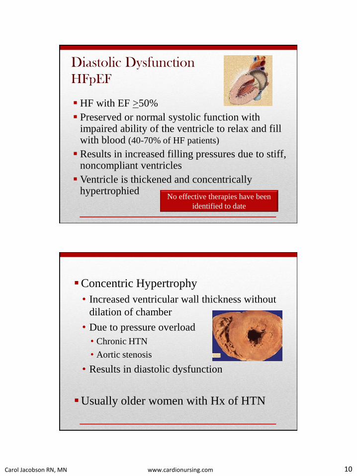
Diastolic Dysfunction
HFpEF
HF with EF >50%
Preserved or normal systolic function with impaired ability of the ventricle to relax and fill with blood (40-70% of HF patients)
Results in increased filling pressures due to stiff, noncompliant ventricles
Ventricle is thickened and concentrically hypertrophied
No effective therapies have been
identified to date
Concentric Hypertrophy
• Increased ventricular wall thickness without
dilation of chamber
• Due to pressure overload
• Chronic HTN
• Aortic stenosis
• Results in diastolic dysfunction
Usually older women with Hx of HTN

Carol Jacobson RN, MN www.cardionursing.com 11
Causes of Diastolic Dysfunction
Hypertension – most important cause
• Present in 60 – 90% of patients with HFpEF
CAD, obesity, diabetes, and AF are highly
prevalent in patients with HFpEF
Hypertrophic cardiomyopathy
Infiltrative diseases
• Sarcoidosis
• Amyloidosis
Aortic stenosis
Normal heart Hypertrophied heart
(diastolic HF)
Dilated heart
(systolic HF)

Carol Jacobson RN, MN www.cardionursing.com 12
Major Risk Factors for Developing HF
Hypertension (2-3 fold increased risk)
• Most important modifiable risk factor
• Long-term treatment of both systolic and diastolic hypertension reduces the risk of HF by about 50%
Atherosclerotic disease (coronary, cerebral, or peripheral)
• MI (8-10 fold increased risk)
Diabetes (2-5 fold increased risk, especially in women)
• Markedly increases the likelihood of developing HF in patients without structural heart disease
• Adversely affects the outcomes of patients with established HF
Metabolic syndrome
Metabolic Syndrome (Insulin Resistance Syndrome)
“The Deadly Quartet” (presence of any 3 of the following):
• Abdominal obesity
• Waist circumference in men > 40 inches and in women > 35 inches
• Hyperglycemia
• Fasting glucose > 100 mg/dl
• Dyslipidemia
• Triglycerides > 150 mg/dl
• HDL < 40 mg/dl in men and < 50 mg/dl in women
• Hypertension: BP > 130/85 mmHg
>40% incidence in people over 40 in the US!
Appropriate treatment of hypertension, diabetes and dyslipidemia
can significantly reduce the development of HF.

Carol Jacobson RN, MN www.cardionursing.com 13
Other Contributors to HF
Obesity - every 1 Kg/M2 increase in BMI is associated with 5% increased risk of HF in men and 7% in women.
Recent pregnancy – peripartum cardiomyopathy
Tachycardia induced cardiomyopathy
Takotsubo cardiomyopathy (stress induced)
Family History – familial cardiomyopathy
Drugs & Toxins – alcohol, cocaine, IV drugs, chemo, tobacco, NSAIDs, cobalt, anabolic steroids, many others
Connective tissue & systemic disorders – lupus, scleroderma, sarcoidosis, amyloidosis
Myocarditis – viral, bacterial, fungal, parasitic
NYHA Functional Classification in Patients With HF
Class I No limitation of physical activity. Ordinary physical activity
does not cause symptoms*.
Class II Slight limitation of physical activity. Comfortable at rest, but
ordinary physical activity results in symptoms.
Class III IIIA: Marked limitation of physical activity. Comfortable at
rest, but less than ordinary activity causes symptoms.
IIIB: Marked limitation of physical activity. Comfortable at
rest, but minimal exertion causes symptoms. Dyspnea with less
than one block walking.
Class IV Unable to carry on any physical activity without discomfort.
Symptoms of cardiac insufficiency present at rest. If any
physical activity is undertaken, discomfort is increased.
* Symptoms = fatigue, palpitations, or dyspnea

Carol Jacobson RN, MN www.cardionursing.com 14
ACC/AHA Stages of Heart Failure
Stage
A Patients at high risk for developing HF but have no structural
disorder of the heart (HTN, atherosclerotic disease, diabetes, metabolic syndrome, obesity,
family history of cardiomyopathy)
Stage
B Patients with structural disorder of the heart but no
symptoms of HF (History of MI, valve disease, LVH, low EF)
Stage
C Patients with past or current symptoms of HF associated with
structural heart disease (Known structural heart disease and signs and symptoms of HF)
Stage
D Patients with end-stage HF who require specialized treatment
such as mechanical circulatory support, continuous inotropic
infusions, cardiac transplant, hospice (Marked symptoms at rest despite maximal therapy, repeated
hospitalizations for HF despite appropriate therapy)
What NYHA class and Stage of HF?
Description NYHA
Class
Stage
of HF
Patient with Hx of HTN who is post MI 2 years ago, EF 30%. Hx of SOB
& fatigue when golfing 18 holes so can now play only 9 holes. Meds:
diuretic, ACEI, beta blocker. Asymptomatic around the house and with
usual activities but notices that he is tired and slightly SOB near the end
of 9 holes of golf.
II C
Patient who has HTN, diabetes, overweight. No history of MI, EF 55%.
Meds: diuretic, Ca++ blocker, ACEI, oral hypoglycemic. Asymptomatic
with all activities but is relatively sedentary.
I A
Patient with Hx of MI, moderate renal failure, and diagnosed with HF for
3 years. Has been managed with diuretics, ACEI, beta blocker,
aldosterone blocker, and has a biventricular pacemaker. EF 20% with this
Rx. Has been progressively more SOB with even minimal activity and
now is symptomatic at rest. Has been hospitalized with ADHF twice in
the past 6 months.
IV D
Patient with aortic valve disease, LVH, and diagnosed HF. EF 26%.
Comfortable while watching TV or reading, gets SOB and fatigued when
walking to bathroom and fixing dinner.
III C

Carol Jacobson RN, MN www.cardionursing.com 15
Pathophysiology of Heart Failure
Physiologic Change Signs & Symptoms
ability of LV to pump
blood
Fatigue, chest pain (if coronary
arteries underperfused)
LVEDV & LVEDP S3 and/or S4 gallop
PWP
Left atrial pressure Atrial arrhythmias
(atrial fib)

Carol Jacobson RN, MN www.cardionursing.com 16
Pathophysiology of Heart Failure
Physiologic Change Signs & Symptoms
pressure in pulmonary
capillaries & pulmonary artery PA pressure & PAD
Leaking of fluid from
pulmonary capillaries
into lungs
Crackles, SOB,
cough, orthopnea,
wheezing, hypoxia
Pathophysiology of Heart Failure
Physiologic Change Signs & Symptoms
right ventricular pressure Sternal heave, RV S3
right atrial pressure CVP, neck veins
Backup of blood into
systemic veins Peripheral edema,
+ abdominojugular test
Carol Jacobson RN, MN www.cardionursing.com 17
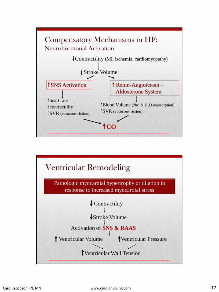
Compensatory Mechanisms in HF: Neurohormonal Activation
Contractility (MI, ischemia, cardiomyopathy)
Stroke Volume
SNS Activation Renin-Angiotensin –
Aldosterone System
heart rate
contractility
SVR (vasoconstriction)
Blood Volume (Na+ & H2O reabsorption)
SVR (vasoconstriction)
CO
Pathologic myocardial hypertrophy or dilation in
response to increased myocardial stress
Contractility
Stroke Volume
Activation of SNS & RAAS
Ventricular Volume Ventricular Pressure
Ventricular Wall Tension
Ventricular Remodeling
Carol Jacobson RN, MN www.cardionursing.com 18
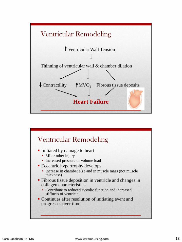
Ventricular Remodeling
Ventricular Wall Tension
Thinning of ventricular wall & chamber dilation
Contractility MVO2 Fibrous tissue deposits
Heart Failure
Ventricular Remodeling
Initiated by damage to heart • MI or other injury
• Increased pressure or volume load
Eccentric hypertrophy develops • Increase in chamber size and in muscle mass (not muscle
thickness)
Fibrous tissue deposition in ventricle and changes in collagen characteristics • Contribute to reduced systolic function and increased
stiffness of ventricle
Continues after resolution of initiating event and progresses over time

Carol Jacobson RN, MN www.cardionursing.com 19
Sympathetic Nervous System
Alpha Receptors Beta Receptors (Arteries & Veins)
Beta 1 Beta 2
(Heart) (Arteries, veins) (Lungs)
Vasoconstriction ↑ heart rate Vasodilation
↑ contractility Bronchodilation
↑ automaticity
↑conduction velocity
↑Renin release
Role of Kidney in HF

Carol Jacobson RN, MN www.cardionursing.com 20
Renin-Angiotensin-Aldosterone System
↓Renal blood flow (↓BP, ↓ Na+, diuresis)
Renin release
Angiotensinogen Angiotensin I
Angiotensin II
Vasoconstriction Aldosterone release ↑Na+ & H2O retention
↑ BP & Organ perfusion
(converting enzyme)
More pressure
More volume
When there is decreased LV
function and cardiac output:
• Kidneys think body is
hypovolemic Na+ & H2O
retention
• Baroreceptors think body is
hypotensive peripheral
vasoconstriction, release of
vasopressin (causes H2O
retention and hyponatremia)
• Endothelin – vasoconstrictor
substance made in myocardium
and vascular endothelium in
response to neurohormones
negative inotrope, peripheral VC,
contributes to remodeling

Carol Jacobson RN, MN www.cardionursing.com 21
Compensatory Mechanisms in Heart
Failure
• ↑ sympathetic NS activity
– Results in tachycardia, ↑ contractility, ↑ SVR
• ↑ Renin-angiotensin-aldosterone activity
– Results in Na+ and H2O retention, ↑ SVR
• ↑ release of natriuretic peptides (ANP, BNP)
– Attempt to counteract vasoconstriction with
vasodilation, diuresis, and natriuresis
Natriuretic Peptides
ANP synthesized in atria
• Released in response to atrial stretch
• Acute response to increased volume
BNP synthesized in ventricles
• Released in response to prolonged volume overload
• Elevated in heart failure (also higher in women and people >
60 without HF)
• Sensitive marker for severity of HF (↑ in proportion to
NYHA class)
• Help different HF from other causes of SOB
• Causes arterial and venous dilation (preload and afterload
reduction)

Carol Jacobson RN, MN www.cardionursing.com 22
Natriuretic Peptides
Cause smooth muscle relaxation
• Arterial dilation = afterload reduction
• Venous dilation = preload reduction
• ↓MAP, ↓PAP, ↓PWP, ↓RAP
Suppress renin and aldosterone release
Act on kidney to cause diuresis and natriuresis
Function to help maintain compensated state in
heart failure
The Natriuretic Peptide System is Overwhelmed in Acute Decompensated Heart Failure
Vasodilators ANP BNP
Nitric oxide
Vasoconstrictors Aldosterone Endothelin
Epinephrine Vasopressin
Angiotensin II

Carol Jacobson RN, MN www.cardionursing.com 23
BNP Levels to Diagnose HF
BNP < 100 pg/ml
HF very unlikely
(2%)
BNP 100-400 pg/ml
Baseline LV dysfunction,
underlying cor
pulmonale, acute PE?
Yes No
Possible
exacerbation
of HF
(25%)
HF likely
(75%)
BNP > 400 pg/ml
HF very likely
(95%)
Recommendations for Biomarkers in HF
Biomarker, Application Setting COR LOE
Natriuretic peptides (BNP or NT-proBNP)
Diagnosis or exclusion of HF Ambulatory,
Acute I A
Prognosis or severity of HF Ambulatory,
Acute I A
Achieve optimal dosing of GDMT in chronic HF
managed by specialized HF program Ambulatory IIa B
Guidance of acutely decompensated HF therapy Acute IIb C
Biomarkers of myocardial injury (Troponins)
Additive risk stratification Acute,
Ambulatory I A
Biomarkers of myocardial fibrosis (Galectin-3)
Additive risk stratification Ambulatory IIb B
Acute IIb A
Carol Jacobson RN, MN www.cardionursing.com 24
Evaluation for clinical manifestations of HF with a routine
history and physical examination is recommended in
patients with the following medical conditions or test
findings that place patient at high risk for HF:
• Family history of cardiomyopathy in a first degree relative (parent, sibling, child)
• Sleep-disordered breathing
• Abnormal ECG (LVH, LBBB, pathologic Q waves)
• Cardiomegaly on chest X-ray
• Hypertension
• Diabetes
• Obesity
• CAD (after MI, PCI, CABG)
• Peripheral arterial disease or
cerebrovascular disease
• Valvular heart disease
• History of exposure to cardiac
toxins
Carol Jacobson RN, MN www.cardionursing.com 25
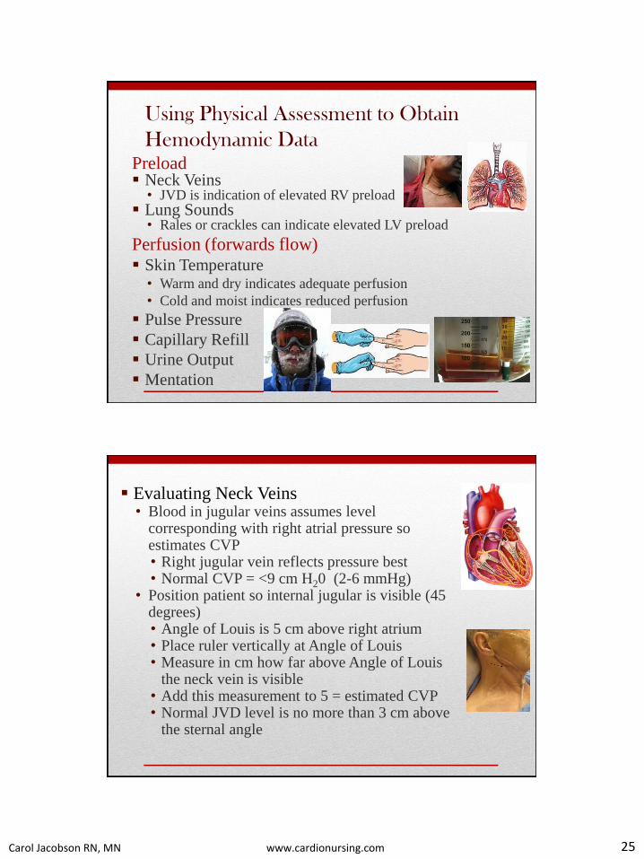
Using Physical Assessment to Obtain
Hemodynamic Data Preload Neck Veins
• JVD is indication of elevated RV preload Lung Sounds
• Rales or crackles can indicate elevated LV preload
Perfusion (forwards flow) Skin Temperature
• Warm and dry indicates adequate perfusion
• Cold and moist indicates reduced perfusion
Pulse Pressure
Capillary Refill
Urine Output
Mentation
Evaluating Neck Veins • Blood in jugular veins assumes level
corresponding with right atrial pressure so estimates CVP • Right jugular vein reflects pressure best • Normal CVP = <9 cm H20 (2-6 mmHg)
• Position patient so internal jugular is visible (45 degrees) • Angle of Louis is 5 cm above right atrium • Place ruler vertically at Angle of Louis • Measure in cm how far above Angle of Louis
the neck vein is visible • Add this measurement to 5 = estimated CVP • Normal JVD level is no more than 3 cm above
the sternal angle

Carol Jacobson RN, MN www.cardionursing.com 26
CVP = 12 cm H2O
Right atrium
Angle of Louis 5 cm
Level of neck vein
7 cm
Angle of Louis
Neck veins are distended whenever CVP is high or something
interferes with RV filling: RV failure, cardiac tamponade, restrictive pericarditis, restrictive
cardiomyopathy, tension pneumothorax
• Abdominojugular reflux – seen in RV failure • Press on upper right quadrant for 30-60 seconds while
watching neck veins
• Rise in neck veins of 1 cm or more = positive
abdominojugular reflux
• Kussmaul’s sign (paradoxical elevation of jugular
venous pressure during inspiration)
• Normally the neck veins empty during inspiration as
intrathoracic pressure drops and venous return to the
heart increases
• In “restrictive” disease (pericardial effusion, constrictive
pericarditis, cardiomyopathy, diastolic HF) the heart cannot
handle the increased venous return so neck veins elevate
when venous return increases during inspiration

Carol Jacobson RN, MN www.cardionursing.com 27
Evaluating Blood Pressure
Systolic BP is affected by
• LV stroke volume
• Peak rate of LV ejection
• Distensibility of blood vessel walls
Diastolic BP is affected by peripheral vascular
resistance (arteriolar tone)
• A rise in DBP is the first BP change – due to
compensatory vasoconstriction
Pulse Pressure
• Difference between systolic BP and diastolic BP
• Normal = 40 mmHg (120/80 = 40 mmHg PP)
• Influenced by stroke volume and arterial compliance
(beat-to-beat change reflects SV)
• ↓ PP is an early sign of ↓ cardiac output
• PP < 30 mmHg is sign of advanced heart failure
• PP can be increased due to high stroke volume
(↑preload, ↑contractility) or high compliance (vasodilation)
• PP can be decreased due to low stroke volume
(↓preload, ↓contractility) or low compliance (vasoconstriction)

Carol Jacobson RN, MN www.cardionursing.com 28
Evaluation of pulse
• Pulsus alternans is diagnostic of LV failure
• Alternating strong and weak pulse during regular rhythm
• Can hear it with slow release of BP cuff - every other beat
heard with strong pulse, eventually every beat heard
Assessing Volume Status
• Paroxysmal nocturnal
dyspnea or orthopnea
• Dyspnea on exertion
• Daily weights
• Vital signs
• Assess for orthostatic BP
and HR changes
• Rales, crackles
• S3 gallop
• Elevated jugular venous
pressure
• Hepatic enlargement and
tenderness
• Positive hepatojugular
reflux
• Edema
• Ascites

Carol Jacobson RN, MN www.cardionursing.com 29
Four major assessment findings suggest severity of the
cardiac dysfunction:
• Resting sinus tachycardia
• Narrow pulse pressure
• A decrease in cardiac output should be suspected when the pulse pressure
is reduced below 30 mmHg.
• Diaphoresis
• Peripheral vasoconstriction
• Cool, pale, and sometimes cyanotic extremities
Three major manifestations of volume overload in
patients with HF:
• Pulmonary congestion (crackles, orthopnea, dyspnea)
• Peripheral edema (weight gain)
• Elevated jugular venous pressure
Careful physical examination with determination of
vital signs and evaluation for these signs of HF
Elevated cardiac filling
pressures and fluid overload
• Elevated jugular venous pressure
• S3 gallop
• Rales
• Hepatojugular reflux
• Ascites
• Edema
Cardiac enlargement • Laterally displaced or prominent
apical impulse
Reduced cardiac output • Narrow pulse pressure
• Cool extremities
• Tachycardia with pulsus alternans
Arrhythmia • Irregular pulse suggestive of atrial
fibrillation or frequent ectopy
HFSA 2010 Comprehensive Heart Failure Practice Guideline. J Card Fail 2010;16:475-539

Carol Jacobson RN, MN www.cardionursing.com 30