hearing in mole crickets (orthoptera: gryllotalpidae) at sonic and
TRANSCRIPT
1967The Journal of Experimental Biology 201, 1967–1979 (1998)Printed in Great Britain © The Company of Biologists Limited 1998JEB1498
HEARING IN MOLE CRICKETS (ORTHOPTERA: GRYLLOTALPIDAE) AT SONICAND ULTRASONIC FREQUENCIES
ANDREW C. MASON*, TIMOTHY G. FORREST† AND RON R. HOYDepartment of Neurobiology and Behavior, S. G. Mudd Hall, Cornell University, Ithaca, NY 14853, USA
*e-mail: [email protected]†Present address: Department of Biology, University of North Carolina at Asheville, 1 University Heights, Asheville, NC 28804, USA
Accepted 3 April; published on WWW 21 May 1998
We have studied auditory responses in two species ofmole cricket (Scapteriscus borelliiand S. abbreviatus) todetermine (1) whether they show sensitivity to ultrasound,(2) whether their hearing (at both low and highfrequencies) is based on the same neural circuitry as thatof true crickets, and (3) whether ultrasound sensitivity indifferent mole cricket species varies with their ability to fly.
S. borellii are sensitive to ultrasonic frequencies. Thereis evidence of a segregation of frequency bands inprothoracic auditory neurons. There are two pairs ofomega neurons (ONs) with similar morphology to ON1 oftrue crickets. The two pairs of ONs differ in tuning. Onepair has two sensitivity peaks: at the frequency of thecalling song of this species (3 kHz), and in the ultrasonicrange (25 kHz). The other pair lacks the high-frequencysensitivity and responds exclusively to frequencies in therange of the species song. These two types are notmorphologically distinguishable. In S. abbreviatus, only oneclass of ON was found. S. abbreviatusONs are narrowlytuned to the frequency of the species’ calls. A T-neuron had
the best ultrasonic frequency sensitivity in S. borellii. Thiscell showed a broad tuning to ultrasonic frequencies andwas inhibited by low-frequency stimuli. A morphologicallysimilar neuron was also recorded in S. abbreviatus, butlacked the high-frequency sensitivity peak of that in S.borellii.
We also assessed the responses of flying S. borellii toultrasound using field playbacks to free-flying animals. Theattractiveness of broadcast calling song was diminished bythe addition of an ultrasound signal, indicating that S.borellii avoid high-frequency sound.
The results indicate that mole crickets process low-frequency auditory stimuli using mechanisms similar tothose of true crickets. They show a negative behaviouralresponse to high-frequency stimuli, as do true crickets, butthe organization of ultrasound-sensitive auditory circuitryin mole crickets differs from that of true crickets.
Key words: Orthoptera, mole cricket, Scapteriscus borellii,Scapteriscus abbreviatus, hearing, ultrasound, evolution.
Summary
3;sir
hat
ntat
t,d
r,irleescy
Auditory processing in ensiferan insects has beextensively studied from the perspective of both intraspecacoustic communication (Kalmring et al. 1997) and thedetection of predators (Hoy, 1992; Libersat and Hoy, 199The majority of these studies have concentrated on tcommon families of Ensifera, the true crickets (Gryllidae) athe katydids (Tettigoniidae). Comparative studies of othfamilies in this suborder of the Orthoptera have been few(Ball and Field, 1981; Cokl et al.1995; Field et al.1980; Jeramet al.1995; Mason, 1991; Mason and Schildberger, 1993) ahave been confined to the tettigoniid clade (Gwynne, 1995
This work has established that, despite consideravariation between the Gryllidae and the Tettigoniidae peripheral auditory anatomy (Bailey, 1993; Ball et al. 1989)and signal characteristics (Bennet-Clark, 1989; Kalmring et al.1997; Morris et al. 1988, 1994), these distantly relateensiferan families (Gwynne, 1995) show a remarkabsimilarity in the central neural circuitry devoted to processi
Introduction
enific
1).wonderer
nd).blein
dle
ng
intraspecific acoustic signals (Mason and Schildberger, 199Wohlers and Huber, 1982). The true crickets and katydidappear to be less similar, however, in the organization of theultrasound-sensitive auditory circuitry. Specifically,ultrasound-elicited acoustic startle responses (ASRs), whichave been associated with the avoidance of echolocating bpredators in both these families, are mediated by differecentral auditory neurons in true crickets and katydids (Libersand Hoy, 1991; Nolen and Hoy, 1984).
The mole crickets (Gryllotalpidae) are the ensiferan familymost closely related to the true crickets (Gwynne, 1995) bualthough mole cricket acoustic behaviour has been studie(Bennet-Clark, 1987; Daws et al. 1996; Forrest, 1980, 1983,1986, 1991; Forrest and Green, 1991; Ulagaraj and Walke1973; Walker and Forrest, 1989), less is known about thehearing. Suga (1968) found that the hearing of several mocricket species was most sensitive to ultrasound frequenciand speculated that this was related to the high-frequen
1968
n
nas
reKore
uststicrupda
anda
g
re
r
fsde
inle
es
esk
d
he
g
ofptheter
fellathmeleseting
A. C. MASON, T. G. FORREST ANDR. R. HOY
content in their songs. Subsequent studies of mole cricsound production have found their songs to contain exclusivlow frequencies (Bennet-Clark, 1989; Forrest, 1983), so tthis interpretation is not supported. Many species of mcricket are frequent nighttime fliers, however, and are likelybe subject to predation by bats. Because mole crickets, untrue crickets, have no social signals that contain higfrequency components (cf. true cricket courtship calls; BennClark, 1989; Libersat et al. 1994), the most plausible contexin which ultrasound hearing could have evolved in this grois for the detection of non-social (predator-related) cues. have therefore undertaken a study of auditory sensitivity aanatomy in mole crickets with the following aims. (1) Tdetermine the relative sensitivity of mole crickets to high- alow-frequency sound, and to characterize central auditneurons on the basis of their possible roles in processintraspecific songs and ultrasound. (2) Another goal wtherefore to compare the central auditory anatomy of mcrickets and true crickets, in particular to establish whethhigh-frequency hearing in the two groups is based on simneural elements. (3) To determine whether there behavioural correlates or interspecific differences betweflying and flightless mole cricket species that are consistwith the use of high-frequency hearing for the detection of baThis would be expected if high-frequency hearing in this groevolved solely in the context of bat predation anindependently of its origin in true crickets.
Materials and methodsAnimals
We used adult Scapteriscus borellii(Giglio-Tos) and S.abbreviatus(Scudder) of either sex in all experiments. S.borellii were collected from the field. As adults, members this species make dispersal flights each night during thbreeding seasons. S. borelliiwere collected by attracting themto traps broadcasting their species’ calling song during thnightly dispersal flight (see below). We obtained specimensS. abbreviatus, a flightless species, from a laboratory colony the University of Florida.
Hearing
Acoustic stimuli
Acoustic stimuli were tone pulses of 20 ms duration wi1 ms rise/fall times. Typically, single pulses were delivereda rate of 2 s−1, but in some cases we created trains of 10 pulwith varying repetition rates and presented these at a rat1 s−1. Stimuli were generated using either of two systems. We used a microcomputer and D/A board (IBM PCcompatible, Data Translation DT2821) to set a voltagcontrolled oscillator (Tektronix FG501), the output of whicwas connected to a custom-built pulse shaper that wmodulated by a square-pulse generator (Tucker Davis TG(2) Stimulus pulses were synthesized with a DSP bo(Tucker Davis APOS II) and output through a D/A interfac(Tucker Davis DA3-2). With both systems, the tone puls
ketelyhatole tolikeh-et-tupWend
ondoryingas
oleer
ilarareenentts.
upd
ofeir
eir ofat
th atsese of(1)-e-has6).
ardees
were attenuated (Tucker Davis PA4), amplified (HarmaKardon HK660) and broadcast via loudspeakers (Realisticpiezo tweeter or 4 inch woofer) placed 50 cm from the positioof the preparation. The frequency range of both systems w1–70 kHz. Stimulus levels at the position of the animal wecalibrated with continuous tones, using a microphone (B&type 4135 or 4138) and sound level meter (B&K type 2209) measuring amplifier (B&K type 2606). Unless otherwisindicated, sound intensities are given in dB SPL (re 20µPa).
Neurophysiology
Auditory neurons were located using a search stimulconsisting of two temporally offset tone pulses of differenfrequencies (3 and 25 kHz). We recorded responses to acoustimuli from interneurons in the prothoracic ganglion. Afteremoving the wings, we mounted the animals ventral side on a metal platform using low-melting-point wax. We removethe ventral cuticle of the prothorax, lifted the ganglion onto chlorided silver spoon that served as reference electrode, softened the ganglionic sheath using an enzyme (Sigmpronase) to facilitate electrode penetration. Recordinelectrodes were electrolyte-filled (0.1 mol l−1 LiCl), thin-walled (1.0 mm o.d.) glass micropipettes the tips of which wefilled with 2.5 % Lucifer Yellow (Sigma). Electrode resistancevaried from 50 to 100 MΩ. Neural responses were amplified(Axoclamp 2A), digitized (Data Translation DT2821 or TuckeDavis AD3 and APOS II) and stored on disk. Followingrecording, we stained cells with Lucifer Yellow by injection o0.5–2.5 nA of hyperpolarizing current. Ganglia in which cellhad been stained were removed, fixed in 4 % paraformaldehyfor 12–24 h, dehydrated in an alcohol series, and clearedmethyl salicylate. Filled neurons were photographed as whomounts using a Leitz Dialux 20 or captured as digital imagusing a BioRad MRC-600 confocal microscope.
Recordings of the summed activity in the neck connectivwere made from some animals using silver wire hooelectrodes.
Behaviour
We tested the behavioural significance of ultrasounsensitivity in S. borellii in a field study. We used two soundtraps (Walker, 1982) to test whether ultrasound influenced tphonotactic behaviour of flying S. borellii. In this species,individuals of both sexes are attracted to conspecific callinsong during evening dispersal flights.
The two traps were placed 10m apart at the University Florida’s Green Acres Farm (Alachua Co., FL, USA). Each traconsisted of a 1.5m diameter sheet metal funnel. Centred in opening of each funnel was a Motorola piezoelectric horn tweethat broadcast simulated calling songs of male S. borellii. Flyingcrickets attracted to the songs and landing near the speakerinto the funnel and were collected in a 19 l plastic bucket beneeach funnel (Fig. 1). The Motorola speakers broadcast the sasynthetic calling song: a 2.7kHz carrier having a 50% duty cycmodulated at 50Hz with 20% raised-cosine ramps on the onand offset of each pulse. However, the two broadcasts of call
1969Hearing in mole crickets
tsityndofchc
at
s’es.-s
tsdnchheea
eeir
Na).ess ofnde.ve.iceser
il,ess.
ry
reg
7 m10 m
2.7 kHz40 kHz
SpeakerFunnel
Bucket
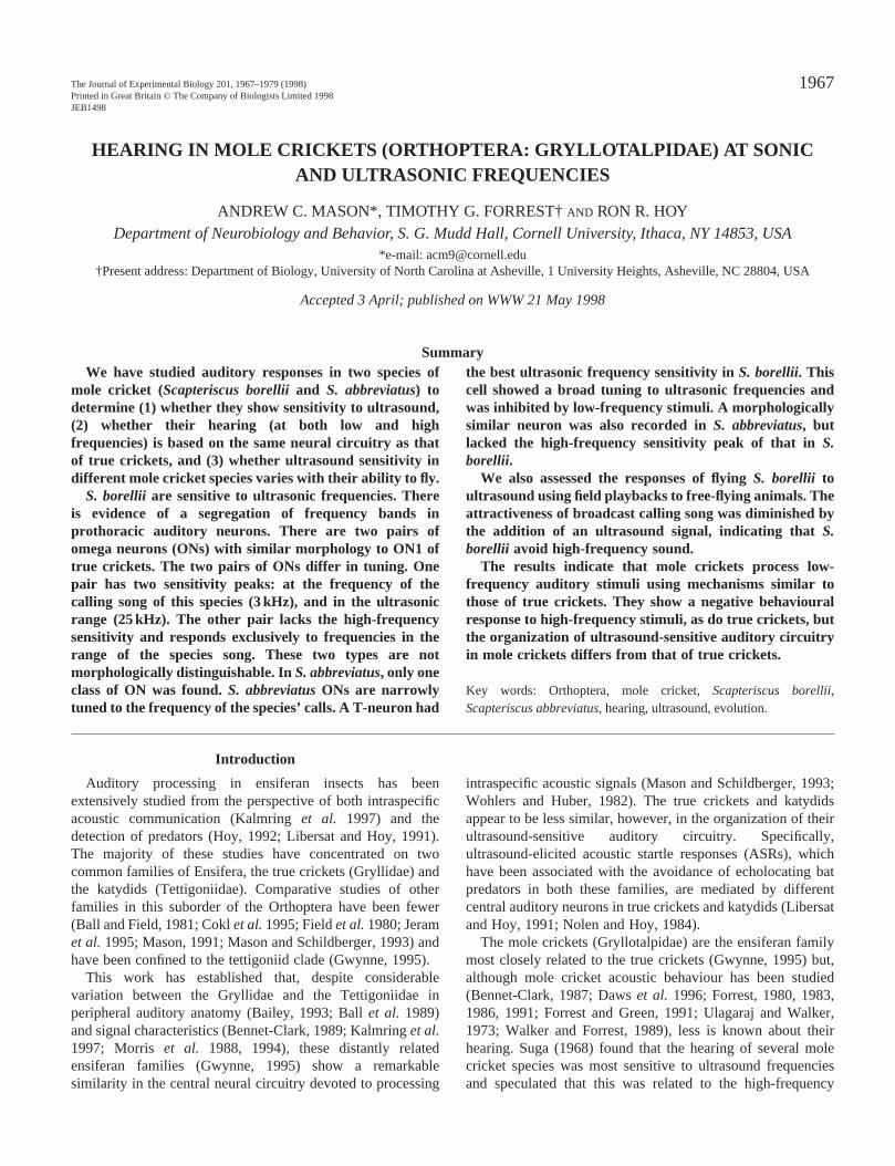
Fig. 1. Schematic diagram of the sound-trapping apparatus usemeasure the effect of ultrasound on flying Scapteriscus borrelliiinthe field. Both speakers broadcast synthetic calling song (2.7 kone 6 dB louder than the other. The louder speaker also broadultrasound (40 kHz) with a temporal pattern similar to that of tsynthetic calling song. Flying S. borelliiattracted to the calling songfrom one of the speakers landed in the funnel and were capturethe bucket. With no effect of ultrasound, the louder source of calsong was a more attractive stimulus, such that one would predictonly 17 % of mole crickets collected should be caught in the quitrap (Forrest, 1980; Forrest and Raspet, 1994). We measuredresponses of S. borellii to ultrasound by recording the proportion omole crickets actually captured in the quieter trap as a function ofenergy of ultrasound added to the louder trap.
song differed in level by 6dB (106 and 100dB SPL at 15cadjusted at each speaker). The proportion of crickets attractethe low-intensity trap is predicted to be approximately 17(Forrest and Raspet, 1994). Empirical results support prediction (16%, N=2979; Forrest, 1980). Along with the highintensity calling song, we simultaneously broadcast a 5duration ultrasound stimulus (40kHz carrier with 1ms raiscosine ramps) from a Panasonic piezoelectric ultrasotransducer (P-9934). The level of the ultrasound was independent variable in the experiment, and we measuredattraction of mole crickets to the two calling songs as a functof the energy of ultrasound relative to the calling song. If tphonotactic behaviour of flying mole crickets is negativeinfluenced by ultrasound (bat predation), we predicted that relative number of crickets collected at the trap broadcasting lintensity calling song would increase significantly as wincreased the level of the ultrasound in the other trap. All stimwere computer-generated using custom-built software and plaback through 16-bit D/A converters (TDT, Quikki D/Aconverter) at a sampling frequency of 100kHz. Anti-aliasifilters with a roll-off of more than 90dB per octave were usedremove aliased frequencies from the broadcast. The sopressure levels of the calling song and ultrasound were calibrprior to each broadcast using a Larsen-Davis model 2520 1/4microphone located 15cm above each speaker. The harmonic distortion of the broadcast system was below −40dBrelative to the broadcast signals.
On each of seven nights (28 April to 4 May 1996), broadcafrom the two traps began at about sunset and continthroughout the nightly flying period (Forrest, 1983). Each nigthe broadcast of ultrasound and high-intensity calling song w
m,d to%this-msedundthe theionhelytheow-euliyed
ng toundatedinchtotal
stsuedht,as
randomly assigned to one of the traps. To control for the effecof trap positions, the speakers broadcasting the high-intenscalling song and ultrasound were switched to the other trap athe experiment continued until approximately equal numbers crickets were caught while ultrasound was broadcast from eatrapping location. Results were analyzed using logistiregression (SAS Institute, 1985).
ResultsCharacterized neurons
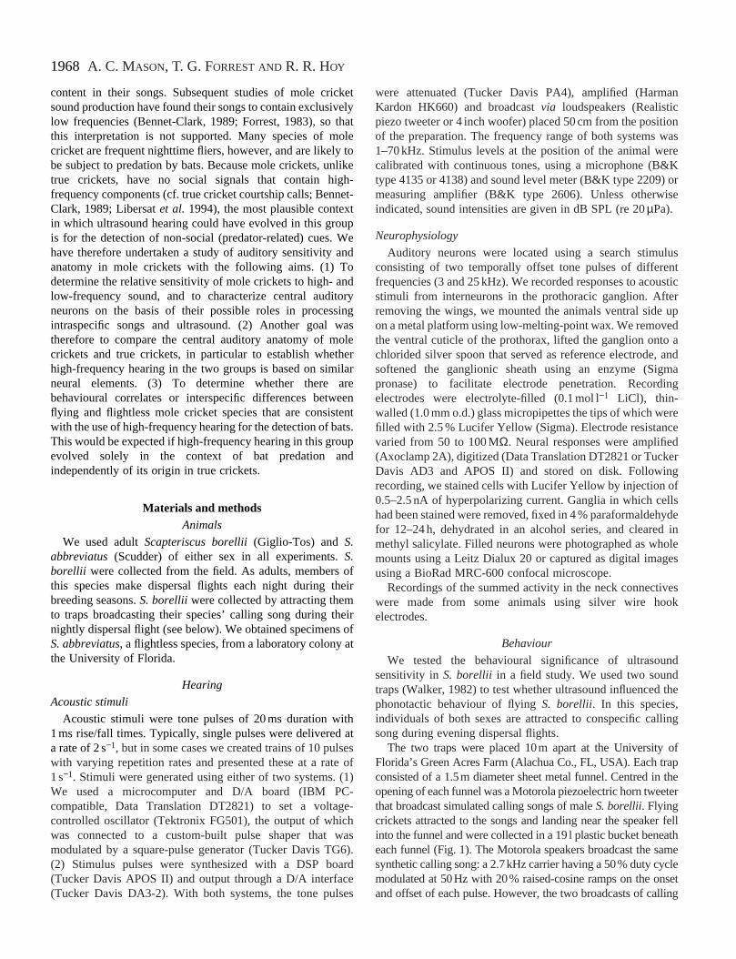
Fig. 2 shows the anatomy of auditory interneuron types thwere recorded and stained in both S. borellii and S.abbreviatus. By analogy with Gryllus spp., we will designateneurons in Scapteriscususing ‘canonical’ names. Omeganeurons (ONs; Fig. 2A) and T-neurons (TNs; Fig. 2B) in S.borellii (but not S. abbreviatus) responded to high-frequencystimuli. Detailed data for these will be presented below.
All descending neurons were narrowly tuned to the speciecall frequencies and had similar responses in the two speciAll have bilateral projections in the auditory neuropil and somacontralateral axons. The cell shown in Fig. 2C is identifiable aDN1, which has previously been described in true cricke(Wohlers and Huber, 1982) and haglids (Mason anSchildberger, 1993). DN1 has its main dendritic projection ithe soma-contralateral auditory neuropil. A secondary branarises from the axon and crosses the midline posterior to tdendritic region to project to the soma-ipsilateral neuropil. Thneuron shown in Fig. 2D (mDN) has a medially located somand similar projection areas to DN1, but differs primarily in theorigin of the secondary branch, which in mDN arises from thprimary neurite anterior to the main dendritic projection. Thestwo neurons are also distinguished by the nature of theresponses to acoustic stimuli. Auditory neurons similar to mDhave been recorded in haglids (A. C. Mason, unpublished datDN1 shows tonic responses with bursts of spikes for tone pulsand accurate copying of pulse trains, whereas the responsemDN are phasic, with only a single spike per sound pulse apoor temporal following. The third descending neuron typ(pDN, Fig. 2E) lies entirely in the posterior half of the ganglionThe soma of pDN lies near the base of the posterior connectiThe neurite gives rise to a large soma-ipsilateral dendritbranch and a mid-line crossing segment which branchcontralaterally into the descending axon and a smallprojection which is nearly symmetrical with the main dendriticbranch. Despite apparently less overlap with auditory neuroppDN receives strong auditory input, responding to tone pulswith bursts of spikes and accurately copying temporal pattern
Response properties of omega and T-neurons
Scapteriscus borellii
Both sexes of S. borellii engage in nightly dispersal flightsduring their breeding seasons (Forrest, 1980). The auditoresponses of S. borellii indicated strong ultrasound sensitivityin this species. While some prothoracic interneurons wetuned only to frequencies in the range of the species’ callin
d to
Hz),casthe
d inling thateter thef the
1970
vee
ft
t
yd
ed
h
A. C. MASON, T. G. FORREST ANDR. R. HOY
A
B
C D E
200µm
Fig. 2. Anatomy of prothoracicauditory interneurons recorded inboth Scapteriscus borelliiand S.abbreviatus. (A) Omega neurons(ONs). The left-hand (LON) andmiddle (HON) examples are from S.borellii, the right-hand ON is fromS. abbreviatus. (B) T-neurons (hfTs)with high-frequency sensitivity in S.borellii (left) and only weakauditory input in S. abbreviatus(right). (C–E) Descending neurons:DN1 (C), mDN (D) and pDN (E).
song, others exhibited tuning curves with two sensitivpeaks: one corresponding to the species’ call and the othethe ultrasonic range (20–30 kHz).
Omega neurons. Two classes of omega neurons wediscovered in the prothoracic ganglion of S. borellii, as in truecrickets, which also possess two bilateral pairs of omeneurons, ON1 and ON2 (Wohlers and Huber, 1982). Unlike tcrickets, there was no consistent morphological differenbetween the two pairs of omega cells in S. borellii. However,double stains of both cells in a single preparation confirmed these were distinct cell types. The omega neurons of S. borelliiwere anatomically similar to ON1 of true crickets. Their axoconnect the auditory neuropils of the two hemiganglia, and bdendritic and axonal arborizations extend laterally from thregion towards the leg nerves (Fig. 2A).
In S. borellii, the two omega cell types differed in thefrequency tuning (Fig. 3A). One type showed two sensitivpeaks, at calling song frequencies and at ultrasonic frequenAbsolute sensitivity at low frequencies was similar in boomega cell classes, with thresholds at the species’ sfrequency (3 kHz) of approximately 50 dB SPL. For the low
ityr in
re
garuece
that
nsothis
iritycies.thong-
frequency-tuned omega cells (LONs, N=12), thresholdsincreased continuously for higher frequencies and were abo80 dB SPL in the ultrasonic range. High-frequency-sensitivomega cells (HONs, N=13) had W-shaped threshold curves,with absolute thresholds of approximately 60 dB SPL forfrequencies of 20–30 kHz.
Both ON types responded to trills with phasic bursts ospikes that copied the temporal pattern of the stimulus arepetition rates up to 50 pulses s−1 (Fig. 3C) and respondedtonically to long-duration tones at song frequencies (data noshown). But while LONs showed weak excitation for someultrasound frequencies (up to 25–30 kHz), they did not coptrills or long tones at these frequencies (Fig. 3C). HONs copietemporal patterns equally well at audio and ultrasonicfrequencies (Fig. 3C). Omega neurons of both types respondto variations in stimulus amplitude with a wide dynamic range(over 50 dB at low frequencies) with responses varying in botspike number and latency (Fig. 3B,D). HONs had dynamicranges of approximately 30 dB for 25 kHz stimuli (Fig. 3B)and responded with fewer spikes per stimulus than for lowfrequencies but with a similar latency shift (Fig. 3E).

1971Hearing in mole crickets
dnyion
s
icialm
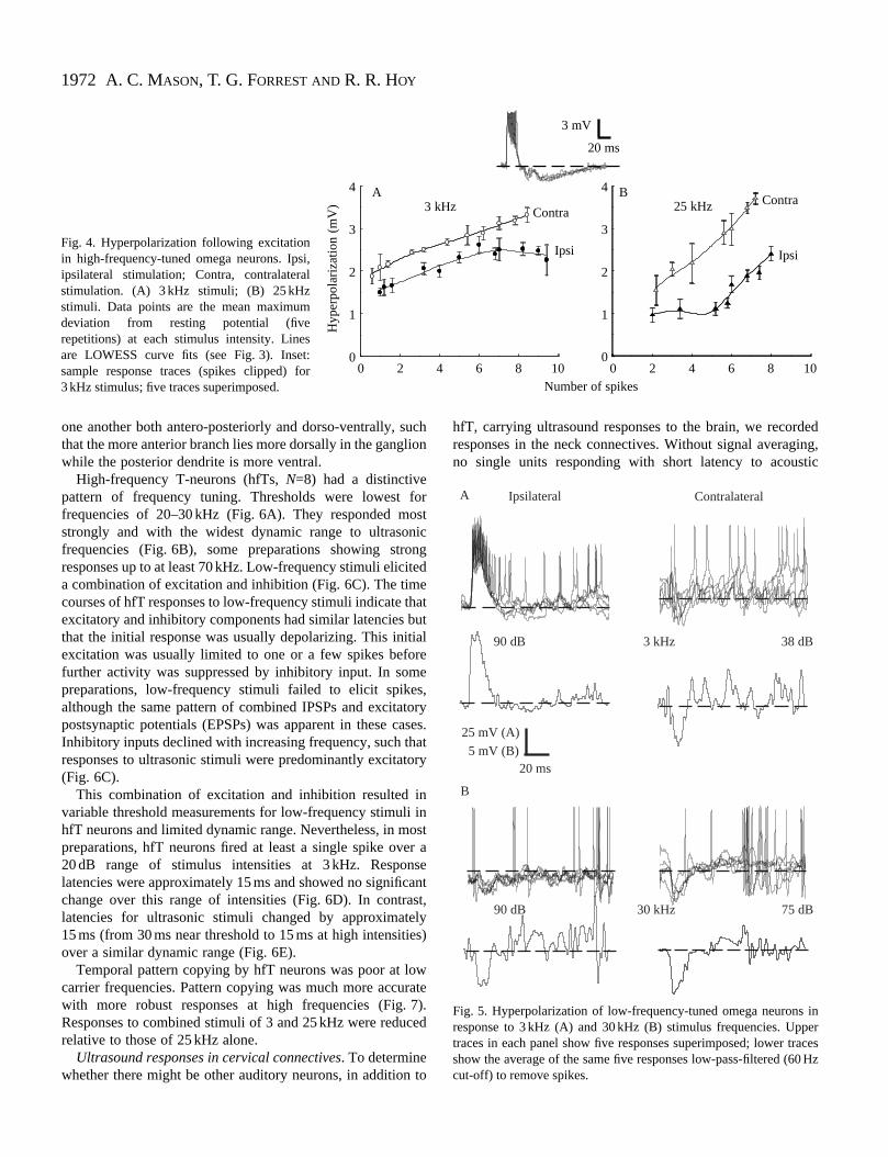
In some ON recordings, inhibitory potentials were visiblIn HONs, inhibition was not frequency-dependent and wpresent over a wide range of intensities. The depth hyperpolarization was greater for contralateral than ipsilateral stimuli at all intensities and had a wider dynamrange. Contralaterally evoked inhibitory postsynappotentials (IPSPs) were also larger when stimuli eliciting eqnumbers of spikes were compared (Fig. 4).
In LONs, inhibitory potentials in response to low frequenciwere more easily observed for contralateral stimulus presenta
1 30
40
50
60
70
80
90
100
0 20 10
15
20
25
30
35
40
Thr
esho
ld (
dB S
PL)
Lat
ency
(m
s)
Freque
90
A
C
D
2 3 4 5 6
Fig. 3. Response characteristics of omeganeurons in Scapteriscus borellii. In allpanels, data shown in blue are for low-frequency-tuned omega neurons (LONs)and data shown in red are for high-frequency-tuned omega neurons (HONs).(A) Mean frequency–tuning curves (±S.D.)for the two types of omega cell.(B) Intensity–response functions for 3 kHz(circles) and 25 kHz (triangles; HONsonly). Data points are global means foreach cell type (N=12 LONs, N=13 HONs,five repetitions of each stimulus valueaveraged within individual cells). Lineswere generated by LOWESS curve-fittingwith the stiffness parameter set to 0.6(Wilkinson et al. 1992). (C) Temporalpattern copying by the two ON types. Onthe left are sample traces (spikes clipped)showing responses to trains at 50 pulses s−1
of 3 and 25 kHz tone bursts (upper, LON;lower, HON). On the right is a raster plotshowing spike times for responses to twopulse repetition rates (33 and 50 pulses s−1).Responses of both HONs and LONs areshown for 3 kHz stimuli and for HONsonly for 25 kHz stimuli. (D,E) Responselatency plotted against stimulus intensityfor 3 kHz (D) (circles) and 25 kHz (E)(triangles; HONs only). Data points aremeans of five repetitions, and data frommultiple preparations are combined. Lineswere fitted with a power function.
e.asof
foric
ticual
estion
at low intensities. In contrast, high-frequency stimuli evokeinhibitory potentials in some preparations in the absence of aexcitatory response, for both ipsi- and contralateral presentatand over a wide range of intensities (Fig. 5).
High-frequency-tuned T-neurons. One type of auditory unitwith a single sensitivity peak in the ultrasound range warecorded in S. borellii. This was a T-neuron (Fig. 2B left),characterised by two large soma-contralateral dendritbranches and soma-contralateral axons in the medconnectives. The two dendritic branches are displaced fro
10 30 500
2
4
6
8
10
12
14
16
10
0 20 40 6010
15
20
25
30
35
40
40 60
Num
ber
of s
pike
s
3 kHz 25 kHz
ncy (kHz)
0 50 100 150 200
Latency (ms)dB SPL, 50 pulses s−1
Rep
etiti
on r
ate
(pul
ses
s−1 ) 50
33
50
33
25 kHz3 kHz
25 kHz
3 kHz
B
E
Intensity (dB relative to threshold)
Intensity (dB relative to threshold)
20 mV30 ms
7 20 30 40 60 80
1972
forsticng
tedehatut
ialree
s,toryases.at
ory
n inostr aseantst,lyes)
wrate7).ced
to
eding,tic
A. C. MASON, T. G. FORREST ANDR. R. HOY
0 2 4 6 8 100
1
2
3
4
0 2 4 6 8 100
1
2
3
4
3 kHz 25 kHzA B
Number of spikes
Ipsi
Contra
Ipsi
Contra
Hyp
erpo
lari
zatio
n (m
V)
20 ms
3 mV
Fig. 4. Hyperpolarization following excitationin high-frequency-tuned omega neurons. Ipsi,ipsilateral stimulation; Contra, contralateralstimulation. (A) 3 kHz stimuli; (B) 25 kHzstimuli. Data points are the mean maximumdeviation from resting potential (fiverepetitions) at each stimulus intensity. Linesare LOWESS curve fits (see Fig. 3). Inset:sample response traces (spikes clipped) for3 kHz stimulus; five traces superimposed.
Ipsilateral Contralateral
90 dB 3 kHz 38 dB
A
B
90 dB 30 kHz 75 dB
25 mV (A)
5 mV (B)20 ms
Fig. 5. Hyperpolarization of low-frequency-tuned omega neurons inresponse to 3 kHz (A) and 30 kHz (B) stimulus frequencies. Uppertraces in each panel show five responses superimposed; lower tracesshow the average of the same five responses low-pass-filtered (60 Hzcut-off) to remove spikes.
one another both antero-posteriorly and dorso-ventrally, sthat the more anterior branch lies more dorsally in the gangwhile the posterior dendrite is more ventral.
High-frequency T-neurons (hfTs, N=8) had a distinctivepattern of frequency tuning. Thresholds were lowest frequencies of 20–30 kHz (Fig. 6A). They responded mostrongly and with the widest dynamic range to ultrasonfrequencies (Fig. 6B), some preparations showing stroresponses up to at least 70 kHz. Low-frequency stimuli elicia combination of excitation and inhibition (Fig. 6C). The timcourses of hfT responses to low-frequency stimuli indicate texcitatory and inhibitory components had similar latencies bthat the initial response was usually depolarizing. This initexcitation was usually limited to one or a few spikes befofurther activity was suppressed by inhibitory input. In sompreparations, low-frequency stimuli failed to elicit spikealthough the same pattern of combined IPSPs and excitapostsynaptic potentials (EPSPs) was apparent in these cInhibitory inputs declined with increasing frequency, such thresponses to ultrasonic stimuli were predominantly excitat(Fig. 6C).
This combination of excitation and inhibition resulted ivariable threshold measurements for low-frequency stimulihfT neurons and limited dynamic range. Nevertheless, in mpreparations, hfT neurons fired at least a single spike ove20 dB range of stimulus intensities at 3 kHz. Responlatencies were approximately 15 ms and showed no significchange over this range of intensities (Fig. 6D). In contralatencies for ultrasonic stimuli changed by approximate15 ms (from 30 ms near threshold to 15 ms at high intensitiover a similar dynamic range (Fig. 6E).
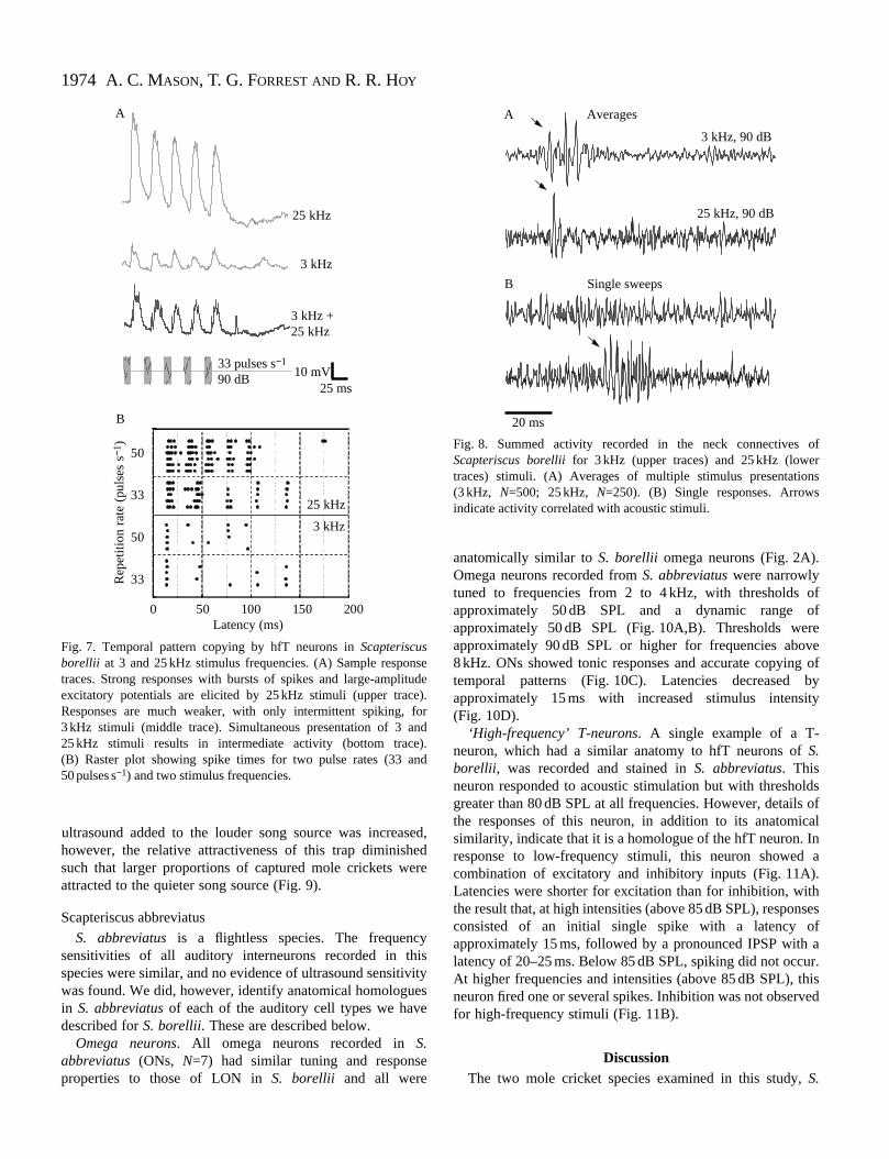
Temporal pattern copying by hfT neurons was poor at locarrier frequencies. Pattern copying was much more accuwith more robust responses at high frequencies (Fig.Responses to combined stimuli of 3 and 25 kHz were redurelative to those of 25 kHz alone.
Ultrasound responses in cervical connectives. To determinewhether there might be other auditory neurons, in addition
uchlion
hfT, carrying ultrasound responses to the brain, we recordresponses in the neck connectives. Without signal averagno single units responding with short latency to acous

1973Hearing in mole crickets
edted
eirns adendwoerof
stimuli were detectable in the summed activity of the ascendconnectives. Signal averaging revealed short-laten(15–20 ms) responses to both audio and ultrasonic stimuli (F8). Averaged responses to audio stimuli had greater amplitand duration than high-frequency responses (Fig. 8A). Higfrequency responses consisted of a single or double peak inaveraged trace. These results suggest the presence of muunits carrying ascending low-frequency responses, but oone or a few cells carrying ascending ultrasound respon(Suga, 1968).
In single sweeps, bursts of large-amplitude spikes wlonger latency (35–40 ms) could be detected, but only response to high-frequency stimuli (Fig. 8B). These respon
0 10 10
15
20
25
30
35
40
3 kHz90 dB
1 40
50
60
70
80
90
100
Thr
esho
ld (d
B S
PL)
Lat
ency
(ms)
A
C
D
Frequen
2 3 4 5 6 7
Fig. 6. Response characteristics of high-frequency-sensitive T-neurons (hfTs) inScapteriscus borellii. (A) Mean tuningcurve (± S.D.) for hfT cells.(B) Intensity–response functions for 3 kHz(circles), 25 kHz (squares) and 40 kHz(triangles). Data points are global means(N=8 cells, five repetitions of each stimulusvalue averaged within individual cells).Lines are LOWESS curve fits (see Fig. 3).(C) Sample response traces for 3, 25 and40 kHz. For 3 kHz, both five single tracessuperimposed (upper) and their average(lower) are shown. Only single traces areshown for 25 and 40 kHz. Responses to3 kHz show brief excitation followed by apronounced IPSP. High-frequency responsesare only excitatory. (D,E) Response latencyplotted against stimulus intensity for 3 kHz(D) and 25 kHz (E). Data points are meansof five repetitions. Lines were fitted with apower function.
ingcyig.
udeh- theltiplenlyses
ithin
ses
habituated rapidly and therefore did not appear in averagresponses. This may represent descending activity associawith ultrasound startle responses (Hoy et al.1989).
Behavioural responses to ultrasound. Free-flying S. borelliiof both sexes were attracted to sound traps broadcasting thspecies’ calling song. A total of 203 crickets were collected ithe traps over the seven nights of the experiment. There wasignificant relationship between the intensity of ultrasounadded to the louder song playback and the relativattractiveness of the quieter song source. With no ultrasouadded to the broadcast, the proportions attracted to the ttraps were as predicted by their relative broadcast pow(Forrest, 1980; Forrest and Raspet, 1994). As the intensity
10
15
20
25
30
35
40
20 30
,
25 kHz,90 dB
40 kHz,90 dB
3 kHz 25 kHz
10 0 10 20 30 400
2
4
6
8
10
12
Num
ber o
f spi
kes
B
E
cy (kHz) Intensity (dB relative to threshold)
Intensity (dB relative to threshold)
25 mV
25 ms
0 10 20 30
20 30 40 60 808
3 kHz
25 kHz
40 kHz
1974
ff
e ofbyy
ds ofalna
esfar.ised
A. C. MASON, T. G. FORREST ANDR. R. HOY
3 kHz +25 kHz
3 kHz
25 kHz
A
0 50 100 150 200
Rep
etiti
on r
ate
(pul
ses
s−1 )
Latency (ms)
50
33
50
33
25 kHz
3 kHz
B
33 pulses s−1
90 dB10 mV
25 ms
Fig. 7. Temporal pattern copying by hfT neurons in Scapteriscusborellii at 3 and 25 kHz stimulus frequencies. (A) Sample respotraces. Strong responses with bursts of spikes and large-amplexcitatory potentials are elicited by 25 kHz stimuli (upper tracResponses are much weaker, with only intermittent spiking, 3 kHz stimuli (middle trace). Simultaneous presentation of 3 a25 kHz stimuli results in intermediate activity (bottom trace(B) Raster plot showing spike times for two pulse rates (33 a50 pulses s−1) and two stimulus frequencies.
A Averages
B Single sweeps
25 kHz, 90 dB
3 kHz, 90 dB
20 ms
Fig. 8. Summed activity recorded in the neck connectives ofScapteriscus borelliifor 3 kHz (upper traces) and 25 kHz (lowertraces) stimuli. (A) Averages of multiple stimulus presentations(3 kHz, N=500; 25 kHz, N=250). (B) Single responses. Arrowsindicate activity correlated with acoustic stimuli.
ultrasound added to the louder song source was increahowever, the relative attractiveness of this trap diminishsuch that larger proportions of captured mole crickets weattracted to the quieter song source (Fig. 9).
Scapteriscus abbreviatus
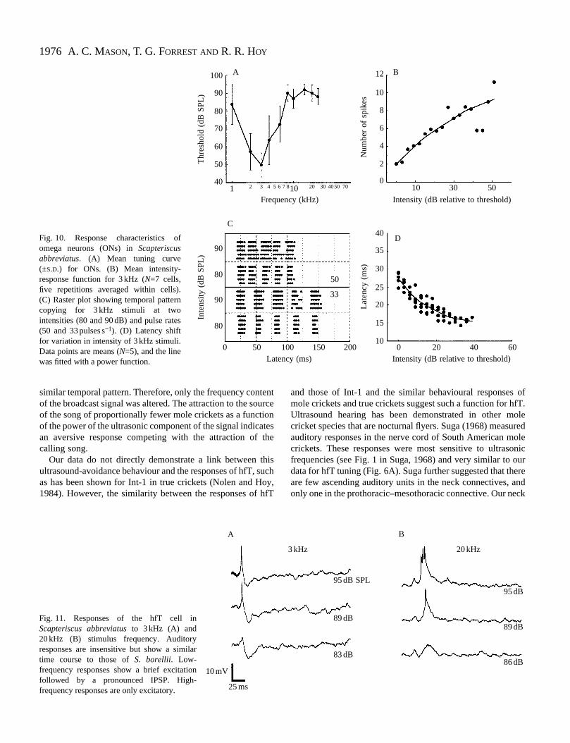
S. abbreviatus is a flightless species. The frequencsensitivities of all auditory interneurons recorded in thspecies were similar, and no evidence of ultrasound sensitiwas found. We did, however, identify anatomical homologuin S. abbreviatusof each of the auditory cell types we havdescribed for S. borellii. These are described below.
Omega neurons. All omega neurons recorded in S.abbreviatus (ONs, N=7) had similar tuning and responsproperties to those of LON in S. borellii and all were
sed,edre
yisvityese
e
anatomically similar to S. borellii omega neurons (Fig. 2A).Omega neurons recorded from S. abbreviatuswere narrowlytuned to frequencies from 2 to 4 kHz, with thresholds oapproximately 50 dB SPL and a dynamic range oapproximately 50 dB SPL (Fig. 10A,B). Thresholds wereapproximately 90 dB SPL or higher for frequencies abov8 kHz. ONs showed tonic responses and accurate copyingtemporal patterns (Fig. 10C). Latencies decreased approximately 15 ms with increased stimulus intensit(Fig. 10D).
‘High-frequency’ T-neurons. A single example of a T-neuron, which had a similar anatomy to hfT neurons of S.borellii, was recorded and stained in S. abbreviatus. Thisneuron responded to acoustic stimulation but with thresholgreater than 80 dB SPL at all frequencies. However, detailsthe responses of this neuron, in addition to its anatomicsimilarity, indicate that it is a homologue of the hfT neuron. Iresponse to low-frequency stimuli, this neuron showed combination of excitatory and inhibitory inputs (Fig. 11A).Latencies were shorter for excitation than for inhibition, withthe result that, at high intensities (above 85 dB SPL), responsconsisted of an initial single spike with a latency oapproximately 15 ms, followed by a pronounced IPSP with latency of 20–25 ms. Below 85 dB SPL, spiking did not occuAt higher frequencies and intensities (above 85 dB SPL), thneuron fired one or several spikes. Inhibition was not observfor high-frequency stimuli (Fig. 11B).
DiscussionThe two mole cricket species examined in this study, S.
nseitudee).fornd).nd
1975Hearing in mole crickets
san
yorsee
he
nlal
is
-
e
estle
lyurt-
re
towsdn of
d-hens
tr
inhral a
66
21
28
3949
−16 −6 4 14 24
0.5
0.4
0.3
0.2
0.1
0
Energy of ultrasound(dB relative to calling song)
Prop
ortio
n at
trac
ted
to tr
apw
ithou
t ultr
asou
nd
Fig. 9. Effect of ultrasound on flying Scapteriscus borelliiin thefield. The plot shows the proportion of individuals attracted to tquieter of two sources broadcasting S. borellii calling song as afunction of the energy of ultrasound added to the louder source. text for further details. Numbers beside symbols give the tonumber of individuals attracted in each experimental treatment. filled symbol represents data from previous experiments (Forr1980; N=2979) which were not included in the regression analyfor this experiment. The equation for the logistic regression wy=[Exp(0.025x−3.844)]/[1+Exp(0.025x−3.844)], P<0.05.
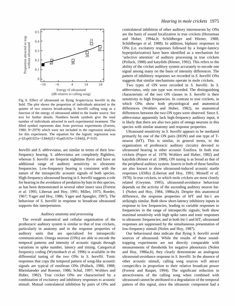
borellii and S. abbreviatus, are similar in terms of their low-frequency hearing. S. abbreviatusare completely flightless,whereas S. borellii are frequent nighttime flyers and have aadditional range of auditory sensitivity to ultrasonfrequencies. Low-frequency hearing is consistent with tnature of the intraspecific acoustic signals of both specHigh-frequency ultrasound hearing in S. borelliisuggests a rolefor hearing in the avoidance of predation by bats in this specas has been demonstrated in several other insect taxa (Foet al. 1995; Libersat and Hoy, 1991; Miller, 1975; Roede1967; Yager and Hoy, 1986; Yager and Spangler, 1997). behaviour of S. borellii in response to broadcast ultrasounsupports this interpretation.
Auditory anatomy and processing
The overall anatomical and cellular organisation of tprothoracic auditory system is similar to that of true crickeparticularly in anatomy and in the response propertiesauditory units that are specialized for intraspecicommunication. Omega neurons (ONs) are able to encodetemporal patterns and intensity of acoustic signals throuvariations in spike number, latency and timing. Categorifrequency coding (Wyttenbach et al. 1996) is available in thedifferential tuning of the two ONs in S. borellii. Tonicresponses that copy the temporal pattern of song-like acousignals are typical of ensiferan ONs (Pollack, 1986, 19Rheinlaender and Roemer, 1986; Schul, 1997; Wohlers Huber, 1982). True cricket ONs are characterised bycombination of excitatory and inhibitory responses to acousstimuli. Mutual contralateral inhibition by pairs of ONs an
nicheies.
ies,rrestr,
Thed
hets, offic thegh
cal
stic88;and aticd
contralateral inhibition of other auditory interneurons by ONare the basis of sound localization in true crickets (Horsemand Huber, 1994a,b; Schildberger and Hörner, 1988;Schildberger et al. 1988). In addition, biphasic responses inONs (i.e. excitatory responses followed by a longer-latenchyperpolarization) have been identified as a mechanism f‘selective attention’ of auditory processing in true cricket(Pollack, 1988) and katydids (Römer, 1992). This refers to thability of the cricket auditory system accurately to encode onsignal among many on the basis of intensity differences. Tpattern of inhibitory responses we recorded in S. borelliiONssuggests that similar mechanisms operate in mole crickets.
Two types of ON were recorded in S. borellii. In S.abbreviatus, only one type was recorded. The distinguishingcharacteristic of the two ON classes in S. borellii is theirsensitivity to high frequencies. In contrast to true crickets, iwhich ONs show both physiological and anatomicadifferences (Wohlers and Huber, 1982), no anatomicdifferences between the two ON types were observed. SinceS.abbreviatusapparently lack high-frequency auditory input, itis likely that there are also two pairs of omega neurons in thspecies with similar anatomy and response properties.
Ultrasound sensitivity in S. borellii appears to be mediatedprimarily by one of the ON pairs (HON) and one type of Tneuron (hfT). This is similar, in general terms, to theorganization of prothoracic auditory circuitry devoted toultrasound hearing in other acoustic Ensifera. In both trucrickets (Popov et al. 1978; Wohlers and Huber, 1982) andkatydids (Römer et al.1988), ON tuning is as broad as that ofthe peripheral auditory system. Insects in both of these familiare also known to show ultrasound-induced acoustic starresponses (ASRs) (Libersat and Hoy, 1991; Moiseff et al.1978). In true crickets, to which mole crickets are most closerelated (Gwynne, 1995), ultrasound-avoidance behaviodepends on the activity of the ascending auditory neuron In1 (Nolen and Hoy, 1984, 1986a,b). Despite this anatomicaldifference, the response properties of hfT and Int-1 astrikingly similar. Both show short-latency inhibitory inputs inresponse to low frequencies, leading to variable responsesfrequencies in the range of intraspecific signals; both shomaximal sensitivity with high spike rates and tonic responseto ultrasonic frequencies; and in both Int-1 and hfT, ultrasounresponses are suppressed by the simultaneous presentatiolow-frequency stimuli (Nolen and Hoy, 1987).
Our behavioural data indicate that flying S. borellii avoidsources of ultrasound. While the results of these sountrapping experiments are not directly comparable witmeasurements of thresholds for negative phonotaxis (Noland Hoy, 1986a,b), they clearly demonstrate an analogouultrasound-avoidance response in S. borellii. In the absence ofother acoustic stimuli, calling song sources will attracconspecifics in proportion to their relative broadcast powe(Forrest and Raspet, 1994). The significant reduction attractiveness of the calling song when combined witultrasound cannot be attributed to a degradation of the tempopattern of this signal, since the ultrasonic component had
he
Seetal
Theest,sisas
1976
offT.olered
olenicurrendck
A. C. MASON, T. G. FORREST ANDR. R. HOY
A B
C
D
90
40
50
60
70
80
100
Thr
esho
ld (
dB S
PL)
12
0
2
4
6
8
10
Num
ber
of s
pike
s
80
90
50
33
80
90
Inte
nsity
(dB
SP
L)
40
10
15
20
25
30
35
Late
ncy
(ms)
1 42 3 5 6 7 810Frequency (kHz)
10 30 50
Intensity (dB relative to threshold)
0 20 40 60Intensity (dB relative to threshold)
50 100 150 2000Latency (ms)
Fig. 10. Response characteristics ofomega neurons (ONs) in Scapteriscusabbreviatus. (A) Mean tuning curve(±S.D.) for ONs. (B) Mean intensity-response function for 3 kHz (N=7 cells,five repetitions averaged within cells).(C) Raster plot showing temporal patterncopying for 3 kHz stimuli at twointensities (80 and 90 dB) and pulse rates(50 and 33 pulses s−1). (D) Latency shiftfor variation in intensity of 3 kHz stimuli.Data points are means (N=5), and the linewas fitted with a power function.
4020 30 50 70
similar temporal pattern. Therefore, only the frequency contof the broadcast signal was altered. The attraction to the soof the song of proportionally fewer mole crickets as a functiof the power of the ultrasonic component of the signal indicaan aversive response competing with the attraction of calling song.
Our data do not directly demonstrate a link between tultrasound-avoidance behaviour and the responses of hfT, sas has been shown for Int-1 in true crickets (Nolen and H1984). However, the similarity between the responses of h
A
10 mV
25 ms
Fig. 11. Responses of the hfT cell inScapteriscus abbreviatusto 3 kHz (A) and20 kHz (B) stimulus frequency. Auditoryresponses are insensitive but show a similartime course to those of S. borellii. Low-frequency responses show a brief excitationfollowed by a pronounced IPSP. High-frequency responses are only excitatory.
enturceontesthe
hisuch
oy,fT
and those of Int-1 and the similar behavioural responsesmole crickets and true crickets suggest such a function for hUltrasound hearing has been demonstrated in other mcricket species that are nocturnal flyers. Suga (1968) measuauditory responses in the nerve cord of South American mcrickets. These responses were most sensitive to ultrasofrequencies (see Fig. 1 in Suga, 1968) and very similar to odata for hfT tuning (Fig. 6A). Suga further suggested that theare few ascending auditory units in the neck connectives, aonly one in the prothoracic–mesothoracic connective. Our ne
B
95 dB SPL
3 kHz 20 kHz
89 dB
95 dB
89 dB
86 dB 83 dB
1977Hearing in mole crickets
s.icne
s.d
enens.g
eg
inofal
orec
rees,l
ynnt
f
:
d
d
ll
connective recordings corroborate these results and sugthat hfT is the only source of ascending ultrasound activiAlso, the facts that single pulses of ultrasound, but not ausound, can elicit a delayed burst of activity in the neconnective and that this activity habituates are consistent wan ultrasound-mediated ASR in S. borellii (Brodfuehrer andHoy, 1989). In contrast, in a non-flying species, S. abbreviatus,high-frequency hearing is absent and hfT neurons show vweak auditory input, while auditory responses to lofrequencies are identical to those of S. borellii.
Evolution
On the basis of comparative morphology, it is probable thmole cricket omega neurons are homologous with thosepreviously studied Ensifera (Mason and Schildberger, 19Römer et al. 1988; Wohlers and Huber, 1982). Two pairs oONs are found in true crickets (Gryllidae; Wohlers and Hub1982) and haglids (Haglidae; Mason and Schildberger, 199Auditory responses have been studied in two other ensifetaxa, katydids (Tettigoniidae) and weta (StenopelmatidaOmega neurons are known from tettigoniids, but only a sinpair of ONs per individual has been described (Römer et al.1988; Schul, 1997). Peripheral auditory responses have brecorded in weta (Field et al.1980), but their central auditoryanatomy is unknown. True cricket ON1 responses are besuited than those of ON2 for a role in processing intraspecacoustic signals, and the function of ON2 is unclear (Horsemand Huber, 1994a,b; Schildberger and Hörner, 1988Schildberger et al. 1988; Wohlers and Huber, 1982). In thcase of S. borellii, both ON types respond similarly to songlike signals, and they differ only in their responses ultrasound. In S. abbreviatus, no functional differentiation ofONs was observed. Thus, we could not determine a clcorrespondence between the two ON subtypes of mole cricand true crickets. In some species of true crickets, howeON1 shows a secondary sensitivity peak at ultrasofrequencies which is similar to that of HON (Atkins anPollack, 1986).
It is more difficult to infer homology between neuronmediating ultrasound-avoidance behaviour in differeensiferan families, since these show more diversity than soprocessing elements. It is possible that hfT is homologousInt-1 of true crickets. In addition to similar acoustic responsthese two neurons show several anatomical similarities: soposition, the location of the midline-crossing segment, tposition of dendritic branches and the location of the ascendaxon in the anterior connective (Casaday and Hoy, 1977).
Regardless of the specific homologies, the diversity ultrasound-processing neurons in different ensiferan familraises interesting evolutionary questions. Hearing and acoucommunication in Ensifera existed prior to the evolution echolocating bats (Novacek, 1985; Otte, 1992; Sharov, 197Ultrasound hearing in relation to bat detection is, thereforesecondary adaptation of the ensiferan auditory system (H1992). Differences in the processing mechanisms associawith ultrasound avoidance in different ensiferan taxa m
gestty.diockith
eryw
at of93;f
er,3).rane).gle
een
tterifican
;e-to
earketsver,nicd
sntng- to
es,maheing
ofiessticof1)., aoy,ted
ay
reflect their independent evolution in already divergent groupGwynne (1995) argues that hearing and acoustcommunication evolved independently in the two maiensiferan clades: the Tettigonioidea, which includes thfamilies Tettigoniidae (katydids), Haglidae andStenopelmatidae (weta), and the Grylloidea, which includeGryllidae (true crickets) and Gryllotalpidae (mole crickets)This obviously implies an independent origin for ultrasounhearing in grylloids and tettigonioids, but within the grylloidsthe origins of ultrasound hearing in different families arunknown. More detailed study of the functional differences ithe organization of the ultrasound auditory pathway in trucrickets and mole crickets could address these questioIdentification of the destination and function of the descendinaxon of hfT in mole crickets would allow comparisons withboth true crickets and katydids. In addition, we havdemonstrated significant differences in ultrasound hearinbetween flying and flightless mole cricket species even withthe same genus. These results highlight the lability specialized sensory systems under varying ecologicconditions (Fullard et al. 1997). The variation among molecricket species we have described suggests that even mdirect tests of the function of identified neurons in conspecificommunication compared with predator avoidance apossible. These data, combined with phylogenetic analyscould help distinguish historical effects from functionaspecializations in the evolution of these sensory systems.
We thank Howard Frank for providing mole cricketspecimens, Jim Lloyd for providing accommodation and TomWalker for providing mole crickets, laboratory facilities andassistance in the field. This manuscript was improved bcomments from members of the Hoy laboratory and aanonymous reviewer. The work was supported by NIH graR01 DC00103 to R.R.H.
ReferencesATKINS, G. AND POLLACK, G. S. (1986). Age dependent occurrence o
an ascending axon on the omega neuron of the cricket, Teleogryllusoceanicus. J. comp. Neurol. 243, 527–534.
BAILEY , W. J. (1993). The tettigoniid (Orthoptera: Tettigoniidae) earmultiple functions and structural diversity. Int. J. Insect Morph.Embryol. 22, 185–205.
BALL , E. AND FIELD, L. H. (1981). Structure of the auditory system ofthe weta Hemideina crassidens(Blanchard 1851) (Orthoptera,Ensifera, Gryllacridoidea, Stenopelmatidae). I. Morphology anhistology. Cell Tissue Res. 217, 321–343.
BALL , E. E., OLDFIELD, B. P. AND RUDOLPH, K. M. (1989). Auditoryorgan structure, development and function. In Cricket Behavior andNeurobiology (ed. F. Huber, W. Loher and T. E. Moore), pp.391–422. Ithaca, London: Cornell University Press.
BENNET-CLARK, H. C. (1987). The tuned singing burrow of molecrickets. J. exp. Biol. 128, 383–410.
BENNET-CLARK, H. C. (1989). Songs and the physics of sounproduction. In Cricket Behavior and Neurobiology(ed. F. Huber,W. Loher and T. E. Moore), pp. 227–261. Ithaca, London: CorneUniversity Press.

1978
:
d
r
s
h
t
n
y
A. C. MASON, T. G. FORREST ANDR. R. HOY
BRODFUEHRER, P. D. AND HOY, R. R. (1989). Integration of ultrasoundand flight inputs on descending neurons in the cricket brain. J. exp.Biol. 145, 157–172.
CASADAY, G. B. AND HOY, R. R. (1977). Auditory interneurons in thecricket Teleogryllus oceanicus:physiological and anatomicalproperties. J. comp. Physiol.A 121, 1–13.
COKL, A., KALMRING, K. AND ROESSLER, W. (1995). Physiology ofatympanate tibial organs in forelegs and midlegs of the cave-livEnsifera, Troglophilus neglectus (Raphidophoridae,Gryllacridoidea). J. exp. Zool. 273, 376–388.
DAWS, A. G., BENNET-CLARK, H. C. AND FLETCHER, N. H. (1996). Themechanism of tuning of the mole cricket singing burrowBioacoustics 7, 81–117.
FIELD, L. H., HILL , K. G. AND BALL , E. E. (1980). Physiological andbiophysical properties of the auditory system of the New Zealaweta Hemideina crassidens (Blanchard 1851) (Ensifera:Stenopelmatidae). J comp. Physiol. A 141, 31–37.
FORREST, T. G. (1980). Phonotaxis in mole crickets: its reproductisignificance. Fla. Ent. 63, 45–53.
FORREST, T. G. (1983). Calling songs and mate choice in mocrickets. In Orthopteran Mating Systems(ed. D. T. Gwynne and G.K. Morris), pp. 185–204. Boulder, CO: Westview Press.
FORREST, T. G. (1986). Bioacoustics, maternal investment adevelopmental strategies in the mole crickets, Scapteriscus acletusand vicinus. PhD thesis, Department of Entomology anNematology, University of Florida, Gainesville.
FORREST, T. G. (1991). Power output and efficiency of sounproduction by crickets. Behav. Ecol. 2, 327–338.
FORREST, T. G., FARRIS, H. E. AND HOY, R. R. (1995). Ultrasoundacoustic startle response in scarab beetles. J. exp. Biol. 198,2593–2598.
FORREST, T. G. AND GREEN, D. M. (1991). Sexual selection and femalchoice in mole crickets (Scapteriscus: Gryllotalpidae) modellingthe effects of intensity and male spacing. Bioacoustics 3, 93–110.
FORREST, T. G. AND RASPET, R. (1994). Models of female choice inacoustic communication. Behav. Ecol. 5, 293–303.
FULLARD, J. H., DAWSON, J. W., OTERO, L.-D. AND SURLYKKE, A.(1997). Bat-deafness in day-flying moths (LepidopterNotodontidae, Dioptinae). J. comp. Physiol. A 181, 477–483.
GWYNNE, D. T. (1995). Phylogeny of the Ensifera (Orthoptera):hypothesis supporting multiple origins of acoustical signallincomplex spermatophores and maternal care in crickets, katyand weta. J. orthopt. Res. 4, 203–218.
HORSEMAN, G. AND HUBER, F. (1994a). Sound localisation in crickets.I. Contralateral inhibition of an ascending auditory interneur(AN1) in the cricket Gryllus bimaculatus. J. comp. Physiol. A 175,389–398.
HORSEMAN, G. AND HUBER, F. (1994b). Sound localisation in crickets.II. Modelling the role of a simple neural network in the prothoracganglion. J. comp. Physiol. A 175, 399–413.
HOY, R. R. (1992). The evolution of hearing in insects as an adaptato predation from bats. In The Evolutionary Biology of Hearing(ed.D. B. Webster, R. R. Fay and A. N. Popper), pp. 115–130. NYork: Springer-Verlag Inc.
HOY, R., NOLEN, T. AND BRODFUEHRER, P. (1989). The neuroethologyof acoustic startle and escape in flying insects.J. exp. Biol. 146,287–306.
JERAM, S., ROESSLER, W., COKL, A. AND KALMRING, K. (1995).Structure of atympanate tibial organs in legs of the cave-liviEnsifera, Troglophilus neglectus (Gryllacridoidea,Raphidophoridae). J. Morph. 223, 109–118.
ing
.
nd
ve
le
nd
d
d
e
a,
ag,dids
on
ic
tion
ew
ng
KALMRING, K., JATHO, M., ROESSLER, W. AND SICKMANN , T. (1997).Acousto-vibratory communication in bushcrickets (OrthopteraTettigoniidae). Ent. Gen. 21, 265–291.
LIBERSAT, F. AND HOY, R. R. (1991). Ultrasonic startle behavior inbushcrickets (Orthoptera, Tettigoniidae). J. comp. Physiol. A 169,507–514.
LIBERSAT, F., MURRAY, J. A. AND HOY, R. R. (1994). Frequency as areleaser in the courtship song of two crickets, Gryllus bimaculatus(de Geer) and Teleogryllus oceanicus: A neuroethological analysis.J. comp. Physiol. A 174, 485–494.
MASON, A. C. (1991). Hearing in a primitive ensiferan: the auditorysystem of Cyphoderris monstrasa (Orthoptera: Haglidae). J. Comp.Physiol. A. 168, 351–363.
MASON, A. C. AND SCHILDBERGER, K. (1993). Auditory interneuronsin Cyphoderris monstrosa(Orthoptera: Haglidae). J. comp.Physiol. A 171, 749–757.
MILLER, L. A. (1975). The behavior of green lacewings, Chrysopacamea, in the presence of ultrasound. J. Insect Physiol. 21,205–219.
MOISEFF, A., POLLACK, G. S. AND HOY, R. R. (1978). Steering responsesof flying crickets to sound and ultrasound: mate attraction anpredator avoidance. Proc. natn. Acad. Sci. U.S.A. 75, 4052–4056.
MORRIS, G. K., KLIMAS, D. E. AND NICKLE, D. A. (1988). Acousticsignals and systematics of false leaf katydids from Ecuado(Orthoptera, Tettigoniidae, Pseudophyllinae). Trans Am. ent. Soc.114, 215–264.
MORRIS, G. K., MASON, A. C., WALL , P. AND BELWOOD, J. J. (1994).High ultrasonic and tremulation signals in neotropical katydid(Orthoptera: Tettigoniidae). J. Zool., Lond. 233, 129–163.
NOLEN, T. G. AND HOY, R. R. (1984). Initiation of behavior by singleneurons: the role of behavioral context. Science 266, 992–994.
NOLEN, T. G. AND HOY, R. R. (1986a). Phonotaxis in flying crickets.I. Attraction to the calling song and avoidance of bat-likeultrasound are discrete behaviors. J. comp. Physiol. A 159,423–440.
NOLEN, T. G. AND HOY, R. R. (1986b). Phonotaxis in flying crickets.II. Physiological mechanisms of two-tone suppression of the higfrequency avoidance steering behavior by the calling song. J. comp.Physiol. A 159, 441–456.
NOLEN, T. G. AND HOY, R. R. (1987). Postsynaptic inhibition mediateshigh-frequency selectivity in the the cricket Teleogryllusoceanicus: implications for flight phonotaxis behavior. J. Neurosci.7, 2081–2096.
NOVACEK, M. J. (1985). Evidence for echolocation in the oldesknown bats. Nature 315, 140–141.
OTTE, D. (1992). Evolution of cricket songs. J. orthopt. Res.1, 25–48.POLLACK, G. S. (1986). Discrimination of calling song models by the
cricket Teleogryllus oceanicus: the influence of sound direction onneural encoding of the stimulus temporal pattern and ophonotactic behavior. J. comp. Physiol. A 158, 549–562.
POLLACK, G. S. (1988). Selective attention in an insect auditorneuron. J. Neurosci. 8, 2635–2639.
POPOV, A. V., MARKOVICH, A. M. AND ANDJAN, A. S. (1978). Auditoryneurons in the prothoracic ganglion of the cricket, Gryllusbimaculatus(DeGeer). I. The large segmental auditory neuron(LSAN). J. comp. Physiol. A 126,183–192.
RHEINLAENDER, J. AND ROEMER, H. (1986). Insect (Tettigoniaviridissima) hearing in the field. I. The use of identified nerve cellsas biological microphones. J. comp. Physiol. A 158, 647–652.
ROEDER, K. D. (1967). Nerve Cells and Insect Behavior.Cambridge:Harvard University Press.
1979Hearing in mole crickets
le
g
s
,
t,
e
RÖMER, H. (1992). Ecology and physiology of selective attention populations of the bushcrickets Tettigonia viridissima. In EighthInternational Meeting on Insect Sound and Vibration,Pommersfelden, Germany.
RÖMER, H., MARQUART, V. AND HARDT, M. (1988). Organization ofa sensory neuropil in the auditory pathway of two groups Orthoptera. J. comp. Neurol. 275, 201–215.
SAS INSTITUTE (1985). SAS/GRAPH User’s Guide, Version 5. Cary,NC: SAS Institute.
SCHILDBERGER, K. AND HÖRNER, M. (1988). The function of auditoryneurons in cricket phonotaxis. I. Influence of hyperpolarization identified neurons on sound localization. J. comp. Physiol. A 163,621–632.
SCHILDBERGER, K., MILDE, J. J. AND HÖRNER, M. (1988). The functionof auditory neurons in cricket phonotaxis. II. Modulation of auditorresponses during locomotion. J. comp. Physiol. A 163, 633–640.
SCHUL, J. (1997). Neuronal basis of phonotactic behaviour Tettigonia viridissima: processing of behaviourally relevant signalby auditory afferents and thoracic interneurons. J. comp. Physiol.A 180, 573–583.
SHAROV, A. G. (1971). Phylogeny of the Orthopteroidea. ITransactions of the Institute of Paleontology, Academy of ScienUSSR, vol. 118, pp. 251. Jerusalem: Israel Program for ScientiTranslation.
in
of
of
y
ins
ncesfic
SUGA, N. (1968). Neural responses to sound in a Brazilian mocricket. J. audit. Res. 8, 129–134.
ULAGARAJ, S. M. AND WALKER, T. J. (1973). Phonotaxis of cricketsin flight: attraction of male and female crickets to male callinsongs. Science 182, 1278–1279.
WALKER, T. J. (1982). Sound traps for sampling mole cricket flight(Orthoptera: Gryllotalpidae: Scapteriscus). Fla. Ent. 65, 105–110.
WALKER, T. J. AND FORREST, T. G. (1989). Mole cricket phonotaxis:effects of intensity of synthetic calling song (OrthopteraGryllotalpidae, Scapteriscus acletus). Fla. Ent. 72, 655–659.
WILKINSON, L., HILL , M., WELNA, J. P. AND BIRKENBEUEL, G. K.(1992). Systat: Statistics. Evanston, IL: Systat Inc. 750pp.
WOHLERS, D. W. AND HUBER, F. (1982). Processing of sound signalsby six types of neurons in the prothoracic ganglion of the crickeGryllus campestrisL. J. comp. Physiol. A 146, 161–173.
WYTTENBACH, R. A., MAY, M. L. AND HOY, R. R. (1996). Categoricalperception of sound frequency by crickets. Science 273,1542–1544.
YAGER, D. D. AND HOY, R. R. (1986). The cyclopean ear: a new sensfor the praying mantis Mantis religiosa. Science 231, 727–729.
YAGER, D. D. AND SPANGLER, H. G. (1997). Behavioral response toultrasound by the tiger beetle Cicindela marutha(Dow) combinesaerodynamic changes and sound production. J. exp. Biol. 200,649–659.