head & neck surgery - connecting repositories · dibella nj, see pearlman nw dolan kd: ......
TRANSCRIPT

HEAD & NECK SURGERY
INDEX FOR V O L U M E 5 I s s u e s 1 - 6 S e p t e m b e r / O c t o b e r 1 9 8 2 - J u l y / A u g u s t 1 9 8 3
A John Wiley & Sons Medical Publication

AUTHOR INDEX TO VOLUME 5
This index lists, in alphabetic order, the names of authors of all articles, letters, and editorials. Füll citation is provided under the first author only, with references made from Joint authors. Letters and editorials are distinguished from articles by the following code: L = letter, E = editorial.
A Allard RHB: The thyroglossal cyst, 134-
146 Angel MF, Stewart A , Pensak M L ,
Pillsbury H R C , Sasaki CT: Mechan-isms of hypercalcemia in patients with head and neck Cancer, 125-129
Appelblatt N H , McClatchey K D : Olfac-tory neuroblastoma: a retrospective clinicopathologic study, 108-113
B Baker H L , Papsidero M J , Batsakis J G ,
Krause C J : Aneurysmal bone cyst of the ethmoid, 177-180
Baker SR, see St. Pierre S Batsakis JG: The pathology of head and
neck tumors: the lymphoepithelial le-sion and Sjögren's Syndrome, part 16, 150-163
Batsakis J G , Hybels R, Crissman J D , Rice DH: The pathology of head and neck tumors: verrucous Carcinoma, part 15, 29-38
Batsakis J G , Kraemer B, Sciubba J J : The pathology of head and neck tumors: the myoepithelial cell and its participation in salivary gland neopla-sia, part 17, 222-233
Batsakis J G , Raymond A K , Rice DH: The pathology of head and neck tumors: papillomas of the upper aerodigestive tracts, part 18, 332-344
Batsakis J G , see Baker H L Berghaus A, see Meyer R Beumer J III, Harrison R, Sanders B,
Kurrasch M : Preradiation dental ex-tractions and the incidence of bone ne-crosis, 514-521
Beutter P, see Laccourreye H Bradley PJ: The complex nasal dermoid,
469-473 Brasnu DF, see Laccourreye H Brooks BS, see Coker N J
c Cantlon G E , Gluckman J L : Sterno-
clavicular Joint hypertrophy following radical neck dissection, 218-222
Carmody RF, see Miller RW Chan P Y M , see Hintz B L Coker N J , Brooks BS, E l Gammal T:
Computed tomography of orbital medial wall fractures, 383-389
Coulthard SW, see Miller RW Crissman JD, see Batsakis J G Crissman JD, see McDonald JS
D DiBella N J , see Pearlman NW Dolan K D : Paranasal sinus radiology,
part 2B: ethmoidal sinuses, 53-64 Dolan K D : Paranasal sinus radiology,
part 3A: sphenoidal sinus, 164-176 Dolan K D : Paranasal sinus radiology,
part 3B: sphenoidal sinus, 237-250 Dolan K D : see Khangure MS Dolan K D , Smoker WRK: Paranasal si
nus radiology, part 4A: maxillary sinuses, 345-362
Dolan K D , Smoker WRK: Paranasal sinus radiology, part 4B: maxillary sinuses, 428-446
Donegan J O , see Gluckman J L Donegan JO, see Myer C M III
E
E l Gammal T, see Coker N J
F Fabian RL, see Harris JP Fanous N: Expression plasty: a new ap-
proach to esthetic surgery, 306-318 Flanigin H , see Lang NP Flores L , see Hintz B L Fried MP, Horowitz Z, Kelly J H , Strome
M : The importance of the pedicle for the survival of a vascularized free Aap:
an experimental study on rats, 130-133
Fries R, see Platz H Fukuda O, see Kobayashi T
G Gerold FP, see Jun M Y Giammara B, see McDonald JS Gittot A , see Tovi F Gluckman J L , McDonough J , Donegan JO:
The free jejunal graft revisited 468-L
Gluckman J L , see Cantlon G E Gluckman J L , see McDonald JS Goldstein J , see Puterman M Goodman M L , see Granich MS Goodman M L , see Joseph MP Granich MS, Pilch BZ, Goodman M L :
Meningiomas presenting in the paranasal sinuses and temporal bone, 319-328
H Handler SD, see Rubinstein J B Harris JP, Fabian RL: Central island
myomucosal tongue Aap, 495-499 Harris JP, South M A : Immunodeficiency
diseases: head and neck manifesta-tions, 114-124
Harris T J , Hinckley D M : Melanoma of the head and neck in Queensland, 197-203
Harrison D F N : Reconstruction after pharyngoesophageal resection, 92 -L
Harrison R, see Beumer J III Harwick RD, see Kaplan IB Hinckley D M , see Harris T J Hintz B L , Kagan AR, Wollins M , Miles
J , Flores L , Nussbaum H , Rao AR, Chan P Y M , Ryoo M C : Local control of Tj vocal cord Cancer with radiation therapy: the importance of tumor character vs. treatment parameters, 204-210
Hokanson J A , see Leipzig B
A u t h o r I ndex H E A D & N E C K S U R G E R Y J u l / A u g 1983 i i i
Horowitz Z, see Fried M P Hudec M , see Platz H Hybels R, see Batsakis J G
J Jackson IT, Laws ER, Martin RD: A
craniofacial approach to advanced re-current Cancer of the central face, 474-488
Johnson FB, see Pearlman N W Joseph MP, Nadol J B , Pilch BZ, Good
man M L : Ectopic parathyroid tissue in the hypopharyngeal mucosa (pyriform sinus), 70-74
Jun M Y , Strong E W , Saltzman EI, Gerold FP: Head and neck Cancer in the elderly, 376-382
K Kagan AR, see Hintz B L Kaplan IB, Harwick RD: Pectoralis ma
jor myocutaneous island Aap revisited: a sentinel vessel simplifying dissec-tion, 452-456
Kelly J H , see Fried M P Kendrick J H , see Lang N P Khangure MS, Dolan K D : High resolu-
tion C T air cisternography in the diag-nosis of small acoustic neuromas, 489-494
Kikawada T, see Kobayashi T Kobayashi T, Kikawada T, Shima K ,
Fukuda O: Ulceration and Stenosis of the hypopharynx and its surgical man-agement, 65-69
Koopmann CF, see Miller RW Kraemer B, see Batsakis J G Krause CJ , see Baker H L Kurrasch M , see Beumer J III
L Laccourreye H , Brasnu DF, Beutter P:
Carcinoma of the laryngeal margin, 500-507
Lam K H : Extralaryngeal spread of Cancer of the larynx: a study of whole Organ sections, 410-424
Lang NP, Kendrick J H , Flanigin H , Wetmore SJ, Suen J Y , Westbrook K C : Surgical management of advanced scalp Cancer, 299-305
Laws ER, see Jackson IT Leipzig B, Hokanson J A : Treatment of
cervical lymph nodes in Carcinoma of the tongue, 3-9
Lore J M , Jr, Pruet CW: Retrieval of the parathyroid glands during thyroidec-tomy, 268-269
M MacDonald DG, see McGregor IA Makek MS, see Obwegeser H L Martin RD, see Jackson IT McClatchey K D , see Appelblatt N H McCurdy J A Jr: A n approach to face-lift
surgery, 211-217 McDonald JS, Crissman JD, Gluckman
J L : Verrucous Carcinoma of the oral cavity, 22-28
McDonald JS, Miller R L , Wagner W, Giammara B: Acral lentiginous mel-anoma of the oral cavity, 257-262
McDonough J , see Gluckman J L McElhinney A J , see Weitz JW McGregor IA, MacDonald DG: Mandibu
lar osteotomy in the surgical approach to the oral cavity, 457-462
Meyer R, Berghaus A: Closure of perfo-rations of the septum including a sin-gle-session method for large defects, 390-400
Meyers A D , see Pearlman NW Miles J , see Hintz B L Miller RL, see McDonald JS Miller RW, Carmody RF, Seeger JS,
Coulthard SW, Smith J R L , Koopmann CF: Digital subtraction angiography: applications in otolaryngology—head and neck surgery, 280-292
Myer C M III, Donegan JO: Traumatic aneurysm of the proximal superficial temporal artery, 181-185
N Nadol JB , see Joseph M P Nahum A M : Cancer of the larynx: pat-
terns of spread, 375-E Nahum A M : Dental extractions and
radiation therapy, 467-E Nahum A M : Head and neck cancer:
management of early lesions, 1 - 2 - E Nahum A M : Head and neck cancer in
young black patients, 279-E Nahum A M : The incidence of melanoma
in Queensland is the highest in the world, 197-E
Nahum A M : When run-of-the-mill Symptoms fail to respond, 9 1 - E
Nussbaum H , see Hintz B L
o Obwegeser H L , Makek MS: Benign
lipoblastoma in the mandible, 251-256
P Papsidero M J , see Baker H L Pearlman NW, Meyers A D , Johnson F B ,
DiBella NJ: Preoperative chemo-radiotherapy in advanced head and neck cancer, 10-14
Pensak M L , see Angel M F Pilch BZ, see Granich MS Pilch BZ, see Joseph M P Pillsbury H R C , see Angel M F Platz H , Fries R, Hudec M , Tjoa A M ,
Wagner RR: Carcinomas of the oral cavity: analysis of various pre-therapeutic classifications, 93-107
Pruet CW, see Lore J M Jr Puterman M , Goldstein J : Primary
lymph nodal Kaposi's sarcoma of the parotid gland, 535-538
R Rankin K V , see Wright J M Rao AR, see Hintz B L Raymond A K , see Batsakis J G Rice D H , see Batsakis J G Rubinstein J B , Handler SD: Orbital and
periorbital cellulitis in children, 15-21
Rush B F Jr , see Slotman G J Ryoo M C , see Hintz B L
S Saltzman EI: Aneurysmal bone cyst of
the ethmoid, 468-L Saltzman EI, see Jun M Y Sanders B, see Beumer J III Sasaki CT, see Angel M F Sciubba J J , see Batsakis J G Seeger JS, see Miller RW Sessions DG: Recent advances in surgery
of the larynx and trachea, 42-52 Shima K, see Kobayashi T Slotman G J , Swaminathan A P , Rush B F
Jr: Head and neck cancer in a young age group: high incidence in black patients, 293-298
Smith AR, van Urk H , Vaandrager M , van der Meulen J C : Treatment of a large hemangioma in the head and neck region, 263-267
Smith J R L , see Miller RW Smith K F , see Webster RC Smith RC, see Webster RC Smoker WRK, see Dolan K D South M A , see Harris J P Stewart A , see Angel M F St. Pierre S, Baker SR: Squamous cell
Carcinoma of the maxillary sinus: analysis of 66 cases, 508-513
Strome M , see Fried MP Strong EW, see Jun M Y Suen J Y , see Lang NP Swaminathan AP, see Slotman G J
T Tjoa A M , see Platz H Tovi F, Gittot A: Sternocleidomastoid
myoperiosteal Aap for the repair of laryngeal and tracheal wall defects, 447-451
V Vaandrager M , see Smith A R van der Meulen J C , see Smith A R van Urk H , see Smith A R
w Wagner RR, see Platz H Wagner W, see McDonald JS Webster RC, Smith RC, Smith K F : Face
lift, part 1: extent of undermining of skin Aaps, 525-534
Weitz JW, Weitz SL, McElhinney A J : A technique for preservation of spinal accessory nerve function in radical neck dissection, 75-78
Weitz SL, see Weitz J W
i v A u t h o r I n d e x H E A D & N E C K S U R G E R Y J u l / A u g 1983

Westbrook K C , see Lang NP Wetmore SJ, see Lang N P Wilson JW, see Wright J M Wollins M , see Hintz B L Wright J M , Rankin K V , Wilson JW:
Traumatic granuloma of the tongue, 3 6 3 - 3 6 6
Au tho r I n d e x H E A D & N E C K S U R G E R Y J u l / A u g 1 9 8 3 v
SUBJECT INDEX TO VOLUME 5
This index gives the first author (in parentheses) and first page of the article, letter, or editorial in which the indexed subject occurs. The reader is referred to the author index for the füll title and coauthors, where appropriate, of the piece. Letters and editorials are distinguished from articles by the following code: L = letter, E = editorial.
A Acoustic neuromas
high resolution computed tomography air cisternography in the diagnosis of small acoustic neuromas (Khan-gure) 489
Aneurysm, traumatic of the proximal superficial temporal
artery (Myer) 181 Angiography, digital subtraction
applications in otolaryngology—head and neck surgery (Miller) 280
Artery, superficial temporal traumatic aneurysm of (Myer)181
B B C G cell-wall preparation
for bovine ocular Carcinoma (Kleinschuster) 401
Black patients high incidence of head and neck cancer
in a young age group (Slotman) 293 Bone cyst, aneurysmal
of the ethmoid (Baker) 177 Book reviews
Ackerman and D e l Regato's Cancer: Diagnosis, Treatment and Progno-sis, 5th Edition (del Regato and Spjut) 545
Aesthetic Plastic Surgery, Volumes I and II (Rees) 372-373
Atlas of C r a n i o m a x i l l o f a c i a l Surgery (Jackson, Munro, Salyer, and Whit-aker) 87
Atlas of Sectional H u m a n Anatomy, V o l u m e 1: H e a d , Neck, T h o r a x (Kor-tike and Sick) 545
Atlas of Sectional H u m a n Anatomy, V o l u m e 2: Abdomen, Pelvis (Kortike and Sick) 545
Basics of Dermatologie Surgery (Steg-man, Tromovitch, and Glogau) 546
Cancer: A Comprehensive Treatise, V o l u m e 1, Etiology: C h e m i c a l and Physical Carcinogenesis, 2nd Edition (Becker, ed) 545
C l i n i c a l and Radiographic I n t e r p r e t a tion of F a c i a l F r a c t u r e s (Gerlock, Sinn, and McBride) 373
Color Atlas of H e a d and Neck Anatomy (McMinn, Hutchings, and Logan) 193
Color Atlas of H u m a n Anatomy (McMinn and Hutchings) 193
A Colour Atlas oföral Cancers (Burkhardt and Maerker) 86-87
Colour Atlas o f O r a l M e d i c i n e (Tyldes-ley) 86-87
T h e Cover-Up: Neckwear for the L a r y n -gectomee and Other Neck Breathers (Kelly and Welborn) 194-195
Disorders of the F a c i a l Nerve: A n a tomy, Diagnosis and M a n a g e m e n t (Graham and House, eds) 86
H e a d and Neck Surgery. F a c e and F a c i a l S k u l l , Volumes 1 and 2 (Naumann, ed) 86
Microscopic and Endoscopic Surgery with the C02 Laser (Andrews and Polanyi, eds) 372
M o d e r n Technics i n Surgery. H e a d and Neck Surgery, I n s t a l l m e n t 1 (Nussbaum, ed) 275
Oculoplastic Surgery (McCord, ed) 275 Operative Surgery: H e a d and Neck,
Parts I and I I , 3rd Edition (Rob and Smith, eds) 194
Psychosocial Aspects of Cancer (Cohen, Cullen, and Martin, eds) 193-194
Radiology o f t h e E a r , Nose, and T h r o a t (Valvassori, Potter, Hanafee, et al.) 276
Spastic Dysphonia: A S u r g i c a l and Voice Therapy Treatment P r o g r a m (Dedo and Shipp) 193
Surgery for Cancer of the L a r y n x and Related Structures (Silver) 87
c Cancer (see also Carcinoma; lesions;
melanoma; neoplasms; tumors)
of the central face, advanced recurrent (Jackson) 474
Cancer, head and neck early management (Nahum) 1-E in the elderly (Jun) 376 mechanisms of hypercalcemia in pa
tients with (Angel) 125 pathology of verrueous Carcinoma
(Batsakis) 29 preoperative chemo-radiotherapy in
advanced tumors (Pearlman) 10 in a young age group, high incidence
in black patients (Slotman) 293 in young black patients (Nahum) 279-E
Cancer, larynx extralaryngeal spread (Lam) 410 patterns of spread (Nahum) 375-E
Cancer, scalp advanced, surgical management of
(Lang) 299 Cancer, vocal cord
local control with radiation therapy (Hintz) 204
Carcinoma (see also cancer; lesions; melanoma; neoplasms; tumors) of the laryngeal margin (Laccourreye)
500 Carcinoma, bovine ocular
intratumoral B C G cell wall preparation therapy and surgery for (Kleinschuster) 401
Carcinoma, oral cavity analysis of various pretherapeutic
Classification (Platz) 93 Carcinoma, squamous cell
of the maxillary sinus (St. Pierre) 508 Carcinoma, tongue
treatment of cervical lymph nodes (Leipzig) 3
Carcinoma, verrueous of the oral cavity (McDonald) 22
Cellulitis in children, orbital and periorbital
(Rubinstein) 15 Chemo-radiotherapy
preoperative, in advanced head and neck cancer (Pearlman) 10
v i S u b j e c t I n d e x H E A D & N E C K S U R G E R Y J u l / A u g 1983
Children (see pediatrics) Computed tomography
high resolution air cisternography in the diagnosis of small acoustic neuromas (Khangure) 489
of orbital medial wall fractures (Coker) 383
C T (see computed tomography) Cysts
aneurysmal bone, of the ethmoid (Baker) 177
thyroglossal (Allard) 134
D Dental extractions
before radiation therapy, incidence of bone necrosis (Beumer) 514
Dermoid, nasal (Bradley) 469 Diagnostic techniques (see computed
tomography)
E Expression plasty
a new approach to esthetic surgery (Fanous) 306
F Face lift surgery (McCurdy) 211
extent of undermining of skin flaps (Webster) 525
Flaps central island myomucosal tongue
(Harris) 495 free, the importance of the pedicle for
the survival of a study on rats (Fried) 130
pectoralis major myocutaneous island (Kaplan) 452
sternocleidomastoid myoperiosteal, for repair of laryngeal and tracheal wall defects (Tovi) 447
Fractures of the orbital medial wall, computed
tomography of (Coker) 383
G Geriatrie patients
head and neck cancer in (Jun) 376 Gland, parotid
primary lymph nodal Kaposi's sar-coma of (Puterman) 535
Glands, parathyroid retrieval during thyroidectomy (Lore)
268 Granuloma, traumatic
of the tongue (Wright) 363
H Hemangioma
of the head and neck (Smith) 263 Hypercalcemia
mechanisms in patients with head and neck cancer (Angel) 125
Hypertrophy of the sternoclavicular Joint following
radical neck dissection (Cantlon) 218
Hypopharynx surgical management for ulceration
and Stenosis of (Kobayashi) 65
I Immunodeficiency disease (Nahum)
91-E head and neck manifestations (Harris)
114
Irradiation (see radiation therapy)
K Kaposi's sarcoma
primary lymph nodal of the parotid gland (Puterman) 535
L Laryngeal margin
Carcinoma of (Laccourreye) 500 Larynx
advances in surgery of (Sessions) 42 cancer of, extralaryngeal spread (Lam)
410 cancer of, patterns of spread (Nahum)
375-E repair of, using sternocleidomastoid
myoperiosteal Aap (Tovi) 447 Lesions
lymphoepithelial, pathology of (Batsakis) 150
pathology of papillomas of the Upper aerodigestive tracts (Batsakis) 332
Lipoblastoma, benign in the mandible (Obwegeser) 251
Lymph nodes treatment in Carcinoma of the tongue
(Leipzig) 3
M Mandible
benign lipoblastoma of (Obwegeser) 251
osteotomy in the surgical approach to the oral cavity (McGregor) 457
Melanoma of the head and neck (Nahum) 197-E of the head and neck, incidence in
Queensland (Harris) 197 of the oral cavity (McDonald) 257
Meningioma of the paranasal sinuses and temporal
bone (Granich) 319
N Nasal dermoid (Bradley) 469 Neck dissection
radical, preservation of spinal acces-sory nerve funetion in (Weitz) 75
Necrosis, bone and preradiation dental extractions
(Nahum) 467-E
preradiation dental extractions and the incidence of (Beumer) 514
Neoplasms (see also cancer; Carcinoma; lesions; tumors) meningioma of the paranasal sinuses
and temporal bone (Granich) 319 lipoblastoma in the mandible (Ob
wegeser) 251 Neoplasms, salivary gland
pathology of (Batsakis) 222 Neuroblastoma, olfactory
a retrospective clinicopathologic study of (Appelblatt) 108
o Olfactory neuroblastoma
a retrospective clinicopathologic study of (Appelblatt) 108
Oncology (see cancer; Carcinoma; lesions; neoplasms; tumors)
Oral cavity acral lentiginous melanoma of
(McDonald) 257 Carcinoma, analysis of pretherapeutic
classifications (Platz) 93 surgical approach using mandibular
osteotomy (McGregor) 457 verrueous Carcinoma of (McDonald) 22
Orbit computed tomography of fractures of
the medial wall of (Coker) 383 Osteonecrosis (see necrosis, bone)
P Papillomas
of the upper aerodigestive tracts, pathology of (Batsakis) 332
Parathyroid tissue ectopic, in the hypopharyngeal mueosa
(pyriform sinus) (Joseph) 70 Pathology
of the lymphoepithelial lesion (Batsakis) 150
of papillomas of the upper aerodigestive tracts (Batsakis) 332
of salivary gland neoplasia (Batsakis) 222
of Sjögren's Syndrome (Batsakis) 150 of verrueous Carcinoma (Batsakis) 29
Pediatrics orbital and periorbital cellulitis
(Rubinstein) 15 Pedicle
importance for the survival of vas-cularized free flaps, a study on rats (Fried) 130
Pharyngoesophageal resection reconstruetion after (Harrison) 92-L
Plastic surgery an approach for face lifts (McCurdy)
211 expression plasty (Fanous) 306 face lift, extent of undermining of skin
flaps (Webster) 525
R Radical neck dissection
sternoclavicular Joint hypertrophy fol-
S u b j e c t I n d e x H E A D & N E C K S U R G E R Y J u l / A u g 1983 v i i
lowing (Cantlon) 218 Radiation therapy
after dental extractions, the incidence of bone necrosis (Beumer) 514
and bone necrosis after dental extractions (Nahum) 467-E
for vocal cord cancer (Hintz) 204 Radiology (see also computed tomog
raphy) of the ethmoidal sinuses, Part 2B
(Dolan) 53 of the maxillary sinuses, Part 4A
(Dolan) 345 of the maxillary sinuses, Part 4B
(Dolan) 428 of the sphenoidal sinuses, Part 3A
(Dolan) 164 of the sphenoidal sinuses, Part 3B
(Dolan) 237 Radiotherapy (see radiation therapy; see
also chemo-radiotherapy) Reconstruction
after pharyngoesophageal resection (Harrison) 92-L
S Salivary gland
neoplasia, pathology of (Batsakis) 222 Scalp
advanced cancer of, surgical management (Lang) 299
Septum perforations of, closure with a single-
session method (Meyer) 390 Sinuses, ethmoidal
radiology of, Part 2B (Dolan) 53 radiology of, Part 4A (Dolan) 345
radiology of, Part 4B (Dolan) 428 squamous cell Carcinoma of (St.
Pierre) 508 Sinuses, paranasal
meningiomas of (Granich) 319 Sinuses, pyriform
ectopic parathyroid tissue in (Joseph) 70
Sinuses, sphenoidal radiology of, Part 3A (Dolan) 164 radiology of, Part 3B (Dolan) 237
Sjögren's Syndrome pathology of (Batsakis) 150
Sternoclavicular joints hypertrophy following radical neck
dissection (Cantlon) 218 Surgical management
of advanced scalp cancer (Lang) 299 of ulceration and Stenosis of the hy-
popharynx (Kobayashi) 65 Surgical techniques (see also plastic
surgery; reconstruction; surgical management; surgery) closure of perforations of the septum
(Meyer) 390 a craniofacial approach to advanced
recurrent cancer of the central face (Jackson) 474
mandibular osteotomy in the surgical approach to the oral cavity (McGregor) 457
pectoralis major myocutaneous island Aap (Kaplan) 452
for the preservation of spinal accessory nerve function in radical neck dissection (Weitz) 75
Surgery (see also plastic surgery; reconstruction; surgical management; sur
gical techniques) for bovine ocular Carcinoma (Klein
schuster) 401 esthetic expression plasty (Fanous)
306 face-lift technique (McCurdy) 211 of the larynx and trachea, advances in
(Sessions) 42
T Temporal bone
meningioma of (Granich) 319 Thyroglossal cysts (Allard) 134 Thyroidectomy
parathyroid gland retrieval during (Lore) 268
Tongue Carcinoma, lymph node treatment
of (Leipzig) 3 traumatic granuloma of (Wright) 363
Trachea advances in surgery of (Sessions) 42 repair using sternocleidomastoid my
operiosteal Aap (Tovi) 447 Tumors (see also cancer; Carcinoma; le
sions; neoplasms) hemangioma of the head and neck
(Smith) 263
V Vocal cords
cancer of (Hintz) 204
X X-ray techniques (see computed tomog
raphy; radiology)
v i i i S u b j e c t I n d e x H E A D & N E C K S U R G E R Y J u l / A u g 1983
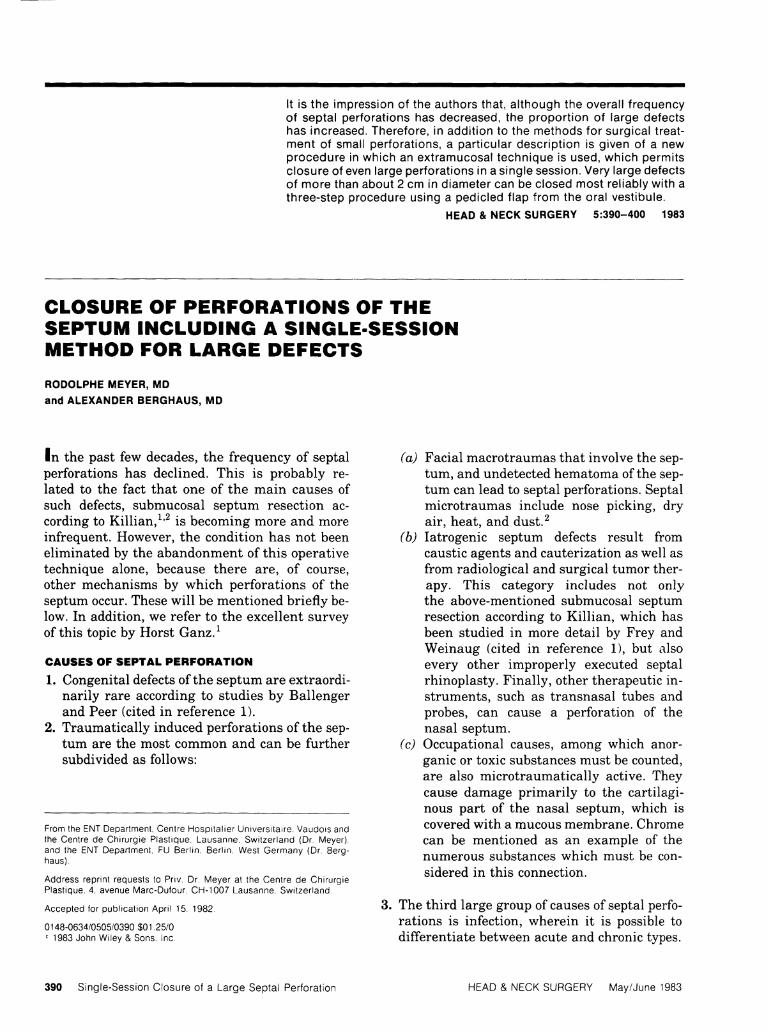
It is the impression of the authors that, a l though the overall f requency of septal perforations has decreased, the proport ion of large defects has increased. Therefore, in addit ion to the methods for surgical treatment of small perforations, a particular descr ipt ion is given of a new procedure in which an extramucosal technique is used, which permits c losure of even large perforations in a Single Session. Very large defects of more than about 2 cm in diameter can be closed most reliably with a three-step procedure using a pedicled flap f rom the oral vestibule.
H E A D & N E C K S U R G E R Y 5 : 3 9 0 - 4 0 0 1 9 8 3
CLOSURE OF PERFORATIONS OF THE SEPTUM INCLUDING A SINGLE-SESSION METHOD FOR LARGE DEFECTS R U D O L P H E M E Y E R , M D
a n d A L E X A N D E R B E R G H A U S , M D
In the past few decades, the frequency of septal perforations has declined. This is probably related to the fact that one of the main causes of such defects, submucosal septum resection ac-cording to Ki l l ian, 1 , 2 is becoming more and more infrequent. However, the condition has not been eliminated by the abandonment of this operative technique alone, because there are, of course, other mechanisms by which perforations of the septum occur. These will be mentioned briefly be-low. In addition, we refer to the excellent survey of this topic by Horst Ganz. 1
CAUSES OF SEPTAL PERFORATION 1. Congenital defects of the septum are extraordi-
narily rare according to studies by Ballenger and Peer (cited in reference 1).
2. Traumatically induced perforations of the septum are the most common and can be further subdivided as follows:
From the ENT Department, Centre Hospital ier Universitaire. Vaudois and the Centre de Chirurgie Plastique, Lausanne. Switzerland (Dr. Meyer), and the ENT Department, FU Berl in. Berl in. West Germany (Dr. Berghaus).
Address reprint requests to Priv. Dr. Meyer at the Centre de Chirurgie Plastique. 4, avenue Marc-Dufour, CH-1007 Lausanne. Switzerland.
Accepted for publ icat ion Apri l 15. 1982
0148-0634/0505/0390 $01.25/0 ' 1983 John Wiley & Sons. Inc.
( a ) Facial macrotraumas that involve the septum, and undetected hematoma of the septum can lead to septal perforations. Septal microtraumas include nose picking, dry air, heat, and dust.2
(b) Iatrogenic septum defects result from caustic agents and cauterization as well as from radiological and surgical tumor therapy. This category includes not only the above-mentioned submucosal septum resection according to Killian, which has been studied in more detail by Frey and Weinaug (cited in reference 1), but also every other improperly executed septal rhinoplasty. Finally, other therapeutic in-struments, such as transnasal tubes and probes, can cause a Perforation of the nasal septum.
(c) Occupational causes, among which anor-ganic or toxic substances must be counted, are also microtraumatically active. They cause damage primarily to the cartilagi-nous part of the nasal septum, which is covered with a mucous membrane. Chrome can be mentioned as an example of the numerous substances which must be con-sidered in this connection.
3. The third large group of causes of septal perforations is infection, wherein it is possible to differentiate between acute and chronic types.
3 9 0 S i n g l e - S e s s i o n C l o s u r e of a L a r g e S e p t a l P e r f o r a t i o n H E A D & N E C K S U R G E R Y M a y / J u n e 1983

(a) Among the acute infections, Perforation of the septum was most commonly caused in former times by scarlet fever (as a result of gangrene), followed by diphtheria, and, in rarer instances, typhus. More common now is the "banal" abscess of the septum, which, in principle, can occur as a result of any manipulation of the septum.
(6) Among the chronic infections, the so-called rhinitis sicca anterior (ulcus septi perfo-rans) must be given particular mention. Heat, dry air, dust and other primarily physical agents of damage are seen as its causes.2 Lues III leads to a connatal or in-herited Perforation, most commonly in the bony part of the nasal septum. It must be noted here that the saddle nose, which is a typical and well-known connatal phenome-non associated with lues, does not ordinar-ily appear until the third year of life. In contrast to the Situation in lues, the bony septum is generally not affected in tuber-culosis, which appears especially as a mucosal lupus of the nose.1 Still to be mentioned is leprosy, which, especially in the lepromatous form, leads to a final stage similar to ozena.
4. Metabolie disorders, such as diabetes mellitus, for instance, are rare causes of perforations of the septum.
5. Tumors also frequently lead to such defects. The therapeutic measures applied in case of a "bleeding polyp" in Kiesselbach's area can eas-ily lead to Perforation.
SYMPTOMS OF SEPTAL PERFORATION The indication for closure of such defects depends on the Symptoms that trouble the patients. The holes in the rear bony part of the septum can usu-ally remain untreated because they do not cause any inconvenience. On the other hand, Symptoms worth mentioning as typical for perforations of the forward cartilaginous part of the septum are: itching; scab formation with impairment of breathing; putrid fetor and dryness of the nose; and recurrent epistaxis. Whistling noises can occur during breathing and even lead to difficulty in falling asleep; however, they occur only in defects that are located well forward, are very small, or have been reduced in size by scab formation.1 Oc-casionally a septum Perforation is bothersome because it is visible, especially, of course, when it leads to a deformity of the columella.
Of the Symptoms mentioned as indications for surgery, particular emphasis should be placed on intense scab formation, recurrent bleeding, whistling noises, and deformity of the nose. Of course, a fresh traumatic Perforation requires im-mediate treatment.
TREATMENT METHODS Basically, the following choices of treatment are available.
1. Conservative therapy. Such treatment, which consists of the application of salves, etc., is in-dicated when an Operation is contraindicated or refused by the patient.
2. Closure with obturators. This method, first proposed by Meyer and, independently, by Link in 1951, and later on by Papangelou (1969) and Von Dishoeck (1975), in which a button consisting of two plastic dises glued to-gether in the middle is inserted into the defect, has been abandoned by us at the present time in favor of surgical procedures.3"6
3. Surgical enlargement of the defect. Although Ganz 1 lists some indications for this procedure, we see this measure at best as a last resort in case of a whistling defect, especially in a case in which the patient refuses a surgical closure. In this regard, it must be pointed out that surgical enlargement of the defect can give rise to new complaints, such as subsequent spontane-ous increase in the size of the Perforation, bleeding and fetor. As a matter of principle, we regard this method with extreme reservation.
4. Surgical closure of the Perforation. In cases with the above-mentioned clinical Symptoms, a total closure of the septal Perforation will always be the goal to be strived for, and the surgical procedure chosen will be partly deter-mined by the size of the defect.
SURGICAL TECHNIQUES
The extramucosal technique that we have been using routinely for years in all rhinoplasties7 is thereby so extraordinarily helpful that the procedure we used in previous years, in which local flaps were used for closure of small and medium sized perforations, is made superfluous in routine cases. This method, which we abandoned about five years ago, consisted in principle of mueo-perichondrial flaps, one of which was cut from the area above the Perforation, and one from the op-posite side in the area below the Perforation, and which were then pushed into place covering the
S i n g l e - S e s s i o n C l o s u r e of a L a r g e S e p t a l Pe r fo ra t i on H E A D & N E C K S U R G E R Y M a y / J u n e 1983 3 9 1

A B
Figure 1. (A) Type of incision used for small perforations (B) .
hole. Good reviews of such techniques can be found in Ganz1 and Masing.2
In contrast, we now close small, medium sized, and even large perforations of the septum using variations of the extramucosal technique de-signed to fit the individual Situation.
Small Defects . In closing small defects, it is sufficient to mobilize a large area of the muco-perichondrium coupled with the use of adaptation sutures free of tension. To make this possible, the transfixion incision is first extended downward running parallel to the lower edge of the apertura piriformis along the floor of the nose to the meatus nasi inferior and then upward somewhat under the concha inferior on the lateral nasal wall. On the other hand, an extension of the incision upward and dorsal leads beneath the roof of the nose to the lower edge of the upper lateral cartilage and then along this to the area of the limen nasi, so that this incision corresponds to the intercartilaginous incision (Fig. 1).
Proceeding from this incision, the mucoperi-chondrium is separated from the septal cartilage over a large area. Then, the mucoperiosteum of the nasal floor is also detached as far as the lower nasal meatus. Finally, the external skin over the upper lateral cartilage is mobilized. This can now be separated at its base in füll length from the cartilaginous septum so that the result of the procedure up to this point is a hose-like structure
consisting of mucoperiosteum and mucoperichon-drium with the attached upper lateral cartilage. The Perforation located therein has collapsed due to the elimination of tension.
The same principle is followed in the other nasal cavity. To mobilize the mucoperiosteum, an incision can be made along the lower edge of the lateral nasal wall extending deep into the bony cavity, but the anterior ethmoidalis artery must not be damaged. The loosely adjoining edges of the perforations in the mucoperiosteum can then be freshened and, following adaptation, sutured.
To reduce the size of the actual cartilage defect in the lamina quadrangularis, we basically use two different methods, depending on the location and nature of the Perforation. If the hole does not lie too far to the rear, below and with its greatest diameter perpendicular to an imaginary line on the floor of the nose, we perform resections of cartilage strips above and below the edges of the Perforation parallel to its largest diameter (Fig. 2). As a result of this procedure, the lamina quadrangularis is divided into two parts: the forward part can be fully mobilized and pushed backward and upward against the second part (Fig. 3). This alone effects a decrease in the size of the Perforation, which can be further closed by inserting a piece of the resected cartilage. To avoid recession of the columella, the other strip of cartilage can be attached to the forward edge of the septum. If the largest diameter of the Perforation is parallel
3 9 2 S i n g l e - S e s s i o n C l o s u r e o f a L a r g e S e p t a l Pe r fo ra t i on H E A D & N E C K S U R G E R Y M a y / J u n e 1983

A B
Figure 2 (A and B). Strip resection for reducing the size of a small defect.
to an imaginary sagittal line at the floor of the nose, then it is possible, using the same procedure, to remove strips of cartilage in front of and behind the Perforation, lengthening the max-imum diameter. In this way, an upper septal plate is separated from a lower. The upper plate can be pushed down onto the second half after mobilization at the dorsum of the nose. The Perforation, which becomes smaller due to this procedure, can now be closed further by inserting a strip of cartilage. It must be remembered that, in this procedure, it is also necessary to lower the bony framework of the nose.
Figure 3. Reduction of the size of the defect by displacement of the septum cartilage and closure of the Perforation in the mucoperiosteum.
S i n g l e - S e s s i o n C l o s u r e of a L a r g e S e p t a l P e r f o r a t i o n
If the Perforation is located lower and farther back (which is more rare) so that the procedure described cannot be used, another method can be helpful for closure of the cartilaginous defect. With this method, after removal of a small strip of cartilage between the lower edge of the Perforation and the crista maxillaris or vomer, the lamina quadrangularis and the mucoperichondrium are rotated into or across the defect from both sides (Fig. 4). This requires a rotation of the mucoperichondrium and the triangulär cartilage on both sides toward the front and downward, whereby the forward lower edge of the upper lat-
Figure 4. Rotation of the mucoperichondrium.
H E A D & N E C K S U R G E R Y M a y / J u n e 1 9 8 3 3 9 3

Figure 5. Closure of the Perforation; the anterior edge of the upper lateral cartilage is shortened.
eral cartilage now reaches into the nasal cavity and becomes bothersome because it decreases the size of the lumen. Therefore, the upper lateral cartilage should be clearly shortened at the forward edge on both sides (Fig. 5). Thereafter, the mobilized and newly relocated flaps are attached with mattress sutures to the inner lining of the nose and additionally supported by tamponade.
For all the procedures mentioned, the access, especially for suturing, can be eased by separat-ing one or both of the wings of the nose, and possi-bly the columella as well, from the skin of the lip and cheek and temporarily folding them upward and backward (Fig. 6). At the end of the Operation, they can be folded back down and sewn in place. If suturing is done carefully, the results of such auxiliary incisions are usually not visible.
Defects up to 2 c m . The extramucosal technique is especially valuable in the treatment of perforations of up to 2 cm in diameter because it permits their closure in a single Session. In contrast to the total number of septal defects, these large perforations have become relatively more common in the past few years. Therefore, it seemed of particular interest to work out a single-session method for closure of such perforations.
To do justice to the unusual extent of such perforations, we begin the Operation in this case on one side with an incision, the course of which cor-responds to that of the hemitransfixion incision, but Starts at a substantially greater distance from the edge of the septum, so far forward that it is located in the skin of the columella, only a few millimeters behind the nasal orifice (Fig. 7). The further course of the incision then corresponds to that already described above, i.e., the incision ex-
3 9 4 S i n g l e - S e s s i o n C l o s u r e of a L a r g e S e p t a l Pe r fo ra t i on
Figure 6. Folding-up of the wings of the nose and columella to facilitate access to the septum.
tends to the rear and upward as an intercar-tilaginous incision and then downward parallel to the lower edge of the apertura piriformis at the floor of the nose, then extends to the lateral nasal wall. Where the incision passes beneath the dorne of the wing, care must be taken to avoid damage to the cartilage.
Beginning at the incision at the edge of the columella, the skin of the lateral columella and the septum membranaceum must be dissected with the utmost care and caution and must not be perforated. For this purpose, we recommend the use of magnifying glasses. When the dissection has reached the forward edge of the septum, the mucoperichondrium of the nasal septum is peeled off, as are the mucoperiosteum of the floor of the nose and bony cavity, along with the external skin over the upper lateral cartilage (Fig. 8). Then the upper lateral cartilage can be separated at its base from the septum, at which point the Perforation located in the mucoperichondrium
Figure 7. Type of incision used for larger perforations (up to 2 cm).
H E A D & N E C K S U R G E R Y M a y / J u n e 1983

Figure 8. Preparation of the mucoperichondrium. Note the coi-lapse of the Perforation of the mucous membrane.
narrows automatically to a slit as the mucous membrane collapses (Fig. 9).
On the other side, the procedure is basically the same, but with the important difference that the vertical course of the incision before the edge of the septum corresponds to that of a hemi-transfixion incision (Fig. 10). Care must be taken not to create a defect connecting the two nasal vestibules. On this side too, the edges of the Perforation in the mucoperichondrium adapt of them-selves after mobilization.
After freshening the edges of the defect in the cartilage, it can be closed by a cartilage trans-plant (Fig. 11), which can be taken without any danger from, for example, the dorso-caudal part of the septum—easily accessible at this point in the
Figure 9. The preparation of the mucoperichondrium is finished.
Operation. It is also possible to cut around a Segment of cartilage in the area beneath the edge of the Perforation and rotate it into the defect.
The edges of the Perforation in the mucoperichondrium, which become loosely adapted spontaneously, are freshened and closed with 5-0 sutures. Likewise, the other incisions in the mucoperichondrium and in the skin of the columella and vestibule are sewn with interrupted sutures of 4-0 or 5-0 nylon (Fig. 12). If dorsal dis-placement of the mucoperichondrium results in too much tension after final suture of the edge of the columella (danger of "hidden columella"), then the loss of skin material can be compensated for by a retroauricular skin transplant. So far, we have not seen any postoperative "hidden columella" in patients who have undergone surgery with this method. We have been using the ex-tended method for three years, and have found nothing more than a Single slight recidivous Perforation in a total of 16 cases (Fig. 13). We suc-cessfully closed this remnant Perforation one year later using the same techniques.
Defects Larger than 2 c m . If the diameter of the Perforation exceeds 2 cm, so that an attempt at surgical closure is not very promising even with this method, then we must fall back upon an-other, substantially more complicated method, which was first mentioned in 1969 by Hertig and Meyer 8 as Meyer's method, and described again by Meyer in 1972.9 In 1971, this procedure was
S i n g l e - S e s s i o n C l o s u r e of a L a r g e S e p t a l Pe r f o ra t i on H E A D & N E C K S U R G E R Y M a y / J u n e 1 9 8 3 3 9 5

Figure 12. After suturing, tho closure ol the Perforation is complete.
3 9 6 S i n g l e - S e s s i o n C l o s u r e of a L a r g e Sep l . j l P f ; r lo r ; i l i ( jh H E A D & N E C K S U R G E R Y M a y / J u n e 1S83

A B
Figure 13. Septum betöre (A) and after (B) closure of the Perforation.
B
Figure 14 (A and B). Oral vestibulär flap for very large defects.
also taken up by Nagel 1 0 and in 1977 by Tardy. 1 1
It is a three-step method in which a spoon-shaped distant flap from the oral vestibule with a piece of cartilage attached is first inserted into the Perforation and then severed from its pedicle after the cartilage fragment, which is covered with mucous membrane on both sides, has grown into the septum.
The surgical procedure consists of the following individual steps:
1. Preparation of the distant flap in the mucous membrane of the oral vestibule. The flap, which will later consist of a pedicle and a piece of cartilage covered on both sides with mucous membrane, is begun in the gingivolabial or gingivobuccal fold of the oral vestibule, directly next to the frenulum above the upper row of teeth. The cartilage is taken from the external ear, which does not result in any sub-stantial deformity. It is then sewn into a pouch of submucosal tissue in the oral vestibule (Fig. 14). Next to this, another flap is cut in the mucous membrane. This flap, however, retains a medial pedicle and is folded under the already existing cartilage-mucous-membrane pouch, so that the cartilage is now covered by mucous membrane on both sides. The defect resulting from the removal of this mucous membrane flap is closed with a simple suture. Between the frenulum and the main part of the distant
S i n g l e - S e s s i o n C l o s u r e of a L a r g e S e p t a l Pe r f o ra t i on H E A D & N E C K S U R G E R Y M a y / J u n e 1983 3 9 7

3 9 8 S i n g l e - S e s s i o n C l o s u r e o f a L a r g e S e p t a l P e r f o r a t i o n H E A D & N E C K S U R G E R Y M a y / J u n e 1983

c Figure 17. Septum before (A ) and after (B and C) closure of the Perforation by oral flap (with detachment of both alar bases).
flap, which is constructed in this manner, the pedicle is prepared by molding a longish horizontal roll of tissue using interrupted incisions and mattress sutures (Fig. 15).
2. After 3 to 5 weeks, during which time the cartilage and the mucous membrane flap grow in, a roughly spoon-shaped flap can be cut and fed into the nasal cavity and to the septum through a tunnel which has been constructed beginning at a point in front of the floor of the nose and next to the spina nasalis anterior. This is the second surgical step. The site of removal is sewn with 4-0 nylon sutures. It is helpful to sever and fold up not only the two
wings of the nose, but also the base of the columella, so that the cartilage fragment covered with mucous membrane on both sides can be sewn into the Perforation without any prob-lem. The incision chosen for this purpose ex-tends to the forward lower edge of the Perforation, so that the portion of the septum located in front of the defect is raised along with the columella, and the Perforation is opened wide. If the mucous membrane on both sides of the flap is now sewn to the local mucous membrane, the columella and the ventral portion of the septum are automatically brought back into their original position (Fig. 16). Thereaf-
S i n g l e - S e s s i o n C l o s u r e of a L a r g e S e p t a l Pe r fo ra t i on H E A D & N E C K S U R G E R Y M a y / J u n e 1983 3 9 9
ter, the columella is again sutured at the philtrum.
3. This new Situation is again left for 3 to 5 weeks. Then the third step can be carried out without the patient's being admitted to the hospital, and the Separation of the pedicle can be performed under local anesthetic. At the same time, the normally necessary thinning out of the three-layered graft can be done.
This method, which we have used sucess-fully since its first description, is always our method of choice for very large perforations. Due to the good prospects of success and the sub-stantial postoperative improvement in the Situation of the patient, we consider this rela-tively complicated and time-consuming Operation suitable and reasonable in selected cases (Fig. 17).
REFERENCES
1. Ganz H: Die Septumperforation und ihre Behandlung, in Schretzenmayr A (ed): A l m a n a c h für O h r e n - , Nasen-, R a c h e n - , and K e h l k o p f k r a n k h e i t e n . München, J F Lehmann, 1976, pp 130-150.
2. Masing H : Versorgung frischer Nasenverletzungen und Chirurgie der inneren Nase, in Naumann H H (ed): Kopf-und H a l s - C h i r u r g i e , Vol. 2(1). Stuttgart, G Thieme, 1974, pp 356-363.
3. Meyer R: Neuerungen in der Nasenplastik. P r a c t i c a Oto-R h i n o - L a r y n g o l o g i c a 13:373-377, 1951.
4. Link R: Zur Plastik des knorpeligen N a s e n g e r ü s t e s . Z L a r y n g o l R h i n o l O t o l 30:81-86, 1951.
5. Papangelou L: Closure of the nasal septum Perforation. A r c h O t o l a r y n g o l 90:528-530, 1969.
6. Van Dishoeck E A , Lashley F O N : Closure of septal Perforation by means of an obturator. Rhinology 13:33-37, 1975.
7. Meyer R, Kesselring U K : Sculpturing and reconstructive procedures in aesthetic functional rhinoplasty. C l i n Plast Swr# 4 ü ) : 1 5 - 3 9 , 1977.
8. Hertig P, Meyer R: Closure of septal defects and sep-tocolumellar reconstruction. Excerpta M e d i c a , International Congress Series No. 206; Oto-Rhino-Laryngology: Proceedings of the Ninth International Congress Mexico, August 10-14: 714-719, 1969.
9. Meyer R: Nasal septal Perforation and nostril Stenosis, in R M Goldwyn (ed): The Unfavorable Result in Plastic Surgery. Boston, Little Brown and Co., 1972, pp 321-333.
10. Nagel F: Unsere Erfahrungen mit dem gestielten Mundvorhoflappen zur Deckung von Nasen-Septumperforatio-nen. L a r y n g o l R h i n o l O t o l (Stuttgart) 50:446-449, 1971.
11. Tardy M E : Practical suggestions on facial plastic surgery: How I do it. Sublabial mucosal flap: Repair of septal perforations. Laryngoscope 87:275-278, 1977.
4 0 0 S i n g l e - S e s s i o n C l o s u r e of a L a r g e Sep ta l P e r f o r a t i o n H E A D & N E C K S U R G E R Y M a y / J u n e ^983