head and neck anatomy - acr
TRANSCRIPT

Head and NeckImaging Overview
Anatomy

Before You Begin
This module, intended for pre-clinical medical students, is part of the core anatomy teaching series. There should be no prerequisite knowledge necessary for medical students to successfully review and understand this module.
Many of the additional module series in our website build off a strong understanding of human anatomy as it presents in imaging. Please refer back to these anatomy modules if you ever need to review.
If material is repeated from another module, it will be outlined as this text is so that you are aware

Introduction
• The Head and Neck includes:• Skull and Cranial Cavity• Face and Scalp• Eyes and Orbits• Ears• Nasal Cavity and Pterygopalatine Fossa• Oral Cavity and Pharynx• Larynx• Neck
• In this module, we will explore basic H&N anatomy identifiable with common imaging modalities

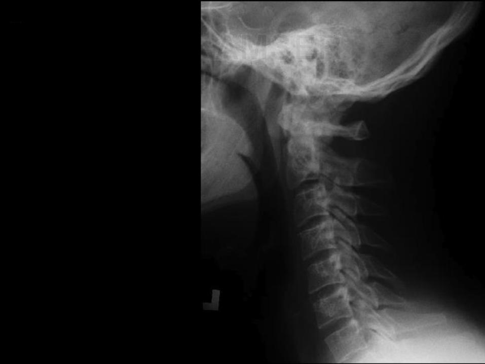
Plain Film Radiographs

Head and Neck Radiographs• Utilize ionizing radiation to
capture images
• Material density determines the degree of X-ray attenuation, and thus, appearance:
Gas (Air)
Soft Tissue (Water)
Metal
Fat
Bone

Basic Osteology Overview

Skull Base Osteology


*
Sella
Turcica
Hyoid
Dorsum
sellae Anterior
clinoid
Mastoid
air cells
Palatine
process
of maxilla
Coronal
Suture

*

*

*Sag. suture
Frontal sinus
Ethmoid air
cells
Crista galli
Dens
Lesser wing
Greater wing
Mandible
MastoidInf. Turbinate


_____
__
____
__
_
____
_
_
__
_

Frontal
sinus
V
Maxilla
Z
N
Frontal
bone
M
S
P
T
N = nasalV = vomerM= mandibleS = sphenoidP = parietalT = temporal


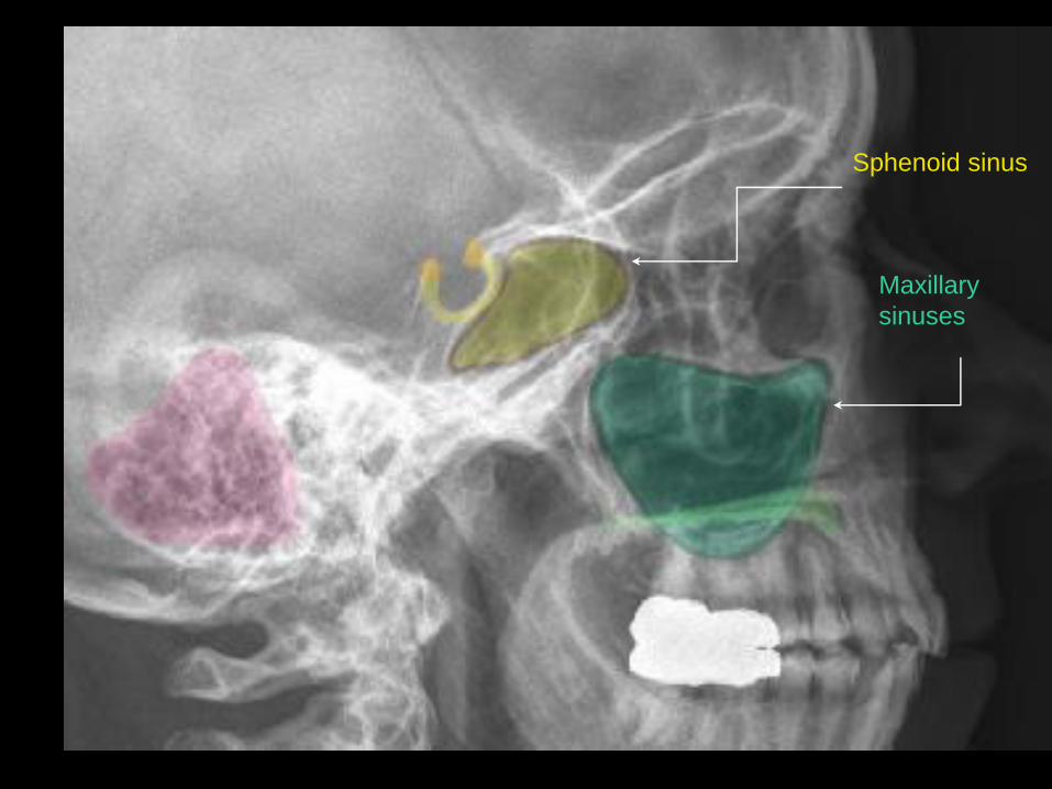
Sphenoid sinus
Maxillary
sinuses


*
Zygomatic arch
Frontal process of
zygo. bone
Maxillary
sinus
Lesser wing
Greater
wing
L. Mastoid

*


L. Maxillary
sinus
Dens
L. Coronoid
processR. Condyle of
mandible
R. Angle of
mandible
*


*Nasopharynx
Soft Palate
Tongue
Mandible
C VI
Atlas
Axis
Dens
Oropharynx

*


*
Trans. Process of C IV
Intervert. foramen
Spinous process
Pedicle

*

*

*
Vestibular fold
Vocal fold
Trachea
Laryngeal ventricle


____
________

Clivus
Occipital
condyle
Lamdoidal
suture
Spinosum
Ovale
Jugular foramen
Lacerum
Optic canal
Sphenoid sinus
IAM
Rotundum

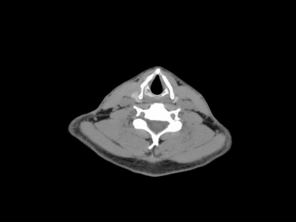
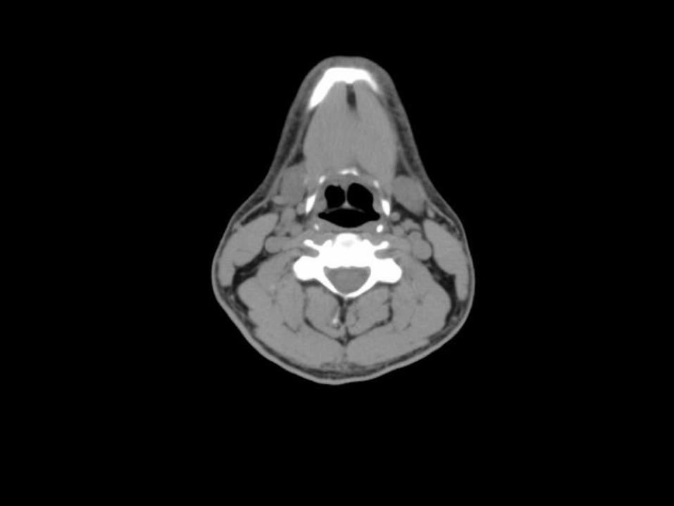
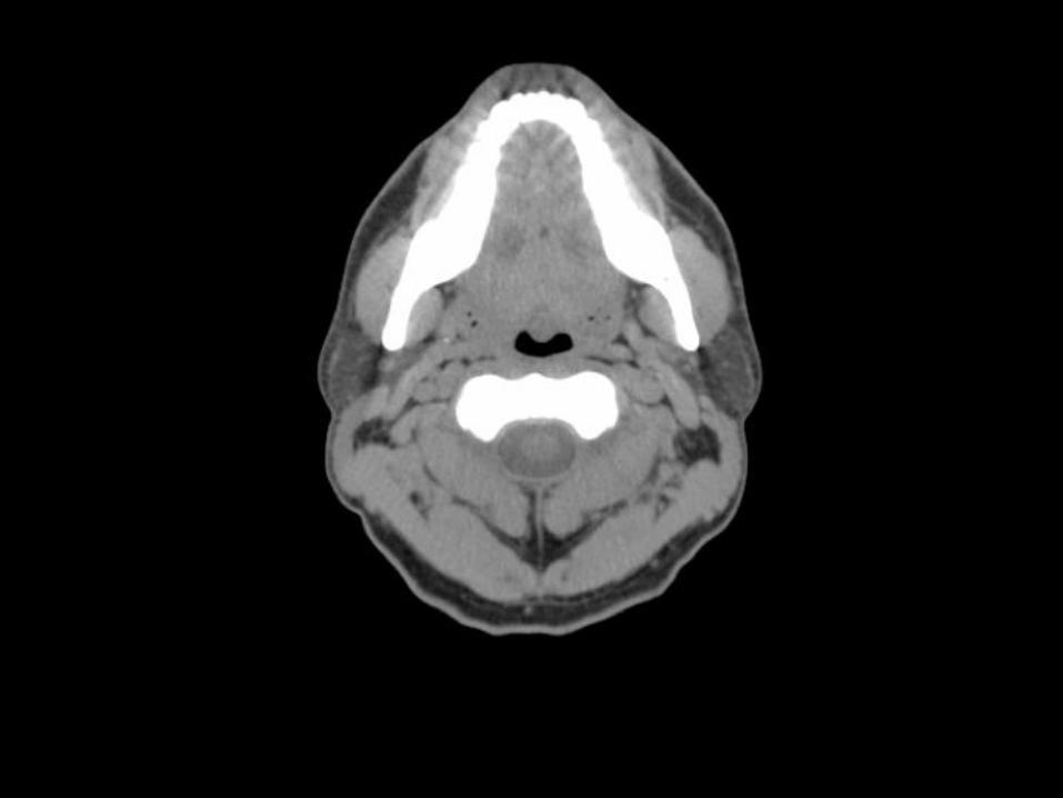
Computed Tomography (CT)

H&N CT
• Utilizes ionizing radiation to produce cross-sectional images
• Digital “windowing” can highlight specific tissues
• Note the patient orientation shown to the left
Anterior
Posterior
Right Left

Muscles of the Head

Muscles of the Neck

First Cervical Vertebrae – Atlas

Second Cervical Vertebrae - Axis

Seventh Cervical Vertebrae

CT Axial Series


*


Vertebral
CC
IJV*
Trapezius
Sternohyoid
SCM
Use airway, apices of lung, and
scapula to estimate level on body.


*


Visceral
compartment
Vascular
compartment
Vertebral
compartment
*
Levator
Scapulae
Semispinalis
Vertebral
CCIJV
Trapezius
Sternohyoid
SCM
Follow Vessels of neck superiorly in axial CT:
CCA, IJV, Vert. art; then follow SCM, then
trapezius


*


*Common Facial
IJVLevator
Scapulae
Semispinalis
Vertebral
Common Carotid
Trapezius
Sternohyoid
SCM
EJV


*

Vertebral level:___

@C3/C4
* Hyoid
EJV
IJV
Levator
Scapulae
Semispinalis
Vertebral
External Carotid
SCM
Internal Carotid
Trapezius


*


*
L. Medial
pterygoid
L. Masseter
L. Parotid
glandPost. Belly of
Digastric m.
DensSCM
Splenius


*


*
L. Medial
pterygoid
L. Masseter
L. Parotid
gland
Splenius
Post. Belly of
Digastric m.


*


Stylomastoid
foramen
*L. Temporalis
L. Lateral
pterygoid
L. Masseter
L. Medial
pterygoid
Maxillary A.


*


Coronoid process
Pterygoid fovea
*L. Temporalis
L. Masseter
L. Lateral pterygoid


*

*

TMJ
Foramen ovale
*L. Temporalis
L. MasseterCoronoid process
L. Mastoid Air
Cells

*

*
____

*
IAC
Cochlea
Auricle
EAC
Horizontal (post.)
Semicircular c.
Internal Carotid
Eustachian tube
Vestibule
Malleus


*


Inferior rectus
*Temporalis


*


Optic nerve
Medial and lateral check ligaments
*Temporalis
Lateral rectus

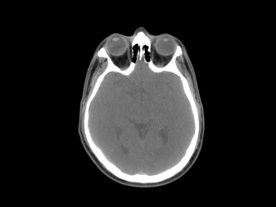
*


Medial rectus Lens
Ophthalmic artery
*Temporalis


*


Superior rectus
*Temporalis
Superior oblique


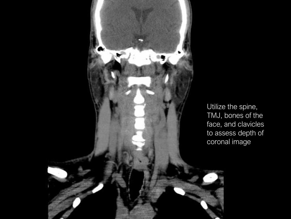
CT Coronal Series
*
Utilize the spine,
TMJ, bones of the
face, and clavicles
to assess depth of
coronal image

*

TMJ*

*

*

Lateral pterygoid
Medial pterygoid
*Nasopharynx

*


R. Masseter
R. Temporalis*
SCM
Pterygoid fossa

*


*Lesser wing
Greater wing
Pterygoid hamulus
Ramus of
mandible
Sphenoidal sinus
Medial pterygoid
attachment
SCM

*

*

*
Inf. Orb.
fissure
Ethmoidal air
cells
Mid. Turbinate
Inf. Turbinate

*


L. Temporalis
L. Masseter
Superior rectusMedial rectus
Inferior rectus
Lateral rectus Optic nerve*


*

Magnetic Resonance Imaging (MRI)

MRI
• Utilizes powerful magnetic fields and radio-frequency pulses to excite (usually hydrogen) atoms within tissues• The energy released by atoms as they return to baseline can be
captured to produce an image
• By varying parameters such as pulse frequency, image qualities can be modulated (i.e. T1 vs T2 weighted)
• MRI can highlight structures that may not be apparent in CT imaging• MRIs are particularly apt when imaging soft tissues
• As they do not rely on X-rays, MRI studies do not expose patients to radiation

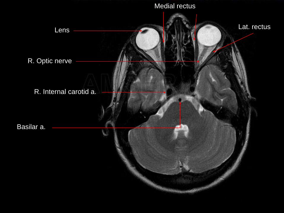
MRI – Axial Series

*

*
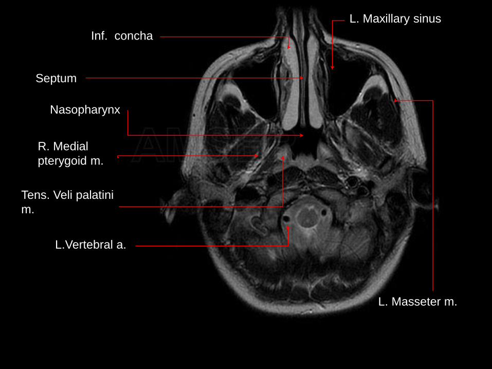
Inf. concha
Septum
L. Maxillary sinus
L.Vertebral a.
Nasopharynx
Tens. Veli palatini
m.
R. Medial
pterygoid m.
L. Masseter m.


*

*

*
Cochlea
Semi-circular
canals
Vestibule
Vestibulocochlear and
Facial nerves

*

*
*
R. Internal carotid a.
Basilar a.
R. Optic nerve
Lens
Medial rectus
Lat. rectus


MRA and Angiography

Vasculature of the Neck

*


*
R. Vertebral A.
R. ICA
R. ECA
R. ICA
R. CCA
R. sup. Cerv. A.

Circle of Willis

*

*

* Ant. Cer. A.Ant. Comm. A.
Middle Cerebral
a.
Int. Carotid a.
Basilar a. Vertebral a.

*


*Ant. Cer. A.
Ant. Comm. A.
Mid. Cer. A.
Int. Car. A.
Basilar a.
Vertebral a.
Post. Cer. A.
Post. Comm. A.

Arteries of the Face

*

*

*Superficial
Temporal a.
Infraorbital a.
Occipital a.
Asc.
Pharyngeal a.
Internal
Carotid a.
External Carotid
a.
Lingual a.
Facial a.
Superior Thyroid. a.
Post. Auricular a.
Maxillary a.
Mid. Meningeal a.

*

*

Superior Thyroid
Lingual
Facial Artery
Occipital
Internal Carotid
Maxillary Artery
Superficial Temporal
Middle Meningeal
*

*

*

Superior
Thyroid a.
Lingual a.
Facial a.
Occipital a.
Internal Carotid a.
Maxillary a.
Superficial
Temporal a.* Post. Auricular a.

*

*

Straight sinus
Transverse
sinus
Sigmoid
sinus
IJV
Superior sagittal
sinus*

*

*

IJV’s
Sigmoid sinus
Trans. sinus
Superior sagittal
sinus
Straight sinus
Great cerebral
vein

END