he visual cortex - tutis · the diagram here shows that visual stimuli activate a structure called...
TRANSCRIPT
0 http://www.tutis.ca/NeuroMD/index.htm 20 February 2013
T he Visual Cortex
1 http://www.tutis.ca/NeuroMD/index.htm 20 February 2013
Chapter contents
Contents
Chapter 2 ........................................................................................................................... 0
T he Visual Cortex ............................................................................................................ 0
Chapter Contents ............................................................................................................. 1
Introduction ..................................................................................................................... 2
Optic Chiasm .................................................................................................................. 2
Where do the eye's ganglion cells project to? ................................................................. 3
To where do LGN neurons project?................................................................................ 4
Primary visual cortex (V1) contains 3 cell types. ........................................................... 5
Why is this important and clinically relevant? ............................................................... 5
What patients with a scotoma (blind area) on their fovea experience? .......................... 6
Describe the structure of primary visual cortex (V1). .................................................... 7
Influence of early development on the structure of visual cortex ................................... 8
What are the synaptic mechanisms for this plasticity? ................................................... 9
Why are two eyes better than one? ............................................................................... 10
How do binocular cells signal disparity? ...................................................................... 10
In summary ................................................................................................................... 11
Where does visual information go to after primary visual cortex? .......................... 12
Where does visual information go next? ...................................................................... 13
What is the function of the inferior temporal cortex (IT)? ........................................... 13
The visual perception of motion ................................................................................... 14
Summary ....................................................................................................................... 15
Practice problems .......................................................................................................... 15
2 http://www.tutis.ca/NeuroMD/index.htm 20 February 2013
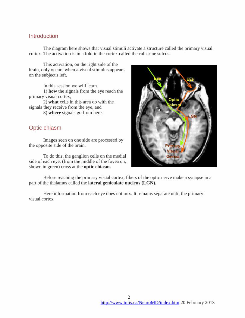
Introduction The diagram here shows that visual stimuli activate a structure called the primary visual
cortex. The activation is in a fold in the cortex called the calcarine sulcus. This activation, on the right side of the
brain, only occurs when a visual stimulus appears on the subject's left.
In this session we will learn 1) how the signals from the eye reach the
primary visual cortex, 2) what cells in this area do with the
signals they receive from the eye, and 3) where signals go from here.
Optic chiasm Images seen on one side are processed by
the opposite side of the brain. To do this, the ganglion cells on the medial
side of each eye, (from the middle of the fovea on, shown in green) cross at the optic chiasm.
Before reaching the primary visual cortex, fibers of the optic nerve make a synapse in a
part of the thalamus called the lateral geniculate nucleus (LGN). Here information from each eye does not mix. It remains separate until the primary
visual cortex
3 http://www.tutis.ca/NeuroMD/index.htm 20 February 2013
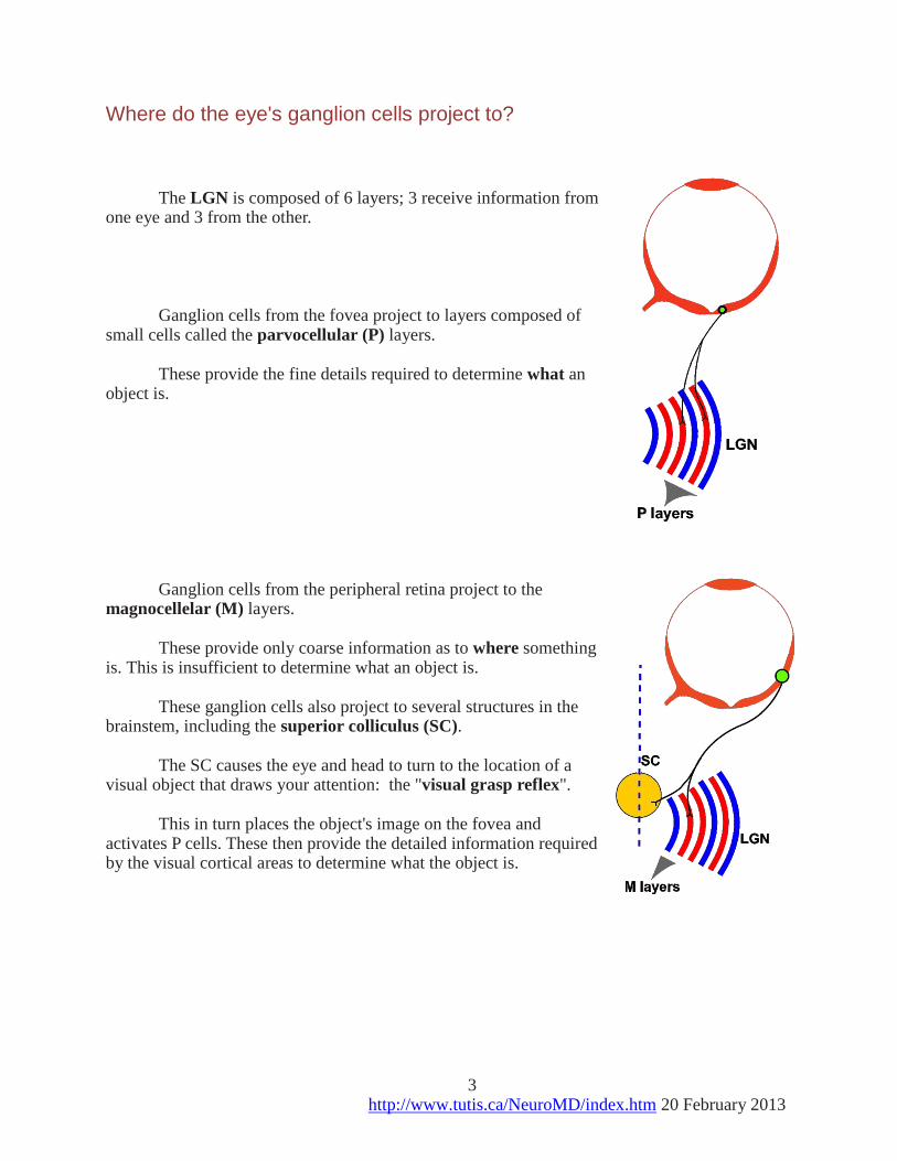
Where do the eye's ganglion cells project to? The LGN is composed of 6 layers; 3 receive information from
one eye and 3 from the other. Ganglion cells from the fovea project to layers composed of
small cells called the parvocellular (P) layers. These provide the fine details required to determine what an
object is. Ganglion cells from the peripheral retina project to the
magnocellelar (M) layers. These provide only coarse information as to where something
is. This is insufficient to determine what an object is. These ganglion cells also project to several structures in the
brainstem, including the superior colliculus (SC). The SC causes the eye and head to turn to the location of a
visual object that draws your attention: the "visual grasp reflex". This in turn places the object's image on the fovea and
activates P cells. These then provide the detailed information required by the visual cortical areas to determine what the object is.
4 http://www.tutis.ca/NeuroMD/index.htm 20 February 2013
To where do LGN neurons project?
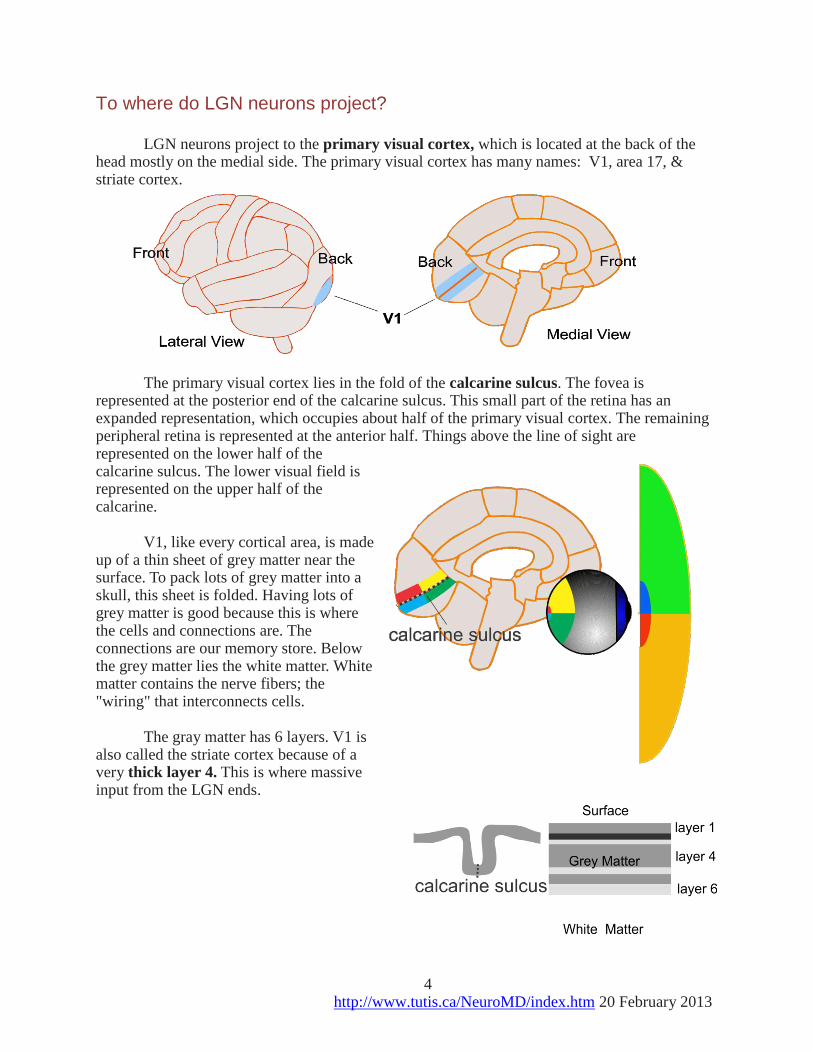
LGN neurons project to the primary visual cortex, which is located at the back of the
head mostly on the medial side. The primary visual cortex has many names: V1, area 17, & striate cortex.
The primary visual cortex lies in the fold of the calcarine sulcus. The fovea is
represented at the posterior end of the calcarine sulcus. This small part of the retina has an expanded representation, which occupies about half of the primary visual cortex. The remaining peripheral retina is represented at the anterior half. Things above the line of sight are represented on the lower half of the calcarine sulcus. The lower visual field is represented on the upper half of the calcarine.
V1, like every cortical area, is made
up of a thin sheet of grey matter near the surface. To pack lots of grey matter into a skull, this sheet is folded. Having lots of grey matter is good because this is where the cells and connections are. The connections are our memory store. Below the grey matter lies the white matter. White matter contains the nerve fibers; the "wiring" that interconnects cells.
The gray matter has 6 layers. V1 is
also called the striate cortex because of a very thick layer 4. This is where massive input from the LGN ends.

5 http://www.tutis.ca/NeuroMD/index.htm 20 February 2013
Primary visual cortex (V1) contains 3 cell types 1. Layer 4 cells, whose receptive fields are round like those of LGN and ganglion cells, 2. simple cells with elongated
receptive fields and thus maximally activated by a line of a particular orientation, and
3. complex cells whose receptive fields are similar to those of simple cells except that the line can lie over a larger area of the retina and these fire more to moving lines. How are simple and complex receptive fields produced?
Several ganglion cells, whose receptive fields lie along a
common line, converge by way of the LGN onto a simple cell. The one shown here fires maximally for a horizontal line and minimally for a vertical line. Each simple cell is selective for one particular orientation.
Complex cells: Several simple cells of the same orientation converge onto a complex cell.
Why is this important and clinically relevant? These studies show how the visual system can
construct complex representations from simple features. The receptive field of a simple cell is tuned to a very
particular stimulus. The complex cell generalizes this particular stimulus over a larger area.
We will see that, further in the visual stream, one finds cells that respond only to particular faces that are generalized over the whole retina.
The discovery of simple and complex cells is the work
of David Hubel, a medical student from McGill, who grew up in Windsor, Canada and who together with Torsten Wiesel, was awarded the 1981 Nobel Prize in Physiology or Medicine. They showed how changes in the organization of these cells can lead to a form of blindness called amblyopia.

6 http://www.tutis.ca/NeuroMD/index.htm 20 February 2013
What patients with a scotoma (blind area) on their fovea experience? When viewing a face against a striped background, patients do not see the face (because
it is in their blind area) but see stripes where the face should be. This "filling in" is produced by cortical simple and complex cells. These cells are producing a similar
filling now as you read this. You have a blind spot on your retina but are not aware of a black area on this page. The blind spot is where the optic nerve leaves the back of the eye.
To find your blind spot, close your left eye and look at some object on a blank wall. Hold a pencil, preferably with a red tip, at arm's length and at eye level. Move the pencil to the right. At about 20 degrees to the right the tip should disappear. Now raise the pencil. The tip of the pencil should reappear and there is no empty gap in the pencil where the blind spot was. Your visual system fills in the image of the pencil.
You can also look at the red x below while paying attention to the dot on the right. It
may be difficult at first to keep from taking a peek at the dot. Remember to close your left eye.
In the first row, when you move the page to between 1 and 2 feet away from you, the dot should disappear.
In the second row, the
gap in the line should disappear.
In the third, the face
should disappear but the lines should be visible where the face was. You should see what the patient with the scotoma in the retina saw.
7 http://www.tutis.ca/NeuroMD/index.htm 20 February 2013
Describe the structure of primary visual cortex (V1). V1 is a map of the retina composed of a grid
(1 mm by 1mm) of hypercolumns. Each hypercolumn analyses information from one small region of retina. Adjacent hypercolumns analyze information from adjacent areas of retina. The map is distorted. The fovea is over-represented. The area devoted to the fovea is
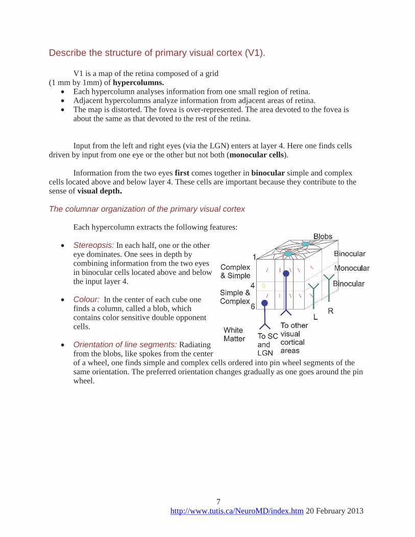
about the same as that devoted to the rest of the retina. Input from the left and right eyes (via the LGN) enters at layer 4. Here one finds cells
driven by input from one eye or the other but not both (monocular cells). Information from the two eyes first comes together in binocular simple and complex
cells located above and below layer 4. These cells are important because they contribute to the sense of visual depth.
The columnar organization of the primary visual cortex
Each hypercolumn extracts the following features:
Stereopsis: In each half, one or the other eye dominates. One sees in depth by combining information from the two eyes in binocular cells located above and below the input layer 4.
Colour: In the center of each cube one finds a column, called a blob, which contains color sensitive double opponent cells.
Orientation of line segments: Radiating from the blobs, like spokes from the center of a wheel, one finds simple and complex cells ordered into pin wheel segments of the same orientation. The preferred orientation changes gradually as one goes around the pin wheel.
8 http://www.tutis.ca/NeuroMD/index.htm 20 February 2013
Influence of early development on the structure of visual cortex
What is the effect of early visual deprivation in one eye?
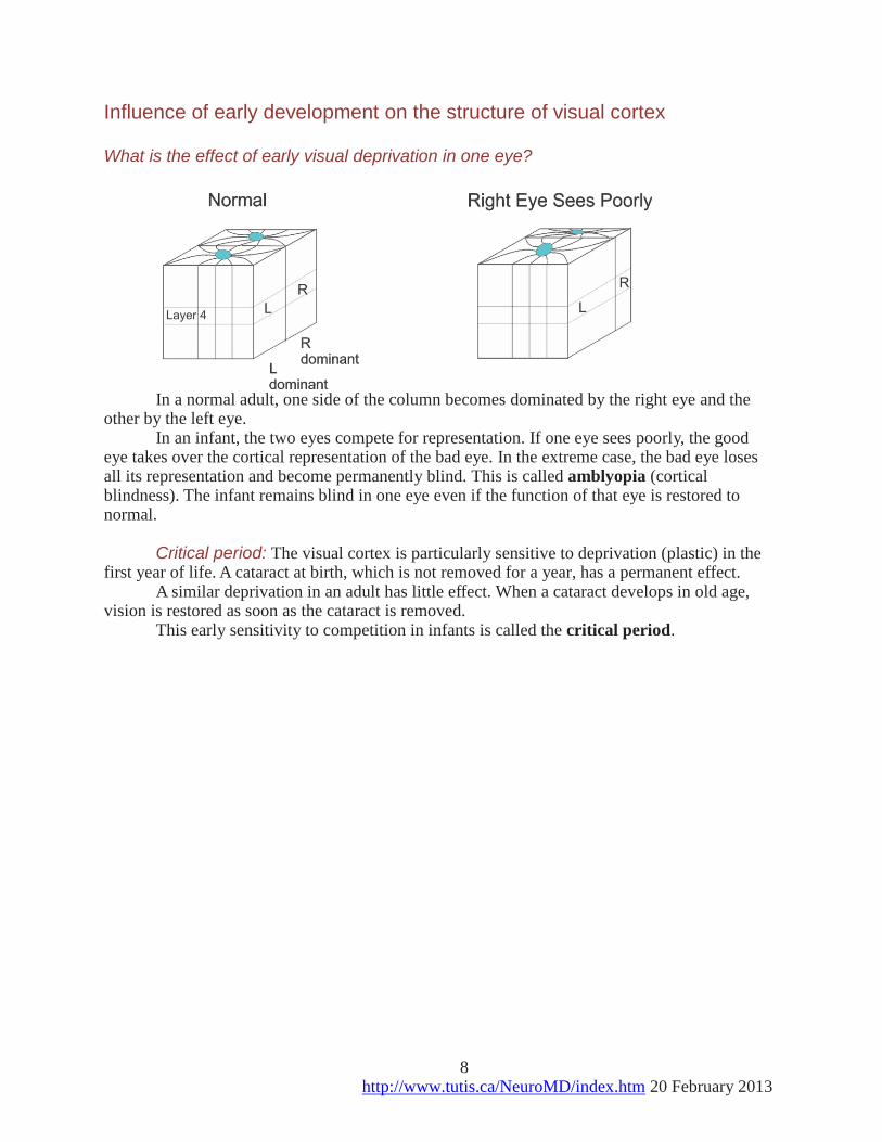
In a normal adult, one side of the column becomes dominated by the right eye and the other by the left eye.
In an infant, the two eyes compete for representation. If one eye sees poorly, the good eye takes over the cortical representation of the bad eye. In the extreme case, the bad eye loses all its representation and become permanently blind. This is called amblyopia (cortical blindness). The infant remains blind in one eye even if the function of that eye is restored to normal.
Critical period: The visual cortex is particularly sensitive to deprivation (plastic) in the
first year of life. A cataract at birth, which is not removed for a year, has a permanent effect. A similar deprivation in an adult has little effect. When a cataract develops in old age,
vision is restored as soon as the cataract is removed. This early sensitivity to competition in infants is called the critical period.
9 http://www.tutis.ca/NeuroMD/index.htm 20 February 2013
What is the effect of early strabismus?
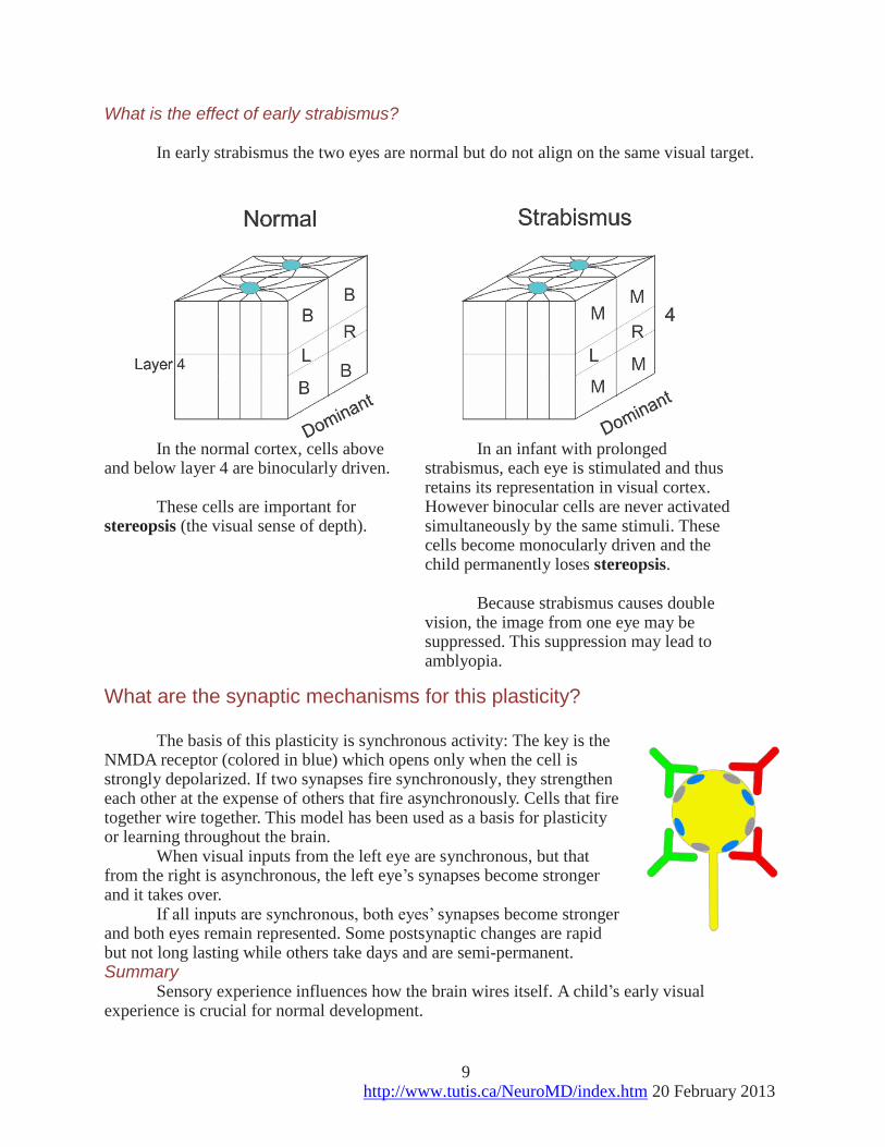
In early strabismus the two eyes are normal but do not align on the same visual target.
In the normal cortex, cells above and below layer 4 are binocularly driven.
These cells are important for
stereopsis (the visual sense of depth).
In an infant with prolonged strabismus, each eye is stimulated and thus retains its representation in visual cortex. However binocular cells are never activated simultaneously by the same stimuli. These cells become monocularly driven and the child permanently loses stereopsis.
Because strabismus causes double
vision, the image from one eye may be suppressed. This suppression may lead to amblyopia.
What are the synaptic mechanisms for this plasticity? The basis of this plasticity is synchronous activity: The key is the
NMDA receptor (colored in blue) which opens only when the cell is strongly depolarized. If two synapses fire synchronously, they strengthen each other at the expense of others that fire asynchronously. Cells that fire together wire together. This model has been used as a basis for plasticity or learning throughout the brain.
When visual inputs from the left eye are synchronous, but that from the right is asynchronous, the left eye’s synapses become stronger and it takes over.
If all inputs are synchronous, both eyes’ synapses become stronger and both eyes remain represented. Some postsynaptic changes are rapid but not long lasting while others take days and are semi-permanent. Summary
Sensory experience influences how the brain wires itself. A child’s early visual experience is crucial for normal development.
10 http://www.tutis.ca/NeuroMD/index.htm 20 February 2013
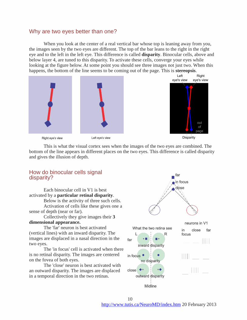
Why are two eyes better than one? When you look at the center of a real vertical bar whose top is leaning away from you,
the images seen by the two eyes are different. The top of the bar leans to the right in the right eye and to the left in the left eye. This difference is called disparity. Binocular cells, above and below layer 4, are tuned to this disparity. To activate these cells, converge your eyes while looking at the figure below. At some point you should see three images not just two. When this happens, the bottom of the line seems to be coming out of the page. This is stereopsis.
This is what the visual cortex sees when the images of the two eyes are combined. The bottom of the line appears in different places on the two eyes. This difference is called disparity and gives the illusion of depth.
How do binocular cells signal disparity?
Each binocular cell in V1 is best
activated by a particular retinal disparity. Below is the activity of three such cells. Activation of cells like these gives one a
sense of depth (near or far). Collectively they give images their 3
dimensional appearance. The 'far' neuron is best activated
(vertical lines) with an inward disparity. The images are displaced in a nasal direction in the two eyes.
The 'in focus' cell is activated when there is no retinal disparity. The images are centered on the fovea of both eyes.
The 'close' neuron is best activated with an outward disparity. The images are displaced in a temporal direction in the two retinas.
11 http://www.tutis.ca/NeuroMD/index.htm 20 February 2013
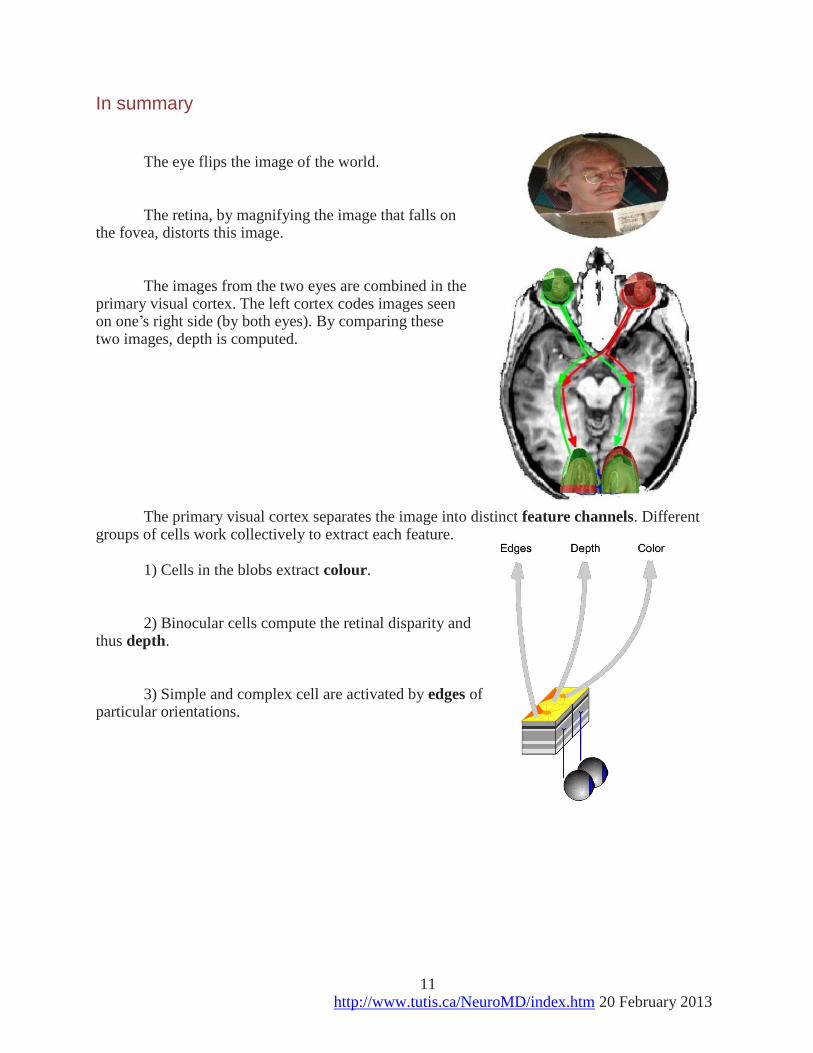
In summary The eye flips the image of the world. The retina, by magnifying the image that falls on
the fovea, distorts this image. The images from the two eyes are combined in the
primary visual cortex. The left cortex codes images seen on one’s right side (by both eyes). By comparing these two images, depth is computed.
The primary visual cortex separates the image into distinct feature channels. Different
groups of cells work collectively to extract each feature. 1) Cells in the blobs extract colour. 2) Binocular cells compute the retinal disparity and
thus depth. 3) Simple and complex cell are activated by edges of
particular orientations.
12 http://www.tutis.ca/NeuroMD/index.htm 20 February 2013
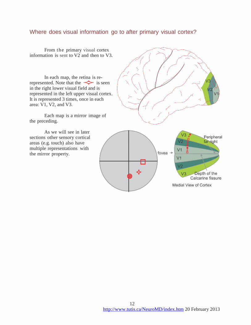
Where does visual information go to after primary visual cortex? From the primary visual cortex
information is sent to V2 and then to V3. In each map, the retina is re-
represented. Note that the is seen in the right lower visual field and is represented in the left upper visual cortex. It is represented 3 times, once in each area: V1, V2, and V3.
Each map is a mirror image of
the preceding. As we will see in later
sections other sensory cortical areas (e.g. touch) also have multiple representations with the mirror property.
13 http://www.tutis.ca/NeuroMD/index.htm 20 February 2013
Where does visual information go next? From V3, information diverges to over 3 dozen higher order visual areas. Each processes some special aspect of visual information. These visual areas are like a
multi-screen cinema. The main difference is that each screen is showing a different attribute of the same movie; some just the motion, others the colors, etc.
Information flows along two main streams. 1) The dorsal stream, to the intra
parietal sulcus in the parietal lobe, is concerned with selecting actions to particular spatial locations. For this reason it is called the "where" stream.
2) The ventral stream, to the inferior part of the temporal lobe, is concerned with the perception and recognition of objects, e.g. faces. This is called the “what" stream.
Lesions in the “what” stream produce deficits in object recognition: visual agnosia. Patients can reach for objects but cannot say what they are.
Patients with lesions in the parietal lobe can exhibit optic ataxia. They can recognize objects but fail to reach for them accurately.
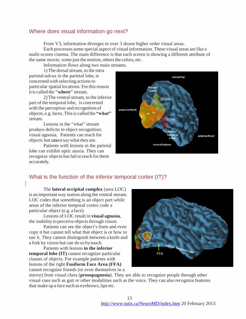
What is the function of the inferior temporal cortex (IT)? The lateral occipital complex (area LOC)
is an important way station along the ventral stream. LOC codes that something is an object part while areas of the inferior temporal cortex code a particular object (e.g. a face).
Lesions of LOC result in visual agnosia, the inability to perceive objects through vision.
Patients can see the object’s form and even copy it but cannot tell what that object is or how to use it. They cannot distinguish between a knife and a fork by vision but can do so by touch.
Patients with lesions in the inferior temporal lobe (IT) cannot recognize particular classes of objects. For example patients with lesions of the right Fusiform Face Area (FFA) cannot recognize friends (or even themselves in a mirror) from visual clues (prosopagnosia). They are able to recognize people through other visual cues such as gait or other modalities such as the voice. They can also recognize features that make up a face such as eyebrows, lips etc.
14 http://www.tutis.ca/NeuroMD/index.htm 20 February 2013
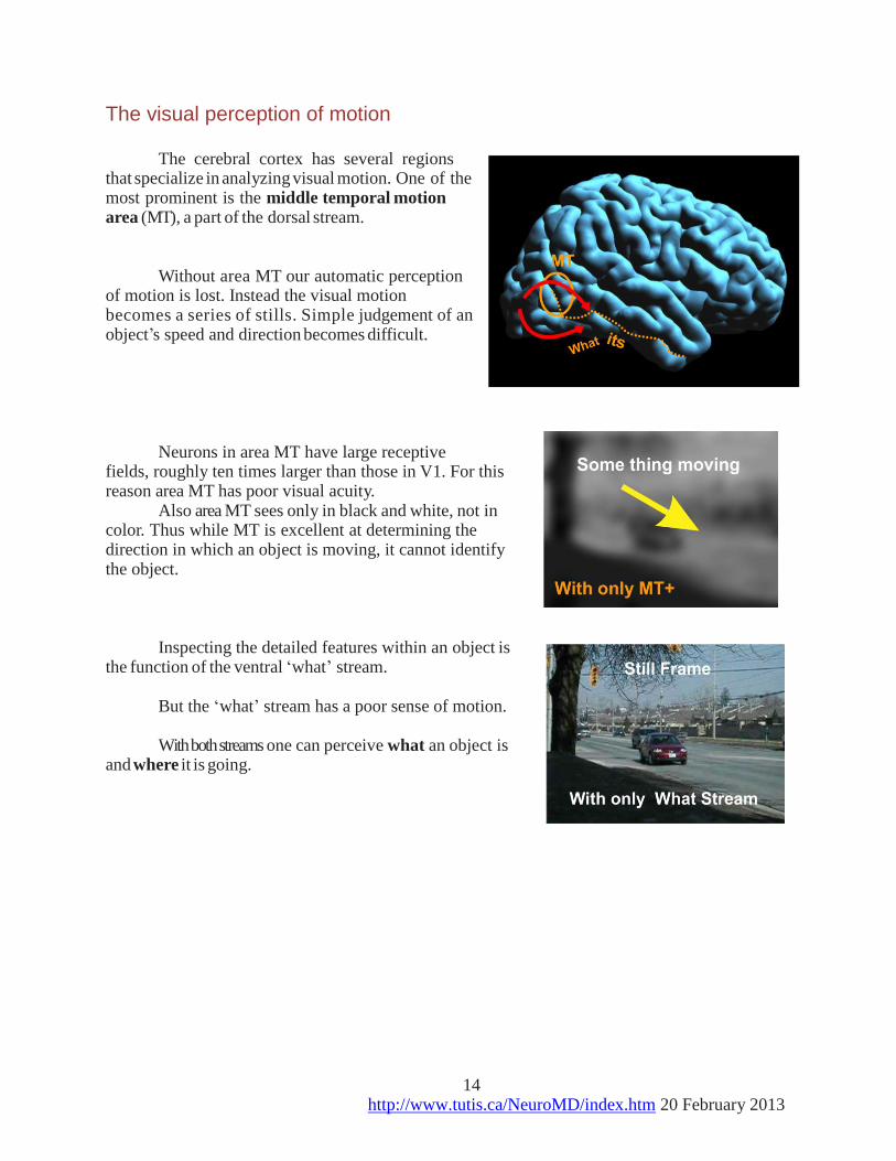
The visual perception of motion The cerebral cortex has several regions
that specialize in analyzing visual motion. One of the most prominent is the middle temporal motion area (MT), a part of the dorsal stream.
Without area MT our automatic perception
of motion is lost. Instead the visual motion becomes a series of stills. Simple judgement of an object’s speed and direction becomes difficult.
Neurons in area MT have large receptive
fields, roughly ten times larger than those in V1. For this reason area MT has poor visual acuity.
Also area MT sees only in black and white, not in color. Thus while MT is excellent at determining the direction in which an object is moving, it cannot identify the object.
Inspecting the detailed features within an object is
the function of the ventral ‘what’ stream. But the ‘what’ stream has a poor sense of motion. With both streams one can perceive what an object is
and where it is going.

15 http://www.tutis.ca/NeuroMD/index.htm 20 February 2013
Summary Visual information from V1 divides
along two streams: 1) a dorsal “Where”' stream: concerned
with the spatial relationship between objects and
2) a ventral “What” stream: concerned with object recognition.
The ‘What’ stream sees in detail
because it gets most of its input from P ganglion cells in the fovea.
The ‘Where’ stream gets less detailed input from M ganglion cell in the peripheral retina and excels at coding the locations of objects around us.
Practice problems
1. Strabismus in early childhood will produce
a) a loss of orientation columns in primary visual cortex (V1).
b) a loss of cells in the blob region of V1.
c) a loss of binocularly driven cells in layer 4 of V1.
d) a loss of binocularly driven cells in layers above and below layer 4 of V1.
e) no permanent changes in visual ability.
2. Your patient has developed a sudden loss in his ability to read. The right side of any word
that he looks at is not visible with either eye but he can see things in his peripheral vision.
His problem is
a) macular degeneration.
b) compression of the optic chiasm.
c) a stroke affecting the posterior end of the calcarine sulcus on the right side.
d) a stroke affecting the anterior end of the calcarine sulcus on the right side.
e) a stroke affecting the posterior end of the calcarine sulcus on the left side.

16 http://www.tutis.ca/NeuroMD/index.htm 20 February 2013
Answers
1. d)
2. e)
See also http://www.tutis.ca/NeuroMD/L2V123/V123Prob.swf