hd scanning: velocities and volume flow
TRANSCRIPT

Cindy Sturt, MD, FACS, RVT
West Orange, NJ
April 27, 2018
HD Scanning:
Velocities and Volume Flow
Non-Invasive Lab
Symposium
VolumeFlow

500,000 Americans on dialysis
National Kidney Foundation
•20-25% annual mortality
•65% 5 year mortality

Life line of
dialysis
patients
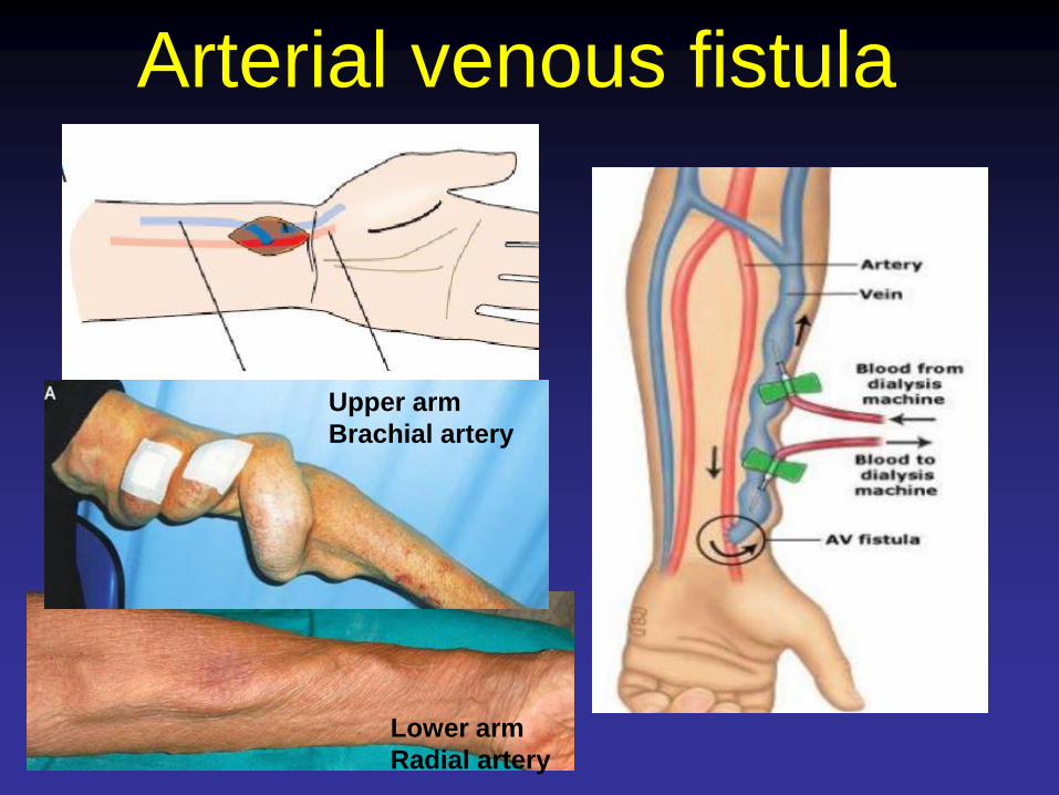
Arterial venous fistula
Upper arm
Brachial artery
Lower arm
Radial artery

Arterial Venous
Grafts
Life line
Straight Looped
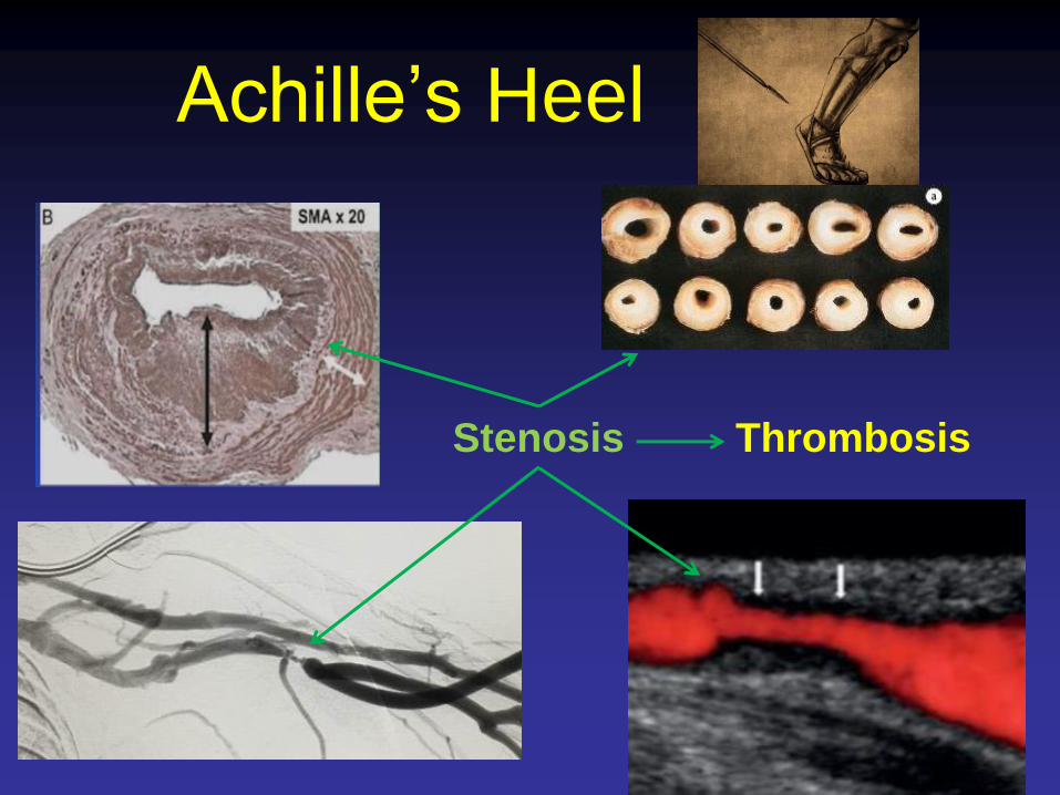
Achille’s Heel
Stenosis Thrombosis

Intervention (to prevent thrombosis)
Balloon angioplasty

Intervention (to prevent thrombosis)
Stenting

AV access
StenosisLifeline Achilles heel
Angioplasty

Surveillance
• Surveillance
• Access flow
• Recirculation
• Venous pressure
• Ultrasound
Dialysis
Unit
• Clinical Monitoring
• Bleeding, difficult cannulations, arm swelling,
chest wall collateralization, increased
pulsatility, decreased thrill

• Access flow decrease <600cc/min or <
1000cc/min with >25% decrease over 4 month
prior
• Increased venous pressure during dialysis
– >150mmHg or trend of persistent increasing pressure
over time

Semin Dial. 2015 March; 28(2):E23-E29.
Our results add to the uncertainty of access blood
flow monitoring as a surveillance method of
hemodialysis accesses.

UltrasoundAVF
Inflow artery
anastomosis
Color Doppler
B mode
Spectral Doppler

Ultrasound - Carotid Circulation
Zwiebel WL et al. 2000
Thrush A. Harshorne et al. 2005
ICA
Normal Doppler Spectra
CCA
ECA

Ultrasound – Mesenteric circulation
SMA: PSV > 275cm/s 70% stenosis
Celiac: PSV > 250cm/s 70% stenosis

Ultrasound – peripheral circulation
2011 ACC/AHA guidelines
for management of PVD –
ultrasound is useful in
diagnosing location and
severity of stenoses.

Ultrasound – Dialysis Circuit
Tortuosity
Aneurysms/pseudoaneurysm
Angulation of anastomosis
Compliance of vein/grafts different from arteries
Diameter of vein varies along its length

•Grayscale/color doppler
•inflow
•anastomosis
•outflow
•+/- subclavian vein
•Spectral waveforms and velocity
•Inflow
•Anastomosis
•proximal
•outflow (beyond anastomosis
•subclavian vein
•Blood flow volume from at least one site.
•Abnormalities require additional images, waveforms, velocity measurements.
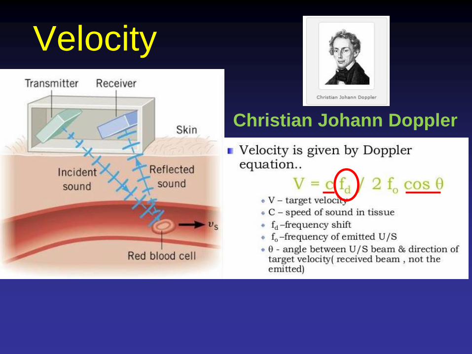
Velocity
Christian Johann Doppler

Low resistance
venous outflow
(AV access)
Inflow arteryNormal brachial artery:
Triphasic
High resistance
Brachial artery – after access creation
Monophasic flow
Large diastolic component

Normal Velocity
100-300cm/s
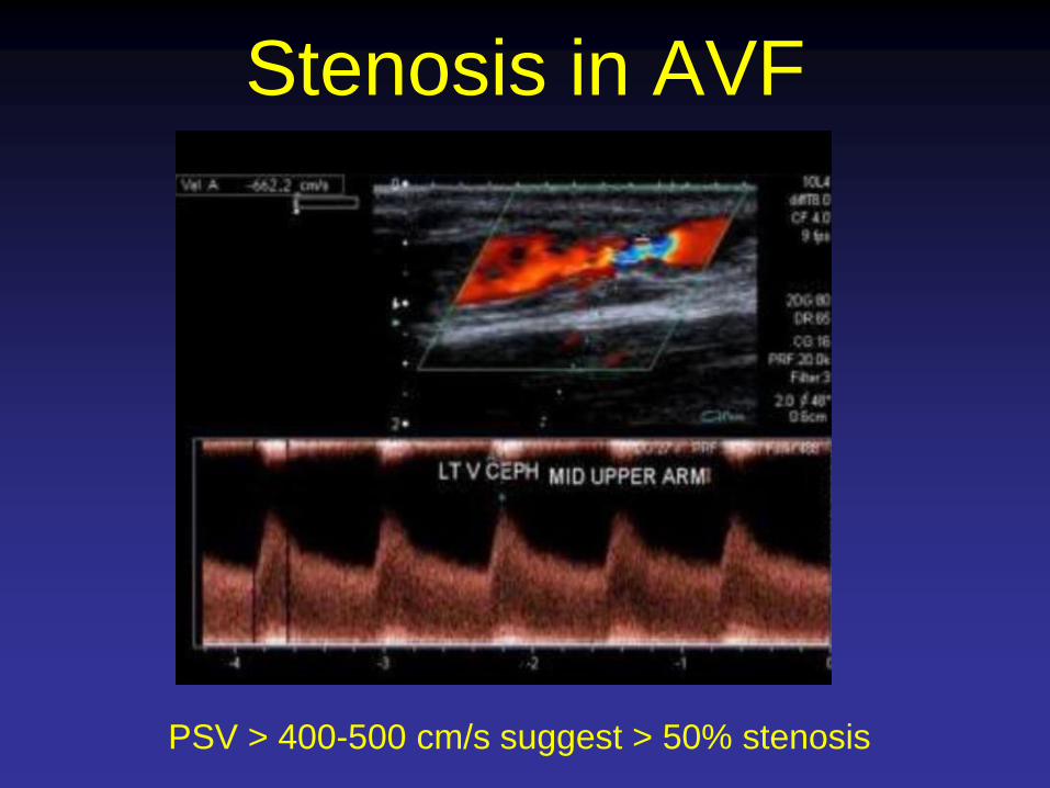
PSV > 400-500 cm/s suggest > 50% stenosis
Stenosis in AVF

Stenosis at anastomosis
PSV > 600 cm/s suggest stenosis?
angulation
angulation

Occlusion of AVF
Triphasic brachial artery
waveform in association with an
occluded brachiocephalic fistula
Occlusion of brachiocephalic fistula.
Thrombus within the vein

Volume Flow
Volume (cc/min)= Area (cm2) x Velocity (cm/s)
Diameter
(longitudinal)
velocity

Volume flow
•Straight segment
•5cm away from anastomosis/stenoses/major abnormalities
•Some recommend measuring at brachial artery

Volume Flow
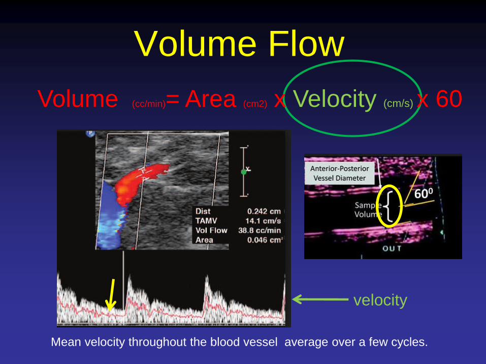
Volume (cc/min)= Area (cm2) x Velocity (cm/s) x 60
Diameter
(longitudinal)
•Longitudinal view
•Systole
•Measure in non-aneurysmal area

Volume Flow
Volume (cc/min)= Area (cm2) x Velocity (cm/s) x 60
Sample volume needs
to include the whole
diameter of the vessel
and not just the middle
Parabolic flow•rbcs in the middle of the vessel travel faster
•rbcs in at the periphery of the vessel travels slower
Volume Flow
velocity
Mean velocity throughout the blood vessel average over a few cycles.
Volume (cc/min)= Area (cm2) x Velocity (cm/s) x 60

Volume Flow
Conclusion
•Ultrasound can be very important in the
surveillance of dialysis access.
•When used accurately, can identify stenoses.
•Need to better understand and come up with
appropriate criteria to indicate stenosis.
•Probably cannot be used in isolation and
clinical monitoring is important.

Thank You


Peak systolic velocities
Normal duplex of peripheral
arteries. High resistance
waveforms. High resistance
flow leading to triphasic
waveforms.

Volume Flow
Triphasic brachial artery
waveform in association with an
occluded brachiocephalic fistula
Occlusion of brachiocephalic fistula.
Thrombus within the vein
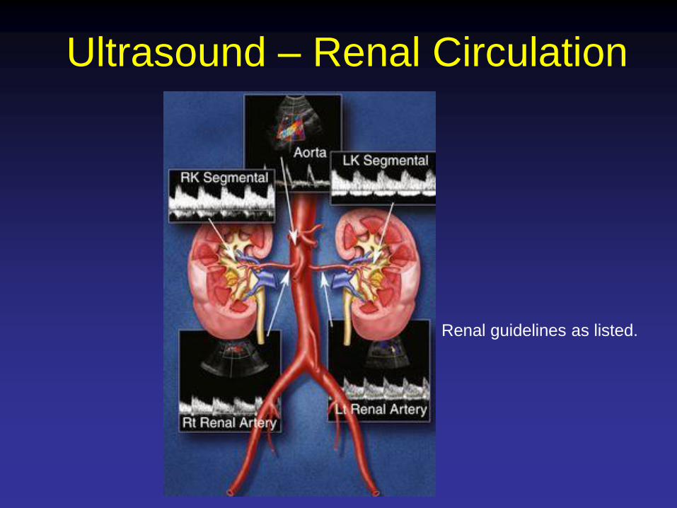
Ultrasound – Renal Circulation
Renal guidelines as listed.