hassel most ern 2014
DESCRIPTION
Hasselmo et al. work in 2014 at Boston UniversityTRANSCRIPT

NeuroImage 85 (2014) 656–666
Contents lists available at ScienceDirect
NeuroImage
j ourna l homepage: www.e lsev ie r .com/ locate /yn img
Review
Theta rhythm and the encoding and retrieval of space and time
Michael E. Hasselmo ⁎, Chantal E. SternCenter for Memory and Brain, Department of Psychology and Graduate Program for Neuroscience, Boston University, 2 Cummington Mall, Boston, MA, 02215, USA
⁎ Corresponding author. Fax: +1 617 358 3296.E-mail address: [email protected] (M.E. Hasselmo).
1053-8119/$ – see front matter © 2013 Elsevier Inc. Allhttp://dx.doi.org/10.1016/j.neuroimage.2013.06.022
a b s t r a c t
a r t i c l e i n f oArticle history:Accepted 4 June 2013Available online 14 June 2013
Keywords:HippocampusEntorhinal cortexMemoryResonanceGrid cellsPlace cellsElectroencephalograph
Physiological data demonstrates theta frequency oscillations associated with memory function and spatialbehavior. Modeling and data from animals provide a perspective on the functional role of theta rhythm,including correlations with behavioral performance and coding by timing of spikes relative to phase of oscil-lations. Data supports a theorized role of theta rhythm in setting the dynamics for encoding and retrievalwithin cortical circuits. Recent data also supports models showing how network and cellular theta rhythmic-ity allows neurons in the entorhinal cortex and hippocampus to code time and space as a possible substratefor encoding events in episodic memory. Here we discuss these models and relate them to current physiolog-ical and behavioral data.
© 2013 Elsevier Inc. All rights reserved.
Contents
Introduction . . . . . . . . . . . . . . . . . . . . . . . . . . . . . . . . . . . . . . . . . . . . . . . . . . . . . . . . . . . . . . . . 657Experimental data on theta rhythm . . . . . . . . . . . . . . . . . . . . . . . . . . . . . . . . . . . . . . . . . . . . . . . . . . . . . 657
Theta rhythm in humans and animals . . . . . . . . . . . . . . . . . . . . . . . . . . . . . . . . . . . . . . . . . . . . . . . . . . 657Behavioral correlates of theta rhythm . . . . . . . . . . . . . . . . . . . . . . . . . . . . . . . . . . . . . . . . . . . . . . . . . . 657Functional coding by phase of theta rhythm . . . . . . . . . . . . . . . . . . . . . . . . . . . . . . . . . . . . . . . . . . . . . . . 657
Theta rhythm may provide separate phases of encoding and retrieval . . . . . . . . . . . . . . . . . . . . . . . . . . . . . . . . . . . . . 658Physiological data supporting the SPEAR model . . . . . . . . . . . . . . . . . . . . . . . . . . . . . . . . . . . . . . . . . . . . . 659
Phasic membrane potential dynamics . . . . . . . . . . . . . . . . . . . . . . . . . . . . . . . . . . . . . . . . . . . . . . . 659Phasic changes in inhibition . . . . . . . . . . . . . . . . . . . . . . . . . . . . . . . . . . . . . . . . . . . . . . . . . . . 659Phasic changes in synaptic input . . . . . . . . . . . . . . . . . . . . . . . . . . . . . . . . . . . . . . . . . . . . . . . . . 659Phasic changes in long-term potentiation . . . . . . . . . . . . . . . . . . . . . . . . . . . . . . . . . . . . . . . . . . . . . 659
Network data relating to encoding and retrieval . . . . . . . . . . . . . . . . . . . . . . . . . . . . . . . . . . . . . . . . . . . . . 659Theta rhythm and models of the phase coding of space . . . . . . . . . . . . . . . . . . . . . . . . . . . . . . . . . . . . . . . . . . . . 660
Support for phase coding involving theta rhythm . . . . . . . . . . . . . . . . . . . . . . . . . . . . . . . . . . . . . . . . . . . . 660Spiking frequency and theta phase precession . . . . . . . . . . . . . . . . . . . . . . . . . . . . . . . . . . . . . . . . . . . 660Dependence on oscillations . . . . . . . . . . . . . . . . . . . . . . . . . . . . . . . . . . . . . . . . . . . . . . . . . . . 660Intracellular membrane properties . . . . . . . . . . . . . . . . . . . . . . . . . . . . . . . . . . . . . . . . . . . . . . . . 661
Current issues for phase coding models . . . . . . . . . . . . . . . . . . . . . . . . . . . . . . . . . . . . . . . . . . . . . . . . . 661Intracellular membrane potential properties . . . . . . . . . . . . . . . . . . . . . . . . . . . . . . . . . . . . . . . . . . . 661Spiking properties of neurons . . . . . . . . . . . . . . . . . . . . . . . . . . . . . . . . . . . . . . . . . . . . . . . . . . 661Oscillations in rats and bats . . . . . . . . . . . . . . . . . . . . . . . . . . . . . . . . . . . . . . . . . . . . . . . . . . . 662Movement direction input . . . . . . . . . . . . . . . . . . . . . . . . . . . . . . . . . . . . . . . . . . . . . . . . . . . . 662
Theta rhythm and episodic memory . . . . . . . . . . . . . . . . . . . . . . . . . . . . . . . . . . . . . . . . . . . . . . . . . . . . . 662Acknowledgments . . . . . . . . . . . . . . . . . . . . . . . . . . . . . . . . . . . . . . . . . . . . . . . . . . . . . . . . . . . . . 663Conflict of interest . . . . . . . . . . . . . . . . . . . . . . . . . . . . . . . . . . . . . . . . . . . . . . . . . . . . . . . . . . . . . 663References . . . . . . . . . . . . . . . . . . . . . . . . . . . . . . . . . . . . . . . . . . . . . . . . . . . . . . . . . . . . . . . . . 663
rights reserved.
657M.E. Hasselmo, C.E. Stern / NeuroImage 85 (2014) 656–666
Introduction
Theta frequency oscillations appear in electroencephalographic(EEG) recordings from scalp electrodes and depth electrodes in humansubjects, but controversy continues over the functional role of thetafrequency oscillations (commonly referred to as theta rhythm). Under-standing the functional role of theta rhythm will benefit from attentionto data on the role of theta rhythm in animals. Here we will reviewdata on theta rhythm in humans and animals and recent theories linkingtheta rhythm to mechanisms of memory and spatial navigation.
Experimental data on theta rhythm
Theta rhythm in humans and animals
In early EEG studies in humans (Niedermeyer, 1999), Hans Bergerused the Greek letter alpha to designate 8–12 Hz frequencies observedfirst in resting participants, then used beta for 12–30 Hz frequencies inmore attentive participants. Subsequently, gamma (30 to 100 Hz), anddelta (below 4 Hz) were named. The 4 to 7 Hz band was designatedtheta (Walter and Dovey, 1944) to stand for thalamus (Niedermeyer,1999) because thalamic lesions in monkeys shifted cortical dynamicsfrom alpha (8–12 Hz) to theta (4–7 Hz). Early studies showed thattheta in the cortical EEG correlates with developmental age and patho-logical conditions, but intracranial electrodes implanted to detectseizure activity also show cortical theta rhythm associated with perfor-mance of memory tasks in humans (Guderian et al., 2009; Kahana et al.,1999, 2001; Lega et al., 2011; Raghavachari et al., 2001, 2006; Rizzuto etal., 2006; Sederberg et al., 2003). The data in humans includes recordingsduring virtual navigation tasks showing increases in power of theta anddelta frequency oscillations associated with spatial navigation andmove-ment speed (Caplan et al., 2003; Ekstromet al., 2005;Watrous et al., 2011,in press). Studies in humans also show a relationship ofmemory functionto theta rhythm in scalp EEG recordings (Jacobs et al., 2006; Klimesch,1999; Klimesch et al., 1994, 1996), and in magnetoencephalographic(MEG) recordings (Jensen and Tesche, 2002; Osipova et al., 2006) thatinclude effects of virtual movement on theta rhythm in MEG (Cornwellet al., 2008; de Araujo et al., 2002; Kaplan et al., 2012). Note that manyof these studies show power changes in low theta and delta ranges, incontrast to higher theta frequencies found in animals. This article willfocus on data from animals, as the topic of theta rhythm in humans isaddressed in other reviews in this special issue by Ranganath, Ekstromand Sederberg and Polyn.
Data from animals suggests functional roles of theta rhythm. Earlystudies of local field potentials (LFPs) in animals found prominentoscillations in the theta frequency range in the hippocampus (Greenand Arduini, 1954). Fig. 1 shows theta rhythm recorded from the hippo-campus. Theta rhythm refers to frequencies from 3 to 10 Hz in rodentsbecause similar mechanisms appear to underlie this full range of fre-quencies (Buzsaki, 2002). Theta rhythm LFP oscillations also appear inrat entorhinal cortex (Alonso and Garcia-Austt, 1987; Brandon et al.,2011; Mitchell and Ranck, 1980) and medial prefrontal cortex (Jonesand Wilson, 2005; Lee et al., 2005).
Behavioral correlates of theta rhythm
Theta rhythm power increases with a range of behaviors includingattention to predators in rabbits (Green and Arduini, 1954; Sainsburyet al., 1987b), and voluntary movement in rats (Bland and Oddie,2001; Kelemen et al., 2005; Lenck-Santini et al., 2008; Shin, 2011;Vanderwolf, 1969; Whishaw and Vanderwolf, 1973) including runningon a track (Hinman et al., 2011; O'Keefe and Nadel, 1978; Skaggs et al.,1996), running wheel (Buzsaki et al., 1983; Hyman et al., 2003b), ortreadmill (Brankack et al., 1993; Fox et al., 1986). Frequency and ampli-tude of rat theta increases with running speed (Hinman et al., 2011,2013; Jeewajee et al., 2008; Maurer et al., 2005; Rivas et al., 1996;
Whishaw and Vanderwolf, 1973) and with jumping (Lenck-Santiniet al., 2008; Vanderwolf, 1969), suggesting a role in coding of velocityand location.
Theta rhythm in the hippocampus correlates with learning andmemory function (Berry and Thompson, 1978; Givens and Olton,1990; Seager et al., 2002; Vertes and Kocsis, 1997; Winson, 1978). Con-ditioning of eye blink responses to air puff or jawmovements to rewardoccurs more rapidly in animals with greater power of pre-stimulustheta rhythm (Berry and Thompson, 1978), and when training occursduring periods of theta rhythm (Griffin et al., 2004; Seager et al.,2002). Theta rhythm also appears during conditioning of fear responses(Sainsbury et al., 1987a; Seidenbecher et al., 2003; Whishaw, 1972).In contrast, theta rhythm during passive rotation does not seem toincrease with cognitive demands (Kelemen et al., 2005).
Lesions of the medial septum and fornix that reduce theta power inthe hippocampus (Rawlins et al., 1979) and entorhinal cortex (Mitchellet al., 1982) cause memory impairments in tasks including delayedspatial alternation (Aggleton et al., 1995; Givens and Olton, 1990),delayed non-match to position (Markowska et al., 1989), delayedresponse (Numan and Quaranta, 1990), spatial reversal (M'Harzi et al.,1987), the Morris water maze (Martin et al., 2007) and the 8-arm radialmaze (Mitchell et al., 1982). Reduction in hippocampal theta rhythmcorrelateswith impairments inmemory (Winson, 1978) that are specificfor recently experienced episodes but not for highly familiar memories(Givens and Olton, 1994; M'Harzi et al., 1987). Temporary inactivationof the medial septum impairs spatial memory and reduces theta rhythmin both the hippocampus (Brioni et al., 1990; Chrobak et al., 1989;Mizumori et al., 1990) and entorhinal cortex (Jeffery et al., 1995),and spatial memory performance after septal inactivation can be recov-ered by stimulation of the fornix at theta rhythm (McNaughton et al.,2006b), supporting a link between theta rhythm and behavioralencoding of space.
Functional coding by phase of theta rhythm
An ongoing controversy concerns whether neurons code informa-tion not just by the rate of spiking but also by the time of spiking (tem-poral coding, also known as phase coding). Support for temporal orphase coding comes from the spike timing relative to theta rhythm.Hippocampal place cells respond selectively when a rat visits a specificlocation (O'Keefe, 1976; O'Keefe and Dostrovsky, 1971; Skaggs et al.,1996). When a rat runs through a place cell firing field, the place cellinitially spikes at late phases of the theta cycle, and then shifts to pro-gressively earlier phases as the rat continues through the place field(O'Keefe and Recce, 1993). This phenomenon, termed theta phase pre-cession, has been replicated in numerous recordings (e.g. Huxter et al.,2003, 2008; Lenck-Santini et al., 2008; Mehta et al., 2002; Mizuseki etal., 2009; Skaggs et al., 1996) showing that the phase of spikes relativeto network oscillations codes space. This code could contribute to acognitive map allowing association of items with locations (Arleo andGerstner, 2000; Burgess et al., 1997; Erdem and Hasselmo, 2012;Hasselmo, 2012; Hasselmo and Eichenbaum, 2005; Jensen andLisman, 2000; Redish and Touretzky, 1998). In humans, hippocampalplace cells have been shown with depth electrodes in participantsperforming virtual navigation tasks (Ekstrom et al., 2003), but thetaphase precession has not yet been shown in humans.
A phase code for space also appears in recordings of grid cells inthe entorhinal cortex in rats (Hafting et al., 2008; Moser and Moser,2008). Grid cells respond as a rat visits a regular array of locationsin the environment described as falling on the vertices of tightlypacked equilateral triangles (Fyhn et al., 2004; Hafting et al., 2005,2008). Each time a rat passes through the firing field of a grid cell,the spiking starts at late phases of the LFP theta cycle, and shifts toearlier phases of the theta cycle (Climer et al., 2013; Hafting et al.,2008). Data supporting grid cells in humans comes from researchbased on a six-fold rotational symmetry of fMRI activation during

Encoding Retrieval
CA3
Entorhinal cortex
CA1 CA3 CA1
EEG s. pyr.
Entorhinal cortex
A) B)
Networkdynamics
Strong LTP No LTP
Fig. 2.Model of separate phases of encoding and retrieval (SPEAR) during different phasesof hippocampal theta rhythm (Hasselmo et al., 2002a). A. Encoding. At the peak of the LFPin region CA1 stratum pyramidale (s. pyr), synaptic input from entorhinal cortex is strong(thick arrows), driving activity in CA3 and CA1. Synapses from CA3 to CA1 have weaktransmission (thin arrows), causing less retrieval, but can undergo long-termpotentiation(LTP) to encode new associations. B. Retrieval. At the trough of the LFP, synaptic inputfrom entorhinal cortex is weaker (thin arrows), but synaptic transmission from CA3 isstrong, allowing retrieval of previously stored associations to drive spiking in regionCA1. LTP is weak (No LTP) to prevent encoding of retrieved activity as new.
Entorhinalcortex
CA1
CA1
Neocortex
MedialSeptum
CA3
fornix
A) Human hippocampus B) Rat hippocampus
s. pyr
s. rad.
s. lac-mol
CA1
s. rad.
CA3
Entorhinal cortex
s. lac-mol
s. pyrGABA int
MedialSeptum
Time (two theta cycles)
Laminarposition
D) Hippocampus
E)a a
b b
c c
d d
Trough Peak Trough Peak
Laye
r
C) Theta rhythm
II
CA3
NeocortexDentate gyrus
CA1
VEntorhinalcortex
III
Fig. 1. Theta rhythm in the hippocampus. A. Location and anatomy of the hippocampus in the human brain. B. Anatomy of the hippocampus in the rat brain showing the input fromthe medial septum via the fornix. C. Theta rhythm in the EEG recorded in stratum lacunosum-moleculare (s. lac-mol) of hippocampal region CA1 of the rat. D. The medial septuminput to GABA cells in the hippocampus paces theta rhythm (Buzsaki, 2002). Gray arrows show synaptic input from entorhinal cortex and CA3 causing synaptic currents in regionCA1. E. Schematic based on current source density data (Buzsaki et al., 1986; Brankack et al., 1993) during two cycles of theta rhythm shows a source (“a” outward current) in thepyramidal layer (s. pyr) at the same phase as a sink (“b” inward current) appears due to entorhinal input in stratum lacunosum moleculare (s. lac-mol). At the opposite phase oftheta rhythm, a current sink (“c”) occurs due to CA3 input in stratum radiatum (s. rad) and a sink (“d”) appears due to spiking in stratum pyramidale (s. pyr).
658 M.E. Hasselmo, C.E. Stern / NeuroImage 85 (2014) 656–666
virtual navigation (Doeller et al., 2010) and unit recording fromdepth electrodes in human entorhinal cortex (Kahana, personalcommunication).
Theta rhythm may provide separate phases of encoding andretrieval
The behavioral data indicates a role of theta rhythm in the encodingof new information, but the mechanisms for this role are not known.Modeling shows how specific physiological processes at differentphases of theta rhythm could enhance encoding by separating thedynamics of encoding and retrieval on different phases of the thetarhythm (Hasselmo et al., 2002a).
Memory requires separation of information arriving from the exter-nal world, to be encoded as new, from the information retrieved frominternal circuits, which must be treated as old. While speaking with afriend, you might remember something they said yesterday, but youmust respond to what they said to you in the current moment, notyour memory of what they said to you yesterday. Physiological dataon theta rhythm inspired a model (Hasselmo et al., 2002a) in whichthe changes in synaptic current at different phases of theta rhythmmediate separate phases of encoding and retrieval that repeat onevery cycle (Fig. 2), referred to here as the SPEAR model (SeparatePhases of Encoding And Retrieval). This model has inspired furtherresearch described below.
In the SPEAR model, during the encoding phase of each theta cycle(Fig. 2A), external input from entorhinal cortex is strong (Hasselmoet al., 2002a), setting a new pattern of depolarization in the postsynapticdendrites in region CA1 for encoding. During this encoding phase, synap-tic input from region CA3 is weaker so that it does not drive the postsyn-aptic depolarization. However, synaptic modification at the synapses inregion CA1 is strong during this phase, allowing synapses with NMDAreceptors to strengthen, encoding associations between the presynapticactivity in CA3 and the postsynaptic activity induced in CA1 neurons by
input from entorhinal cortex. Physiological data shows that long-termpotentiation is strongest at this phase of the EEG (Holscher et al., 1997;Huerta and Lisman, 1995; Hyman et al., 2003a).
During the retrieval phase of each theta cycle, external input fromentorhinal cortex is weaker, but the excitatory input from region CA3 isstronger. The stronger input from CA3 means postsynaptic activity inregion CA1 is driven by the spread of activity across previously modifiedsynapses, retrieving previously stored associations. At this time, the cellbody receives the least inhibition, allowing retrieval to drive the spikingoutput of the neurons. Long-term potentiation is reduced during thistime, so that the retrieval activity is not stored as a new event.
Simulations show that separate phases of encoding and retrieval alloweffective separation of new external input from prior retrieval (Hasselmo

659M.E. Hasselmo, C.E. Stern / NeuroImage 85 (2014) 656–666
et al., 2002a). Encoding and retrieval can overlap, but too much overlapcould cause problems. If encoding dynamics happen during the retrievalphase, then you maymistake current sensory input for a retrieved mem-ory (déjà vu), or you might mistake your retrieved memory from yester-day as a newevent happening today andnew informationmay distort oldmemories. If retrieval is allowed during the encoding phase, the spread ofretrieval activity causes postsynaptic spiking to occur during induction oflong-term potentiation, resulting in formation of incorrect associationsbetween old and new events, causing a breakdown in network function(Hasselmo et al., 2002b; Hasselmo, 2012).
Physiological data supporting the SPEAR model
Physiological data on theta rhythm are consistent with theencoding/retrieval model, including membrane potential dynamics,changes of inhibition, changes in synaptic transmission, and changesin LTP.
Phasic membrane potential dynamicsHippocampal neurons fire at different rates on different phases of
theta rhythm (Fox et al., 1986; O'Keefe and Recce, 1993; Skaggs et al.,1996), possibly due to phasic changes in membrane potential (Fox,1989; Fujita and Sato, 1964; Kamondi et al., 1998). At one phase, thedendrites are depolarized (Kamondi et al., 1998) by entorhinal input,allowing encoding,while the cell body is hyperpolarized, preventing spik-ing due to interference from retrieval of previous associations (Hasselmoet al., 2002a). At the opposite phase, cell bodies are depolarized duringinput from region CA3, allowing spiking output for retrieval of previouslystored associations. Consistent with this, spiking in region CA1 occurs at aphase following spiking in region CA3 and not during spiking in entorhi-nal layer III (Mizuseki et al., 2009).
Phasic changes in inhibitionMembrane potential changes could arise due to different morpho-
logical classes of inhibitory interneurons that spike at different phasesof theta rhythm (Klausberger and Somogyi, 2008; Klausberger et al.,2003). Modeling suggests functional roles for different phases of inter-neuron firing in separating encoding and retrieval (Cutsuridis andHasselmo, 2012; Kunec et al., 2005). Inhibitory axo-axonic and basketcells could inhibit the cell bodies and axons of excitatory cells to reducespiking output during encoding (Cutsuridis andHasselmo, 2012). At theopposite, retrieval phase, oriens lacunosum-moleculare cell spikinginhibits the layer where entorhinal input contacts the distal dendrites,reducing external input during retrieval of associations at previouslymodified synapses in stratum radiatum (Hasselmo et al., 2002a;Kunec et al., 2005).
Phasic changes in synaptic inputCurrent source density analysis (Brankack et al., 1993; Buzsaki et al.,
1986) shows systematic changes in synaptic currents during theta.During one phase, strong excitatory currents in stratum lacunosum-moleculare (Brankack et al., 1993; Buzsaki et al., 1986) could allowencoding by driving depolarization throughout the dendritic tree toform associations with coincident synaptic input from region CA3,even though spiking driven by retrieval is reduced by inhibition at thecell body (Kamondi et al., 1998). At the opposite phase, stronger excit-atory currents in stratum radiatum (Brankack et al., 1993) could reflectpreviously strengthened synapses from region CA3 driving retrievalthat causes spiking of CA1 pyramidal cells (Csicsvari et al., 1999; Foxet al., 1986; Skaggs et al., 1996) based on the pattern of previouslyencoded associations (Hasselmo et al., 2002a).
Phasic changes in synaptic input could also arise from differences inpresynaptic inhibition of synaptic transmission (Wyble et al., 2000) caus-ing changes in size of synaptic potentials (Villarreal et al., 2007; Wybleet al., 2000) and population spiking (Buzsaki et al., 1981; Rudell et al.,1980; Villarreal et al., 2007), possibly due to presynaptic inhibition
caused by GABAB receptors (Hasselmo and Fehlau, 2001; Molyneauxand Hasselmo, 2002). These phasic changes allow synaptic transmissionin stratum radiatum to be weak when induction of long-term potentia-tion is strong during encoding (Hasselmo et al., 2002a).
Phasic changes in long-term potentiationThe separation of encoding and retrieval is consistent with phasic
changes in long-term potentiation (LTP). LTP is stronger in dentategyrus when a tetanus is delivered on positive phases of theta (Orret al., 2001; Pavlides et al., 1988). In intracellular slice preparations ofregion CA1 in rat hippocampus showing theta rhythm, stimulation onthe peak of theta causes LTP, while stimulation on the trough causeslong-term depression (Huerta and Lisman, 1995). The phase changein LTP induction also occurs in anesthetized rats (Holscher et al.,1997), and awake, behaving animals (Hyman et al., 2003a) indicatingthat LTP can be induced at the synapses from region CA3when synaptictransmission is weak at these CA3–CA1 synapses but postsynaptic den-drites are depolarized by entorhinal input. At this phase, the cell body ishyperpolarized, preventing spiking due to retrieval, but dendritic spikescan underlie LTP even when the soma is hyperpolarized (Golding et al.,2002). Thus, extensive physiological data are consistentwith separationof encoding and retrieval on different phases of theta rhythm.
Network data relating to encoding and retrieval
The SPEAR model helps in understanding impairments of memoryencoding during loss of theta rhythm (Givens and Olton, 1994; Winson,1978). In rats, fornix lesions that reduce theta rhythm cause an increasein the number of erroneous visits to a previously rewarded arm in aT-maze task (M'Harzi et al., 1987). Without theta to separate encodingand retrieval, strong synaptic transmission may mediate retrieval of thememory for food at the now unrewarded location at the phase whenlong-term potentiation enhances synaptic strength, thereby slowing theextinction of the old food memory (Hasselmo et al., 2002a). The loss ofthe encoding phase could also underlie the impairments in memorytasks associated with inactivation of the medial septum (Chrobak et al.,1989).
The SPEAR model is consistent with data showing that the phase oftheta rhythm correlates with sniffing (Macrides et al., 1982), that thetarhythm shows phase reset during stimulus encoding (Givens, 1996),and phase resetting enhances induction of long-term potentiation inrats (McCartney et al., 2004). EEG oscillations in human subjects showphase reset to different phases of theta rhythm during behavioral trialsrequiring item encoding versus response to a retrieval probe (Rizzutoet al., 2006). In rats performing a delayed non-match to sample task,spiking occurs at different phases of theta for match (retrieval) versusnon-match (encoding) stimuli (Manns et al., 2007). Studies of gammafrequency oscillations in rats also support the model. Gamma appearson specific phases of theta in hippocampus (Bragin et al., 1995) andentorhinal cortex (Chrobak and Buzsaki, 1998; Tort et al., 2009). Consis-tent with the encoding phase of the model, high frequency gammaoscillations are coherent between entorhinal cortex and region CA1 atone phase of theta (Colgin et al., 2009). At a different phase of theta,region CA1 shows coherence of low frequency gamma with regionCA3 (Colgin et al., 2009) consistent with a retrieval phase.
Because the retrieval phase is associated with long-term depressionof synapses, the SPEARmodel has the property of retrieval-induced for-getting, similar to another model using oscillations to strengthen targetmemories and weaken competing memories (Norman et al., 2006,2007), consistent with EEG data in humans (Newman and Norman,2010). Behavioral studies show thatmodulatory separation of encodingand retrieval may also occur over longer time courses, as indicated bydata showing enhanced pattern completion that persists after periodsof retrieval (Duncan et al., 2012).

660 M.E. Hasselmo, C.E. Stern / NeuroImage 85 (2014) 656–666
Theta rhythm and models of the phase coding of space
The extensive data on theta phase precession (Huxter et al., 2003,2008; Mehta et al., 2002; Mizuseki et al., 2009; O'Keefe and Recce,1993; Skaggs et al., 1996) inspired a range of models addressing howthe phase of spiking relative to theta rhythm codes the spatial locationof an animal (Geisler et al., 2007; Jensen and Lisman, 1996; O'Keefeand Recce, 1993; Tsodyks et al., 1996; Wallenstein and Hasselmo,1997; Samsonovich and McNaughton, 1997). The first paper on thetaphase precession proposed that it arose from oscillatory interference(O'Keefe and Recce, 1993) between a baseline oscillation (e.g. net-work theta rhythm) and a velocity-controlled oscillator (e.g. singlecell membrane potential) pushed to higher frequency by runningspeed. Consistent with this, data shows that average frequency ofplace cell spiking (detected by autocorrelation of spike trains) ishigher than the frequency of network theta rhythm, which couldcause the higher frequency spiking to systematically shift to earlierphases relative to the baseline.
The models of theta phase precession also address mechanisms ofplace cell generation. The oscillatory interference model (OIM) (O'KeefeandRecce, 1993) generates spatialfiringfields because the sumof twoos-cillations shows a transition from zero amplitude when the oscillationsare out of phase to a peak amplitude due to constructive interferencewhen the oscillations are close in phase, followed by a decrease as theygo back out of phase. As the oscillations continuously shift in and out ofphase, this would produce multiple firing fields. This could be seen asthe model of O'Keefe and Recce predicting the multiple firing fields ofgrid cells that were reported many years later in the entorhinal cortex(Hafting et al., 2005; Moser and Moser, 2008). When grid cells were dis-covered, Burgess, Barry and O'Keefe showed how the model could be ex-tended to simulate the repeating array of grid cell firing fields as the ratmoves through the environment due to interaction of multiple oscillatorsregulated by velocity relative to preferred directions of movement(Burgess, 2008; Burgess et al., 2005, 2007). In this model, velocity shiftsthe frequency of the oscillators, such that the relative phase of oscillationscodes the spatial position of the animal, and summation of interfering os-cillators generates grid cellfiringfields as shown in Fig. 3. Thismodel linksthe circuit dynamics of theta rhythm to the coding of space for behavior(Blair et al., 2007, 2008; Burgess, 2008; Burgess et al., 2005, 2007;
0
A) Grid cell model B) Oscillato
Fig. 3. Theta phase precession in the oscillatory interference model (OIM) of grid cells. A. Thgrid cell at constant velocity. B. In the model (Burgess et al., 2007) the velocity alters the frphase (solid lines) relative to baseline phase (dashed lines) that is proportional to the intSum of the oscillations. The model replicates experimental data (Harvey et al., 2009) showin(shifts backward in phase) relative to network field potential oscillations.
Hasselmo, 2008; Hasselmo and Brandon, 2012; Hasselmo et al., 2007;Welday et al., 2011). Thismodel is referred to here as theOIM (OscillatoryInterference Model).
Despite the wealth of data, some researchers still argue that thetaphase precession is an epiphenomenon not central to the cognitivefunction of cortical circuits, focusing on the role of attractor dynamicsand rate coding in representation of space. However, recent modelshave shown that attractor dynamics and oscillatory interference arenot incompatible, merging them to address experimental data ongrid cells and place cells. This section will first address experimentaldata supporting the oscillatory interference model (OIM), and thenwill address data that presents a problem for this model.
Support for phase coding involving theta rhythm
Spiking frequency and theta phase precessionThe OIM explicitly requires theta phase precession (Burgess et al.,
2007) and predicted the experimental data showing theta phase pre-cession of entorhinal grid cell spiking (Hafting et al., 2008), and the dif-ference in slope of theta phase precession at different dorsal to ventralpositions in entorhinal cortex and hippocampus (Hafting et al., 2008;Kjelstrup et al., 2008). This contrasts with models of grid cells usingattractor dynamics, which do not automatically generate theta phaseprecession. TheOIM also predicted the relationship of the intrinsic spik-ing frequency of grid cells tested by autocorrelograms (Burgess, 2008)to rat running speed and the spacing of grid cell firing fields (Jeewajeeet al., 2008; Stensola et al., 2012). Consistent with the model, modula-tion of spiking frequency by the cosine of head direction has beenshown in theta rhythmic neurons of the hippocampus and medial sep-tum (Blair et al., 2008; Welday et al., 2011).
Dependence on oscillationsThe OIM motivated tests of whether grid cells depend upon theta
rhythm, using blockade of entorhinal theta rhythm via pharmacologicalinactivation of the medial septum (Brandon et al., 2011; Koenig et al.,2011). The blockade of theta rhythmwas accompanied by loss of spatialperiodicity of grid cells, without loss of head direction cell selectivity.Conjunctive grid cells that showed both grid cell spatial periodicityand head direction selectivity lost their grid cell spatial periodicity
Shiftedoscillation
Baselineoscillation
Spiking
Pattern
Time 6
ry interference
Sum
is example shows the model as a rat runs straight through the firing field of a simulatedequency of the Shifted oscillation relative to the Baseline oscillation, causing a shift inegral of velocity (location). The relative phase can be read out by spiking due to theg that spikes occur at the peak of membrane potential oscillations and spiking precesses

661M.E. Hasselmo, C.E. Stern / NeuroImage 85 (2014) 656–666
but not their head direction sensitivity during loss of theta rhythm(Brandon et al., 2011).
Intracellular membrane propertiesThe OIM predicted (Burgess et al., 2007) that the difference in spac-
ing between firing fields of grid cells at different dorsal to ventral posi-tions in the medial entorhinal cortex (Hafting et al., 2005; Sargolini etal., 2006) should be associated with a difference in intrinsic frequencyof neurons (Fig. 4). Intracellular recordings in stellate cells showed a cor-responding difference in frequency of resonance and subthresholdmembrane potential oscillations at different anatomical positions inme-dial entorhinal cortex (Giocomo et al., 2007) that has been extensivelyreplicated (Boehlen et al., 2010; Dodson et al., 2011; Giocomo andHasselmo, 2008a, 2008b, 2009; Heys et al., 2010; Pastoll et al., 2012;Shay et al., 2012), building on the initial work showing resonance(Erchova et al., 2004; Haas and White, 2002). The OIM also accountsfor the appearance of grid cells in medial but not lateral entorhinal cor-tex (Hargreaves et al., 2005) in correlation with membrane potentialresonance appearing in medial but not entorhinal cortex neurons(Canto and Witter, 2012; Shay et al., 2012). Physiologically, resonanceis tested by intracellular injection of oscillatory current that increasesin frequency. The frequency that causes a maximal voltage change inthe neuron is the resonance frequency, which also reflects the timecourse of depolarizing rebound from hyperpolarization.
These resonance properties correlated with grid field spacing canarise from the hyperpolarization-activated cation current (h current)that causes a depolarizing rebound from hyperpolarization (Dicksonet al., 2000; Fransén et al., 2004). Mice with knockout of the HCN1subunit of the h current channel show a decrease in resonance frequen-cy (Giocomo andHasselmo, 2009) and an increase in spacing of grid cellfiring fields (Giocomo et al., 2011), supporting the link to molecularmechanisms. However, grid cell firing patterns were not abolished byHCN1 knockout, indicating the role of other mechanisms (e.g. HCN2subunits). Activation of muscarinic acetylcholine receptors decreasesthe magnitude of h current (Heys and Hasselmo, 2012), and the fre-quency of resonance (Heys et al., 2010). The OIM shows how thiscould underlie the increase of grid cell spacing in novel environments(Barry et al., 2012b, 2012c), because acetylcholine levels increasein novel environments (Acquas et al., 1996). The h current has alsobeen implicated in generation of theta rhythm in the hippocampus(Rotstein et al., 2005).
Current issues for phase coding models
Some data do not support the OIM. Aspects of the data supportcomplementary models of grid cells that involve attractor dynamics
Dorsal –high frequency
Ventral– low frequency
A) In vitro data
Ento-rhinalcortex
CA1
CA1
Neocortex
CA3
Input current
Fig. 4. Intrinsic resonance frequency of neurons and spacing of grid cell firing fields. A. Expecells in response to an input current sweeping through increasing frequencies (bottom tracventral cells (Giocomo and Hasselmo, 2008b; Giocomo et al., 2007; Shay et al., 2012). B. Thisin data from awake behaving rats (Hafting et al., 2005). C. The oscillatory interference mod
(Burak and Fiete, 2009; Fuhs and Touretzky, 2006; Guanella et al.,2007; McNaughton et al., 2006a, 2006b), or self-organization of affer-ent input (Kropff and Treves, 2008; Mhatre et al., 2010; Si et al.,2012). These points of discussion include the following areas ofexperimental data.
Intracellular membrane potential propertiesThe OIM correctly predicts differences in intrinsic frequency, but an
OIM involving interactions of membrane potential within a single neu-ron cannot function. Oscillations of different phase within a neuronwillsynchronize (Remme et al., 2010). An alternate OIM used the influenceof velocity on spiking frequency of neurons (Hasselmo, 2008; Hasselmoand Brandon, 2008). Single neuron persistent spiking is too variablein phase (Zilli et al., 2009), but this problem was overcome by usingpopulations of spiking neurons (Zilli and Hasselmo, 2010).
The OIM requires a linear change in oscillation frequency withdepolarization driven by running speed. Recordings from neurons inslices of entorhinal cortex show that depolarization does not cause a lin-ear shift in membrane potential oscillations (Yoshida et al., 2011), butmembrane potential resonance does shows a systematic linear changein resonance frequency with depolarization (Shay et al., 2012). Theproperties of resonance motivated development of a newer variant ofthe oscillatory interference model (Fig. 5) in which the rebound depo-larization correlated with resonance regulates the movement of gridcell spiking, and which can replicate theta cycle skipping properties ofentorhinal neurons (Brandon et al., 2013).
Recently, the use of virtual visual worlds allowed intracellularrecordings from grid cells in head fixed mice. Consistent with theOIM, these recordings show oscillations in membrane potential dur-ing running that shift in phase relative to network theta rhythm(Schmidt-Hieber and Häusser, 2013). These recordings also show adepolarizing shift in membrane potential that correlates with the fir-ing field of a neuron (Domnisoru et al., 2013; Schmidt-Hieber andHäusser, 2013) that could arise from attractor dynamics that couldoccur at the same time as OIM mechanisms.
Spiking properties of neuronsData supports the need for network attractor dynamics to generate
modules of grid cells with shared orientation (Hafting et al., 2005;Stensola et al., 2012) and shared size and spacing of firing fields(Barry et al., 2007; Stensola et al., 2012). Consistent with the OIM, theMoser lab showed that grid cells share intrinsic frequency within mod-ules (Stensola et al., 2012), but argued that the predicted correlation ofintrinsic frequencywith grid cell spacing does not occur in all rats. How-ever, the correlation does appear when all cells are pooled, and theinconsistent examples usually involve smaller numbers of cells.
Dorsal
Ventral
B) In vivo data C) ModelDorsal
Ventral
rimental data shows that the intrinsic membrane potential resonance of layer II stellatee) shows a peak amplitude at a higher frequency in dorsal cells and lower frequency indecrease in frequency scales with the increase in spacing between grid cell firing fieldsel can link the in vitro data in A to the in vivo data on grid cell firing fields in B.

C) Stellate cells
D) Pyramidal cells
E) Inhibitory interneurons
a
b
c
d
e
f
g
S6
S1
P1
P6
A) Circuit
Stellatecells
Pyrcells
Medseptum
Neu
ron
#N
euro
n #
B) Phase coding
MS1
MS6
20Time
Fig. 5. A–E. Neuronswith resonance properties can interact in oscillatory attractor dynamics to generate codingof spatial location by relative phase similar to theOIM. A. The circuit involves apopulation of resonant stellate cells with excitatory connections with pyramidal cells (Pyr Cells) that receive phase-specific oscillatory input from medial septal neurons (Med Septum).B. Stellate cell numbers S1–S3 start out active and then show rebound activity that activates pyramidal cells P1-P4 according to the phase of Medial Septum (MS) input to pyramidalcells, and engages excitatory feedback between the stellates and pyramidals. Stellate cells rebound slightly faster than the frequency of MS input, activating pyramidal cells at earlier phases,causing a progressive shift in attractor dynamics to different populations. C–E. A similar model uses resonance to generate the theta cycle skipping properties of entorhinal neurons. A set offive stellate cells (C) respond to a hyperpolarizing pulse with rebound spikes (Ca) that excites a set of pyramidal cells (D) causing spiking (Db) that activates a set of inhibitory interneurons(E). The spiking of the interneurons (Ec) causes hyperpolarization in another set of stellate cells to cause rebound spiking (Cd) that induces spiking in a different groupof pyramidal cells (De)and interneurons (Ef). These interneurons cause inhibition that induces rebound spiking in the first set of stellate cells (Cg) to start the cycle again. The firing on alternate cycles of thetarhythm resembles theta cycle skipping observed in unit recording from entorhinal cortex (Brandon et al., 2013; Deshmukh et al., 2010).
662 M.E. Hasselmo, C.E. Stern / NeuroImage 85 (2014) 656–666
Attractor models do not require theta phase precession, but preces-sion can be generated in attractor models by adding rhythmic reboundproperties (Navratilova et al., 2012). Attractor dynamics and oscillatorydynamics are compatible and can be combined inmanyways (Hasselmoand Brandon, 2012). For example, a shift in intrinsic resonance frequen-cy can shift an attractor due to the interaction between neurons withdifferent oscillation phases as shown in Fig. 5B.
Oscillations in rats and batsAs noted above, grid cell spatial periodicity disappears during the
blockade of theta rhythm in rats (Brandon et al., 2011; Koenig et al.,2011). However, the role of theta rhythm may be species specific, asgrid cells have been demonstrated in crawling bats that show onlybrief bouts of theta rhythmicity in the field potential and do not showtheta rhythm in the spiking autocorrelograms (Yartsev et al., 2011).The absence of theta rhythmicity in autocorrelograms could be due tothe low firing rate of grid cells recorded in crawling bats (Barry et al.,2012a). Recordings in slices show that bat medial entorhinal cortexneurons do not reveal the same distribution of theta frequency reso-nance that appears in rodents (Heys et al., 2013), though bat cells doshow lower frequency resonance that could still underlie oscillatoryinterference.
Movement direction inputAn important problem for all of these models concerns the consis-
tent use of velocity to shift grid cell firing in both oscillatory interfer-ence models and attractor models. These models cite data on changesin firing rate with running speed in hippocampus (Maurer et al.,2005; O'Keefe et al., 1998) and entorhinal cortex (Sargolini et al.,2006; Wills et al., 2012), and neurons that change firing rate withhead direction relative to the preferred head direction of a neuron,as if responding to the compass direction of the head (Sargolini et al.,2006; Taube et al., 1990). However, recent data (Raudies, Brandon,Chapman, Hasselmo, unpublished) shows that the spiking response ofmost head direction cells and conjunctive grid by head direction cells
does not correlate with the actual movement direction of the animalwhen it differs from head direction. As an alternative to currentmodels,head direction may allow tracking of the direction and speed of changein the spherical angle of sensory stimuli for updating of grid cell firing,with grid cells of different spacing responding to features at differentvertical positions or distances within the visual field.
Theta rhythm and episodic memory
Theta rhythm may contribute to encoding the where and when ofepisodic memory. In addition to coding spatial location, neurons inthe hippocampus and entorhinal cortex also respond selectively at con-sistent time points within the trials of a behavioral task (Kraus et al., inpress; MacDonald et al., 2011; Pastalkova et al., 2008). These responseshave been referred to as “time cells” (MacDonald et al., 2011). The firingof time cells could allow events or items to be associated with a specifictime point coded by neural activity as well as a specific location codedby place cells. Previous modeling shows that the same frameworkused for modeling grid cells with theta rhythm could contribute to cod-ing of time intervals (Hasselmo, 2009, 2012) consistent with phase pre-cession relative to jumping time (Lenck-Santini et al., 2008). This use ofoscillations to code time intervals resembles previous models of codingof time intervals (Brown et al., 2000; Miall, 1989). Fig. 6A shows howneurons rhythmically spiking near theta frequency could generate theresponse of a time cell.
Models of episodic memory show how the coding of time and spaceby theta rhythm could account for episodic retrieval (Hasselmo, 2009,2012) that could underlie the hippocampal activity found in functionalmagnetic resonance imaging studies of navigation (Brown and Stern,2013; Brown et al., 2010). In this framework, an episodic memoryinvolves continuous movement through time and space along a spatio-temporal trajectory that induces a continuous shift in relative phase indifferent populations of neurons in the entorhinal cortex (Hasselmo,2009, 2012). Neurons that shift frequency based on velocity can gener-ate grid cell firing responses that code location, whereas neurons that
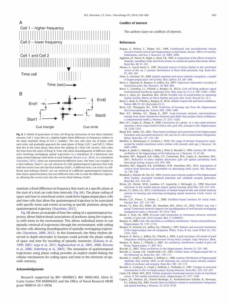
B
Start1
End1
Cue triangle Cue circle
Start2
End2
Time (sec)
Cell 1 and Cell 2
Time cell
Cell 1 – higher frequency
Cell 2 – lower frequency
A
Fig. 6. A. Model of generation of time cell firing by interaction of two theta rhythmicneurons. Cell 1 (top) fires at a slightly higher fixed difference in frequency relative tothe theta rhythmic firing of Cell 2 (middle). The two cells start out of phase witheach other and gradually approach the same phase of firing (Cell 1 and Cell 2). Whenthey fire at the same phase, they drive the spiking of a Time Cell (arrow), that codesthe time from the onset of firing. B. Time cells allow disambiguation of different mem-ories involving overlapping spatial trajectories in a simulation of a behavioral taskusing virtual hallways with items in each hallway (Brown et al., 2010). In a simulation(Hasselmo, 2012), items are represented by different icons. One item (cue triangle) ina start hallway (Start1) can cue retrieval of a full spatiotemporal trajectory that endswith the correct turn into the final hallway (End1). A different item (cue circle) in a dif-ferent start hallway (Start2) can cue retrieval of a different spatiotemporal trajectorythat shares spatial locations, but uses different time cells to code the different trajecto-ry, allowing the correct turn into the correct final hallway (End2).
663M.E. Hasselmo, C.E. Stern / NeuroImage 85 (2014) 656–666
maintain a fixed difference in frequency that starts at a specific phase atthe start of a trial can code time intervals (Fig. 6A). The phase coding ofspace and time in entorhinal cortex could drive hippocampal place cellsand time cells that allow the spatiotemporal trajectory to be associatedwith specific items and events occurring at specific positions along thespatiotemporal trajectory (Hasselmo, 2012).
Fig. 6B shows an example of how the coding of a spatiotemporal tra-jectory allows bidirectional associations of positions along the trajecto-ry with items in the environment. This allows individual items to cueepisodic retrieval of trajectories through the environment, with codingby time cells allowing disambiguation of spatially overlapping trajecto-ries (Hasselmo, 2009, 2012). In this framework, the theta rhythm ob-served in depth electrodes in humans could provide the phase codingof space and time for encoding of episodic memories (Kahana et al.,1999, 2001; Lega et al., 2011; Raghavachari et al., 2001, 2006; Rizzutoet al., 2006; Sederberg et al., 2003). The simulation of spatiotemporaltrajectories using phase coding provides an explicit model linking thecellular mechanisms for coding space and time to the elements of epi-sodic memory.
Acknowledgments
Research supported by R01 MH60013, R01 MH61492, Silvio O.Conte Center P50 MH094263 and the Office of Naval Research MURIgrant N00014-10-1-0936.
Conflict of interest
The authors have no conflicts of interest.
References
Acquas, E., Wilson, C., Fibiger, H.C., 1996. Conditioned and unconditioned stimuliincrease frontal cortical and hippocampal acetylcholine release: effects of novelty,habituation, and fear. J. Neurosci. 16, 3089–3096.
Aggleton, J.P., Neave, N., Nagle, S., Hunt, P.R., 1995. A comparison of the effects of anteriorthalamic, mamillary body and fornix lesions on reinforced spatial alternation. Behav.Brain Res. 68, 91–101.
Alonso, A., Garcia-Austt, E., 1987. Neuronal sources of theta rhythm in the entorhinalcortex of the rat. I. Laminar distribution of theta field potentials. Exp. Brain Res.67, 493–501.
Arleo, A., Gerstner, W., 2000. Spatial cognition and neuro-mimetic navigation: a modelof hippocampal place cell activity. Biol. Cybern. 83, 287–299.
Barry, C., Hayman, R., Burgess, N., Jeffery, K.J., 2007. Experience-dependent rescaling ofentorhinal grids. Nat. Neurosci. 10, 682–684.
Barry, C., Ginzberg, L.L., O'Keefe, J., Burgess, N., 2012a. Grid cell firing patterns signalenvironmental novelty by expansion. Proc. Natl. Acad. Sci. U. S. A. 109, 17687–17692.
Barry, C., Heys, J.G., Hasselmo, M.E., 2012b. Possible role of acetylcholine in regulatingspatial novelty effects on theta rhythm and grid cells. Front. Neural Circ. 6, 5.
Barry, C., Bush, D., O'Keefe, J., Burgess, N., 2012c. Models of grid cells and theta oscillations.Nature 488, E1–E2 (discussion E2-3).
Berry, S.D., Thompson, R.F., 1978. Prediction of learning rate from the hippocampalelectroencephalogram. Science 200, 1298–1300.
Blair, H.T., Welday, A.C., Zhang, K., 2007. Scale-invariant memory representationsemerge from moire interference between grid fields that produce theta oscillations:a computational model. J. Neurosci. 27, 3211–3229.
Blair, H.T., Gupta, K., Zhang, K., 2008. Conversion of a phase- to a rate-coded positionsignal by a three-stage model of theta cells, grid cells, and place cells. Hippocampus18, 1239–1255.
Bland, B.H., Oddie, S.D., 2001. Theta band oscillation and synchrony in the hippocampalformation and associated structures: the case for its role in sensorimotor integration.Behav. Brain Res. 127, 119–136.
Boehlen, A., Heinemann, U., Erchova, I., 2010. The range of intrinsic frequencies repre-sented by medial entorhinal cortex stellate cells extends with age. J. Neurosci. 30,4585–4589.
Bragin, A., Jando, G., Nadasdy, Z., Hetke, J., Wise, K., Buzsaki, G., 1995. Gamma (40–100 Hz)oscillation in the hippocampus of the behaving rat. J. Neurosci. 15, 47–60.
Brandon, M.P., Bogaard, A.R., Libby, C.P., Connerney, M.A., Gupta, K., Hasselmo, M.E.,2011. Reduction of theta rhythm dissociates grid cell spatial periodicity fromdirectional tuning. Science 332, 595–599.
Brandon, M.P., Bogaard, A.R., Schultheiss, N.W., Hasselmo, M.E., 2013. Segregation ofcortical head direction cell assemblies on alternating theta cycles. Nat. Neurosci.16, 739–748.
Brankack, J., Stewart, M., Fox, S.E., 1993. Current source density analysis of the hippocampaltheta rhythm: associated sustained potentials and candidate synaptic generators.Brain Res. 615, 310–327.
Brioni, J.D., Decker, M.W., Gamboa, L.P., Izquierdo, I., McGaugh, J.L., 1990. Muscimolinjections in the medial septum impair spatial learning. Brain Res. 522, 227–234.
Brown, T.I., Stern, C.E., 2013. Contributions of medial temporal lobe and striatal memorysystems to learning and retrieving overlapping spatial memories. Cereb. Cortex (inpress).
Brown, G.D., Preece, T., Hulme, C., 2000. Oscillator-based memory for serial order.Psychol. Rev. 107, 127–181.
Brown, T.I., Ross, R.S., Keller, J.B., Hasselmo, M.E., Stern, C.E., 2010. Which way was Igoing? Contextual retrieval supports the disambiguation of well learned overlappingnavigational routes. J. Neurosci. 30, 7414–7422.
Burak, Y., Fiete, I.R., 2009. Accurate path integration in continuous attractor networkmodels of grid cells. PLoS Comput. Biol. 5, e1000291.
Burgess, N., 2008. Grid cells and theta as oscillatory interference: theory and predictions.Hippocampus 18, 1157–1174.
Burgess, N., Donnett, J.G., Jeffery, K.J., O'Keefe, J., 1997. Robotic and neuronal simulationof the hippocampus and rat navigation. Philos. Trans. R. Soc. Lond. B Biol. Sci. 352,1535–1543.
Burgess, N., Barry, C., Jeffery, K.J., O'Keefe, J., 2005. A grid and place cell model of pathintegration utilizing phase precession versus theta. Comput. Cogn. Neurosci. Meet.
Burgess, N., Barry, C., O'Keefe, J., 2007. An oscillatory interference model of grid cellfiring. Hippocampus 17, 801–812.
Buzsaki, G., 2002. Theta oscillations in the hippocampus. Neuron 33, 325–340.Buzsaki, G., Leung, L.W., Vanderwolf, C.H., 1983. Cellular bases of hippocampal EEG in
the behaving rat. Brain Res. 287, 139–171.Buzsaki, G., Czopf, J., Kondakor, I., Kellenyi, L., 1986. Laminar distribution of hippocampal
rhythmic slow activity (RSA) in the behaving rat: current-source density analysis,effects of urethane and atropine. Brain Res. 365, 125–137.
Buzsaki, G., Grastyan, E., Czopf, J., Kellenyi, L., Prohaska, O., 1981. Changes in neuronaltransmission in the rat hippocampus during behavior. Brain Res. 225, 235–247.
Canto, C.B., Witter, M.P., 2012. Cellular properties of principal neurons in the rat entorhinalcortex. II. The medial entorhinal cortex. Hippocampus 22, 1277–1299.
Caplan, J.B., Madsen, J.R., Schulze-Bonhage, A., Aschenbrenner-Schiebe, R., Newman,E.L., Kahana, M.J., 2003. Human theta oscillations related to sensorimotor integrationand spatial learning. J. Neurosci. 23, 4725–4736.
664 M.E. Hasselmo, C.E. Stern / NeuroImage 85 (2014) 656–666
Chrobak, J.J., Buzsaki, G., 1998. Gamma oscillations in the entorhinal cortex of the freelybehaving rat. J. Neurosci. 18, 388–398.
Chrobak, J.J., Stackman, R.W., Walsh, T.J., 1989. Intraseptal administration of muscimol pro-duces dose-dependentmemory impairments in the rat. Behav. Neural Biol. 52, 357–369.
Climer, J.R., Newman, E.L., Hasselmo, M.E., 2013. Phase coding by grid cells in unconstrainedenvironments: two-dimensional phase precession. Eur. J. Neurosci. (in press).
Colgin, L.L., Denninger, T., Fyhn, M., Hafting, T., Bonnevie, T., Jensen, O., Moser, M.B.,Moser, E.I., 2009. Frequency of gamma oscillations routes flow of information inthe hippocampus. Nature 462, 353–357.
Cornwell, B.R., Johnson, L.L., Holroyd, T., Carver, F.W., Grillon, C., 2008. Humanhippocampal and parahippocampal theta during goal-directed spatial navigationpredicts performance on a virtual Morris water maze. J. Neurosci. 28, 5983–5990.
Csicsvari, J., Hirase, H., Czurko, A., Mamiya, A., Buzsaki, G., 1999. Oscillatory coupling ofhippocampal pyramidal cells and interneurons in the behaving Rat. J. Neurosci. 19,274–287.
Cutsuridis, V., Hasselmo, M., 2012. GABAergic contributions to gating, timing, and phaseprecession of hippocampal neuronal activity during theta oscillations. Hippocampus22, 1597–1621.
De Araujo, D.B., Baffa, O., Wakai, R.T., 2002. Theta oscillations and human navigation: amagnetoencephalography study. J. Cogn. Neurosci. 14, 70–78.
Deshmukh, S.S., Yoganarasimha, D., Voicu, H., Knierim, J.J., 2010. Theta modulation inthe medial and the lateral entorhinal cortices. J. Neurophysiol. 104, 994–1006.
Dickson, C.T., Magistretti, J., Shalinsky, M.H., Fransen, E., Hasselmo, M.E., Alonso, A.,2000. Properties and role of I(h) in the pacing of subthreshold oscillations in entorhinalcortex layer II neurons. J. Neurophysiol. 83, 2562–2579.
Dodson, P.D., Pastoll, H., Nolan, M.F., 2011. Dorsal-ventral organization of theta-like activityintrinsic to entorhinal stellate neurons is mediated by differences in stochastic currentfluctuations. J. Physiol. 589, 2993–3008.
Doeller, C.F., Barry, C., Burgess, N., 2010. Evidence for grid cells in a human memorynetwork. Nature 463, 657–661.
Domnisoru, C., Kinkhabwala, A.A., Tank, D.W., 2013. Membrane potential dynamics ofgrid cells. Nature 495, 199–204.
Duncan, K., Sadanand, A., Davachi, L., 2012. Memory's penumbra: episodic memorydecisions induce lingering mnemonic biases. Science 337, 485–487.
Ekstrom, A.D., Kahana, M.J., Caplan, J.B., Fields, T.A., Isham, E.A., Newman, E.L., Fried, I.,2003. Cellular networks underlying human spatial navigation. Nature 425, 184–188.
Ekstrom, A.D., Caplan, J.B., Ho, E., Shattuck, K., Fried, I., Kahana, M.J., 2005. Humanhippocampal theta activity during virtual navigation. Hippocampus 15, 881–889.
Erchova, I., Kreck, G., Heinemann, U., Herz, A.V., 2004. Dynamics of rat entorhinal cor-tex layer II and III cells: characteristics of membrane potential resonance at restpredict oscillation properties near threshold. J. Physiol. 560, 89–110.
Erdem, U.M., Hasselmo, M., 2012. A goal-directed spatial navigation model usingforward trajectory planning based on grid cells. Eur. J. Neurosci. 35, 916–931.
Fox, S.E., 1989. Membrane potential and impedance changes in hippocampal pyramidalcells during theta rhythm. Exp. Brain Res. 77, 283–294.
Fox, S.E., Wolfson, S., Ranck Jr., J.B., 1986. Hippocampal theta rhythm and the firing ofneurons in walking and urethane anesthetized rats. Brain Res. 62, 495–508.
Fransén, E., Alonso, A.A., Dickson, C.T., Magistretti, J., Hasselmo, M.E., 2004. Ionicmechanisms in the generation of subthreshold oscillations and action potentialclustering in entorhinal layer II stellate neurons. Hippocampus 14, 368–384.
Fuhs, M.C., Touretzky, D.S., 2006. A spin glass model of path integration in rat medialentorhinal cortex. J. Neurosci. 26, 4266–4276.
Fujita, Y., Sato, T., 1964. Intracellular records from hippocampal pyramidal cells inrabbit during theta rhythm activity. J. Neurophysiol. 27, 1012–1025.
Fyhn, M., Molden, S., Witter, M.P., Moser, E.I., Moser, M.B., 2004. Spatial representationin the entorhinal cortex. Science 305, 1258–1264.
Geisler, C., Robbe, D., Zugaro, M., Sirota, A., Buzsaki, G., 2007. Hippocampal place cellassemblies are speed-controlled oscillators. Proc. Natl. Acad. Sci. U. S. A. 104, 8149–8154.
Giocomo, L.M., Hasselmo, M.E., 2008a. Computation by oscillations: implications of ex-perimental data for theoretical models of grid cells. Hippocampus 18, 1186–1199.
Giocomo, L.M., Hasselmo, M.E., 2008b. Time constants of h current in layer II stellatecells differ along the dorsal to ventral axis of medial entorhinal cortex. J. Neurosci.28, 9414–9425.
Giocomo, L.M., Hasselmo, M.E., 2009. Knock-out of HCN1 subunit flattens dorsal-ventral frequency gradient of medial entorhinal neurons in adult mice. J. Neurosci.29, 7625–7630.
Giocomo, L.M., Zilli, E.A., Fransen, E., Hasselmo, M.E., 2007. Temporal frequency ofsubthreshold oscillations scales with entorhinal grid cell field spacing. Science315, 1719–1722.
Giocomo, L.M., Hussaini, S.A., Zheng, F., Kandel, E.R., Moser, M.B., Moser, E.I., 2011. Gridcells use HCN1 channels for spatial scaling. Cell 147, 1159–1170.
Givens, B., 1996. Stimulus-evoked resetting of the dentate theta rhythm: relation toworking memory. Neuroreport 8, 159–163.
Givens, B.S., Olton, D.S., 1990. Cholinergic and GABAergic modulation of the medialseptal area: effect on working memory. Behav. Neurosci. 104, 849–855.
Givens, B., Olton, D.S., 1994. Local modulation of basal forebrain: effects on workingand reference memory. J. Neurosci. 14, 3578–3587.
Golding, N., Staff, N., Spruston, N., 2002. Dendritic spikes as a mechanism for cooperativelong-term potentiation. Nature 418, 326–331.
Green, J.D., Arduini, A.A., 1954. Hippocampal electrical activity and arousal. J. Neurophysiol.17, 533–557.
Griffin, A.L., Asaka, Y., Darling, R.D., Berry, S.D., 2004. Theta-contingent trial presentationaccelerates learning rate and enhances hippocampal plasticity during trace eyeblinkconditioning. Behav. Neurosci. 118, 403–411.
Guanella, A., Kiper, D., Verschure, P., 2007. A model of grid cells based on a twistedtorus topology. Int. J. Neural Syst. 17, 231–240.
Guderian, S., Schott, B.H., Richardson-Klavehn, A., Duzel, E., 2009. Medial temporal thetastate before an event predicts episodic encoding success in humans. Proc. Natl. Acad.Sci. U. S. A. 106, 5365–5370.
Haas, J.S., White, J.A., 2002. Frequency selectivity of layer II stellate cells in the medialentorhinal cortex. J. Neurophysiol. 88, 2422–2429.
Hafting, T., Fyhn, M., Molden, S., Moser, M.B., Moser, E.I., 2005. Microstructure of aspatial map in the entorhinal cortex. Nature 436, 801–806.
Hafting, T., Fyhn, M., Bonnevie, T., Moser, M.B., Moser, E.I., 2008. Hippocampus-independentphase precession in entorhinal grid cells. Nature 453, 1248–1252.
Hargreaves, E.L., Rao, G., Lee, I., Knierim, J.J., 2005. Major dissociation between medialand lateral entorhinal input to dorsal hippocampus. Science 308, 1792–1794.
Harvey, C.D., Collman, F., Dombeck, D.A., Tank, D.W., 2009. Intracellular dynamics ofhippocampal place cells during virtual navigation. Nature 461, 941–946.
Hasselmo, M.E., 2008. Grid cell mechanisms and function: contributions of entorhinalpersistent spiking and phase resetting. Hippocampus 18, 1213–1229.
Hasselmo, M.E., 2009. A model of episodic memory: mental time travel along encodedtrajectories using grid cells. Neurobiol. Learn. Mem. 92, 559–573.
Hasselmo, M.E., 2012. How we remember: brain mechanisms of episodic memory.MIT Press, Cambridge, MA.
Hasselmo, M.E., Brandon, M.P., 2008. Linking cellular mechanisms to behavior: entorhinalpersistent spiking and membrane potential oscillations may underlie path integra-tion, grid cell firing, and episodic memory. Neural Plast. 2008, 658323.
Hasselmo, M.E., Brandon, M.P., 2012. A model combining oscillations and attractordynamics for generation of grid cell firing. Front. Neural Circ. 6, 30.
Hasselmo, M.E., Eichenbaum, H., 2005. Hippocampal mechanisms for the context-dependent retrieval of episodes. Neural Netw. 18, 1172–1190.
Hasselmo, M.E., Fehlau, B.P., 2001. Differences in time course of ACh and GABAmodulationof excitatory synaptic potentials in slices of rat hippocampus. J. Neurophysiol. 86,1792–1802.
Hasselmo, M.E., Bodelon, C., Wyble, B.P., 2002a. A proposed function for hippocampaltheta rhythm: separate phases of encoding and retrieval enhance reversal ofprior learning. Neural Comput. 14, 793–817.
Hasselmo, M.E., Wyble, B.P., Cannon, R.C., 2002b. From spike frequency to free recall;how neural circuits perform encoding and retrieval. In: Parker, A., Bussey, T.J.,Wilding, E. (Eds.), The Cognitive Neuroscience of Memory: Encoding and Retrieval.Psychology Press, London.
Hasselmo, M.E., Giocomo, L.M., Zilli, E.A., 2007. Grid cell firingmay arise from interferenceof theta frequency membrane potential oscillations in single neurons. Hippocampus17, 1252–1271.
Heys, J.G., Hasselmo, M.E., 2012. Neuromodulation of I(h) in layer II medial entorhinalcortex stellate cells: a voltage-clamp study. J. Neurosci. 32, 9066–9072.
Heys, J.G., Giocomo, L.M., Hasselmo, M.E., 2010. Cholinergic modulation of the resonanceproperties of stellate cells in layer II of medial entorhinal cortex. J. Neurophysiol. 104,258–270.
Heys, J.G., MacLeod, K.M., Moss, C.F., Hasselmo, M.E., 2013. Bat and rat neurons dif-fer in theta frequency resonance despite similar coding of space. Science 340,363–367.
Hinman, J.R., Penley, S.C., Long, L.L., Escabi, M.A., Chrobak, J.J., 2011. Septotemporal varia-tion in dynamics of theta: speed and habituation. J. Neurophysiol. 105, 2675–2686.
Hinman, J.R., Penley, S.C., Escabi, M.A., Chrobak, J.J., 2013. Ketamine disrupts theta synchronyacross the septotemporal axis of the CA1 region of hippocampus. J. Neurophysiol. 109,570–579.
Holscher, C., Anwyl, R., Rowan, M.J., 1997. Stimulation on the positive phase of hippo-campal theta rhythm induces long-term potentiation that can be depotentiated bystimulation on the negative phase in area CA1 in vivo. J. Neurosci. 17, 6470–6477.
Huerta, P.T., Lisman, J.E., 1995. Bidirectional synaptic plasticity induced by a singleburst during cholinergic theta oscillation in CA1 in vitro. Neuron 15, 1053–1063.
Huxter, J., Burgess, N., O'Keefe, J., 2003. Independent rate and temporal coding inhippocampal pyramidal cells. Nature 425, 828–832.
Huxter, J.R., Senior, T.J., Allen, K., Csicsvari, J., 2008. Theta phase-specific codes for two-dimensional position, trajectory and heading in the hippocampus. Nat. Neurosci.11, 587–594.
Hyman, J.M., Wyble, B.P., Goyal, V., Rossi, C.A., Hasselmo, M., 2003a. Stimulationin hippocampal region CA1 in behaving rats yields LTP when delivered tothe peak of theta and LTD when delivered to the trough. J. Neurosci. 23,11725–11731.
Hyman, J.M., Wyble, B.P., Goyal, V., Rossi, C.A., Hasselmo, M.E., 2003b. Stimulation inhippocampal region CA1 in behaving rats yields long-term potentiation whendelivered to the peak of theta and long-term depression when delivered to thetrough. J. Neurosci. 23, 11725–11731.
Jacobs, J., Hwang, G., Curran, T., Kahana, M.J., 2006. EEG oscillations and recognitionmemory: theta correlates of memory retrieval and decision making. Neuroimage32, 978–987.
Jeewajee, A., Barry, C., O'Keefe, J., Burgess, N., 2008. Grid cells and theta as oscillatoryinterference: electrophysiological data from freely moving rats. Hippocampus 18,1175–1185.
Jeffery, K.J., Donnett, J.G., O'Keefe, J., 1995. Medial septal control of theta-correlated unitfiring in the entorhinal cortex of awake rats. Neuroreport 6, 2166–2170.
Jensen, O., Lisman, J.E., 1996. Hippocampal CA3 region predicts memory sequences:accounting for the phase precession of place cells. Learn. Mem. 3, 279–287.
Jensen, O., Lisman, J.E., 2000. Position reconstruction from an ensemble of hippocampalplace cells: contribution of theta phase coding. J. Neurophysiol. 83, 2602–2609.
Jensen, O., Tesche, C.D., 2002. Frontal theta activity in humans increases with memoryload in a working memory task. Eur. J. Neurosci. 15, 1395–1399.
Jones, M.W., Wilson, M.A., 2005. Theta rhythms coordinate hippocampal–prefrontalinteractions in a spatial memory task. PLoS Biol. 3, e402.

665M.E. Hasselmo, C.E. Stern / NeuroImage 85 (2014) 656–666
Kahana, M.J., Sekuler, R., Caplan, J.B., Kirschen, M., Madsen, J.R., 1999. Human thetaoscillations exhibit task dependence during virtual maze navigation. Nature 399,781–784.
Kahana, M.J., Seelig, D., Madsen, J.R., 2001. Theta returns. Curr. Opin. Neurobiol. 11, 739–744.Kamondi, A., Acsady, L., Wang, X.J., Buzsaki, G., 1998. Theta oscillations in somata and
dendrites of hippocampal pyramidal cells in vivo: activity-dependent phase-precessionof action potentials. Hippocampus 8, 244–261.
Kaplan, R., Doeller, C.F., Barnes, G.R., Litvak, V., Duzel, E., Bandettini, P.A., Burgess, N., 2012.Movement-related theta rhythm in humans: coordinating self-directed hippocampallearning. PLoS Biol. 10, e1001267.
Kelemen, E., Moron, I., Fenton, A.A., 2005. Is the hippocampal theta rhythm related tocognition in a non-locomotor place recognition task? Hippocampus 15, 472–479.
Kjelstrup, K.B., Solstad, T., Brun, V.H., Hafting, T., Leutgeb, S., Witter, M.P., Moser, E.I., Moser,M.B., 2008. Finite scale of spatial representation in the hippocampus. Science 321,140–143.
Klausberger, T., Somogyi, P., 2008. Neuronal diversity and temporal dynamics: theunity of hippocampal circuit operations. Science 321, 53–57.
Klausberger, T., Magill, P.J., Marton, L.F., Roberts, J.D., Cobden, P.M., Buzsaki, G., Somogyi,P., 2003. Brain-state- and cell-type-specific firing of hippocampal interneurons in vivo.Nature 421, 844–848.
Klimesch, W., 1999. EEG alpha and theta oscillations reflect cognitive and memoryperformance: a review and analysis. Brain Res. Brain Res. Rev. 29, 169–195.
Klimesch,W., Schimke, H., Schwaiger, J., 1994. Episodic and semanticmemory: an analysisin the EEG theta and alpha band. Electroencephalogr. Clin. Neurophysiol. 91, 428–441.
Klimesch,W., Doppelmayr,M., Russegger, H., Pachinger, T., 1996. Theta band power in thehuman scalp EEG and the encoding of new information. Neuroreport 7, 1235–1240.
Koenig, J., Linder, A.N., Leutgeb, J.K., Leutgeb, S., 2011. The spatial periodicity of gridcells is not sustained during reduced theta oscillations. Science 332, 592–595.
Kraus, B.J., Robinson, R.J., White, J.A., Eichenbaum, H., Hasselmo, M.E., 2013. Hippocampal“time cells”: time versus path integration. Neuron 78, 1090–1101.
Kropff, E., Treves, A., 2008. The emergence of grid cells: intelligent design or justadaptation? Hippocampus 18, 1256–1269.
Kunec, S., Hasselmo, M.E., Kopell, N., 2005. Encoding and retrieval in the CA3 region ofthe hippocampus: a model of theta-phase separation. J. Neurophysiol. 94, 70–82.
Lee, M.G., Hassani, O.K., Alonso, A., Jones, B.E., 2005. Cholinergic basal forebrain neuronsburst with theta during waking and paradoxical sleep. J. Neurosci. 25, 4365–4369.
Lega, B.C., Jacobs, J., Kahana, M., 2011. Human hippocampal theta oscillations and theformation of episodic memories. Hippocampus 22, 748–761.
Lenck-Santini, P.P., Fenton, A.A., Muller, R.U., 2008. Discharge properties of hippocampalneurons during performance of a jump avoidance task. J. Neurosci. 28, 6773–6786.
MacDonald, C.J., LePage, K.Q., Eden, U.T., Eichenbaum, H., 2011. Hippocampal “timecells” bridge the gap in memory for discontiguous events. Neuron 71, 737–749.
Macrides, F.H., Eichenbaum, H., Forbes, W.B., 1982. Temporal relationship between sniffingand limbic theta rhythm during odor discrimination reversal learning. J. Neurosci. 2,1705.
Manns, J.R., Zilli, E.A., Ong, K.C., Hasselmo, M.E., Eichenbaum, H., 2007. Hippocampal CA1spiking during encoding and retrieval: relation to theta phase. Neurobiol. Learn.Mem. 87, 9–20.
Markowska, A.L., Olton, D.S., Murray, E.A., Gaffan, D., 1989. A comparative analysis of therole of fornix and cingulate cortex in memory: rats. Exp. Brain Res. 74, 187–201.
Martin, M.M., Horn, K.L., Kusman, K.J., Wallace, D.G., 2007. Medial septum lesionsdisrupt exploratory trip organization: evidence for septohippocampal involvementin dead reckoning. Physiol. Behav. 90, 412–424.
Maurer, A.P., Vanrhoads, S.R., Sutherland, G.R., Lipa, P., McNaughton, B.L., 2005. Self-motionand the origin of differential spatial scaling along the septo-temporal axis of thehippocampus. Hippocampus 15, 841–852.
McCartney, H., Johnson, A.D., Weil, Z.M., Givens, B., 2004. Theta reset produces optimalconditions for long-term potentiation. Hippocampus 14, 684–687.
McNaughton, B.L., Battaglia, F.P., Jensen, O., Moser, E.I., Moser, M.B., 2006a. Pathintegration and the neural basis of the ‘cognitivemap’. Nat. Rev. Neurosci. 7, 663–678.
McNaughton, N., Ruan, M., Woodnorth, M.-A., 2006b. Restoring theta-like rhythmicity inrats restores initial learning in the Morris water maze. Hippocampus 16, 1102–1110.
Mehta, M.R., Lee, A.K., Wilson, M.A., 2002. Role of experience and oscillations intransforming a rate code into a temporal code. Nature 417, 741–746.
M'Harzi, M., Palacios, A., Monmaur, P., Willig, F., Houcine, O., Delacour, J., 1987. Effectsof selective lesions of fimbria-fornix on learning set in the rat. Physiol. Behav. 40,181–188.
Mhatre, H., Gorchetchnikov, A., Grossberg, S., 2010. Grid cell hexagonal patterns formedby fast self-organized learning within entorhinal cortex. Hippocampus 22, 320–334.
Miall, R., 1989. The storage of time intervals using oscillating neurons. Neural Comput.1, 359–371.
Mitchell, S.J., Ranck Jr., J.B., 1980. Generation of theta rhythm in medial entorhinalcortex of freely moving rats. Brain Res. 189, 49–66.
Mitchell, S.J., Rawlins, J.N., Steward, O., Olton, D.S., 1982.Medial septal area lesions disrupttheta rhythm and cholinergic staining in medial entorhinal cortex and produce im-paired radial arm maze behavior in rats. J. Neurosci. 2, 292–302.
Mizumori, S.J.Y., Perez, G.M., Alvarado, M.C., Barnes, C.A., Mcnaughton, B.L., 1990.Reversible inactivation of the medial septum differentially affects 2 forms of learningin rats. Brain Res. 528, 12–20.
Mizuseki, K., Sirota, A., Pastalkova, E., Buzsaki, G., 2009. Theta oscillations provide temporalwindows for local circuit computation in the entorhinal–hippocampal loop. Neuron64, 267–280.
Molyneaux, B.J., Hasselmo,M.E., 2002. GABA(B) presynaptic inhibition has an in vivo timeconstant sufficiently rapid to allow modulation at theta frequency. J. Neurophysiol.87, 1196–1205.
Moser, E.I., Moser, M.B., 2008. A metric for space. Hippocampus 18, 1142–1156.
Navratilova, Z., Giocomo, L.M., Fellous, J.M., Hasselmo, M.E., McNaughton, B.L., 2012.Phase precession and variable spatial scaling in a periodic attractor map model ofmedial entorhinal grid cells with realistic after-spike dynamics. Hippocampus 22,772–789.
Newman, E.L., Norman, K.A., 2010. Moderate excitation leads to weakening of perceptualrepresentations. Cereb. Cortex 20, 2760–2770.
Niedermeyer, E., 1999. The normal EEG of the waking adult. In: Niedermeyer, E., Lopes daSilva, F. (Eds.), Electroencephalography: Basic Principles, Clinical Applications andRelated Fields. Lippincott Williams & Wilkins, Baltimore, MD, pp. 149–173.
Norman, K.A., Newman, E.L., Detre, G., Polyn, S., 2006. How inhibitory oscillations cantrain neural networks and punish competitors. Neural. Comput. 18, 1577–1610.
Norman, K.A., Newman, E.L., Detre, G., 2007. A neural network model of retrieval-inducedforgetting. Psychol. Rev. 114, 887–953.
Numan, R., Quaranta Jr., J.R., 1990. Effects of medial septal lesions on operant delayedalternation in rats. Brain Res. 531, 232–241.
O'Keefe, J., 1976. Place units in the hippocampus of the freely moving rat. Exp. Neurol.51, 78–109.
O'Keefe, J., Dostrovsky, J., 1971. The hippocampus as a spatial map. Preliminaryevidence from unit activity in the freely-moving rat. Brain Res. 34, 171–175.
O'Keefe, J., Nadel, L., 1978. The Hippocampus as a Cognitive Map. Oxford UniversityPress, Oxford, UK.
O'Keefe, J., Recce, M.L., 1993. Phase relationship between hippocampal place units andthe EEG theta rhythm. Hippocampus 3, 317–330.
O'Keefe, J., Burgess, N., Donnett, J.G., Jeffery, K.J., Maguire, E.A., 1998. Place cells, navigationalaccuracy, and the human hippocampus. Philos. Trans. R. Soc. Lond. B Biol. Sci. 353,1333–1340.
Orr, G., Rao, G., Houston, F.P., McNaughton, B.L., Barnes, C.A., 2001. Hippocampal synapticplasticity is modulated by theta rhythm in the fascia dentata of adult and aged freelybehaving rats. Hippocampus 11, 647–654.
Osipova, D., Takashima, A., Oostenveld, R., Fernandez, G., Maris, E., Jensen, O., 2006.Theta and gamma oscillations predict encoding and retrieval of declarative memory.J. Neurosci. 26, 7523–7531.
Pastalkova, E., Itskov, V., Amarasingham, A., Buzsaki, G., 2008. Internally generated cellassembly sequences in the rat hippocampus. Science 321, 1322–1327.
Pastoll, H., Ramsden, H.L., Nolan, M.F., 2012. Intrinsic electrophysiological properties ofentorhinal cortex stellate cells and their contribution to grid cell firing fields. Front.Neural Circ. 6, 17.
Pavlides, C., Greenstein, Y.J., Grudman, M., Winson, J., 1988. Long-term potentiation inthe dentate gyrus is induced preferentially on the positive phase of theta-rhythm.Brain Res. 439, 383–387.
Raghavachari, S., Kahana, M.J., Rizzuto, D.S., Caplan, J.B., Kirschen, M.P., Bourgeois, B.,Madsen, J.R., Lisman, J.E., 2001. Gating of human theta oscillations by a workingmemory task. J. Neurosci. 21, 3175–3183.
Raghavachari, S., Lisman, J.E., Tully, M., Madsen, J.R., Bromfield, E.B., Kahana, M.J., 2006.Theta oscillations in human cortex during a working-memory task: evidence forlocal generators. J. Neurophysiol. 95, 1630–1638.
Rawlins, J.N., Feldon, J., Gray, J.A., 1979. Septo-hippocampal connections and thehippocampal theta rhythm. Exp. Brain Res. 37, 49–63.
Redish, A.D., Touretzky, D.S., 1998. The role of the hippocampus in solving the Morriswater maze. Neural Comput. 10, 73–111.
Remme, M.W., Lengyel, M., Gutkin, B.S., 2010. Democracy-independence trade-off inoscillating dendrites and its implications for grid cells. Neuron 66, 429–437.
Rivas, J., Gaztelu, J.M., Garcia-Austt, E., 1996. Changes in hippocampal cell dischargepatterns and theta rhythm spectral properties as a function of walking velocityin the guinea pig. Exp. Brain Res. 108, 113–118.
Rizzuto, D.S., Madsen, J.R., Bromfield, E.B., Schulze-Bonhage, A., Kahana, M.J., 2006.Human neocortical oscillations exhibit theta phase differences between encodingand retrieval. Neuroimage 31, 1352–1358.
Rotstein, H.G., Pervouchine, D.D., Acker, C.D., Gillies, M.J., White, J.A., Buhl, E.H.,Whittington, M.A., Kopell, N., 2005. Slow and fast inhibition and anH-current interactto create a theta rhythm in a model of CA1 interneuron network. J. Neurophysiol. 94,1509–1518.
Rudell, A.P., Fox, S.E., Ranck, J.B., 1980. Hippocampal excitability phase-locked to thetheta rhythm in walking rats. Exp. Neurol. 68, 87–96.
Sainsbury, R.S., Harris, J.L., Rowland, G.L., 1987a. Sensitization and hippocampal type 2theta in the rat. Physiol. Behav. 41, 489–493.
Sainsbury, R.S., Heynen, A., Montoya, C.P., 1987b. Behavioral correlates of hippocampaltype 2 theta in the rat. Physiol. Behav. 39, 513–519.
Samsonovich, A., McNaughton, B.L., 1997. Path integration and cognitive mapping in acontinuous attractor neural network model. J. Neurosci. 17, 5900–5920.
Sargolini, F., Fyhn, M., Hafting, T., McNaughton, B.L., Witter, M.P., Moser, M.B., Moser,E.I., 2006. Conjunctive representation of position, direction, and velocity in entorhinalcortex. Science 312, 758–762.
Schmidt-Hieber, C., Häusser, M., 2013. Cellular mechanisms of spatial navigation in themedial entorhinal cortex. Nat. Neurosci. 16, 325–331.
Seager, M.A., Johnson, L.D., Chabot, E.S., Asaka, Y., Berry, S.D., 2002. Oscillatory brainstates and learning: Impact of hippocampal theta-contingent training. Proc. Natl.Acad. Sci. U. S. A. 99, 1616–1620.
Sederberg, P.B., Kahana, M.J., Howard, M.W., Donner, E.J., Madsen, J.R., 2003. Theta andgamma oscillations during encoding predict subsequent recall. J. Neurosci. 23,10809–10814.
Seidenbecher, T., Laxmi, T.R., Stork, O., Pape, H.C., 2003. Amygdalar and hippocampaltheta rhythm synchronization during fear memory retrieval. Science 301, 846–850.
Shay, C.F., Boardman, I.S., James, N.M., Hasselmo, M.E., 2012. Voltage dependence of sub-threshold resonance frequency in layer II of medial entorhinal cortex. Hippocampus22, 1733–1749.

666 M.E. Hasselmo, C.E. Stern / NeuroImage 85 (2014) 656–666
Shin, J., 2011. The interrelationship between movement and cognition: theta rhythmand the P300 event-related potential. Hippocampus 21, 744–752.
Si, B., Kropff, E., Treves, A., 2012. Grid alignment in entorhinal cortex. Biol. Cybern. 106,483–506.
Skaggs, W.E., McNaughton, B.L., Wilson, M.A., Barnes, C.A., 1996. Theta phase precessionin hippocampal neuronal populations and the compression of temporal sequences.Hippocampus 6, 149–172.
Stensola, H., Stensola, T., Solstad, T., Froland, K., Moser, M.B., Moser, E.I., 2012. Theentorhinal grid map is discretized. Nature 492, 72–78.
Taube, J.S., Muller, R.U., Ranck Jr., J.B., 1990. Head-direction cells recorded fromthe postsubiculum in freely moving rats. I. Description and quantitative analysis.J. Neurosci. 10, 420–435.
Tort, A.B., Komorowski, R.W., Manns, J.R., Kopell, N.J., Eichenbaum, H., 2009. Theta-gammacoupling increases during the learning of item-context associations. Proc. Natl. Acad.Sci. U. S. A. 106, 20942–20947.
Tsodyks, M.V., Skaggs, W.E., Sejnowski, T.J., McNaughton, B.L., 1996. Population dy-namics and theta rhythm phase precession of hippocampal place cell firing: a spik-ing neuron model. Hippocampus 6, 271–280.
Vanderwolf, C.H., 1969. Hippocampal electrical activity and voluntary movement in therat. Electroencephalogr. Clin. Neurophysiol. 26, 407–418.
Vertes, R.P., Kocsis, B., 1997. Brainstem-diencephalo-septohippocampal systemscontrolling the theta rhythm of the hippocampus. Neuroscience 81, 893–926.
Villarreal, D.M., Gross, A.L., Derrick, B.E., 2007. Modulation of CA3 afferent inputs bynovelty and theta rhythm. J. Neurosci. 27, 13457–13467.
Wallenstein, G.V., Hasselmo, M.E., 1997. GABAergicmodulation of hippocampal populationactivity: sequence learning, place field development, and the phase precession effect.J. Neurophysiol. 78, 393–408.
Walter, W., Dovey, V., 1944. Electro-encephalography in cases of sub-cortical tumour.J. Neurol. Neurosurg. Psychiatry 7, 57–65.
Watrous, A.J., Fried, I., Ekstrom, A.D., 2011. Behavioral correlates of human hippocampaldelta and theta oscillations during navigation. J. Neurophysiol. 105, 1747–1755.
Watrous, A.J., Lee, D.J., Izadi, A., Gurkoff, G.G., Shahlaie, K., Ekstrom, A.D., 2013s. Acomparative study of human and rat hippocampal low-frequency oscillationsduring spatial navigation. Hippocampus (in press).
Welday, A.C., Shlifer, I.G., Bloom, M.L., Zhang, K., Blair, H.T., 2011. Cosine directionaltuning of theta cell burst frequencies: evidence for spatial coding by oscillatoryinterference. J. Neurosci. 31, 16157–16176.
Whishaw, I.Q., 1972. Hippocampal electroencephalographic activity in the Mongoliangerbil during natural behaviours and wheel running and in the rat during wheelrunning and conditioned immobility. Can. J. Psychol. 26, 219–239.
Whishaw, I.Q., Vanderwolf, C.H., 1973. Hippocampal EEG and behavior: changes inamplitude and frequency of RSA (theta rhythm) associated with spontaneousand learned movement patterns in rats and cats. Behav. Biol. 8, 461–484.
Wills, T.J., Barry, C., Cacucci, F., 2012. The abrupt development of adult-like grid cellfiring in the medial entorhinal cortex. Front. Neural Circ. 6, 21.
Winson, J., 1978. Loss of hippocampal theta rhythm results in spatial memory deficit inthe rat. Science 201, 160–163.
Wyble, B.P., Linster, C., Hasselmo, M.E., 2000. Size of CA1-evoked synaptic potentialsis related to theta rhythm phase in rat hippocampus. J. Neurophysiol. 83,2138–2144.
Yartsev, M.M., Witter, M.P., Ulanovsky, N., 2011. Grid cells without theta oscillations inthe entorhinal cortex of bats. Nature 479, 103–107.
Yoshida, M., Giocomo, L.M., Boardman, I., Hasselmo, M.E., 2011. Frequency ofsubthreshold oscillations at different membrane potential voltages in neurons atdifferent anatomical positions on the dorsoventral axis in the rat medial entorhinalcortex. J. Neurosci. 31, 12683–12694.
Zilli, E.A., Hasselmo, M.E., 2010. Coupled noisy spiking neurons as velocity-controlledoscillators in a model of grid cell spatial firing. J. Neurosci. 30, 13850–13860.
Zilli, E.A., Yoshida, M., Tahvildari, B., Giocomo, L.M., Hasselmo, M.E., 2009. Evaluationof the oscillatory interference model of grid cell firing through analysis andmeasured period variance of some biological oscillators. PLoS Comput. Biol. 5,e1000573.