harvard medical school - login...
TRANSCRIPT
1
Director, Computational Imaging and Bioinformatics Lab (CIBL)
Dana-Farber Harvard Cancer Center
Dana-Farber Cancer Institute, Brigham and Women’s Hospital
Harvard Medical School
Radiomics:
The Promise of Imaging for Precision Medicine
Hugo Aerts
HARVARD MEDICAL SCHOOL
Disclosure Information 2016 AAPM meeting Washington, DC
Dr. Hugo Aerts
I have the following financial relationships to disclose:
Shareholder: Genospace LLC, Sphera Inc.
Grant/Research support from: NIH-NCI, EU, KWF, Kaye Award, DFCI TR, BWH
Objectives
- Describe the motivation and methodology for Computational
Imaging & Radiomics
- Describe biomarker quantification studies in Radiomics and
Imaging-Genomics (Radiogenomics)

2
Imaging for precision medicine Advantages of Imaging:
- Performed non-invasively
- Provides 3D picture of the entire cancer
- Already performed in clinical practice
- Multiple times during treatment for diagnosis, staging, radiation oncology planning, response assessment
- Captures a cancer’s appearance over time and space
Disadvantages of Imaging:
- Probes the cancer at the macroscopic level
- Often qualitative not quantitative
- Very heterogeneous acquisition protocols:
- comparisons between patients difficult
- comparisons same patient in time difficult
- Storage of only reconstructed images (not the raw data)
Representative CT images of lung cancer
Tumors are different
Medical imaging can capture these phenotypic differences
Multi-level patient data
Radiomic Data
Clinical Data
Genomic Data
*Lambin et al. Eur J Cancer 2012
3
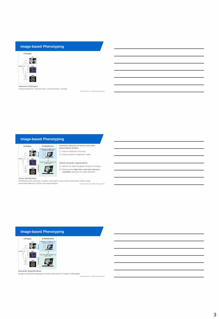
Image-based Phenotyping
PAGE 7 *accepted Aerts HJ, JAMA Oncology 2016
Important Challenges:
Image Acquisition, reconstruction, standardization, storage
Image-based Phenotyping
PAGE 8 *accepted Aerts HJ, JAMA Oncology 2016
Tumor Identification
Identifying tumor presence, location, and extend using visual assessment and/or using
automated detection (CADe) and segmentation.
Automatic detection of tumors and other
abnormalities (CADe):
1) Improve diagnostic accuracy.
2) Improve speed of diagnostic reads.
(Semi) automatic segmentation:
1) Method for high throughput analysis of images.
2) Reducing the high intra- and inter-observer
variability observed for target definition.
Image-based Phenotyping
PAGE 9 *accepted Aerts HJ, JAMA Oncology 2016
Semantic Quantification
Image-based phenotyping by visual assessment of expert radiologists
4
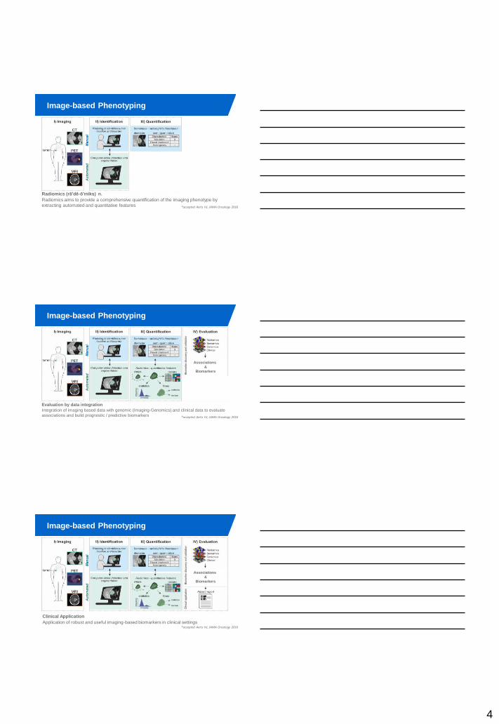
Image-based Phenotyping
PAGE
10
Radiomics (rā'dē-ō'mĭks) n.
Radiomics aims to provide a comprehensive quantification of the imaging phenotype by
extracting automated and quantitative features
*accepted Aerts HJ, JAMA Oncology 2016
Image-based Phenotyping
PAGE
11 *accepted Aerts HJ, JAMA Oncology 2016
Evaluation by data integration
Integration of imaging based data with genomic (Imaging-Genomics) and clinical data to evaluate
associations and build prognostic / predictive biomarkers
Associations
&
Biomarkers
Image-based Phenotyping
PAGE
12 *accepted Aerts HJ, JAMA Oncology 2016
Clinical Application
Application of robust and useful imaging-based biomarkers in clinical settings
Associations
&
Biomarkers

5
Image-based Phenotyping
(Radiomics and Imaging-Genomics examples)
HARVARD MEDICAL
SCHOOL
Early Grade Classification in Meningioma Patients Combining Radiomics and Semantics Data
Thibaud Coroller
Meningioma
Early Grade Classification in Meningioma Patients Combining Radiomics and Semantics Data
Thibaud Coroller
Study Design
*Coroller et al. submitted

6
Early Grade Classification in Meningioma Patients Combining Radiomics and Semantics Data
Thibaud Coroller
Univariate prediction (n=175)
* Fisher exact test * Noether test
Early Grade Classification in Meningioma Patients Combining Radiomics and Semantics Data
Thibaud Coroller
Random Forest Classifiers
*training dataset (n=131), validation dataset (n=44) *Coroller et al. submitted
Early Grade Classification in Meningioma Patients Combining Radiomics and Semantics Data
Thibaud Coroller
Conclusions meningioma
• Clinical model could not predicted grade
• Radiographic features did predicted grade
– Semantic (simple, intuitive)
– Radiomic (reproducible, high throughout)
• Combined model (sem. + rad.)
significantly improved grade classification
*Coroller et al. submitted

7
Imaging-Genomics in GBM
Methods: Manual delineations
PAGE 20 *Grossmann accepted BMC Cancer
Prognostic value of volumetric features
*Grossmann accepted BMC Cancer
8
*n=96 GBM patients
from TCGA-GBM cohort
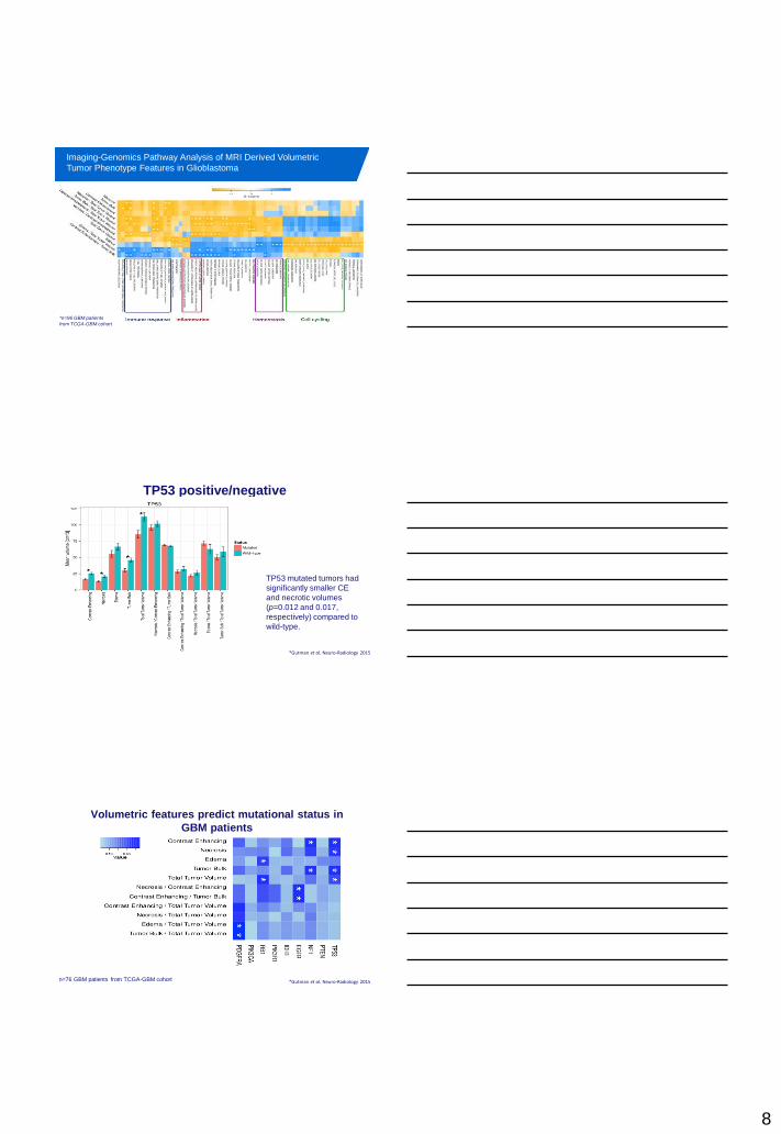
Imaging-Genomics Pathway Analysis of MRI Derived Volumetric
Tumor Phenotype Features in Glioblastoma
TP53 positive/negative
TP53 mutated tumors had
significantly smaller CE
and necrotic volumes
(p=0.012 and 0.017,
respectively) compared to
wild-type.
*Gutman et al. Neuro-Radiology 2015
n=76 GBM patients from TCGA-GBM cohort
Volumetric features predict mutational status in
GBM patients
*Gutman et al. Neuro-Radiology 2015
9
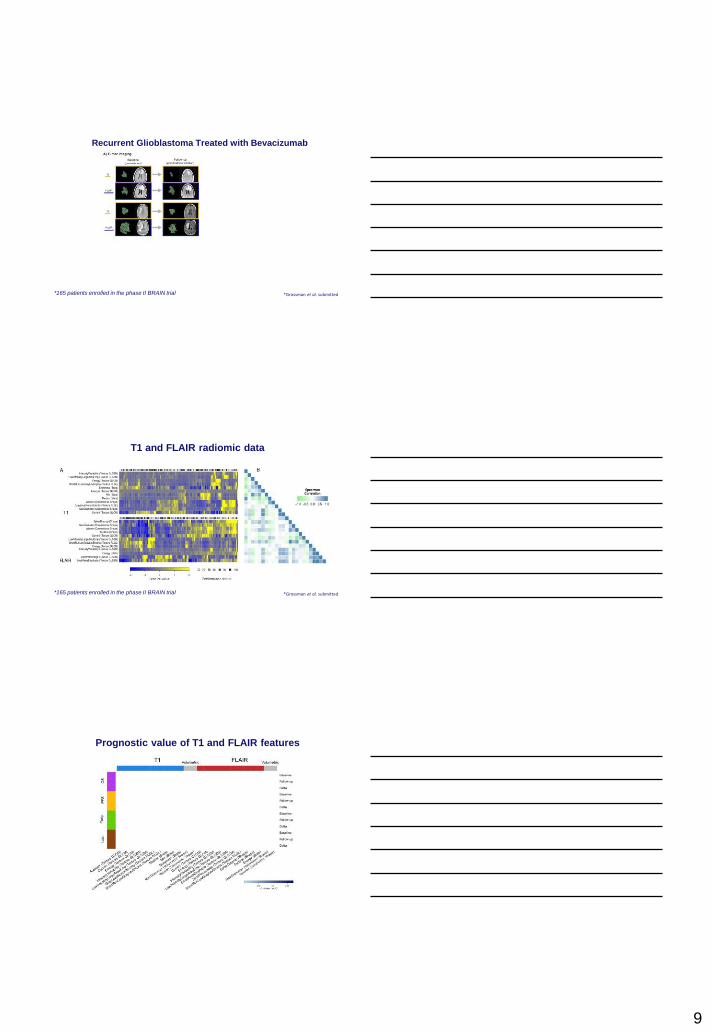
*165 patients enrolled in the phase II BRAIN trial *Grossman et al. submitted
Recurrent Glioblastoma Treated with Bevacizumab
T1 and FLAIR radiomic data
*Grossman et al. submitted *165 patients enrolled in the phase II BRAIN trial
Prognostic value of T1 and FLAIR features

10
Prognostic value of T1 and FLAIR features
Multivariable survival and progression models derived from
T1-weighted baseline imaging
*Grossman et al. submitted
These markers showed strong stratification power in independent validation data (hazard-ratio > 2;
log-rank p ≤ 0.001) after adjusting for age, sex, and baseline Karnofsky performance status.
Radiomics: Current Status
- Imaging moves towards a computational data science (bioinformatics)
- Due to advances in imaging, quantitative imaging is currently possible
- Large retrospective and prospective potential
- Large number of imaging features defined & successfully implemented
- Feature extraction pipelines implemented in 3D-Slicer (Python / Matlab)
- Radiomics signatures are prognostic across cancer types
- Radiomics are strongly connected with genomic patterns
- Integration of multiple datasets to improve performance

11
PAGE
31
01/08/201
6
• Philippe Lambin
• Ralph Leijenaar
• Sara Cavalho
• Emmanuel Rios-Velazques, PhD
• Stephen Yip, PhD
• Elizabeth Huynh, PhD
• Patrick Grossmann, MSc
• Thibaud Coroller, MSc
• Chintan Parmar, MSc • Robbert Gillies
• Yuhua Gu
• Virendra Kumar
• Olya Grove
• Benjamin Haibe-Kains
• Nehme Hachem
• Brian Alexander
• Tina Kapur
• Raymound Mak
• Fiona Fennessy
• Mike Makrigiorgos
• Ross Berbecco
• John Quackenbush
• Gisele Cruz
• Nabgha Farhat
• Ron Kikinis
• Aedin Culhane
www.cibl-harvard.org
• Ying Hou, MD
• Matthew Wagar, MSc
• Vivek Narayan, BSc
• Roman Zeleznik, BSc