handling of upper gi cancer specimens - … ugi...handling of upper gi cancer specimens ......
TRANSCRIPT

Handling of Upper GI Cancer Specimens
Prof Ray McMahon
Histopathology Department
Manchester Royal Infirmary
Bryan Warren School
Sarajevo
November 2016

Epidemiology
Classification
Role of Barrett‟s oesophagus
Handling of specimens
Resections
EMRs
Newer techniques
Pathology of oesophageal cancer


Oesophageal Cancer: epidemiology
• Majority are SCC
• Adenocarcinoma on the increase
associated with Barrett‟s
oesophagus
• Variants
• Prognosis related to stage (TNM)

Oesophageal Cancer (SCC)
• Alcohol: spirits, maize-based beer, mate
• Tobacco: smoke or chew
• Diet: Iran – bread and tea, lack of vitamins
• Chemoprevention (Linxian): no benefit
• Infections: fungal, HPV
• Exogenous factors: lye, radiation
• Genetic factors: tylosis (17q25), ALDH2
polymorphism, MTHF reductase
• Associations: achalasia, Plummer-Vinson
syndrome, coeliac disease
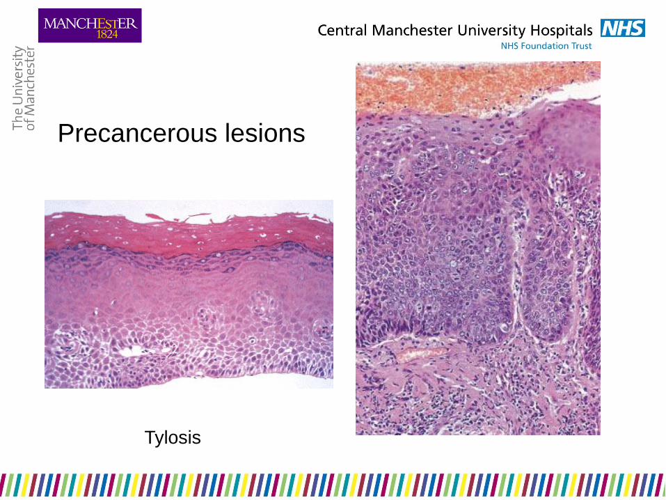
Precancerous lesions
Tylosis

Oesophageal Cancer:
clinical
• Early cancers
asymptomatic
• Dysphagia from
invasion into wall with
narrowing of lumen
• Pain from involvement
of adjacent structures
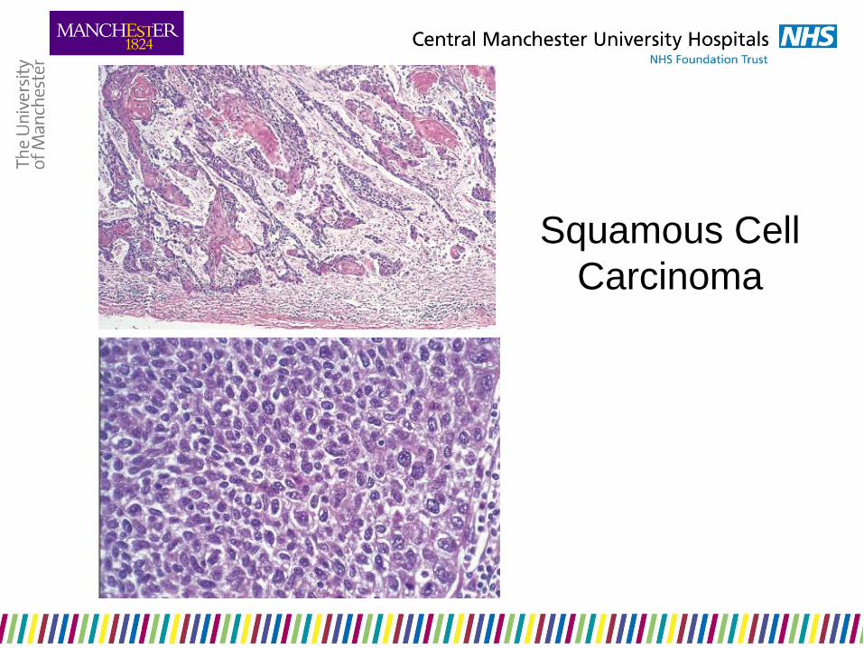
Squamous Cell
Carcinoma

Verrucous Carcinoma
• Well differentiated
• Slowly growing
• Rarely metastatic
• Extensive local spread

Superficial Spreading Squamous
Carcinoma
• In situ or invasive carcinoma
confined to mucosa and submucosa
• > 20mm
• High frequency of lymph node
involvement
• Poor prognosis
Soga J et al 1982 Cancer 50: 1641–1645

Basaloid Squamous Carcinoma
• Elderly males
• Solid, discrete nests
• Small, mitotically active cells
• Microcystic spaces or
necrosis
• Stromal hyalinisation
– May be confused with adenoid
cystic carcinoma
• Poor prognosis
Abe K et al 1996 Am J Surg Pathol 20: 453-61

Spindle Cell Carcinoma
• Polypoid carcinoma,
carcinosarcoma, pseudosarcoma
• Biphasic pattern
– Mesenchymal metaplasia
• Present early
• Better prognosis


Small Cell Carcinoma
• Uncommon
• Identical to lung counterpart
– TTF-1 positive
• Rarely associated with ectopic
hormone syndrome
• May be combined with squamous
carcinoma or adenocarcinoma


Adenocarcinoma
• Increasing in frequency
• Males > females
• Smoking, alcohol, obesity
• Barrett‟s oesophagus
• p53, c-erbB-2, COX-2 upregulation
• Heterotopic gastric mucosa - inlet
patch

Adenocarcinoma arising in CLO

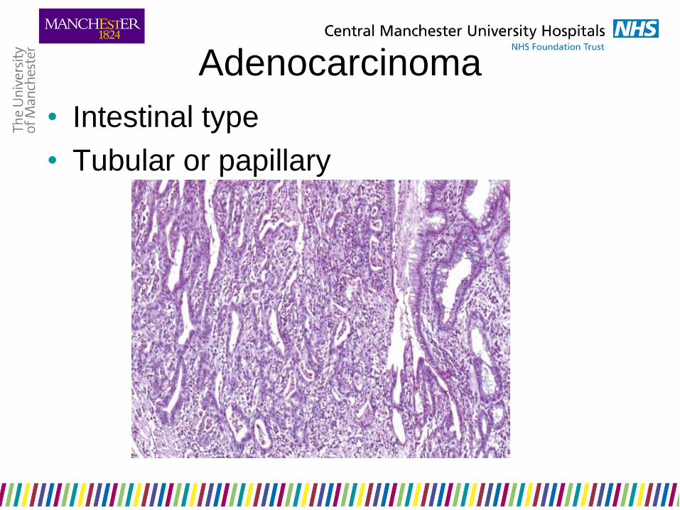
Adenocarcinoma
• Intestinal type
• Tubular or papillary
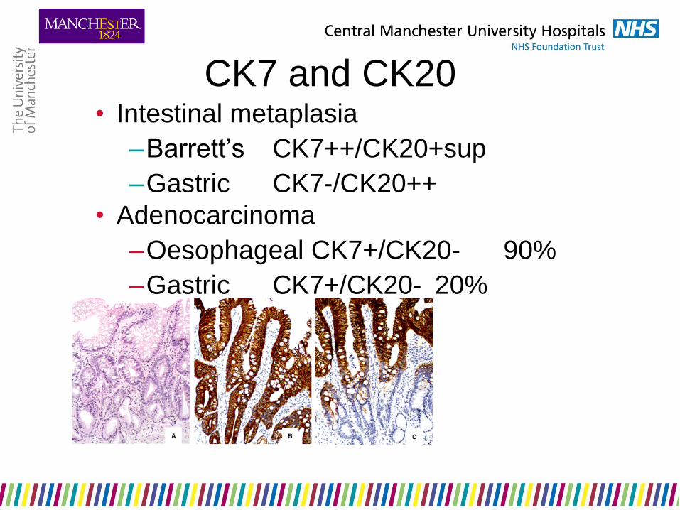
CK7 and CK20 • Intestinal metaplasia
–Barrett‟s CK7++/CK20+sup
–Gastric CK7-/CK20++
• Adenocarcinoma
–Oesophageal CK7+/CK20- 90%
–Gastric CK7+/CK20- 20%
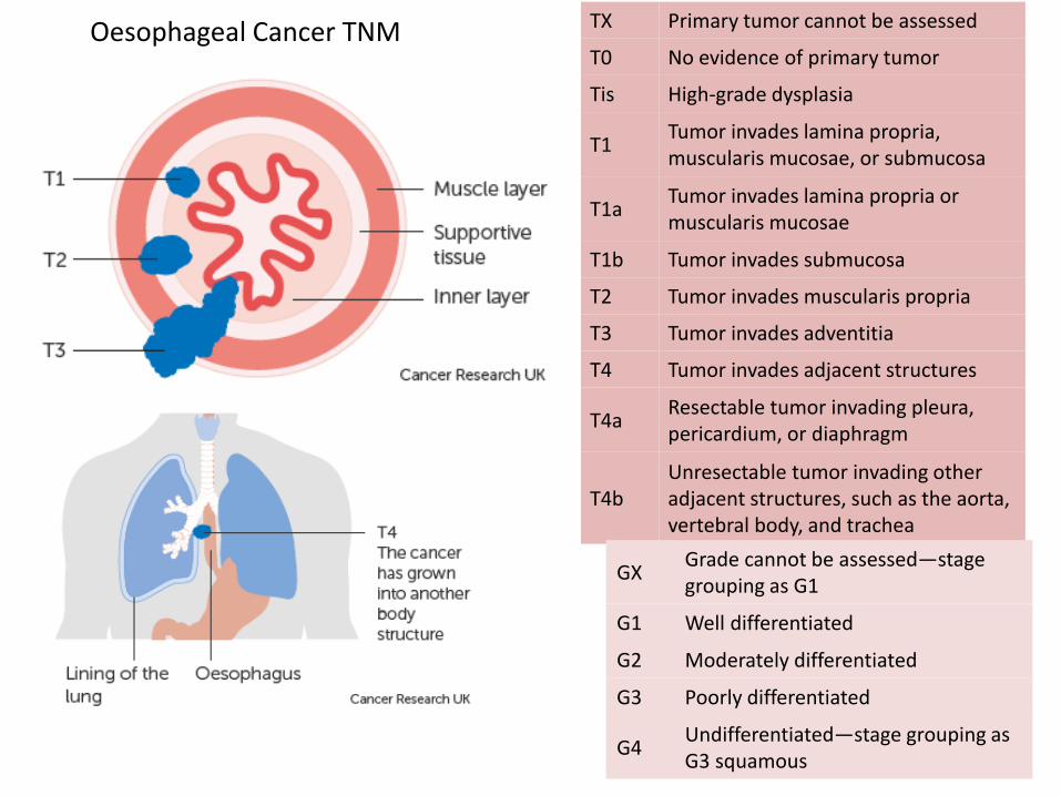
TX Primary tumor cannot be assessed
T0 No evidence of primary tumor
Tis High-grade dysplasia
T1 Tumor invades lamina propria, muscularis mucosae, or submucosa
T1a Tumor invades lamina propria or muscularis mucosae
T1b Tumor invades submucosa
T2 Tumor invades muscularis propria
T3 Tumor invades adventitia
T4 Tumor invades adjacent structures
T4a Resectable tumor invading pleura, pericardium, or diaphragm
T4b Unresectable tumor invading other adjacent structures, such as the aorta, vertebral body, and trachea
GX Grade cannot be assessed—stage grouping as G1
G1 Well differentiated
G2 Moderately differentiated
G3 Poorly differentiated
G4 Undifferentiated—stage grouping as G3 squamous
Oesophageal Cancer TNM

NX Regional lymph node(s) cannot be assessed
N0 No regional lymph node metastasis
N1 Metastasis in 1-2 regional lymph nodes
N2 Metastasis in 3-6 regional lymph nodes
N3 Metastasis in 7 or more regional lymph nodes
Oesophageal Cancer TNM

Type I
1-5cm above
GOJ
Low
oesophageal
carcinoma

Type II
1cm above
to 2cm
below GOJ
True cardia
carcinoma

Type III
2-5cm below
the
endoscopic
cardia
Sub-cardiac
carcinoma

Siewert‟s Classification
Type I adenocarcinomas of the distal third of the oesophagus (1-5cm above cardia)
low oesophageal carcinoma
Type II adenocarcinomas straddling the gastro-oesophageal junction (1cm above to 2 cm below cardia)
true cardiac tumour
Type III subcardial gastric adenocarcinomas that grow proximally to involve the GOJ (2-5 cm below cardia)
subcardiac carcinoma

Siewert‟s Classification Type I adenocarcinomas of the distal third of the oesophagus
(1-5cm above GOJ)
staged by oesophageal rules
Type II adenocarcinomas straddling the gastro-oesophageal junction (1cm above to 2 cm below cardia)
staged by oesophageal rules
Type III subcardial gastric adenocarcinomas that grow proximally to involve the GOJ (2-5 cm below cardia)
staged by gastric rules
In TNM7, tumours with an „epicentre‟ within 5cm of the OGJ
which extend into the oesophagus are classified according to the oesophageal carcinoma scheme
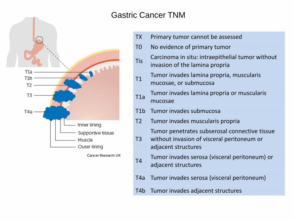
TX Primary tumor cannot be assessed
T0 No evidence of primary tumor
Tis Carcinoma in situ: intraepithelial tumor without invasion of the lamina propria
T1 Tumor invades lamina propria, muscularis mucosae, or submucosa
T1a Tumor invades lamina propria or muscularis mucosae
T1b Tumor invades submucosa
T2 Tumor invades muscularis propria
T3 Tumor penetrates subserosal connective tissue without invasion of visceral peritoneum or adjacent structures
T4 Tumor invades serosa (visceral peritoneum) or adjacent structures
T4a Tumor invades serosa (visceral peritoneum)
T4b Tumor invades adjacent structures
Gastric Cancer TNM

NX Regional lymph node(s) cannot be assessed
N0 No regional lymph node metastasis
N1 Metastasis in 1-2 regional lymph nodes
N2 Metastasis in 3-6 regional lymph nodes
N3 Metastasis in seven or more regional lymph nodes
N3a Metastasis in 7-15 regional lymph nodes
N3b Metastasis in 16 or more regional lymph nodes
Gastric Cancer TNM
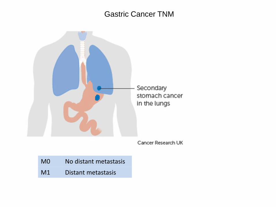
M0 No distant metastasis
M1 Distant metastasis
Gastric Cancer TNM


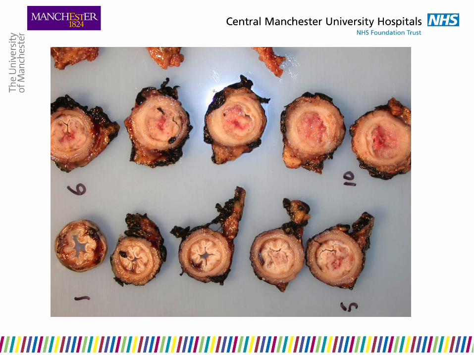
Specimen Choices
• Open and ink CRM
• Leave unopened
with a foam or
paper wick and ink
CRM
• Slice transversely
first
• Look for lymph
nodes first

Effects of Chemoradiotherapy on
Epithelial Tumours
• Tumour
– Vacuolation
and
eosinophilia
– Nuclear
pyknosis,
apoptosis
– Necrosis
• Stromal
– Fibrosis, hyalinisation
– Inflammation
– Giant cell granulomatous
reaction
– Calcification, malakoplakia
– Residual acellular
elements
– keratin
– mucus pools


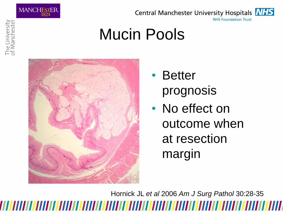
Mucin Pools
• Better
prognosis
• No effect on
outcome when
at resection
margin
Hornick JL et al 2006 Am J Surg Pathol 30:28-35
ypTNM staging
• e.g. ypT2 N0
• Categorises the extent of tumour actually present at
the time of examination.
• Ignore residual acellular elements eg mucus pools,
keratin
• NOT an estimate of disease prior to neoadjuvant
therapy
Pathological Response
• Complete or partial
• Quantitation if partial
• Requires widespread sampling
– Serial slices of site of the previous
tumour

Complete Response


Partial Response
• Reduction in tumour volume
• Requires comparison with untreated
tumour
– Pre-treatment biopsy
– ? representative of the whole lesion
• Microscopic foci only
• Relative proportions of tumour and
fibrosis

Mandard Classification Relative Proportion of Tumour: Fibrosis
TRG1 No residual cancer
TRG2 Rare residual cancer cells
TRG3 Fibrosis „outgrowing‟ cancer
TRG4 Cancer „outgrowing‟ fibrosis
TRG5 Absence of regression Significant correlation with disease free survival for TRG1-3 vs TRG4-5
Mandard AM et al 1994 Cancer 73:2680-6

TRG3 TRG4

Pathological Response • Mandard Classification
– ? Too complex
– ? Reproducible
• Simpler System
– No residual tumour
– Minimal residual disease
• only occasional microscopic tumour foci are
identified with difficulty
– No marked regression
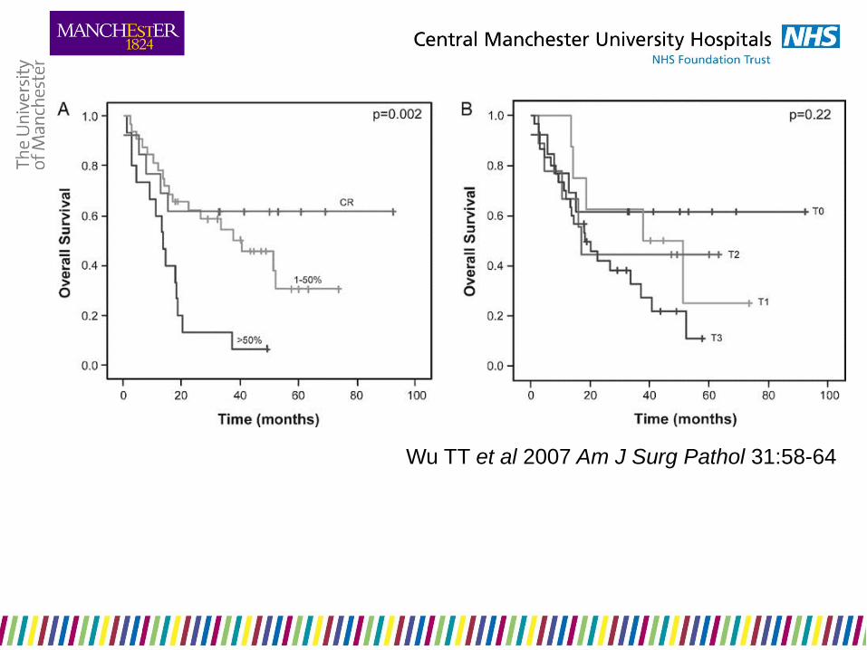
Wu TT et al 2007 Am J Surg Pathol 31:58-64
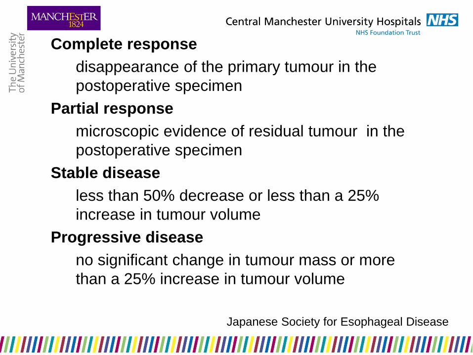
Complete response
disappearance of the primary tumour in the
postoperative specimen
Partial response
microscopic evidence of residual tumour in the
postoperative specimen
Stable disease
less than 50% decrease or less than a 25%
increase in tumour volume
Progressive disease
no significant change in tumour mass or more
than a 25% increase in tumour volume
Japanese Society for Esophageal Disease

More than 10 grading systems available (Mandard,
Japanese, Dworak, Wheeler, Becker, Junker
and Mueller, Rubbia-Brandt, Ryan, Le Sodan,
Schneider, Lowy, Mansourd)
This tells us none is entirely reliable
Relies on complete embedding of abnormal area
(presumed tumour bed)
‘As there is no national or international
consensus, (RCPath) cannot be prescriptive
and suggest that regression system to be used
should be determined locally by MDT involving
pathologist’ RCPath Dataset; Grabsch, Mapstone and Novelli, in preparation

Margin Involvement
• Proximal margin involvement
– Predicts local recurrence
– May be discontinuous with the main
tissue mass
– Always sample histologically
• Distal margin involvement
• Circumferential margin involvement

Margin Involvement
Number median survival
R2 17 6.2 months
R1 57 17.5 months
R0 82 51.9 months
p<0.0001
Number 5 year survival
R1/2 46 0
R0 137 48
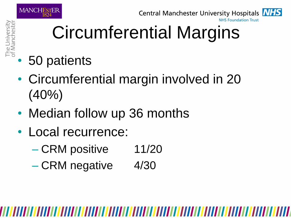
Circumferential Margins
• 50 patients
• Circumferential margin involved in 20
(40%)
• Median follow up 36 months
• Local recurrence:
– CRM positive 11/20
– CRM negative 4/30

Circumferential Margins
• 135 patients
• Circumferential margin involved in 64 (47%)
– (tumour within 1mm)
• Median survival:
– CRM positive 21 months
– CRM negative 39 months
– Effect only seen when “low metastatic burden”
(<25% nodes positive)

Circumferential Margins
• 329 patients
• Circumferential margin involved in 20%
• 5 year survival:
– CRM positive 22%
– CRM negative 29% difference not significant

Circumferential Margins
• 249 patients
• Circumferential margin involved in 32%
• Median survival:
– CRM positive 18 months
– CRM negative 37 months p<0.0001
• CRM status had a greater prognostic effect in T3
tumours with a low metastatic lymph node burden
(p=0.04).
Griffiths EA et al 2006 Eur J Surg Oncol 32: 413-9

Oesophageal Cancer: clinical
• Surveillance is the application of a test
that allows detection at a stage when
intervention may improve outcome
• Benefit from surveillance is limited
• Endoscopy with quadrantic biopsies
every 2cm + macroscopically abnormal
areas
• Patches of dysplasia easily missed
• Annual progression of 0.6% to
adenocarcinoma
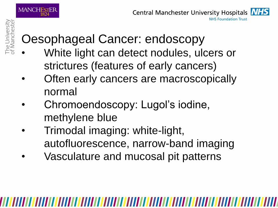
Oesophageal Cancer: endoscopy • White light can detect nodules, ulcers or
strictures (features of early cancers)
• Often early cancers are macroscopically
normal
• Chromoendoscopy: Lugol‟s iodine,
methylene blue
• Trimodal imaging: white-light,
autofluorescence, narrow-band imaging
• Vasculature and mucosal pit patterns

Lugol‟s iodine

Oesophageal Cancer: endoscopy • magnification and high resolution
endoscopy • chromo-endoscopy • auto-fluorescence endoscopy • narrow band imaging • microscopic tools: confocal microscopy;
multiphoton microscopy • in situ molecular analysis: FISH • spectroscopic analysis: fluorescence,
light scattering, optical coherence, Raman (inelastic) spectroscopy

Oesophageal Cancer: endoscopy • Confocal fluorescence microscopy: high
negative predictive value but poor sensitivity
• Elastic scattering spectroscopy: changes in
subcellular components during malignant
transformation; high sensitivity but poor
specificity
• Optical coherence tomography: similar to
ultrasound (3mm depth): in vivo studies not
convincing

Standard endoscopic view of CLO with HGD: no lesion identified
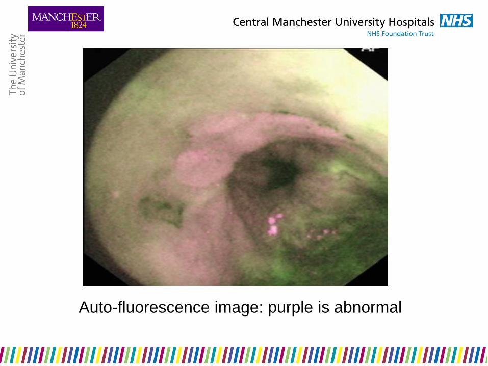
Auto-fluorescence image: purple is abnormal

Narrow band image of same abnormal area

Chromo-endoscopy with indigo carmine dye-spray:
HGD on histology

Low grade dysplasia in EMR/ER – makes
histological assessment easier

69F. Long history of achalasia. Nodule close to OGJ. Multiple
biopsies at three endoscopic sittings had provided equivocal
results. EMR.

Oesophageal EMR
• Olympus/Keymed – inject submucosa and form
pseudopolyp, direct snare
• Cook Duette band EMR/ER – band ligation and
snare
• methodology and subsequent therapy likely
influences how important margins are
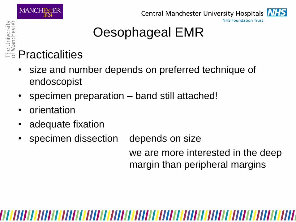
Oesophageal EMR
Practicalities
• size and number depends on preferred technique of
endoscopist
• specimen preparation – band still attached!
• orientation
• adequate fixation
• specimen dissection depends on size
we are more interested in the deep
margin than peripheral margins

• close collaboration with
endoscopist and assistant to
identify important landmarks
• resect en bloc if possible
• keep specimen(s) intact
• pin out on cork
• mark margins
• embed whole specimen(s) for
histology

Oesophageal EMRs

Histological assessment of EMR
Intramucosal pathology
Submucosal involvement by
carcinoma
Referral for surgery
Endoscopic treatment
and/or survelliance
The rationale for EMR/ER in CLO
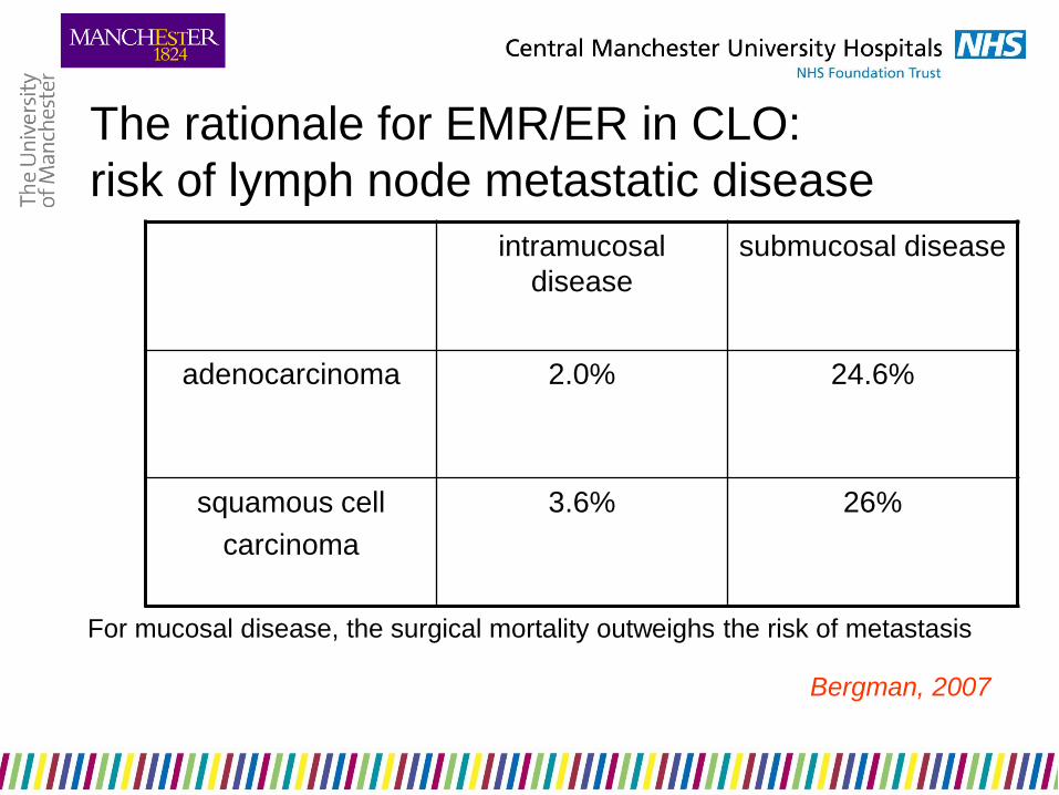
The rationale for EMR/ER in CLO:
risk of lymph node metastatic disease
intramucosal
disease
submucosal disease
adenocarcinoma 2.0% 24.6%
squamous cell
carcinoma
3.6% 26%
For mucosal disease, the surgical mortality outweighs the risk of metastasis
Bergman, 2007

What are the diagnostic pathological issues in
oesophageal EMR?

HGD versus intramucosal carcinoma

Entrapped and submucosal glands mimicking
submucosal adenocarcinoma

Reporting oesophageal EMRs
• is it Barrett‟s?
• are there treatment effects?
• neoplasia diagnosis
• depth of spread
• lymphovascular spread
• peripheral margins status
• deep margin status
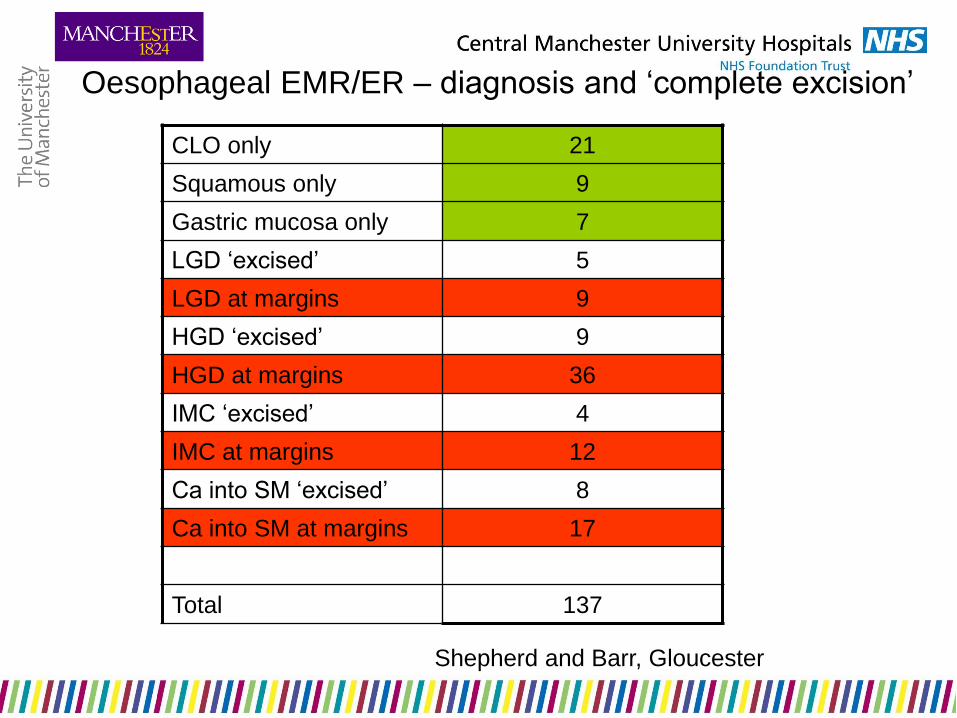
Oesophageal EMR/ER – diagnosis and „complete excision‟
CLO only 21
Squamous only 9
Gastric mucosa only 7
LGD „excised‟ 5
LGD at margins 9
HGD „excised‟ 9
HGD at margins 36
IMC „excised‟ 4
IMC at margins 12
Ca into SM „excised‟ 8
Ca into SM at margins 17
Total 137
Shepherd and Barr, Gloucester

Low grade dysplasia at margins
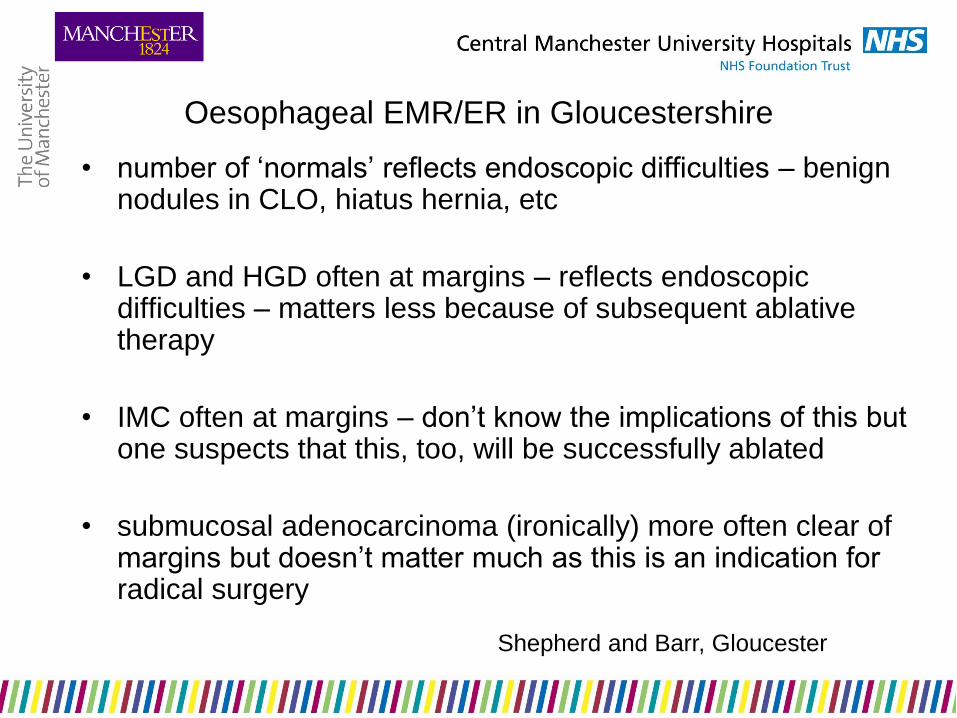
Oesophageal EMR/ER in Gloucestershire
• number of „normals‟ reflects endoscopic difficulties – benign nodules in CLO, hiatus hernia, etc
• LGD and HGD often at margins – reflects endoscopic difficulties – matters less because of subsequent ablative therapy
• IMC often at margins – don‟t know the implications of this but one suspects that this, too, will be successfully ablated
• submucosal adenocarcinoma (ironically) more often clear of margins but doesn‟t matter much as this is an indication for radical surgery
Shepherd and Barr, Gloucester

CLO and the double muscularis mucosae and
entrapment of dysplastic epithelium
Takubo et al, 1991, Lewis et al, 2008

Oesophageal EMR
pathology is important, mainly to confirm or
refute:
• the presence of malignancy
• if present, depth of malignancy
margins matter less (but this does depend on
subsequent management strategy)
THANK YOU FOR YOUR ATTENTION
ANY QUESTIONS?