handling and sterilization of surgical...
TRANSCRIPT
1
Handling and Sterilization of Surgical Instrumentation
There are many regulatory requirements for sterilization, including federal, state and professional associations that guide our practice in situations where instruments need to be sterilized. The following agencies play a prominent role in sterilization: the Centers for Medicare and Medicaid Services (CMS), the Departments of Public Health (DPH) and the Joint Commission Accreditation Hospitals (JCAHO) now known as “The Joint Commission”. The Occupational Safety and Health Administration (OSHA) and U.S. Food and Drug Administration (FDA) also have roles in the sterile environment.
Professional associations include: the Association for Advancement of Medical Instrumentation (AAMI) which consists of over 100 technical committees and working groups that produce standards and recommended practices with technical information for medical devices. AAMI has been involved in standards approved on an international level with international technical committees. The American National Standards Institute (ANSI), who represents national consensus, approves many American national standards.
The Association for Operating Room Nurses (AORN) is also very involved with the policies and procedures of handling and sterilization of instrumentation. With the increase of ambulatory surgical centers, perioperative nurses are increasingly responsible for the processing of and sterilization of instrumentation. It is extremely important for staff to be familiar with the methods of sterilization available in each facility.
2
Nurses topped the Gallup Honesty and Ethics of Professionals survey for the 11th consecutive year for 2010. In the Operating Room, patients expect nurses to keep them safe. Everyone in the operating room must advocate for the patient to keep them safe. Patient safety is everyone’s responsibility in the perioperative setting, and surgical site infections is considered a “never event”. In 2003, the Joint Commission produced the Surgical Care Improvement Project (SCIP), which identified core measures to improve surgical site infection rates. CMS has implemented nonpayment for adverse events and conditions “not present on admission”. CMS also has the ability to hand out severe punishment, including the immediate closure of a facility. Not only is infection or disease a life-or-death situation for patients, it also has become a life-or-death situation for hospitals and surgery centers.
The SCIPS core measures that the Joint Commission and CMS produced together for infections, is to improve surgical site infection rates. SCIP measures collected data based on these implementations to show improvement in preventing these types of infections. This information is available to patients, which allows them options for choosing their health care provider based of these SCIP measures.
According to Society for Healthcare Epidemiology of America (SHEA), approximately 500,000 surgical site infections (SSI) occur every year in the United States. This is a cost of $10 billion annually. Every patient with an SSI has an additional 7- to –ten day hospital stay with additional pain and suffering. Each patient with an SSI, may face additional surgeries including irrigation and debridements, possible removal of implants with placement of antibiotics. When evaluating the morbidity of patients with SSI’s, it was found that 77% of the deaths of the patients with SSI’s were directly related to their infection.
3
Even though processing of surgical instrumentation is a team effort, it is the nurses who are responsible for being the patient advocates. Everyone who comes in contact with a sterile item is part of the O.R.’s checks and balance system to prevent a patient from being contaminated by unsterile instruments or items. However, in the O.R. setting, it is the nurse who is held responsible for monitoring all aspects of aseptic technique, including monitoring the sterility of instruments and devices. It is everyone’s responsibility to check packages for being outdated or compromised before being opened on to a sterile field.
What is Sterilization?
Sterilization is the process that eliminates or kills all forms of life, including transmissible agents. Sterilization removes and destroys all microorganisms from an object. These agents include fungi, bacteria, viruses, spore forms, parasites, etc. Sterilization can happen in many ways including heat - the most common form - and also chemical, irradiation, high pressure, and filtration. Sterilization definition also includes disabling or destruction of infectious proteins such as prions related to Transmissible Spongiform Encephalopalopathies (TES).
The history of sterilization can be traced back to the times of ancient Rome. Back then, medical instruments were put through flame (heat) sterilization. Aristotle, a famed philosopher, recommended to Alexander the Great, King of Macedonia, that he should boil the water before giving it to his troops. In the following reference, AORN talks about sterilization in biblical times when God instructed Moses: “anything else that can withstand fire must be put through the fire, and then it will be clean. But it must also be purified with the water of
4
cleansing. And whatever cannot withstand fire must be put through that water.” This helps to prove that even back then, they understood the concept of clean and sterile.
William Henry (1774-1836) was a Manchester physician who studied contagious diseases. He believed that these diseases were spread by chemicals that were rendered harmless by heating. Henry used heat to disinfect clothing during an outbreak of cholera in 1831.
Surgical instruments have been around since prehistoric times when humans used several instruments for surgical purposes. In the Neolithic times, Shamans used trephines for round crainiotomies to release evil spirits and other types of head trauma.
The Sushruta Samhita is a Sanskrit text on all of the major concepts of ayurvedic medicine with innovative chapters on surgery. Cataract surgery was performed by Sushruta around 800 BC. Sushruta was considered the “Father of surgery.” The Sanshrit text, as it’s preserved, dates back to the 3rd or 4th century AD. In the Antiquity, surgeons and physicians in Greece and Rome developed instruments that were made of bronze, iron and silver. We still use many of these instruments today. Examples include scalpels, forceps, and curettes.
In Medieval times, Abulcasis (Abu al-Qasim al-Zahrawi) is known as the “Father of Modern surgery” in the West because of his book titled “On Surgery”, one of his thirty volumes of medicine.
In more modern times, the “History of Sterilization and Disinfection” was written by Joseph Lister, (1827-1912) who is known for pioneering antiseptic techniques in surgery. Lister would use carbolic acid sprays to decontaminate surgical wounds as he worked. It was the phenol
5
irritation to the surgeon’s hands that prompted them to wear rubber gloves. The practice of wearing gloves continues today for different reasons. Lister studied microbiology of air to control wound infections associated with surgery. Because of Lister’s sterilization methods, many German soldier lives were saved. Because of Lister, German surgeons began to practice antiseptic surgery.
Today our main focus is on preventing wound infections. With the increase in antibiotic resistance, and with more and more bacteria being discovered, we really must increase the focus of our practice to prevent these types of infections. Let’s review some of the bacteria that are causing serious life threatening situations.
Bacteria are one-celled organisms without a true nucleus or cell organelles, belonging to the kingdom Procaryotae (Monera). The cytoplasm is surrounded by a ridged wall composed of carbohydrates and other chemicals that provide the basis for gram stain. Bacteria can either be gram positive or negative. Millions of nonpathogenic bacteria live on human skin. These bacteria are considered normal flora. Bacteria that cause disease are called pathogens. Bacteria reproduce by binary fission, however some bacteria can exchange genetic material.
Bacteria can be one of three shapes, spherical or ovoid which occur in single cells or pairs, rod-shaped which are called bacilli, or spiral bacteria which are rigid. What makes bacteria dangerous is the fact it mutates like all living things. The environment dictates those changes based on beneficial mutations which have survival value. Hand hygiene (washing, cleaning nails) is the most important action that health care workers can do to prevent the spread of infection. Health care associated infections account for an estimated 1.7 million infections
6
and 99,000 associated deaths each year, with 32% UTIs, 22% SSIs, 15% pneumonia, and 14% blood stream infections, according to AORN. According to the Associated Professional Infection Control Committee (APIC) economic survey of associated health care infection for 2009, there were 100,000 deaths in the U.S. with a cost of $20 billion to the nation’s health care system.
Beta-Lactamase/ Extended-Spectrum Beta-Lactamases (ESBLs)
Beta-lactamase are enzymes that are produced by some bacteria and are responsible for their resistance to beta-lactam antibiotics like penicillins, cephamysins and carbapenems (ertapenem). The two most common bacteria are Escherichia coli (E.coli) and Klebsiella pnuemoniae.
Cephalosporins are common in their molecular structure to beta-lactamase; they both have four-atom rings, these are known as beta-lactam. The lactamase enzyme breaks open the ring which deactivates the molecule’s antibacterial properties.
Extended-Spectrum Beta-Lactamases (ESBLs) are enzymes that can be produced by bacteria, making them resistant to cephalosporins e.g. cefuroxime, cefotaxime, ceftriaxone and ceftazidime as well as monobactums e.g. aztreonam. Extended-spectrum are third generation antibiotics. These antibiotics are widely used in many hospitals. At this time they do not affect the cephamycins which are cefotetan or

7
cefoxitin. They also do not affect carbapenems including meropenem or imipenem.
ESBLs were first described in the mid 1980’s and were mostly found in Klebsiella species, mostly in hospitals and often in intensive care units, usually with patients with illnesses that make them opportunistic for bacterial infections. At that time, it is suggested that ESBLs, because of molecular analysis, may have derived from mutations. This problem was not a big issue at the time, however, now we have a new class of ESBL’s called CTX-M enzymes, which is detected among Escherichia coli (E. coli) bacteria.
E. coli is able to resist Penicillins and cephalosporins. These CTX-M enzymes are rapidly expanding. Missing the presence of ESBL could result in treatment failure. It is difficult on occasion to detect these ESBL’s because they do have different activity levels.
Other types of infections are caused by E. coli which could lead to bacteremia, which is a potential life-threatening blood infection. K. pneumonia, causes bacterial pneumonia or wound infections in addition to UTIs. Patients with weak immune systems, other illnesses, children and elderly are at increased risk for bacterema.
The National Committee for Clinical Laboratory Standards (NCCLS) developed both microdilution and disk diffusion screening tests. These tests have indicated that cefpodoxime and ceftazidime show the highest sensitivity of ESBL. Another problem is some ESBLs contain β-lactamases that can mask ESBL production.
Beta-lactam antibiotics are used to treat a broad spectrum of Gram ⁺ and Gram⁻ bacteria. Examples of the many different bacteria would be
8
Enterobactoer, K. pneumonia, K. oxytoca, E. coli, Enterobacteriaceae (Salmonella), Proteus, Morganella, Mirabilis, Psuedomonas aeruginosa, Citobacter, andvSerratia, which all produce ESBLs.
MRSA
What is MRSA?
It has been brought to the forefront of many people’s minds lately because it’s been a subject of many news features. Why has MRSA been featured? Because of the spread of the “super disease” and new cases. Health care workers are more and more concerned about its transmission process and there is an increasing fear of being exposed to the disease due to their ability to spread.
Staphylococcus aureus is a common cause of healthcare associated infections reported to the National Healthcare Safety Network (NHSN). The data reveals that Coagulase-negative staphylococci is the leading infection rate at 15%, while Staphylococcus aureus is 14%. Staphylococcus aureus is the most common cause of surgical site infections at 30% and causing ventilator associated pneumonia at 24%. Of all the healthcare associated S. aureus infections, it is estimated that 49-65% are caused by Methicillin resistant strains.
MRSA: Methicillin Resistant Staphylococcus Aureus is a type of “staph” bacteria that does not react to certain beta-lactam antibiotics called antimicrobial-resistant and will normally cause skin infections. Mutations may be beneficial to bacteria, and may not be to humans
9
because mutation provides resistance to the potentially lethal effects of antibiotics against bacteria.
MRSA can cause other infections that CAN BE FATAL! MRSA occurs most frequently with patients who undergo invasive procedures. Examples are catheters or surgery and with patients who have weakened immune systems. MRSA in the healthcare setting commonly cause bloodstream infections, surgical site infections as well as pneumonia.
History of Methicillin-resistance:
Methicillin-resistance in S. aureus was first identified in the 1960’s, usually among hospitalized patients.
•In 1974, MRSA infections accounted for about 2% of the total number of staph infections.
• By 1995 it was up to 22%; in 2004 it was 63%. The CDC estimates that each year approximately 27 million surgical procedures are performed.
•The CDC estimated 94,360 invasive MRSA cases occurred in the US in 2005. Of these cases, 18,650 (20%) were associated with death.
• In 2006-2007 MRSA was viewed as stabilizing at 56%
When dealing with the serious MRSA disease that is predominantly delivered by healthcare exposures, about 85% are associated with healthcare. When dealing with the two-thirds outside of the hospital infections, about one-third of those happened during a hospitalization.
10
About 14% of all infections occurred in persons without obvious exposures to healthcare. The overall rates of disease were consistently highest among persons older than 65, black and also males.
MRSA is resistant to antibiotics including methicillin, oxacillin, penicillin and amoxicillin including cephalosporins (e.g., cephalexin). Since these strong drugs are no longer effective against MRSA, these infections are sometimes called multidrug resistant organisms (MDROs). According to the CDC, high prevalence influences unfavorable antibiotic prescribing, which possibly could contribute to the increased spread of bacterial resistance.
MRSA is seen most frequently among patients who undergo invasive medical procedures or often occur with people who have weakened immune systems and are in hospitals and/or healthcare facilities. This includes nursing homes, dialysis centers and prisons. MRSA in healthcare settings commonly causes serious and potentially life threatening infections such as bloodstream infections, surgical site infections or pneumonia.
What is a surgical site infection?
An infection that occurs at the site of surgery within thirty days of an operation or within one year of an operation if a foreign body (e.g., artificial heart valve, joint or mesh) is implanted as part of the surgery. Most surgical site infections, (approximately 70%) are superficial infections which involve the skin only. The remaining, more serious infections may involve tissues under the skin, organs or implanted material.
11
An example of this would be orthopedic surgery. According to the CDC, who estimates that more than 4 million orthopedic surgeries are performed each year, and over 500,000 of these surgeries involve the knee. Typically depending on the type of surgery, less than 1% of surgeries result in surgical site infection. Of these infected cases, 50% are caused by MRSA.
This infection spreads because of skin-to-skin contact or touching personal items from a person who has infected skin. MRSA can be spread from touching a surface or item that has been in contact with someone with MRSA. In the case of MRSA, patients who already have an MRSA infection or who carry the bacteria on their bodies but do not have any symptoms are the most common sources of transmission.
Colonization of MRSA:
Colonization of MRSA generally proceeds to infection and in this case colonization can be long lasting. This means it could last from months to years in some subpopulations.
MRSA infections that occur in otherwise healthy people who have not recently (usually within the last year) been in the hospital or had surgery, are known as community-associated MRSA infections (CA-MRSA). In the community at large, these infections are usually skin and soft tissue infections (SSTIs) such as pimples, furuncles (abscessed hair follicles or “boils”), Carbuncles (coalesced masses of furuncles), abscesses and other pus-filled lesions. The role of MRSA in cellulitis without abscess or purulent drainage is less clear since cultures are
12
rarely obtained. However these infections may also lead to more serious illness, such as pneumonia.
Major strides have been made in recent years to reduce the numbers of MRSA infections in healthcare settings.
What to look for:
When considering a patient has an MRSA infection, you will find skin with a red, swollen and painful area. This area of skin will be warm to the touch, and possibly full of pus or other drainage. Additionally, the patient may also present with a fever.
The CDC encourages an MRSA in the differential diagnosis of SSTI’s compatible with S. aureus infections, especially those that are purulent (fluctuant or palpable fluid-filled cavity, yellow or white center, central point or “head” draining pus. It may be possible to aspirate pus with a syringe). A patient may complain of a “spider bite,” this should raise suspicion of a Staphylococcus aureus infection.
How is MRSA spread in the healthcare setting?
Although MRSA can come from the environment and be transmitted to people, the most common method of transmission is from person-to- person. The main mode of transmission in the healthcare setting from patients is through human hands, especially healthcare workers’ hands. Health care workers’ hands may become contaminated with MRSA bacteria by contact with infected or colonized patients. If appropriate hand washing with soap and water, or use of an alcohol-based hand rub
13
is not performed then the bacteria could be spread by a healthcare worker who has come in contact with MRSA to a patient. It is also appropriate to ask all visitors to wash their hands before visiting patients. When possible, it is best for patients if friends and relatives do not visit while a patient is ill.
Colonization means the growth of microorganisms, especially bacteria, in a particular body site. A patient who has acquired MRSA colonization during a hospital stay has increased risk for MRSA infections after discharge from the hospital or a transfer to a long term acute admission. These MRSA carriers can transmit the disease as they move through and across the healthcare facilities.
If appropriate hand washing with soap and water or using an alcohol-based hand sanitizer is not performed, the bacteria can be spread when the healthcare worker touches other patients.
Let’s discuss MRSA:
The expensive results of antimicrobial resistance bacteria, along with MRSA bacteria, is becoming resistant to the most commonly prescribed antimicrobial antibiotics and treatments. In some cases this means no antibiotics are effective against these mutated “Super” bacteria. However at this time, MRSA for healthcare-associated treatment still exits.
People with antibiotic-resistant organisms like MRSA are more likely to have extended and more expensive hospital stays. These patients are at higher likelihood of complications and possible serious health issues resulting from this infection. Extended treatments create a greater
14
burden and expense to the healthcare system. Because of this issue, the CDC, State and Local health departments, and other health partners nationwide are collaborating to prevent MRSA infections in the healthcare settings.
Of the pathogens that are causing the antibiotic resistant infections, most strains are associated with MRSA infections and are usually caused by traditional strains associated within the healthcare community. However, the strains traditionally associated with the community transmission are now being identified in the healthcare system as well.
One test to know if you are dealing with MRSA is to culture patients who are suspected to have colonized or have MRSA. Cultures can be expensive to a facility, however, it is less costly that other tests and it is more commonly practiced. It does however take 72 hours to identify if MRSA is present. Start treating patients as if they are positive while waiting for results. This way there is less chance of spreading if a patient is positive.
Another test is the Polymerase chain reaction test. This is a very fast way of testing patients. On the negative side, it is very expensive. Additionally it is a more difficult test for lab personnel to perform. Another issue with this test is which body site to use; possible site are wounds, axillae and groin.
The CDC recommends testing patients who are in high risk areas like ICU, however, anywhere in the facility would be appropriate.
Preventing MRSA:
15
It is very important that Healthcare providers review frequently updated policies and procedures when dealing with MRSA.
There are ways to prevent infection in MRSA colonized patients. The CDC calls these Core Prevention Strategies. Implement contact precautions for patients with MRSA during hospital stay. Recognize previously colonized patients. Rapidly reporting MRSA lab results and making sure to give this information during handoff reports. Provide MRSA education for all healthcare providers, including all staff members who interact with patient’s care.
Hand hygiene is one of the most important parts of the prevention efforts. This prevents transmission of MRSA by the hands of healthcare professionals. Make sure soap and water, as well as alcohol-based hand creams or gels are easily available to the entire staff including family and visitors. Educate not only health care professionals, but include the patients and family. Observe how the health care providers put these practices into action. Make sure all employees are following policies and procedures correctly. Always do what the CDC calls “just in time feedback” when staff members are not washing their hands according to policy.
Contact precautions are another core prevention to put in place with someone with or suspected of MRSA. Use a gown and gloves prior to entering patient’s room. Remove this personal protective equipment (PPE) prior to leaving a patient’s room to prevent spread. Put these patients in their own room, or if confirmed MRSA, put with another confirmed colonized/infected patient. Always use dedicated disposable items, blood pressure cuffs and stethoscopes are examples. Leave the
16
IV poles and pumps in the rooms for the entire stay. These patients could be in the hospital for months.
Education is a huge part of the core prevention measure. Education helps improve adherence to hand hygiene by health care workers and patients, including family and friends. It also helps to improve interventions, including contact precautions. Understanding this problem helps to encourage behavioral change.
What can patients do to protect themselves?
There are several things a patient can do to protect themselves from MRSA. It is important for patients to maintain a healthy weight. If a patient smokes, educate the importance of quitting at least 30 days prior to surgery. If a patient has diabetes, they should work with their doctor to keep blood sugar levels under control, especially prior to surgery. Make sure patients take a shower or bath prior to surgery, at least the day before. Make sure patients do not shave an area prior to surgery. Explain to the patient hair may be clipped if necessary in surgery.
Patients can also ask that doctors make sure to use antibiotics correctly prior to and after their surgery. Patients need to be proactively involved with their care. They can watch to make sure staff is washing hands prior to touching them.
Decolonization therapy for MRSA carriers is one way to try and suppress or possibly eliminate colonization. This is the use of topical and/or systemic agents. This therapy may reduce risk of subsequent infections in MRSA carriers as well as decrease transmission. One of the
17
problems with decolonization is determining which body parts to target, whether it is just the nares, or the whole body. Then the question becomes whether an intra-nasal mupirocin can be used only, or just a chlorhexidine bath. The other option is to do both. There are also oral agents available now. The worry would be an emergence of mupirocin resistance.
Prevention is our main goal with MRSA and prevention in surgery is an Operating Room nurses goal. Health care facilities should put prevention measures in place which can affect surgical site infections. Active surveillance testing is one of the strategies used. Another more controversial method is chlorhexidine bathing. There are also impregnated prepackaged wash cloths that some surgeons are having patients use prior to surgery.
It is the Operating Room nurses responsibility to post contact precautions signs on doors when necessary. It is also extremely important to pass this information on to each other in our hand off reports and briefings. This information should be written on the O.R. room count boards for all staff entering the room to see. When possible, have the patients bed completely cleaned while a surgical case is in progress. Make sure to communicate information about MRSA to environmental services personnel to wear protective equipment. Make sure to completely clean the patient of all body fluids before they leave the Operating Room suite.
Again, communicate all information to recovery room staff so that they are prepared to receive the patient appropriately attired, and if possible, separated from other recovery room patients. This will ensure
18
we help prevent surgical site infection throughout the perioperative phase.
Post Surgical Infection Prevention:
Once a patient is discharged, it is very important that the patient takes home the MRSA prevention information. Make sure they know that everyone is to wash their hands for at least 15 seconds when they wash their hands. Keep hand sanitizer available at all times after surgery. Do not use sanitizer when hands are visibly soiled (dirty).
When educating a patient and patient’s family, remind them it is important for everyone to wash their hands 15 seconds prior to fixing meals, and before eating these meals. Always wash your hand after using the toilet. Do not share hand towels, always use fresh linins. Wash hands after handling dirty clothes, towels, and linins. Wash all items in contact with patient in hot water to kill any contaminates that could possibly be present. Once home from surgery, Patients should not share items, such as razors, clothing or exercise equipment. Everything should be wiped down prior to use. Always keep wounds covered with clean, dry bandages. It is important to keep all shared items and surfaces clean for the surgical patient. These important precautions will help to keep the surgical patient from contacting MRSA after surgery.
19
Group A Streptococcal (GAS) Disease
Group A Streptococcus (GAS) is a beta-hemolytic streptococci bacterium often found in the throat and on the skin. Some people may be carriers of streptococci in their throats and or skin and may never have any symptoms of illness. Most GAS infections are relatively mild illnesses. Examples include strep throat, pharyngitis, tonsillitis, sinusitis, otitis media and pneumonia. Skin issues include cellulitis, scarlet fever, erysipelas, necrotizing fasciitis and impetigo. Impetigo is a bacterial infection of the skin caused by streptococci or staphylococci and marked by a yellow-to-red, weeping or pustular lesion. These lesions are usually around the nose, mouth, and cheeks or on the extremities. There are several million cases of strep throat and impetigo reported each year. Group A Streptococcus infection may have immunologic sequelae such as rheumatic fever and acute glomerulonephritis.
Rheumatic fever can develop approximately 18 days after a bout of strep throat, and it can cause heart disease with or without joint pain. Syndenham shorea, a disorder where the muscles of the torso, arms and legs move involuntarily in a dancing or jerky manner, can occur months later.
Occasionally, these bacteria can cause severe and even life-threatening diseases including sepsis. When GAS disease is spread to parts of the body where this bacteria is normally not found, it can become severe and life-threatening. Examples include when it’s found in places such as muscle, blood (bacteremia) or lungs. When found in these places the infections are termed invasive GAS disease. There are about 9,000-11,500 reported cases of invasive GAS disease each year in the U.S.
20
There are two forms of this infection that are the most severe kinds of this disease. The first would be Toxic Shock Syndrome (TSS). TSS is most commonly related to tampon usage. The bacteria strains that caused exotoxin to be produced were Staphylococcus aureus and Group A Streptococci, which in turn caused TSS. TSS has also been linked with not only vaginal tampons, but has included contraceptive sponges, diaphragms and surgical wound packing. Approximately 10-15 percent of patients with invasive Group A Streptococcal disease die from the infection. This relates to approximately 1,000 to 1,800 deaths annually in the United States.
This infection usually presents with a fever of 102° (38.9°C) or greater, diffuse, macular (flat), erythematous rash, followed by 1 to 2 weeks of peeling of the skin. The peeling usually occurs in the palms of the hands and soles of the feet. The patients may have hypotension or orthostatic syncope.
Patients could have involvement in one of the three or more organ systems.
•When the gastrointestinal system is involved, the patient may have vomiting or diarrhea at the onset of the illness. If the muscular system is involved, they may have severe myalgia (pain or tenderness).
•The mucous membrane may include any or all of these areas: vagina, opharyngeal, or conjunctival. A patient may have issues with hyperemia, unusual amount of blood in a part, including hepatic and hematological (platelet) problems.
•When the central nervous system is involved, the patient may experience disorientation or alteration in consciousness without focal
21
neurological signs when fever and hypotension are absent. Culture results are usually negative when taken from blood, throat and cerebrospinal fluid.
The second very serious form is necrotizing fasciitis, most commonly known as the “flesh eating disease” which is a rapidly aggressive spreading bacteria. Even though it is the least common of this disease, it is potentially deadly, destroying muscle, fat and skin tissue.
Streptococcal toxic shock syndrome (STSS) results in rapid drop in blood pressure and organs (e.g. kidney, liver and lungs) begin to fail. STSS is not the same as TSS, they are different bacteria. Approximately 25% of patients with necrotizing fasciitis and more than 35% with STSS die, according to the CDC. Aggressive and early surgical intervention is often needed for a person with necrotizing fasciitis to remove the damaged tissue and to try and stop the disease from spreading. Amputation of limbs may need to occur.
GAS is spread through direct contact of persons who are infected. The bacteria come from the mucus of the nose or throat and from infected wounds or sore from an infected person’s skin. Patients who have strep throat or skin infections are most likely to spread the infection. However, a person may have the bacteria without any symptoms, but could still pass on the bacteria. When a patient is treated with antibiotics for 24 hours or longer, it usually eliminates the possibility of spreading bacteria. Always remember to reinforce with patients to finish the entire course of antibiotics as directed.
Invasive Group A Streptococcal disease can get past a person’s defenses when they have sores or breaks in skin, and this allows the bacteria into
22
the tissue. People with chronic illness or an immune deficiency may allow virulent strains to cause severe disease more so than in others.
Persons with cancer, diabetes, chronic heart or lung disease or those who use steroids or chemotherapy or have suppressed immune systems are at higher risk. Persons who have open wounds, surgical wounds , chicken pox, who are elderly, and those who have a history of alcohol or drug abuse are also at higher risk for this disease. Patients who are burn victims are also at very high risk. This disease may occur in patients who are otherwise healthy and have no known risk factors.
Once a patient has GAS infections, it can be treated with many different antibiotics. For STSS and necrotizing fasciitis, high doses of penicillin and clindamycin are recommended. Supported care in ICU may also be necessary.
How do we stop the spread of Group A streptococcal infections. It can be as easy as washing ones hands. Good hand washing practices helps to stop the spread of many diseases. Remind anyone who is coughing and sneezing to wash their hands often. Always wash your hands before preparing and eating foods. Persons with sore throats should be seen by a doctor to be tested for strep throat. If results are positive, stay home with treatment for at least 24 hours to prevent spreading.
All wounds should be watched for signs of infection and kept clean and dressed properly. Patients with strep throat, but more often with GAS skin infections, can also develop inflammation of the kidneys. This rarely happens in the United States because of prompt intervention. If signs of infection arise, seek medical attention immediately to prevent a GAS infection. At the time of surgery, most patients receive a dose of antibiotics at the time of incision. Make sure to document this information correctly.
23
Mycobacterium Tuberculosis
Tuberculosis (TB) is a bacteria that could have a class of its own, however, this lesson will just hit on some important points related to drug resistance. TB is a bacteria that attacks not only the lungs, but also kidneys, spine and brain. TB is spread through the air from one person to another. It is usually passed when an infected person coughs, sneezes, speaks or sings. According to the CDC, it cannot be spread by kissing or sharing a toothbrush.
Not every patient infected with TB becomes ill. In fact, most people are able to fight off the TB bacteria from growing. This is called Latent TB Infection (LTBI). About 5 to 10 percent of patients with (LTBI) who do not receive treatment, will develop TB. TB sometimes is discovered through the tuberculin skin test or special TB blood test. You could have the disease for years before it becomes active. If the TB bacteria is able to become active, possibly because of a weakened immune system, and then begins to multiply, eventually the patient will become sick.
Extensively drug-resistant tuberculosis (XDR-TB) is caused by Mycobacterium Tuberculosis. XDR TB is a rare type of multidrug resistant tuberculosis (MDR TB). The first line of medication historically used to treat TB - Isoniazid and Rifampin - are no longer effective against MDR TB. XDR TB is also resistant to the best second-line medications including Fluroquinolones and at least three of the injectable drugs being Amikacin, Kanamycin, and Capreomycin. At this time, patients have bad outcomes due to less effective treatments.
24
Today, patients with weak immune systems are at higher risk of death once infected with TB. Symptoms of a patient with TB include: Feeling ill, or a prolonged cough – one lasting for three weeks or longer. A patient may experience chest pain, weakness, fatigue, weight loss (due to suppressed appetite), possible chills, and fever. Some patients may complain of night sweating. A patient may cough up phlegm, possibly with blood. Symptoms will vary when a patient is affected in a different part of the body.
High risk persons that have these conditions, including babies and young children, are:
1. HIV-infected 2. Substance abuse 3. Silicosis: a form of pneumonoconiosis which are inhaled. 4. Diabetes mellitus 5. Severe kidney disease 6. Low body weight 7. Organ transplants 8. Head and neck cancer 9. Patients on corticosteroids or taking rheumatoid arthritis.
The CDC has a tremendous amount of information about TB if more information is needed.
25
C. Diff
Clostridium Difficile is a bacterium found in feces that causes diarrhea as well as other serious intestinal conditions such as pseudomembranous colitis. About 30% of people have C. Diff as one of the normal germs in their intestine that help digest food. Other diseases that result from C. Diff are serious intestinal conditions such as toxic megacolon and perforations of the colon, sepsis and death in rare cases. C. Diff is a spore-forming, gram-positive anaerobic bacillus that produces two exotoxins. It is a common cause of antibiotic-associated diarrhea.
Symptoms for C. Diff are watery diarrhea, at least three bowel movements per day for two or more days. Other symptoms are loss of appetite, fever, nausea, and abdominal pain or tenderness. Treatment for C. Diff is usually 10 days of antibiotics and has few side-effects. In some cases, it may be necessary to have multiple treatments.
To test for C. Diff a stool culture can be done, although it is very difficult. Antigen detection can also be done, but it must be done in combination with toxin testing to verify diagnosis.
Patients in good health usually do not get C. Diff disease. Patients with other illnesses or conditions requiring prolonged antibiotics are at greater risk. The elderly or immunocompromised patients are also at greater risk of C. Diff. Patients who have had gastrointestinal surgery or intestinal manipulation, usually are at greater risk. Patients usually become infected after coming in contact with items or surfaces contaminated with feces then touch their mouth or mucous
26
membranes. Health care workers can spread the bacteria to other patients or contaminate surfaces if they do not wash their hands after contact with a patient’s contaminated feces.
Patients with C. Diff should be placed on contact precautions and their room should be cleaned regularly with disinfectants because surfaces harbor the bacterium and is a source of contamination. If possible, place these patients in private rooms because of surface contamination of the C. Diff spores. It is recommended to clean with Hypochlorite-based disinfectant for environmental surface disinfection.
Always wash hands with soap and water, especially after using the restroom. Always wash hands prior to preparing or eating food. Alcohol-based disinfectants are not effective against C. Diff and should not be used to disinfect environmental surfaces.
Treatment options for C. Diff include Metronidazole or oral Vacomycin. Even with treatment, the patient may still remain colonized.
Klebsiella Pneumoniae (K. Pneumonia)
T. A. Edwin Klebs was a German Bacteriologist and American Pathologist (1834-1913). He identified and treated Klebsiella, which is a genus of gram-negative, encapsulated bacilli of the family Enterobacteriaceae.
Edwin Klebs also demonstrated the presence of bacteria in wounds. K. pneumoniae is a species that may cause sinusitis, bronchitis or pneumonia.
27
K. pneumoniae in today’s healthcare setting has caused infections that include pneumonia, bloodstream infections, wound or surgical site infections and meningitis. Klebsiella is joining the list of bacteria that have developed antibiotic resistance.
Carbapenems is the most recent class of antibiotics that Klebsiella has formed resistance to. When Klebsiella pneumoniae bacteria produce an enzyme known as carbapenemase, they are also known as KPC producing organisms or Carvapenem-Resistant Klebsiella pneumoniae (CRKP). Carbapenem antibiotics are often the last line of defense against gram-negative infections that are resistant to other antibiotics.
Other Resistant Bacteria
Burkholderia Cepacia (B. Cepacia): A group or “complex” bacteria which is found in water or soil and is often resistant to common antibiotics. It does not pose great risk to the healthy population. It is usually a problem for patients with weakened immune systems. Patients who have cystic fibrosis (CF) or chronic lung diseases are at higher risk. B. Cepacia pneumonia has been reported in patients who were exposed either by person-to-person contact, contaminated surfaces or devices, and just ordinary exposure to the environment.
(VANCOMYCIN-INTERNEDIATE) VISA/Vancomycin Resistant (VRSA): Are specific types of antimicrobial staph bacteria. Most staph is treated with Vancomycin. Today, VISA and VRSA are no longer susceptible.
28
Stretococcus Pneumoniae disease: Resistant to more than one commonly used antibiotic. Invasive disease is usually caused by Pneumococci. S. Pneumoniae, which causes 60,000 cases per year of the invasive disease. Risk groups include people who work at child care centers, and people who recently used antimicrobial agents. Children are also at increased risk.
Resistant Psudomonas Aeruginosa: Commonly found in soil or water. It enters into the body through a cut or other breaks in skin and potentially can become deadly. Mortality rate is 50% of infected patients, which can happen with burn patients, and patients with cystic fibrosis. It causes other illness including UTIs, and bone and joint infections.
Resistant E. Coli: Associated with GI infections and dehydration. Resistant E. Coli can come from animal feces. This strain causes about 3,000 deaths per year in the United States.
Acinetobacter Baumannii: Also found in soil and water, but can be found on the skin on otherwise healthy people. This rarely occurs outside the health care setting, but commonly occurs with patients in the ICU.
Fungi
Fungi are the kingdom of organisms that include yeast, molds and mushrooms. Fungi grow as single cells as in yeast. They can also grow in multicellular filamentous colonies as in molds or mushroom. Fungi do
29
not contain chlorophyll so they are saprophytic or parasitic which means they obtain most of their food from dead organic material.
Most fungi are not pathogenic and are part of the body’s normal flora. Fungi that cause disease come from a group of fungi called Fungi Imperfecti. In patients with AIDS or Immunosuppressive drug therapy, Fungi are a source or opportunistic infections that cause death.
Spores
Spores are cell produced by fungi for reproduction. Spores may remain dormant yet viable for months. Pathogenic spores are usually inhaled rather than ingested. A spore can also be a resistant cell produced by bacteria that can withstand extreme heat, cold or dehydration. These spores can remain viable for decades. Spore-forming bacteria include Tetanus, botulism, and gas gangrene. Spores can be destroyed by steam underpressure (autoclave).
Parasites
A parasite is an organism that lives within or upon and at the expense of another organism resulting in harm to its host. A parasite such as a
30
virus or rickettsia that can reproduce only when within a living cell, although it may survive outside a cell, is called obligate intracellular.
A Parasite that is Internal is a protozzon or worm that lives with the body of a host and it occupies the digestive tract or body cavities. They may also live in organs, blood, tissues or cells.
Virus
A virus is a pathogen composed of a nucleic acid within a protein shell, which can grow and reproduce after infecting a host cell. More than 400 types of viruses that cause illness are known. All of them can attach to cell membranes, enter the cytoplasm, and take over the cells function. Once they take over the cells function they can reproduce their parts, and assemble themselves into mature forms capable of infecting other cells.
A few of the most virulent viruses known are hemorrhagic fever, which has been in the news recently, and Ebola virus. Viruses that cause tumors or malignant neoplasms are Epstien-Barr virus, hepatitis B virus, Papilloma virus, and human herpesvirus 8.
Death can be associated with many viruses.
31
What makes surgery a unique area?
The sterile environment makes surgery a unique area. Surgery has become a fast paced process with new and more complex equipment. This makes it even more important that staff remains educated and up- to-date with new techniques. We have to make split second decisions that will affect our patients for the rest of their lives, be it positive or negative.
The process starts the minute an instrument is purchased. All instruments must be washed and checked for proper function even before being put into circulation. It sounds trivial, but all staff must know and follow manufacturers’ instructions and understand that those instructions might change, and they need to be updated with each new product received. Patient safety is at risk every time instructions are not followed. Perioperative nurses and surgical technologists should also receive the same information as SPD with all new equipment when it comes to processing. When possible, facilities should form a sterilization committee that can review and give staff recommendations on how to deal with complex issues. It should consist of O.R. and SPD staff so it is well represented within the perioperative group. This group can also come up with ideas for verification and documentation of the cleaning process and can follow, if applicable, the manufacturers test procedures.
The perioperative environment includes a sterile processing department (SPD). They are a huge part of our patient safety goals. So let’s start with the cleaning process, which begins with the dirty instruments in decontamination room.
32
The first step is an extremely important one. Instruments should not be returned to SPD covered with dried blood and bioburded. AORN addressed the issue by stating: “You can clean without sterilizing, but you can’t sterilize without cleaning. “ They call this a simple but important concept. However we are discovering it is not so simple. Surgical cases themselves have increased in speed because of innovation of new equipment and systems. It has become harder to stay ahead of the surgeons in some cases, let alone try to keep instruments clean. However it is still a very important step that is being missed in some operating rooms and we need to get back to basics.
Part of the communication that should happen between the scrub personnel and decontamination personnel is proper sorting of instruments being returned for processing. You must clearly identify any instrument or devise that is not properly functioning to be sent out for repair. Make sure all sharps are separated for SPD protection with all disposables thrown away in proper containers. If your facility has a reprocessing program, make sure to deposit disposable equipment (trocars, scissors, graspers, etc) for reprocessing in proper containers. If your facility uses an instrument enzyme cleaner, try to put on instruments before leaving the O.R. If you are not able to accomplish this in the O.R., try to do this for the SPS staff before leaving the decontamination area.
Cleaning and Care of Surgical Instruments and Powered Equipment
Newly purchased instruments or equipment, items returned from repair, and loaner or consignment instrumentation should always be cleaned and inspected for proper function prior to sterilization. It is
33
extremely important to remember this step. It can be a dangerous shortcut, because it can lead to surgical infections. Never believe or trust that another facility has accomplished this task without witnessing it yourself. This will keep our patients safer.
Complex instruments can be hard to clean without manufactures instructions. Have those instructions available to staff, as they can be copied and laminated to keep them cleaner, and are able to be wiped down with disinfectant. This is important for those instruments that come back to the O.R. not properly processed.
Again, it is extremely important to remember instruments or devises with bioburden cannot be disinfected or sterilized. Blood or body fluids that have been allowed to dry on the surface or on the inside of an instrument, especially cannulated items, could possibly lead to transmission of infections. Blood, tissue, and mucous is extremely difficult to remove when it is allowed to dry on instruments and this increases cleaning time dramatically. That’s why it is so important to keep instruments clean of “gross soil” during surgical procedures. Remember during the surgical case to have a basin of water and sterile water soaked sponge to wipe or submerge instruments that need to be cleaned while the case is in progress. Cleaning should begin as soon as possible after an instrument is used. Pre-cleaning and sorting begins in the O.R. at the sterile field with the scrub personnel, keeping instruments free of gross soil.
Remember to never use saline for this process because it can damage instruments due to pitting, which could also harbor contaminates. Saline also causes rust and corrosion, which shortens the life of instruments. It is important to flush instruments that could have blood
34
or material inside of them. An example would be a suction that could have blood clots on the inside from a really bloody case. To prevent aerosolization, staff should flush instruments below the surface of water. Aerosolization could suspend particles of matter in the air which could be inhaled. Keeping instruments clean during a surgical case also assists central processing in the cleaning process. In turn, they are able to get instruments back into circulation fast, since flashing is the exception, in which we will discuss later.
When separating instruments for processing after a surgical case, remember to put the lightweight instruments on top of the heavier instruments. If this is not possible, separate them into different containers. It is important to reduce the risk of staff injuries and instrument damage.
Make sure to communicate to the decontamination staff if this set or specialty instruments will be needed back. This is important to communicate so there are no delays in reprocessing of the higher priority instrumentation. This could end up costing the facility because of delays in a patient’s surgery.
Cleaning itself should be done manually or mechanically, this includes washing by hand and rinsing thoroughly. For delicate instruments, please refer to manuals for proper cleaning methods. Instrumentation with lumens must be cleaned with the proper size brush to remove debris from inside. Power and electrical equipment should also be manually cleaned since it cannot be immersed.
Cleaning defined by AAMI is the removal of contamination from an item to the extent necessary for further processing or for the intended use. AAMI notes: “Cleaning consists of removal, usually with detergent
35
and water, of adherent soil (e.g., blood, protein, and other debris) from the surfaces, crevices, serrations, joints and lumens of instruments, devices and equipment by manual or mechanical process that prepare the items for safe handling or further decontamination.”
Mechanical decontamination includes ultrasonic cleaners, washer-decontaminators, washer-sterilizers, ultrasonic cleaners and cart washers. Always use manufactures recommendations and never overload these machines, they must be able to do the job correctly for ensuring patient safety. It is also important to remember to run your manual cleaners through your mechanical decontaminator daily to keep them clean for the next use.
Cleaning is the most important step to breaking the chain in disease transmission. Make sure to take apart instruments for the cleaning process that have multiple parts. Pay special attention to reamers and biopsy forceps where tissue can be trapped.
Tray Assembly
Once the instrumentation has gone through its terminal cleaning process then it’s on to the tray assembly area.
Ignazio Graziano, an immigrant from Sicily to the U.S., was working in a machine shop that happened to do work with the medical industry. He realized that, especially with orthopedic instruments, there had to be a better way to handle them. In 1984, Ignazio started Jewel Precision, which was the first company to design and manufacture sterilization trays for the medical industry.
36
Before assembly of instrument trays, and after cleaning and decontamination, it is important for instruments to be inspected. This is another check for instruments that may need further cleaning or sent out for repair. Devices at this time should also be checked to see if they are functioning correctly. Make sure that these work areas are equipped with lighted magnifiers. This allows staff to see if there are breaks in insulated equipment or other concerns. While following the recipe sheet for each tray, make sure to organize the tray in a way that the sterilant can come in contact with all exposed surfaces. Steam must be able to touch all surfaces of each instrument. Place integrating indicators in the center of the tray. When using a multilevel tray, place integrating indicators in opposite corners of each level of instruments.
The standard has not changed on the weight of sets, it is still set at 25 lbs. With specialty loaner sets coming and going out of a facility, those sets weighing more than the standard 25 pounds should be flagged and it should be documented in the patients chart if those set are used on that patient for better tracking, in case of infection. The possibility of sterility compromise is greater with these complex heavier sets.
Types of sterilization
There are many types of sterilization as well as many different sterilizers themselves. The first recorded steam sterilizers appearance was that of a pressure cooker. It was invented by Charles Chamberlain, a colleague of Louis Pasteur in 1880. Louis Pasteur (1876), Robert Koch and Wolffhugel, had each developed and used autoclaves themselves. Gaston Poupinel introduced dry heat sterilization, and in 1885 began its use in hospitals.
37
All sterilizers are considered Class II medical devices and are required to follow guidelines for manufacturing and submission to the FDA prior to being marketed for sale.
There are two types of steam sterilization that are often used in Operating Rooms.
The first one is the gravity displacement type sterilizer. This sterilizer works by the chamber, filling with hot steam and as the chamber fills, the pressure pushes the air down the drain. This sterilizer is slowly being replaced with the “Pre-Vac” sterilizer. You will find these types of sterilizers in an OR substerile room within the OR. These types of sterilizers are not only used in hospitals, they are also used in outpatient clinics and dental offices as well.
In a Pre-vac sterilizer or dynamic air removal sterilizer, a vacuum pump pulls the air out prior to the hot steam entering the chamber. These machines operate at a higher temperature than a gravity sterilizer. Not only creating a more efficient and rapid cycle because of the higher temperature 270-275 degrees, but hopefully less air pockets, creating greater opportunity for all instrumentation exposure to the hot steam. Always use a Bowie-Dick biologic test pack to ensure the machine is working correctly daily. The proper way to do this test is in an empty chamber. The test pack should be run for 3.5 to 4 minutes at 270 to 275 degrees Fahrenheit. Make sure to document this result in the sterilization records. If the test fails, repeat the test. If it fails a second time, report the information to a supervisor for possible retesting or servicing. Again, this information will be present to the surveyors when they come through on their visits.
38
Use both of these machines according to manufacturer’s recommendations. It is also important to have proper documentation in place and have it set up according to The Joint Commission and Centers for Medicare and Medicaid Services (CMS) guidelines so that your facility is prepared for their visit. Documentation is extremely important with flash sterilization since the recommendation is to “Never Flash”. Be very clear as to the reason for flashing since both agencies will want an explanation as to why an item was flashed. And also include who was notified as to why the item was flashed.
When documentation is done for sterilization monitoring of loads, it is important to document the following:
• Lot number • Contents of the load • Exposure time and temperature • Operator Identification • Results of Bowie-Dick Testing • Results of biologic testing including the chemical indicators
These records will help with recalls to be able to trace where the instrumentation maybe and where it possibly was used. The number allows the retrieval process to be done efficiently.
Load time should be done according to manufactures recommendations for the machines that your facility is using. Dry times for packaged instruments depending on the loads can be from 30 minutes to 2 hours. The bigger and heavier the load, the longer the cooling time should be.
39
Flashing of Instruments
Flashing is a clear focus for the Joint Commission and CMS. Flash sterilization should NEVER be a substitute for sufficient instrument inventory. This is a topic of issue with O.R.’s with decreasing budgets.
The American National Standards Institute (ANSI)/AAMI, AORN, The Association for Practitioners in infection Control (APIC), and the CDC have set guidelines for flashing. Flashing should only occur when an instrument is contaminated (dropped, contaminated wrapper, or chemical indicator failure) as is a “one of a kind” instrument and only in an emergency.
The most important concern when it comes to flashing is the cleaning and preparation process used on these instruments before being sterilized. The concern is that bioburden and soiled instruments be completely cleaned from the instrument to ensure proper sterilization. OSHA defines decontamination as use of physical or chemical means to remove, inactivate or destroy blood-borne pathogens on a surface to the point where they are no longer capable of transmitting infectious particles. This process renders the item safe for handling, use or disposal.
It is recommended that only trays that have been approved for flash sterilization by the FDA be used to sterilize the item in for safe, easy transport, and low burn risk to staff.
There are several options to prevent burns, the most common are special gloves or towels. Once the item is removed from the flash sterilizer, the concern would be to have a reduced risk of contamination or damage during transport. Once in the room, the next concern is
40
presentation to the sterile field. The recommendation is that the tray NEVER be placed on a nonsterile surface. It should be placed on a sterile impervious drape separate from the back table.
Do not forget your documentation. The sterilizer printout should be reviewed and initialed with room number. The flash log should be filled out with clear reason for flashing and who was notified (manager or charge nurse). Also, complete charting with which sterilizer was used, load number, what was sterilized and did it meet parameters.
Other concerns that address the containers themselves:
1. Are they cleaned, checked and maintained on a daily basis according to manufactures instructions?
2. The ability of staff to tell the difference between flash sterilizer containers and containers used for regular sterilization.
3. Flash containers should be opened immediately, item delivered, not stored for later use.
4. Make sure to follow ANSI/AAMI ST79:2006-A2.2009 guidelines.
Remember, the instruments that have been flashed are hot. Take precautions not to burn the patient. Cool the instrument before use. Cool sterile water from a designated refrigerator is one way of quickly cooling an instrument.
If the scrub person must transport an item from the sterilizer to the room, the Circulating Nurse must direct traffic and the scrub personnel MUST be aware of their surroundings during transport.
41
It is also recommended by AORN that a facility policy and procedure be developed with periop management and the infection control department with the objective of ensuring the best practice possible for aseptic transfer within the physical constraints of the facility.
Ethylene Oxide
Ethylene Oxide is another form of sterilization that has been around since the 1950’s and can be used for heat and/or moister sensitive items. All instruments and tubing must be completely dry for this process. This is a very effective process with a very low temperature. At the end of the sterilization cycle, the load must be aerated before they can be used. Ethylene Oxide, however, is a strong poison to humans, and chronic exposure has been known to cause cancer, reproductive effects, mutagenic changes, neurotoxicity, and sensitization according to OSHA with their update as of April, 2009. Under the Clean Air Act, the first chloroflourocarbons were phased out, which were part of the mixture for ETO sterilizers. These chloroflourocarbons were linked to destruction of the earth’s ozone layer. OSHA also regulates the acceptable vapor levels of ETO due to exposure being an occupational hazard. This has pushed development of other alternative technologies for low temperature sterilization in the health care setting.
Paracetic Acid
Paracetic acid for hospital use came in 1985, although at the time it had been recognized for its disinfection and sterilization properties for over 100 years. Paracetic acid is considered a “just in time” process.
42
Instruments must be moisture tolerant since this is a liquid process. If the item is left in the sterilizer, it is only considered sterile up to 2 hours. This process is usually used for scopes, light cords, and cameras. The FDA has cleared a new version of steris called the Steris System1E.
Hydrogen Peroxide Gas plasma
Low temperature sterilization can also be achieved with hydrogen peroxide gas plasma which was patented in 1987. This was not available in the U.S. till 1993. This process does not require aeration because the by-products are oxygen and water. Gas plasmas are generated in an enclosed chamber under deep vacuum using radio frequency or microwave energy to excite the gas molecules and produce charged particles. Many of these molecules are free radicals. A free radical is an atom with an unpaired electron and is highly reactive. This process requires very specific lumen size regulations. Again, load must be completely dry before starting the cycle. The biological indicator is Bacillus atrophaeus spores. This process has the ability to inactivate a broad range of microorganisms, including resistant bacterial spores.
Ozone
Ozone can also be used for terminal sterilization of heat and moisture sensitive instruments by oxidation. This technology came on board in 2003. Ozone cycles last 4 to 5 hours. Ozone has lumen restrictions as well.
43
Vaporized Hydrogen Peroxide
Vaporized Hydrogen Peroxide is the newest method of low-temperature sterilization. Again, there are some restrictions on lumen length and diameter.
It is the perioperative nurses’ responsibility to have a basic understanding of various types of quality control for sterilized instruments. These should include the mechanical controls such as graphs and printouts. Make sure to understand what you are reading and how to document those items. Also, understand the chemical controls such as indicators, integrators, autoclave tape, pouch dots, container arrows or breakaway lock. Know the difference between each of the colors prior to sterilization and the colors they should turn when a load should be sterile. Biologic controls need to be incubated and read. Understand that parameters have been met for that load to be sterile. It is also everyone’s responsibility to know the steps to follow if an indicator has not turned correctly.
All sterile processing equipment must be properly maintained with preventative maintainence. This should be done on a routine basis to keep equipment working properly to prevent failures. Proper maintenance records should be kept up to date as well.
Moving to the Operating Room
Your case is picked and you are ready to start your case. You spread everything out as you’re checking the physician preference card to make sure you have everything needed for this case. A tray is opened and moisture is found, at this point, the tray should be considered
44
contaminated. Moisture can act as a route with wicking action to allow microorganisms to be drawn into the package via any paper area. You should take steps to document that the tray in contaminated and central processing must be notified to start the recall process.
As you open the sterile items for your case, you must inspect all items for integrity and ALWAYS check the expiration date. Outdated supplies can also play a role in infections. Just handling a product can create an environment in which the packaging could be compromised and contamination could occur.
Things that can affect package sterility:
• Proper humidity, temperature and air exchange. When certain items require specific levels, monitoring must occur and logs may be required.
• How items are stored: Wire racks must be lined properly to protect packaging. Do not aggressively remove sets from racks.
• Moisture from spills or being placed close to a sink area. • Inadequate air exchanges taking place in sterile areas. • Airborne contaminates from things like lint from linen or dust. • Placement of sterile supplies in high traffic areas. Unsterile items
laid or placed on sterile packages. Example: sweaty lead aprons on sterile sets.
Best practice: when opening paper items, put the wrapper up to an O.R. spot light to check for holes. This takes only a moment and is an easy way to check for holes. If your hospital has recycling, then place the wrapper in the recycling bin. We will talk about this option later.

45
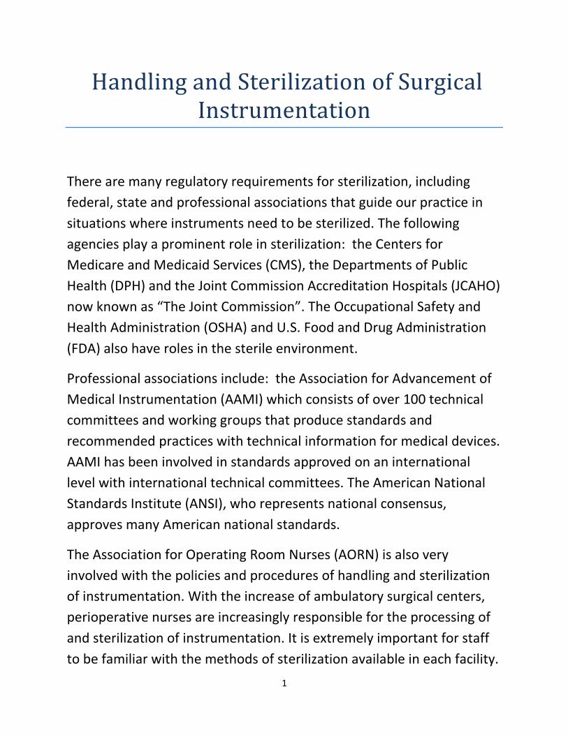
See IMAGE #1 below. This is an example of the how holes can be detected . There are actually two holes and they have been outlined in black. You can see the light pass through them:
IMAGE #1
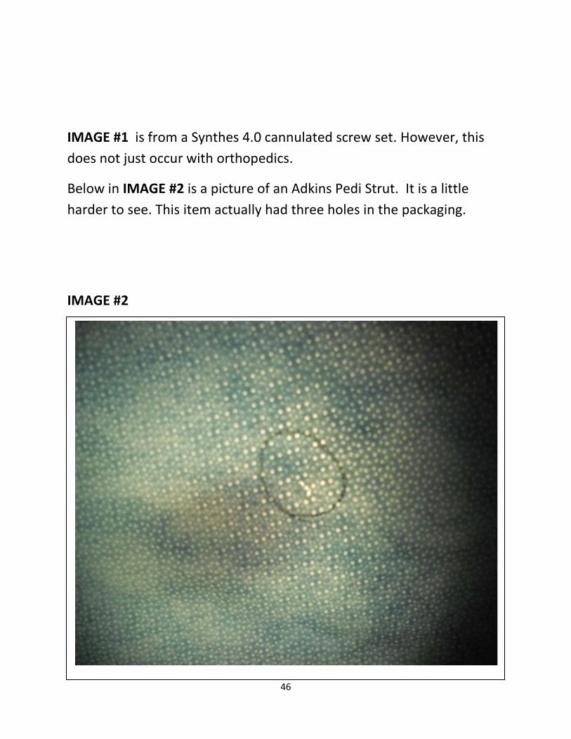
46
IMAGE #1 is from a Synthes 4.0 cannulated screw set. However, this does not just occur with orthopedics.
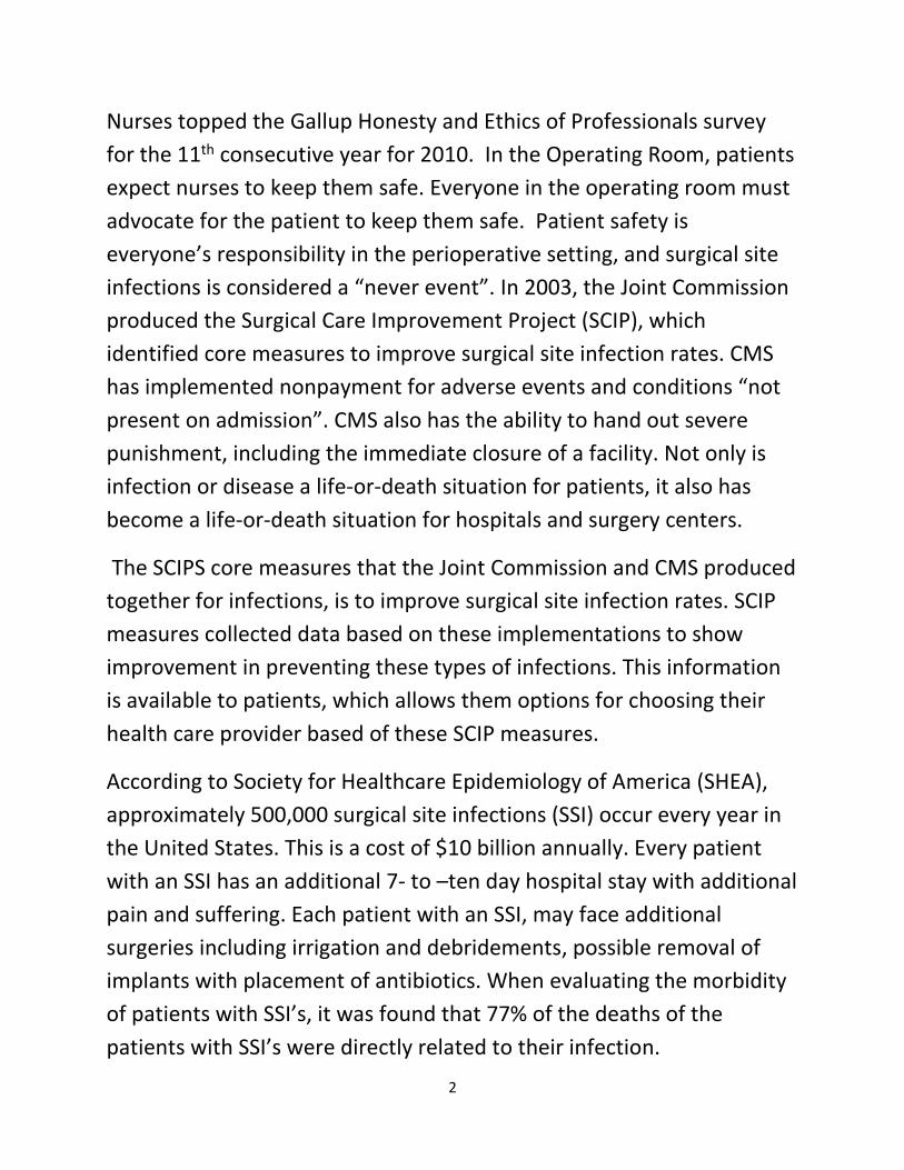
Below in IMAGE #2 is a picture of an Adkins Pedi Strut. It is a little harder to see. This item actually had three holes in the packaging.
IMAGE #2
47
When you turn a wrapper slightly, you can see the light reflecting through these holes.
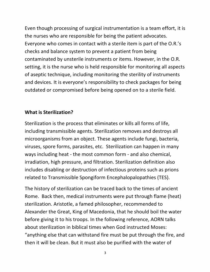
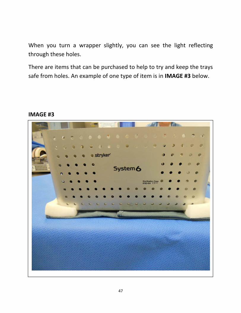
There are items that can be purchased to help to try and keep the trays safe from holes. An example of one type of item is in IMAGE #3 below.
IMAGE #3
48
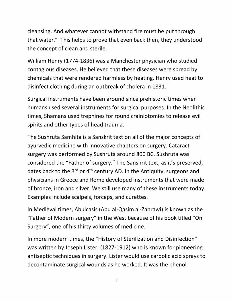
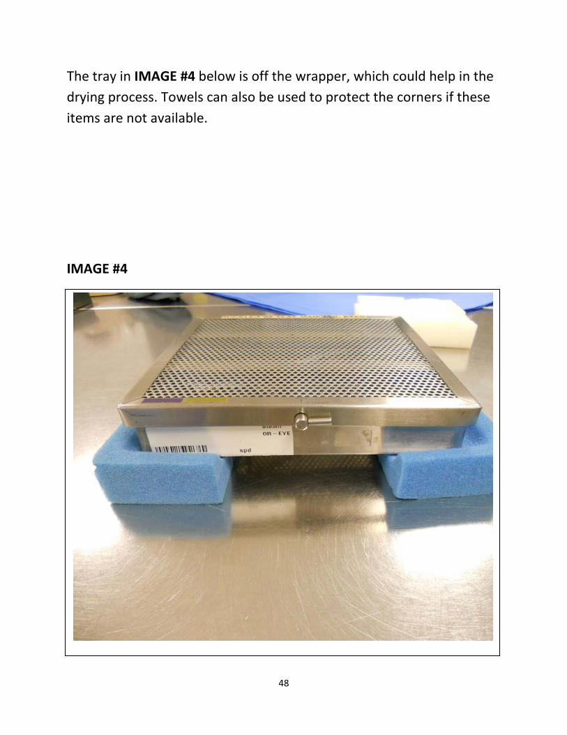
The tray in IMAGE #4 below is off the wrapper, which could help in the drying process. Towels can also be used to protect the corners if these items are not available.
IMAGE #4
49
This helps with sets that are heavy and close to the 25 pound limit.
Peel pouches easily get holes in them from handling, and being put away in an over stocked bin. Just packaging some of the instruments that have sharp points, for example; scissors, gelpis or forceps can easily create holes in packaging. If you are seeing consistent holes in packaging, other packaging options may be needed. Peel packs can also be held up to the light for inspection. If an instrument is opened to the scrub personnel, they should not return to the back table until the package is inspected by the circulating nurse.
Nothing should be brought to the back table until sets are checked for sterility. This means checking the indicator before being brought to the table. A turned indicator does not always mean a set is sterile. Indicators can change if left in a sterilizer for an extended period of time, even if the load is not initiated. Usually these indicators will be a lot lighter in color. If you see this, it should be questioned. Make sure the indicator has turned completely. This is also why it is important to check all chemical indicators, including the tape. Make sure the correct indicator is in the correct package. An example of this would be a Sterrad indicator in a steam sterilized package. Correct indicators must be used for the right load.
During the case, care of the instruments is extremely important to the decontamination process. At all times, keep instruments free of gross blood and debris. This is accomplished with a basin of water and a water soaked sponge throughout the surgical case. At the end of the case, sort instrument appropriately and secure all sharps for everyone’s protection, as well as protecting the instruments. When possible, use
50
enzyme cleaner to start the decontamination process prior to leaving the O.R. suite.
After entering the decontamination area, give a hand-off report to the staff in decontamination. Point out what instruments will be needed to be turned over for the following cases. Point out any broken instruments that need to be sent out for repair. Remind the staff of where sharps are located. Dispose of sharps in sharps container and deposit any items that may be recycled in correct containers.
Loaner Instruments
With any loaner tray, there usually are implants that follow these instruments. Each facility should set up a tracking system and have quality control procedures in place. This includes inpatient and outpatient facilities. Facilities will be held responsible for lost instruments. When possible, loaner trays should be brought in the day before surgery. This is to allow time for policy and procedure to be followed when preparing trays for sterilization. If loaner equipment arrives wrapped, consider it unsterile and break it down to be reprocessed.
A management system of loaner instruments reduces lost instruments and helps to ensure that decontamination and sterilization have been done correctly with communication involving venders. Make sure to follow all manufactures’ recommendations when cleaning and sterilizing another company’s instrumentation. Sterile processing should maintain records, which should include when instruments were received. Additionally, the time instruments were received, the person
51
checking in the instruments, the sets brought in, document any missing instruments and copies of manufactures instructions. It helps if venders have “count sheets” of their sets, if they do not request them.
The Association for the Advancement of Medical Instrumentation recommends a maximum weight of 25 pounds for loaner sets. This weight is to include the weight of the container the instruments are in. This weight makes it difficult to determine the correct drying time to ensure sterility of these sets.
Another problem with heavy instrument sets is that that they are difficult for the staff to lift. This can cause injury to not only SPD staff, but O.R. staff as well. Many injuries are reported due to the lifting of heavy instrument sets.
Each facility should address a return protocol in their policy. It is important for staff to know how instruments should be cleaned and decontaminated before being returned to the vendor. These policies should be strictly enforced.
Implants
According to the FDA, an implant is a device that is placed into a surgically or naturally formed cavity of the human body and is intended to remain there for 30 days or more. In order to protect public health, the FDA may determine that devices placed in subjects for shorter periods are also implants. Implants should NEVER be flashed sterilized per recommendations from the CDC, the Joint Commission, AORN and AAMI. Infections that involve implants are much harder to treat and often lead to more surgeries. In some cases, the implants have to be
52
removed and antibiotic cement is used as spacers or in place of the implant until the patient is infection free. In worse cases, patients die from the infections. Implants are foreign bodies and they increase the risk of surgical site infections (SSI).
If an implant must be flashed, a biological indicator must accompany the implant and all proper paperwork should be sent to sterile processing. The biological should be read at 1 hour and at 2 hours, with results communicated to the circulator in the room. If the surgical case is finished and staff has left the building, report the results to the manager or charge nurse. If the results from this biological are positive, notify the surgeon and infection control immediately.
AAMI recommends a dedicated exception log and form to facilitate dedicated detailed information related to this event. The information should include detailed information of the reason for premature release of the implant for fully traceable information to the patient. This information should include:
• Patient name • Implant prematurely released • Surgeon • Reason for premature release • What could have prevented premature release of this implant
Sterilization loads with implants should only be released by an experienced person at the end of a sterilization cycle. This person should strictly review all information pertaining to this load including monitors and printouts. When possible, this load should be quarantined until the biological is read and the results are negative.
53
Monitoring of each load should include:
• The physical monitoring of each load • Label every package with an external process indicator a Class
One • Place an internal Class One indicator inside each package • Monitor with a process challenge devices (Biological indicator) • Evaluate all quality control measures and data at conclusion of the
sterilization cycle. • Release loads only if all criteria for release are met, which should
be performed by a knowledgeable, experienced professional.
Cleaning and processing of Anesthesia Equipment
If your facility does not have anesthesia technicians, it usually means the perioperative nurse is involved with the anesthesia equipment. Anesthesia equipment that comes in contact with mucous membranes should be sterilized or undergo high-level disinfection. It is also important for anesthesia techs or nurses to follow all manufacturers’ instructions for the products that are used. Whenever the anesthesia department purchases new blades, clean them before use and review the new products instructions.
The Perioperative Area Accountability
The Operating Room suite historically has been a place where no one wanted to venture and was usually passed over by surveyors. This is no
54
longer true. In fact, the O.R. is now under the microscope. The Operating Room is a huge drain on financial recourses, and with public education and scrutiny, the O.R. must change to meet these needs. With educational TV and the internet, patients can watch the surgical process before they have it and understand what should happen.
The Operating Room, whose double doors were closed to everyone, must now open them so everyone can look in and see how we are changing and meeting the needs of our patients. We have to become more efficient and still protect our patients from harm. It has become a true balancing act, to give more with less.
The days of surveyors passing us by because we were out there on our own are long gone. With “wrong site” surgeries, “never events”, and increasing surgical site infections, we are in the fore front more than ever. The Joint Commission surveyors, as well as CMS investigators are here with the staff and asking a lot of questions. They are verifying that education is being done, and ensuring that quality improvement participation is indeed happening. This is why perfect documentation is required.
The Joint Commission Surveyors are perfecting the tracer methodology with every survey. Direct observation and personal contact involved with that patient throughout the perioperative area is a newer experience for everyone. Even though the Joint Commission process is voluntary, it is a requirement for Medicare and Medicaid reimbursement. AORN thoroughly explains how each of these agencies can affect how a hospital or surgery center functions.
55
The FDA, established in 1906, regulates medical products and oversees medical devises; like being illegal to alter a medical device. The FDA also regulates the reuse of items that are labeled “single use only”.
The EPA and OSHA were each established in 1970. OSHA was established by the U.S. Department of Labor, and it works to ensure safe work places for the employee. If an employee does not follow the law, OSHA can invoke severe fines and endanger Joint Commission accreditation. The EPA maintains a central file of Material Safety Data Sheets and manufacture’s instruction for storage and the use and disposal of all products. The hope is that facilities will keep their MSDS up to date and educate the staff so the hospital can remain in compliance with hazardous chemicals. In the perioperative setting, some of these chemicals would be used to clean, disinfect and sterilize the instruments for the O.R.
Another federal entity is AHCA ,whose primary function is to inspect new and renovated facilities construction. They are also involved with OSHA to do annual facility inspections.
The agencies roles in the healthcare industry are to keep workers and patients safe. How can these agencies affect your facility? Extreme consequences by these agencies can include immediate closure. CMS can reduce the amount of funding a facility can receive. State and Federal agencies can levy fines for infractions of statutes/standards.
56
Reprocessing “single use only” Devices
In 1948, the first single-use devices (SUD) were created, however, prior to that, there were only reusable medical supplies. The practice of reprocessing single-use devices (SUD) for hospitals began in the late 1970’s. The U.S. Public Health Services approved the reprocessing of hemodialyzers in the early 1980’s. Reusable items are the items which the original equipment manufacturer (OEM) provides guidance for reuse of an item.
There are two categories of single-use items that require consideration for reprocessing. This creates different processes based on these two:
• Opened and unused: reprocessing, repackaging and re-sterilization.
• Opened and used: involves cleaning, decontamination, functional testing, packaging, and sterilization.
A survey of O.R. managers in 1998, showed that over a four-year period, the reuse of single-use devices almost doubled. Approximately 60% reported reuse of single-used items and about 40% reported use of opened and not used items
In June 2000, the U.S. Government Accounting Office (GAO) issued a report to Congress entitled “Single Use Medical Devices: Little Available Evidence of Harm from Resued, but Oversight Warranted.” The GAO indicated that clinical evidence supports some SUD’s can safely be reprocessed and reused on other patients if appropriate cleaning, testing and sterilization procedures are carefully followed. Hospitals did
57
report significant saving and this also helps reduce the amount of medical devices we are putting in our landfills.
In February 2002, the FDA conducted a survey of hospitals that found 24% of the hospitals were using at least one type of reprocessing SUD, and the majorities were using “third parties”. These items where:
• Sequential devices • Drill Bits, saws, blades and burrs • Biopsy forceps and snares • Endoscopic scissors, graspers, dissectors or clamps • Electrophysiology diagnostic catheters
The Association of Medical Device Reprocessors (AMDR) is a trade-based association representing companies that are third-party reprocessors of single-use medical devices. Membership in this association is voluntary. Members must comply with standards from the Association for the Advancement of Medical Instrumentation (AAMI), The American Hospital Association (AHA), The Association of Operating Room Nurses (AORN) and the Centers for Disease Control and prevention (CDC), state-licensing boards and pertinent legislation.
CMS reimbursement has indicated that if SUDs are reprocessed, according to the FDA guidance document, devices are approved for reimbursement exactly like new devices for both inpatient and outpatients.
Here is the most updated list:
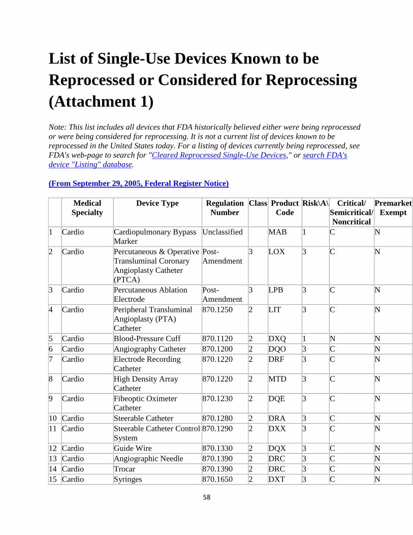
58
List of Single-Use Devices Known to be Reprocessed or Considered for Reprocessing (Attachment 1)
Note: This list includes all devices that FDA historically believed either were being reprocessed or were being considered for reprocessing. It is not a current list of devices known to be reprocessed in the United States today. For a listing of devices currently being reprocessed, see FDA's web-page to search for "Cleared Reprocessed Single-Use Devices," or search FDA's device "Listing" database.
(From September 29, 2005, Federal Register Notice)
Medical Specialty
Device Type Regulation Number
Class Product Code
Risk\A\ Critical/ Semicritical/ Noncritical
Premarket Exempt
1 Cardio Cardiopulmonary Bypass Marker
Unclassified MAB 1 C N
2 Cardio Percutaneous & Operative Transluminal Coronary Angioplasty Catheter (PTCA)
Post- Amendment
3 LOX 3 C N
3 Cardio Percutaneous Ablation Electrode
Post-Amendment
3 LPB 3 C N
4 Cardio Peripheral Transluminal Angioplasty (PTA) Catheter
870.1250 2 LIT 3 C N
5 Cardio Blood-Pressure Cuff 870.1120 2 DXQ 1 N N 6 Cardio Angiography Catheter 870.1200 2 DQO 3 C N 7 Cardio Electrode Recording
Catheter 870.1220 2 DRF 3 C N
8 Cardio High Density Array Catheter
870.1220 2 MTD 3 C N
9 Cardio Fibeoptic Oximeter Catheter
870.1230 2 DQE 3 C N
10 Cardio Steerable Catheter 870.1280 2 DRA 3 C N 11 Cardio Steerable Catheter Control
System 870.1290 2 DXX 3 C N
12 Cardio Guide Wire 870.1330 2 DQX 3 C N 13 Cardio Angiographic Needle 870.1390 2 DRC 3 C N 14 Cardio Trocar 870.1390 2 DRC 3 C N 15 Cardio Syringes 870.1650 2 DXT 3 C N

59
16 Cardio Injector Type Syringe Actuator
870.1670 2 DQF 3 C N
17 Cardio Oximeter 870.2700 2 DQA 3 N N 18 Cardio Tissue Saturation
Oximeter 870.2700 2 MUD 3 C N
19 Cardio Intra-Aortic Balloon System
870.3535 3 DSP 3 C N
20 Cardio Vascular Clamp 870.4450 2 DXC 3 C N 21 Cardio Heart Stabilizer 870.4500 1 MWS 2 C Y 22 Cardio Noncompression Heart
Stabilizer 870.4500 1 MWS 3 C Y
23 Cardio External Vein Stripper 870.4885 2 DWQ 3 C N 24 Cardio Compressible Limb
Sleeve 870.5800 2 JOW 1 N N
25 Dental Bur 872.3240 1 EJL 1 C Y 26 Dental Diamond Coated Bur 872.3240 1 EJL 3 C Y 27 Dental Diamond Instrument 872.4535 1 DZP 3 C Y 28 Dental AC-Powered Bone Saw 872.4120 2 DZH 2 C N 29 Dental Manual Bone Drill and
Wire Driver 872.4120 2 DZJ 2 C N
30 Dental Powered Bone Drill 872.4120 2 DZI 2 C N 31 Dental Intraoral Drill 872.4130 1 DZA 1 C Y 32 Dental Injection Needle 872.4730 1 DZM 3 C Y 33 Dental Metal Orthodontic Bracket 872.5410 1 EJF 3 S Y 34 Dental Plastic Orthodontic
Bracket 872.5470 2 DYW 3 S N
35 ENT Bur 874.4140 1 EQJ 1 C Y 36 ENT Diamond Coated Bur 874.4140 1 EQJ 3 C Y 37 ENT Microdebrider 874.4140 1 EQJ 3 C Y 38 ENT Microsurgical Argon Fiber
Optic Laser Cable, For Uses Other Than Otology, Including Laryngology & General Use In Otolaryngology
874.4490 2 LMS 1 S N
39 ENT Microsurgical Argon Fiber Optic Laser Cable, For Use In Otology
874.4490 2 LXR 1 S N
40 ENT Microsurgical Carbon-Dioxide Fiber Optic Laser Cable
874.4500 2 EWG 1 S N
41 ENT Bronchoscope Biopsy Forceps (Nonrigid)
874.4680 2 BWH 3 C N
42 ENT Bronchoscope Biopsy 874.4680 2 JEK 1 C N

60
Forceps (Rigid) 43 Gastro/Urology Biopsy Forceps Cover 876.1075 1 FFF 1 C Y 44 Gastro/Urology Biopsy Instrument 876.1075 2 KNW 3 C N 45 Gastro/Urology Biopsy Needle Set 876.1075 2 FCG 3 C N 46 Gastro/Urology Biopsy Punch 876.1075 2 FCI 2 C N 47 Gastro/Urology Mechanical Biopsy
Instrument 876.1075 2 FCF 2 C N
48 Gastro/Urology Nonelectric Biopsy Forceps
876.1075 1 FCL 3 C Y
49 Gastro/Urology Cytology Brush For Endoscope
876.1500 2 FDX 2 S N
50 Gastro/Urology Endoscope Accessories 876.1500 2 KOG 2 S N 51 Gastro/Urology Extraction
Balloons/Baskets 876.1500 2 KOG 2 S N
52 Gastro/Urology Endoscopic Needle 876.1500 2 FBK 3 C N 53 Gasro/Urology Simple
Pneumoperitoneum Needle
876.1500 2 FHP 3 C N
54 Gastro/Urology Spring Loaded Pneumoperitoneum Needle
876.1500 2 FHO 3 C N
55 Gastro/Urology Active Electrosurgical Electrode
876.4300 2 FAS 3 S N
56 Gastro/Urology Biliary Sphincterotomes 876.5010, 876.1500
2 FGE 3 C N
57 Gastro/Urology Electric Biopsy Forceps 876.4300 2 KGE 3 C N 58 Gastro/Urology Electrosurgical
Endoscopic Unit (with or without accessories)
876.4300 2 KNS 3 S N
59 Gastro/Urology Flexible Snare 876.4300 2 FDI 3 S N 60 Gastro/Urology Flexible Suction
Coagulator Electrode 876.4300 2 FEH 3 S N
61 Gastro/Urology Flexible Stone Dislodger 876.4680 2 FGO 3 S Y 62 Gastro/Urology Metal Stone Dislodger 876.4680 2 FFL 3 S Y 63 Gastro/Urology Needle Holder 876.4730 1 FHQ 1 C Y 64 Gastro/Urology Nonelectrical Snare 876.4730 1 FGX 1 S Y 65 Gastro/Urology Urological Catheter 876.5130 2 KOD 2 S N 66 Gastro/Urology Single Needle Dialysiss
Set 876.5540 2 LBW,
FIE 3 C N
67 Gastro/Urology Hemodialysis Blood Circuit Accessories
876.5820 2 KOC 2 S N
68 Gastro/Urology Single Needle Dialysis Set 876.5820 2 FIF 3 C N 69 Gastro/Urology Hemorrhoidal Ligator 876.4400 2 FHN 2 C N 70 General Implanted Programmable Post 3 LKK 3 C N

61
Hospital Infusion Pump Amendment 71 General
Hospital Needle Destruction Device
Post Amendment
3 MTV 1 N N
72 General Hospital
Nonpowered Flotation Therapy Mattress
880.5150 1 IKY 2 N Y
73 General Hospital
NonAC-Powered Patient Lift
880.5510 1 FSA 2 N Y
74 General Hospital
Alternating Pressure Air Flotation Mattress
880.5550 2 FNM 1 N Y
75 General Hospital
Temperature Regulated Water Mattress
880.5560 1 FOH 2 N Y
76 General Hospital
Hypodermic Single Lumen Needle
880.5570 2 FMI 3 C N
77 General Hospital
Piston Syringe 880.5860 2 FMF 3 C N
78 General Hospital
Mattress Cover (Medical Purposes)
880.6190 1 FMW 2 N Y
79 General Hospital
Disposable Medical Scissors
880.6820 1 JOK 1 N Y
80 General Hospital
Irrigating Syringe 880.6960 1 KYZ, KYY
1 C Y
81 Infection Control
Surgical Gowns 878.4040 2 FYA 1 C N
82 Lab Blood Lancet 878.4800 1 FMK 1 C Y 83 Neurology Clip Forming/Cutting
Instrument 882.4190 1 HBS 3* C Y
84 Neurology Drills, Burrs, Trephines & Accessories (Manual)
882.4300 2 HBG 3* C N
85 Neurology Drills, Burrs, Trephines & Accessories (Compound, Powered)
882.4305 2 HBF 3* C N
86 Neurology Drills, Burrs, Trephines & Accessories (Simple, Powered)
882.4310 2 HBE 3* C N
87 OB/GYN Oocyte Aspiration Needle 3 MHK 3 C N 88 OB/GYN Laparoscope Accessories 884.1720 1 HET 2 C Y 89 OB/GYN Laparoscope Accessories 884.1720 2 HET 3 C N 90 OB/GYN Laparoscopic Dissectors 884.1720 1 HET 2 C Y 91 OB/GYN Laparoscopic Graspers 884.1720 1 HET 2 C Y 92 OB/GYN Laparoscopic Scissors 884.1720 1 HET 2 C Y 93 OB/GYN Insufflator Accessories
(Tubing, Verres Needle, Kits)
884.1730 2 HIF 3 C Y
94 OB/GYN Laparoscopic Insufflator 884.1730 2 HIF 2 N N

62
95 OB/GYN Endoscopic Electrocautery and Accessories
884.4100 2 HIM 2 N N
96 OB/GYN Gynecologic Electrocautery (and Accessories)
884.4120 2 HGI 2 N N
97 OB/GYN Endoscopic Bipolar Coagulator-Cutter (and Accessories)
884.4150 2 HIN 2 N N
98 OB/GYN Culdoscopic Coagulator (and Accessories)
884.4160 2 HFI 2 N N
99 OB/GYN Endoscopic Unipolar Coagulator-Cutter (and Accessories)
884.4160 2 KNF 2 N N
100 OB/GYN Hysteroscopic Coagulator (and Accessories)
884.4160 2 HFH 2 N N
101 OB/GYN Unipolar Laparoscopic Coagulator (and Accessories)
884.4160 2 HFG 2 N N
102 OB/GYN Episiotomy Scissors 884.4520 1 HDK 1 C Y 103 OB/GYN Umbilical Scissors 884.4520 1 HDJ 1 C Y 104 OB/GYN Biopsy Forceps 884.4530 1 HFB 3 C Y 105 OB/GYN Assisted Reproduction
Needle 884.6100 2 MQE 3 C N
106 Ophthalmic Endoilluminator 876.1500 2 MPA 3* C N 107 Ophthalmic Surgical Drapes 878.4370 2 KKX 2 C N 108 Ophthalmic Ophthalmic Knife 886.4350 1 HNN 3 C Y 109 Ophthalmic Keratome Blade 886.4370 1 HMY,
HNO 3 C N
110 Ophthalmic Phacoemulsification Needle
886.4670 2 HQC 3 C N
111 Ophthalmic Phacoemulsification/ Phacofragmentation Fluidic
886.4670 2 MUS 2 C N
112 Ophthalmic Phacofragmentation Unit 886.4670 2 HQC 1 N N 113 Orthopedic Saw Blades 878.4820 1 GFA,
DWH, GEY, GET
1 C Y
114 Orthopedic Surgical Drills 878.4820 1 GEY, GET
1 C Y
115 Orthopedic Arthroscope Accessories 888.1100 2 HRX 2 C Y 116 Orthopedic Bone Tap 888.4540 1 HWX 1 C Y 117 Orthopedic Burr 888.4540 1 HTT 1 C Y 118 Orthopedic Carpal Tunnel Blade 888.4540 1 LXH 2 C Y

63
119 Orthopedic Countersink 888.4540 1 HWW 1 C Y 120 Orthopedic Drill Bit 888.4540 1 HTW 1 C Y 121 Orthopedic Knife 888.4540 1 HTS 1 C Y 122 Orthopedic Manual Surgical
Instrument 888.4540 1 LXH 1 C Y
123 Orthopedic Needle Holder 888.4540 1 HXK 1 C Y 124 Orthopedic Reamer 888.4540 1 HTO 1 C Y 125 Orthopedic Rongeur 888.4540 1 HTX 1 C Y 126 Orthopedic Scissors 888.4540 1 HRR 1 C Y 127 Orthopedic Staple Driver 888.4540 1 HXJ 1 C Y 128 Orthopedic Trephine 888.4540 1 HWK 1 C Y 129 Orthopedic Flexible Reamers/Drills 886.4070/
878.4820 1 GEY,
HRG 1 C Y
130 Orthopedic External Fixation Frame 888.3040/ 888.3030
2 JEC, KTW, KTT
2 N N
131 Physical Medicine
Nonheating Lamp for Adjunctive Use Inpatient Therapy
890.5500 2 NHN 1 N N
132 Physical Medicine
Electrode Cable 890.1175 2 IKD 1 N Y
133 Physical Medicine
External Limb Component, Hip Joint
890.3420 1 ISL 2 N Y
134 Physical Medicine
External Limb Component, Knee Joint
890.3420 1 ISY 2 N Y
135 Physical Medicine
External Limb Component, Mechanical Wrist
890.3420 1 ISZ 2 N Y
136 Physical Medicine
External Limb Component, Shoulder Joint
890.3420 1 IQQ 2 N Y
137 Plastic Surgery Stapler 878.4800 1 GAG, GEF, FHM, HBT
2 C Y
138 Radiology Isotope Needle 892.5730 2 IWF 3 C N 139 Respiratory Endotracheal Tube
Changer Unclassified 3 LNZ 3 C N
140 Respiratory Anesthesia Conduction Needle
868.5150 2 BSP 3 C N
141 Respiratory Short Term Spinal Needle 868.5150 2 MIA 3 C N 142 Respiratory Respiratory Therapy and
Anesthesia Breathing Circuits
868.5240 1 CAI 2 S Y

64
143 Respiratory Oral and Nasal Catheters 868.5350 1 BZB 1 C Y 144 Respiratory Gas Masks 868.5550 1 BSJ 1 S Y 145 Respiratory Breathing Mouthpiece 868.5620 1 BYP 1 N Y 146 Respiratory Tracheal Tube 868.5730 2 BTR 3 C N 147 Respiratory Airway Connector 868.5810 1 BZA 2 S Y 148 Respiratory CPAP Mask 868.5905 2 BZD 3 S N 149 Respiratory Emergency Manual
Resuscitator 868.5915 2 BTM 2 S N
150 Respiratory Tracheobronchial Suction Catheter
868.6810 1 BSY 3 S Y
151 Surgery AC-Powered Orthopedic Instrument and Accessories
878.4820 1 HWE 2 C N
152 Surgery Breast Implant Mammary Sizer
Unclassified MRD 1 C N
153 Surgery Ultrasonic Surgical Instrument
Unclassified LFL 3 C N
154 Surgery Trocar 874.4420 1 KAB, KBG, KCI
3 C Y
155 Surgery Endoscopic Blades 876.1500 2 GCP, GCR
2 C N
156 Surgery Endoscopic Guidewires 876.1500 2 GCP, GCR
1 C N
157 Surgery Inflatable External Extremity Splint
878.3900 1 FZF 1 N Y
158 Surgery Noninflatable External Extremity Splint
878.3910 1 FYH 1 N Y
159 Surgery Catheter Needle 878.4200 1 GCB 3 C Y 160 Surgery Implantable Clip 878.4300 2 FZP 3 C N 161 Surgery Electrosurgical and
Coagulation Unit with Accessories
878.4400 2 BWA 2 C N
162 Surgery Electrosurgical Apparatus 878.4400 2 HAM 2 C N 163 Surgery Electrosurgical Cutting &
Coagulation Device & Accessories
878.4400 2 GEI, NUJ 2 3 C N
164 Surgery Electrosurgical Device 878.4400 2 DWG 2 C N 165 Surgery Electrosurgical Electrode 878.4400 2 JOS 2 C N 166 Surgery Implantable Staple,
Clamp, Clip for Suturing Apparatus
878.4750 2 GDW 3 C N
167 Surgery Percutaneous Biopsy Device
878.4800 1 MJG 3 C Y

65
168 Surgery Gastro-Urology Needle 878.4800 1 FHR 3 C Y 169 Surgery Aspiration and Injection
Needle 878.4800 1 GAA 3 C Y
170 Surgery Biopsy Brush 878.4800 1 GEE 1 C Y 171 Surgery Blood Lancet 878.4800 1 FMK 1 C Y 172 Surgery Bone Hook 878.4800 1 KIK 1 C Y 173 Surgery Cardiovascular Biopsy
Needle 878.4800 1 DWO 3 C Y
174 Surgery Clamp 878.4800 1 GDJ 1 C Y 175 Surgery Clamp 878.4800 1 HXD 1 C Y 176 Surgery Curette 878.4800 1 HTF 1 C Y 177 Surgery Disposable Surgical
Instrument 878.4800 1 KDC 1 C Y
178 Surgery Disposable Vein Stripper 878.4800 1 GAJ 1 C Y 179 Surgery Dissector 878.4800 1 GDI 1 C Y 180 Surgery Forceps 878.4800 1 GEN 2 C Y 181 Surgery Forceps 878.4800 1 HTD 2 C Y 182 Surgery Gouge 878.4800 1 GDH 1 C Y 183 Surgery Hemostatic Clip Applier 878.4800 1 HBT 2 C Y 184 Surgery Hook 878.4800 1 GDG 1 C Y 185 Surgery Manual Instrument 878.4800 1 MDM,
MDW 1 C Y
186 Surgery Manual Retractor 878.4800 1 GZW 1 C Y 187 Surgery Manual Saw and
Accessories 878.4800 1 GDR,
HAC 1 C Y
188 Surgery Manual Saw and Accessories
878.4800 1 HAC 1 C Y
189 Surgery Manual Surgical Chisel 878.4800 1 FZO 1 C Y 190 Surgery Mastoid Chisel 878.4800 1 JYD 1 C Y 191 Surgery Orthopedic Cutting
Instrument 878.4800 1 HTZ 1 C Y
192 Surgery Orthopedic Spatula 878.4800 1 HXR 1 C Y 193 Surgery Osteotome 878.4800 1 HWM 1 C Y 194 Surgery Rasp 878.4800 1 GAC 1 C Y 195 Surgery Rasp 878.4800 1 HTR 1 C Y 196 Surgery Retractor 878.4800 1 GAD 1 C Y 197 Surgery Retractor 878.4800 1 HXM 1 C Y 198 Surgery Saw 878.4800 1 HSO 1 C Y 199 Surgery Scalpel Blade 878.4800 1 GES 1 C Y 200 Surgery Scalpel Handle 878.4800 1 GDZ 1 C Y 201 Surgery Scissors 878.4800 1 LRW 1 C Y 202 Surgery Snare 878.4800 1 GAE 1 C Y 203 Surgery Spatula 878.4800 1 GAF 1 C Y
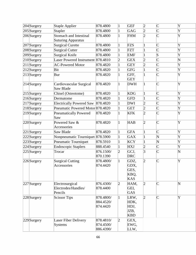
66
204 Surgery Staple Applier 878.4800 1 GEF 2 C Y 205 Surgery Stapler 878.4800 1 GAG 2 C Y 206 Surgery Stomach and Intestinal
Suturing Apparatus 878.4800 1 FHM 2 C Y
207 Surgery Surgical Curette 878.4800 1 FZS 1 C Y 208 Surgery Surgical Cutter 878.4800 1 FZT 1 C Y 209 Surgery Surgical Knife 878.4800 1 EMF 1 S Y 210 Surgery Laser Powered Instrument 878.4810 2 GEX 2 C N 211 Surgery AC-Powered Motor 878.4820 1 GEY 2 C Y 212 Surgery Bit 878.4820 1 GFG 1 C Y 213 Surgery Bur 878.4820 1 GFF,
GEY 1 C Y
214 Surgery Cardiovascular Surgical Saw Blade
878.4820 1 DWH 1 C Y
215 Surgery Chisel (Osteotome) 878.4820 1 KDG 1 C Y 216 Surgery Dermatome 878.4820 1 GFD 1 C Y 217 Surgery Electrically Powered Saw 878.4820 1 DWI 2 C Y 218 Surgery Pneumatic Powered Motor 878.4820 1 GET 2 C Y 219 Surgery Pneumatically Powered
Saw 878.4820 1 KFK 2 C Y
220 Surgery Powered Saw & Accessories
878.4820 1 HAB 2 C Y
221 Surgery Saw Blade 878.4820 1 GFA 1 C Y 222 Surgery Nonpneumatic Tourniquet 878.5900 1 GAX 1 N Y 223 Surgery Pneumatic Tourniquet 878.5910 1 KCY 1 N Y 224 Surgery Endoscopic Staplers 888.4540 1 HXJ 2 C Y 225 Surgery Trocar 876.1500/
870.1390 2 GCJ,
DRC 3 C N
226 Surgery Surgical Cutting Accessories
878.4800/ 874.4420
1 GDZ, GDX, GES, KBQ, KAS
2 C Y
227 Surgery Electrosurgical Electrodes/Handles/ Pencils
876.4300/ 878.4400
2 HAM, GEI, GAS
2 C N
228 Surgery Scissor Tips 878.4800/ 884.4520/ 874.4420
1 LRW, HDK, HDJ, JZB, KBD
2 C Y
229 Surgery Laser Fiber Delivery Systems
878.4810/ 874.4500/ 886.4390/
2 GEX, EWG, LLW,
67
884.4550 HQF, HHR, HQB
Cost savings depends on your facility with staff compliance and face- to-face discussions with potential reprocessing vendors.
Kaiser Roseville, CA, mainly reprocesses compression sleeves, external fixation, open and unused tourniquet cuffs, trocars, ultrasonic scalpels and the total estimated annual savings is $216,799.00.
Recycling
This subject has little to do with sterilization, however it does relate to the packaging of sterile items and how to deal with the item once a surgical case is completed. Most O.R. staff realizes how much waste is generated from one surgical case.
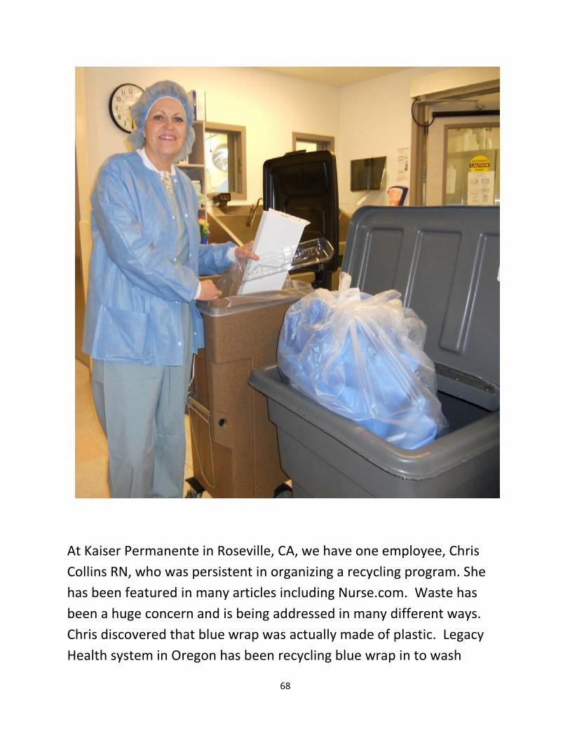
68
At Kaiser Permanente in Roseville, CA, we have one employee, Chris Collins RN, who was persistent in organizing a recycling program. She has been featured in many articles including Nurse.com. Waste has been a huge concern and is being addressed in many different ways. Chris discovered that blue wrap was actually made of plastic. Legacy Health system in Oregon has been recycling blue wrap in to wash

69
buckets, lawn furniture, flower pots, squirt bottles and plastic lumber for picnic tables as well as other products. Chris’s wish is that this Program takes off everywhere, not just in our HMO, and saves recourses for the future generations.
We also have buy-in from the staff not only in our department but from the environmental services staff. The environmental staff empties the bins in the O.R. which collect the blue wrappers and they move them to a larger bin in the hallway. This all gets moved to a grey bin and alarger garbage “can” which collects:
• Plastic bottles • Empty IV bags • Boxes from disposable laparoscopic items • Hard plastic that the sterile items came in
With the push to go fast and get more completed, just remember it takes only one minute out of a case to throw these items in the bin and dispose of them in a much more environmentally friendly way. Take a look at these reported recycling measurements from 2010: Northern California saves 176,038 pounds of waste, Hawaii 9,735, Ohio 734, Oregan 13,038, Southern California 129,154 for a total of 328,699 pounds equaling 164.3 tons of waste.
We should make this a healthy competition and see who can recycle the most, and continue following on Chris’s vision!
Just a few more fun historical facts:
70
Horace Wells (1815-1848) was the 1st practitioner to publically advocate for the use of nitrous oxide as an anesthetic. He wanted to reduce pain during dental procedures. You’ve got to love this man!
William Morton (1819-1868) claimed credit as the discoverer of anesthesia. He performed the first anesthetic surgical procedure in 1846 using ether.
Gustav Neuber - In 1883, the practice of wearing surgical gowns and caps was started by this German surgeon. In 1885, Neuber built a small hospital in Kiel, Germany with his aseptic design including a five-room OR suite. He gave each room a purpose, including one room for “clean procedures” and one for “infected” procedures.
Jan Mikulicz-Radecki (1850-1905) born in present day Ukraine, was credited with using the first mask made of gauze. He was also one of the first to use gloves during surgery.
William Stewart Halsted (1852-1922) An American surgeon and champion for “Strict Aseptic Technique”. Halsted was one of the first surgeons credited with using surgical rubber gloves. After calling his sister who just gave birth, he found out she was bleeding to death. He rushed to her aid and withdrew his blood to give to her. This was one of the first documented blood transfusions and he was able to save her life after he did surgery on her. Halsted was credited for starting the first formal surgical residency.
William Keen (1837-1932) In reference to the abundance of recent surgical inventions, Keen commented: “Antisepsis relieved patients from the terror of death and gave to the surgeons restful nights and joyous days.”
71
Association: An organization of people with a common purpose and having a formal structure.
Bioburden: The number of contaminating microorganisms present on an object. Reduction of bioburden is the goal of infection control programs and protocols.
Biological Indicator: A biological indicator is a device to monitor the sterilization process that consists of a standardized population of bacterial spores known to be resistant to the mode of sterilization being monitored. The spores are in a media cohesive to growth and if not killed in the sterilization process show that load of instrument are not sterile and should be recalled. Biological indicators indicate that all the parameters necessary for sterilization were present when they are negative.
Bowie-Dick Test: Autoclave testing services describes as:
A Bowie-Dick test is used in pre-vacuum type (or dynamic air removal) sterilizers. They are used to detect air leaks and inadequate air removal and consist of folded 100% cotton surgical towels that are clean and preconditioned. A commercially available Bowie-Dick-type test sheet should be placed in the center of the pack.
The test pack should be placed horizontally in the front, bottom section of the sterilizer rack, near the door and over the drain, in an otherwise empty chamber and run at 134°C for 3.5 minutes. The test is used each day the vacuum-type steam sterilizer is used, before the first processed load.
Air that is not removed from the chamber will interfere with steam contact. Smaller, commercially available disposable test packs (or process challenge devices) have been devised to
72
replace the stack of folded surgical towels for testing the efficacy of the vacuum system in a prevacuum sterilizer.
They should be representative of the load and simulate the greatest challenge to the load. Sterilizer vacuum performance is acceptable if the sheet inside the test pack shows a uniform color change. Entrapped air will cause a spot to appear on the test sheet, due to the inability of the steam to reach the chemical indicator. If the sterilizer fails the Bowie-Dick test, do not use the sterilizer until it is inspected by the sterilizer maintenance personnel and passes the Bowie-Dick test.
See the complete recommendations on sterilizer and disinfection at www.cdc.gov “Guideline for Disinfection and Sterilization in Healthcare Facilities, 2008”.
Chemical Indicators: or CI that change color or physical form when exposed to certain temperatures. These would include autoclave tape, special markings on sterilization pouches and bags. This does not show that sterilization has been achieved or that a complete sterilization cycle has occurred. These are just process indicators, and show that the item has passed through a sterilizer.
To continue course, click on link below for the CDC information.
http://www.cdc.gov/ncidod/dhqp/pdf/guidelines/disinfection_nov_2008.pdf
73
References
www.aami.org/standards/
www.absoluteastronomy.com: Sushruta Samhita
Ask.com Encyclopedia
AORN Perioperative Nursing Clinics 2010
AORN online
A short history of the old trip of sterilization Irini Soulatatou: online
Autoclave testing services, Inc: online
www.CDC.com
www.ezinearicles.com
www.FDA.com
WWW.gallup.com
www.jewelprecision.com
The Merriam thesaurus and Webster Dictionary
www.nurses.com
www.osha.gov
www.remierinc.com
www.surgicalholdings.co.uk
Taber’s cyclopedic Dictionary edition 21
University of Leeds: History of sterilization and Disinfection: online

74