hallux rigidus: treatment by cheilectomy* · 2015-06-15 · metatarsal head, occasionally we found...
TRANSCRIPT

( op nght 988 t)s The Journol of Bone and Joint Surger . Ineorporated
400 ThE JOURNAL OF BONE AND JOINT SURGERY
Hallux Rigidus: Treatment by Cheilectomy*BY ROGER A. MANN, M.D.t, OAKLAND, CALIFORNIA, AND THOMAS 0. CLANTON, M.D4, HOUSTON, TEXAS
ABSTRACT: Cheilectomy, the excision of an irreg-ular osseous rim that interferes with motion of a joint,was performed on the distal part of the first metatarsalof twenty-five patients who had hallux rigidus. Relief ofpain was achieved in all but three patients, whose caseswere considered as failures. Joint motion improved byan average of 20 degrees, and it was in an acceptablerange in twenty-three patients. There were no compli-cations other than persistence of swelling in six patients.No patient required additional operative interventionduring an average follow-up of fifty-six months. We con-eluded that cheilectomy is a better method of treatmentfor hallux rigidus than arthrodesis, resection arthro-plasty, or arthroplasty with the use of a flexible implant.
Hallux rigidus is a painful affliction of the first meta-tarsophalangeal joint that is associated with limitation ofmotion, especially dorsiflexion. The original description ofthis condition has been attributed to Davies-Colley8, whocalled it hallux flexus, in a paper that was read before theClinical Society ofLondon in 1887. The name hallux rigiduswas proposed four months later by Cotteri!l6, and it remainsthe most common designation despite the advocacy of othernames such as hallux limitus, hallux do!orosus, metatarsusnon-extensus, dorsal bunion, winkle-picker disease, andmetatarsus primus elevatus. The history of the terminologywas well summarized by Kelikian3#{176}.
The general approach to treatment has not variedgreatly since Davies-Colley suggested resecting the base ofthe proximal phalanx of the great toe5. Most authors39”13.20.27.29.32.34.36.40.43.44.46.54.55 have recommended that form of
resection arthroplasty or arthrodesis of the first metatarso-phalangeal joint. Wedge osteotomy of the proximal phalanxor of the neck of the metatarsal has been advocated foryoung patients’82031324. Resection of the head of the firstmetatarsal was widely advocated in the early literature onhallux rigidus562326, but this procedure has since fallen intodisrepute3. More recently attention has been focused onvarious implants that have been used in the treatment ofpathological conditions of the great toe7 15.24.42.45.50.5 I
While cheilectomy is routinely included in some arthro-plasty procedures for hallux rigidus, it has received scant
*No benefits in any form have been received or will be received froma commercial party related directly or indirectly to the subject ofthis article.No funds were received in support of this study.
t 3300 Webster Street, Suite 1200, Oakland, California 94609.1 University ofTexas Health Science Center at Houston. 6410 Fannin,
Suite 310, Houston, Texas 77030. Please address requests for reprints toDr. Clanton.
attention as an isolated procedure’53 t, and the purpose ofthis paper is to report on a group of patients who weretreated with this procedure.
Materials and Methods
Between January 1976 and December 198 1 , we treatedtwenty-eight patients with thirty-four cheilectomies. Onlytwo other patients who had hallux rigidus were treated op-eratively during this period of time. Those two patients hadan arthrodesis because they had extensive degenerativechanges and desired a single definitive procedure. Manymore patients who had hallux rigidus were seen during thesix-year period, but they either improved with non-operativetreatment or went elsewhere for treatment.
The diagnosis of hailux rigidus was based on a com-plaint of pain in the first metatarsophalangeal joint and thesephysical findings: increased bulk of the joint, especiallydorsally; not infrequently, an associated synovitis; andmarked restriction of dorsiflexion. Plantar flexion alsocaused some pain. We attributed that to stretching of thecapsule of the joint and of the tendon of the extensor hallucislongus over the osteophyte on the dorsal rim of the meta-tarsal head. Radiographs always revealed degenerative ar-thritis of the first metatarsophalangeal joint, and anosteophyte was always present on the dorsal aspect of themetatarsal. No patient had evidence of rheumatoid arthritis,gout, or seronegative spondyloarthropathy.
Twenty-five of the twenty-eight patients were exam-med at follow-up by one of us (R. A. M.) (Table I). Threepatients were lost to follow-up. The average length of fol-low-up was fifty-six months (range, thirty to 100 months).There were five men and twenty women, and the averageage was fifty-six years (range, thirty to eighty years). Sixpatients had the lesion bilaterally. Ten patients had a historyof injury to the great toe, but in only five could the injurybe directly related to the onset of the symptoms. Only onepatient had an occupation that might have contributed to thedevelopment of the symptoms. Ten patients related thesymptoms predominantly to recreational activities.
The pain was severe enough to interfere with the life-style of all of the patients. Discomfort that was producedby pressure of the shoe on the osteophyte caused all patientsto alter their footwear. Eight patients complained of thecosmetic deformity of the dorsal bunion, and four had anulceration of the skin over the bunion.
The preoperative arc of motion of the first metatarso-phalangeal joint averaged 29 degrees (range, 5 to 65 de-grees). There is some disagreement about what constitutes
HALLUX RIGIDUS: TREATMENT BY CHEILECTOMY 401
VOL. 70-A, NO. 3, MARCH 1988
TABLE I
DATA ON PATIENTS
Motion in Degrees
ReliefLevel of
Satisfaction Length ofPreop. Preop. Preop. Plantar Postop. Postop. Postop. PlantarCase Age Side Arc Dorsiflexion Flexion Arc Dorsiflexion Flexion of Pain with Result Follow-up
(Yrs.) (Mos.)
1 56 RL
2525
1010
1515
7050
6040
1010
CompletePartial
SatisfiedSatisfied*
7171
2 51 R 5 15 - 10 45 25 20 Complete Satisfied 96
3 50 R 30 10 20 10 35 - 25 Unchanged Dissatisfied 484 49 R
L3030
1515
1515
5560
5050
510
CompleteComplete
SatisfiedSatisfied
3636
5 61 L 25 10 15 65 50 15 Complete Satisfied 376 64 L 25 15 10 45 30 15 Complete Satisfied 567 71 L 25 15 10 5 15 - 10 Worse Dissatisfied 558 58 R 45 30 15 80 60 20 Complete Satisfied 36
9 30 R 35 20 15 60 40 20 Complete Satisfied 3010 45 R
L5515
405
1510
8080
7070
1010
CompleteComplete
SatisfiedSatisfied
5454
1 1 5 1 RL
4540
4540
00
3040
4045
- 10- 5
CompleteComplete
SatisfiedSatisfied
3939
12 60 L 20 5 15 30 30 0 Complete Satisfied 3013 54 R
L2535
1020
1515
6575
6060
515
CompleteComplete
SatisfiedSatisfied
4646
14 45 RL
3065
2530
535
3565
2545
1020
PartialComplete
Satisfied*Satisfied
4242
15 64 L 15 10 5 25 40 - 15 Complete Satisfied 9616 39 R 15 15 0 40 30 10 Partial Satisfied 10017 69 L 10 10 0 30 15 15 Partial Satisfied 7318 74 R 30 30 0 65 60 5 Complete Satisfied 6619 53 R 30 10 20 25 30 - 5 Complete Satisfied* 4120 53 R 30 10 20 50 45 5 Complete Satisfied 5121 40 R 30 20 10 50 45 5 Partial Satisfied* 7222 80 R 20 25 - 5 30 30 0 Complete Satisfied 6623 63 L 15 5 10 15 30 - 15 Unchanged Dissatisfied 5924 59 R 20 5 15 80 65 15 Partial Satisfied* 34
25 69 R 30 10 20 30 35 - 5 Complete Satisfied* 60
* The patient was satisfied but had reservations.
a normal arc of motion in this jointU724252*. We have con-sidered 100 degrees as the normal arc, which is composedof 70 degrees of dorsiflexion and 30 degrees of plantarflexion. Dorsiflexion was limited to 30 degrees or less inall but two patients (Cases 10 and 1 1). No patient had anyimportant coexisting lesions, such as hallux valgus, on theaffected foot.
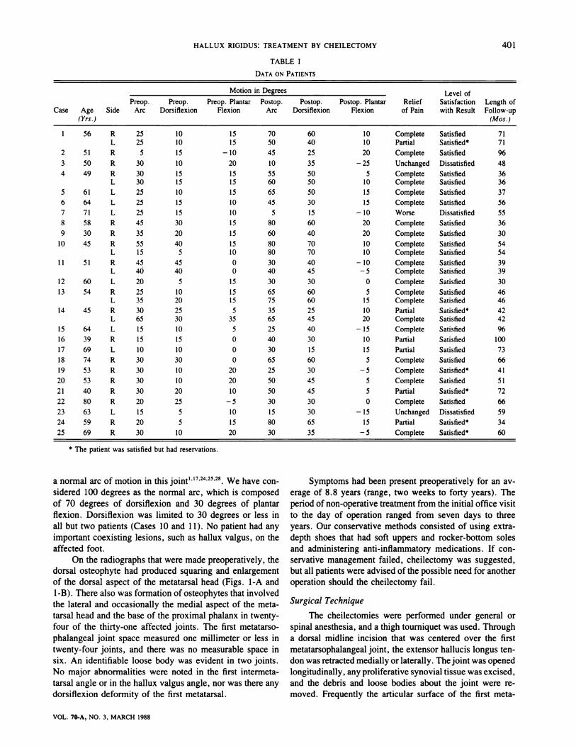
On the radiographs that were made preoperatively, thedorsal osteophyte had produced squaring and enlargementof the dorsal aspect of the metatarsal head (Figs. 1-A and1-B). There also was formation of osteophytes that involvedthe lateral and occasionally the medial aspect of the meta-tarsal head and the base of the proximal phalanx in twenty-four of the thirty-one affected joints. The first metatarso-phalangeal joint space measured one millimeter or less intwenty-four joints, and there was no measurable space insix. An identifiable loose body was evident in two joints.No major abnormalities were noted in the first intermeta-tarsal angle or in the hallux valgus angle, nor was there anydorsiflexion deformity of the first metatarsal.
Symptoms had been present preoperatively for an av-erage of 8. 8 years (range, two weeks to forty years). Theperiod of non-operative treatment from the initial office visitto the day of operation ranged from seven days to threeyears. Our conservative methods consisted of using extra-depth shoes that had soft uppers and rocker-bottom solesand administering anti-inflammatory medications. If con-servative management failed, cheilectomy was suggested,but all patients were advised of the possible need for anotheroperation should the cheilectomy fail.
Surgical Technique
The cheilectomies were performed under general orspinal anesthesia, and a thigh tourniquet was used. Througha dorsal midline incision that was centered over the firstmetatarsophalangeal joint, the extensor hallucis longus ten-don was retracted medially or laterally. Thejoint was openedlongitudinally, any proliferative synovial tissue was excised,and the debris and loose bodies about the joint were re-moved. Frequently the articular surface of the first meta-
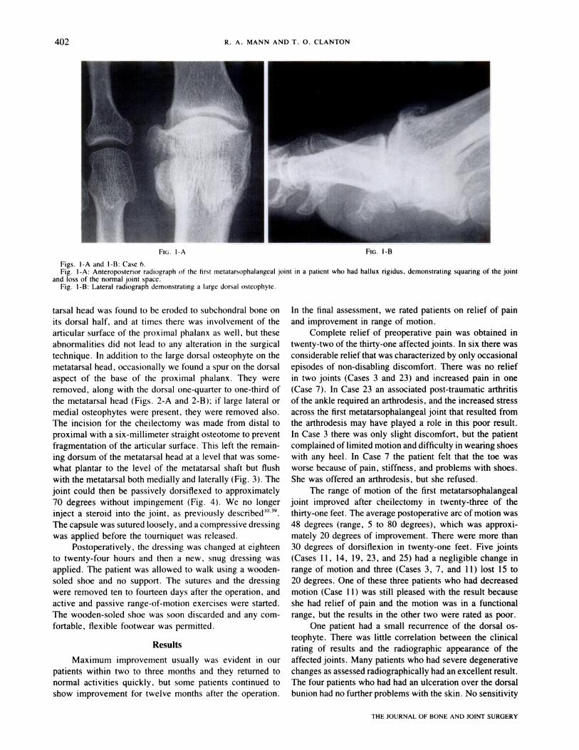
FIG. I-A FIG. 1-B
402 R. A. MANN AND T. 0. CLANTON
THE JOURNAL OF BONE AND JOINT SURGERY
Figs. I-A and 1-B: Case 6.Fig. 1-A: Anteroposterior radiograph of the first rnetatarsophalangeal joint in a patient who had hallux rigidus. demonstrating squaring of the joint
and loss of the normal joint space.Fig. 1-B: Lateral radiograph demonstrating a large dorsal osteophyte.
tarsal head was found to be eroded to subchondral bone onits dorsal half, and at times there was involvement of thearticular surface of the proximal phalanx as well, but theseabnormalities did not lead to any alteration in the surgicaltechnique. In addition to the large dorsal osteophyte on themetatarsal head, occasionally we found a spur on the dorsalaspect of the base of the proximal phalanx. They wereremoved, along with the dorsal one-quarter to one-third ofthe metatarsal head (Figs. 2-A and 2-B); if large lateral ormedial osteophytes were present, they were removed also.The incision for the cheilectomy was made from distal toproximal with a six-millimeter straight osteotome to preventfragmentation of the articular surface. This left the remain-ing dorsum of the metatarsal head at a level that was some-what plantar to the level of the metatarsal shaft but flushwith the metatarsal both medially and laterally (Fig. 3). Thejoint could then be passively dorsiflexed to approximately70 degrees without impingement (Fig. 4). We no longerinject a steroid into the joint, as previously described’039.The capsule was sutured loosely, and a compressive dressingwas applied before the tourniquet was released.
Postoperatively, the dressing was changed at eighteento twenty-four hours and then a new. snug dressing wasapplied. The patient was allowed to walk using a wooden-soled shoe and no support. The sutures and the dressingwere removed ten to fourteen days after the operation, andactive and passive range-of-motion exercises were started.The wooden-soled shoe was soon discarded and any com-fortable, flexible footwear was permitted.
Results
Maximum improvement usually was evident in ourpatients within two to three months and they returned tonormal activities quickly, but some patients continued toshow improvement for twelve months after the operation.
In the final assessment, we rated patients on relief of painand improvement in range of motion.
Complete relief of preoperative pain was obtained intwenty-two of the thirty-one affected joints. In six there wasconsiderable relief that was characterized by only occasionalepisodes of non-disabling discomfort. There was no reliefin two joints (Cases 3 and 23) and increased pain in one(Case 7). In Case 23 an associated post-traumatic arthritisof the ankle required an arthrodesis, and the increased stressacross the first metatarsophalangeal joint that resulted fromthe arthrodesis may have played a role in this poor result.In Case 3 there was only slight discomfort, but the patientcomplained oflimited motion and difficulty in wearing shoeswith any heel. In Case 7 the patient felt that the toe wasworse because of pain, stiffness, and problems with shoes.She was offered an arthrodesis, but she refused.
The range of motion of the first metatarsophalangealjoint improved after cheilectomy in twenty-three of thethirty-one feet. The average postoperative arc of motion was48 degrees (range, 5 to 80 degrees), which was approxi-mately 20 degrees of improvement. There were more than30 degrees of dorsiflexion in twenty-one feet. Five joints(Cases 1 1 , 14, 19, 23, and 25) had a negligible change inrange of motion and three (Cases 3 , 7 , and 1 1) lost 15 to20 degrees. One of these three patients who had decreasedmotion (Case 1 1) was still pleased with the result becauseshe had relief of pain and the motion was in a functionalrange, but the results in the other two were rated as poor.
One patient had a small recurrence of the dorsal os-teophyte. There was little correlation between the clinicalrating of results and the radiographic appearance of theaffected joints . Many patients who had severe degenerativechanges as assessed radiographically had an excellent result.The four patients who had had an ulceration over the dorsalbunion had no further problems with the skin. No sensitivity

Ft . 2-A
Figs. 2-A and 2-B: Illustrations depicting bone that was removed fromthe first metatarsal during the cheilectoniy.
Fig. 2-A: Preoperative lateral view of the dorsal osteophyte.
FIG. 2-B
HALLUX RIGIDUS: TREATMENT BY CHEILECTOMY 403
VOL. 70-A, NO, 3. MARCH 1958
was noted at the site of the incision. Problems with shoespersisted in four patients, but the remaining twenty-onepatients were able to wear ordinary shoes; however, mostof the women avoided heels that were higher than five cen-timeters.
There were no serious complications. Swelling aboutthe first metatarsophalangeal joint without evidence of in-fection persisted for one year in one patient, for four monthsin one, and for six weeks in four, but this complication didnot compromise the result in terms of pain or function. Atthe time of writing, no patient in this series had had anyadditional related surgical procedure.
Discussion
Hallux rigidus is a common and disabling affliction ofthe great toe and it occurs in about one in forty-five mdi-viduals who are more than fifty years old’6. A predisposition
to development of the lesion has been suggested in peoplewho have a long, slender foot2; a pronated foot2’23” ’; a longgreat toe489 4852; pes planus4922, particularly when theperson habitually wears stiff boots6; congenital flattening ofthe first metatarsal head’#{176};metatarsus primus elevatus33; andosteochondritis dissecans of the first metatarsal head’43552.Each of these conditions allegedly increases the stress acrossthe first metatarsophalangeal joint and results in damage tothe articular surface. Then there is reflex spasm in the sur-rounding musculature and subsequent production of osteo-phytic bone926. Although some patients in our seriesmanifested one or more of these alleged predispositions,none seemed to be a common predisposing factor for halluxrigidus.
Hallux rigidus has been described in two age-groups:adolescent patients, who have localized changes in the ar-ticular cartilage; and adult patients, who have more gen-eralized degenerative arthritis of the joint . Kessel andBonney3’ implicated osteochondritis dissecans as a cause ofhallux rigidus in young patients. This idea was substantiatedby Goodfellow’4, who proposed that trauma to a vulnerableepiphysis could result in osteochondritis dissecans of thefirst metatarsal head. No patient in our series was an ado-lescent. The only salient pathological variation in our pa-tients was an increase in the degree of osteoarthriticinvolvement with age. We did not alter our approach on thebasis of age or degree of involvement, although others havesuggested that this should be done’532.
The technique of cheilectomy that we are reporting isessentially unchanged from the procedure that was describedoriginally by DuVries’#{176}in 1959 and was reported by Mannet al.39 in 1979. Authors of previous articles on hallux rig-idus that mentioned exostectomy conveyed little enthusiasmfor the procedure2’3”, but perhaps the unsatisfactory resultswere related to removal of less bone than the amount thatwe have advocated.
Cheilectomy was recently advocated by Gould’5 for thetreatment of hallux rigidus in young patients, and that seriesincluded twelve patients. He thought that toe power andstability were better in the patients who were treated bycheilectomy than in a simultaneously reported group of pa-tients who were treated with an implant. Our previous seriesoftwenty patients who were treated by cheilectomy included
Removal of the osteophyte and a portion of the normal articular surface of the dorsal aspect of the first metatarsal.
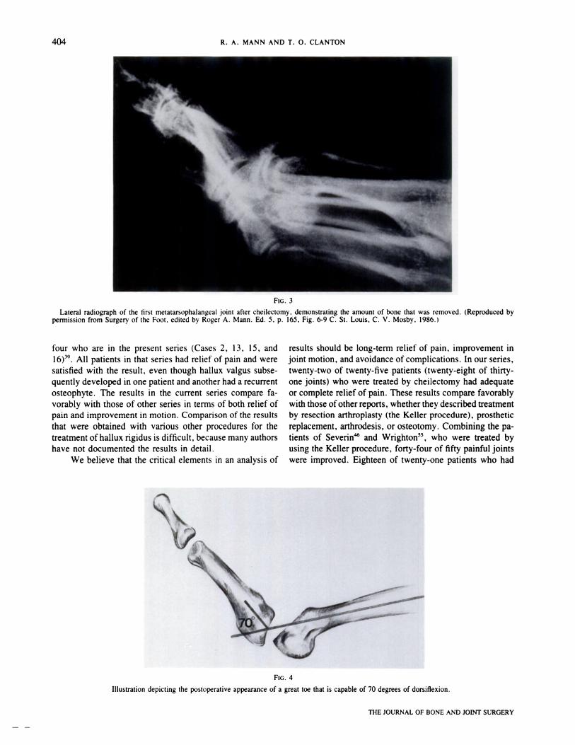
FIG. 3
FIG. 4
404 R. A. MANN AND T. 0. CLANTON
THE JOURNAL OF BONE AND JOINT SURGERY
Lateral radiograph of the first metatarsophalangeal joint after cheilectomy, demonstrating the amount of bone that was removed. (Reproduced bypermission from Surgery of the Foot. edited by Roger A. Mann. Ed. 5. p. 165, Fig. 6-9 C. St. Louis, C. V. Mosby, 1986.)
four who are in the present series (Cases 2, 13, 15, andl6) . All patients in that series had relief of pain and weresatisfied with the result, even though hallux valgus subse-quently developed in one patient and another had a recurrentosteophyte. The results in the current series compare fa-vorably with those of other series in terms of both relief ofpain and improvement in motion. Comparison of the resultsthat were obtained with various other procedures for thetreatment ofhallux rigidus is difficult, because many authorshave not documented the results in detail.
We believe that the critical elements in an analysis of
results should be long-term relief of pain, improvement injoint motion, and avoidance ofcomplications. In our series,twenty-two of twenty-five patients (twenty-eight of thirty-one joints) who were treated by cheilectomy had adequateor complete relief of pain. These results compare favorablywith those ofother reports, whether they described treatmentby resection arthroplasty (the Keller procedure), prostheticreplacement, arthrodesis, or osteotomy. Combining the pa-tients of Severin and Wrighton55, who were treated byusing the Keller procedure, forty-four of fifty painful jointswere improved. Eighteen of twenty-one patients who had
Illustration depicting the postoperative appearance of a great toe that is capable of 70 degrees of dorsiflexion.

HALLUX RIGIDUS: TREATMENT BY CHEILECTOMY 405
VOL. 70-A, NO. 3. MARCH 1988
hallux rigidus and were treated by arthrodesis had relief ofpain’24345. The use of implant arthroplasty relieved pain inseventy-six of eighty-five patients7244245475’ , and the useof osteotomy relieved pain in nine of ten joints3t . This com-parison of results suggests that relief of pain is remarkablysimilar for the several operations, but joint motion and sta-bility are retained with cheilectomy, and no foreign materialis inserted.
Decreased dorsiflexion of the first metatarsophalangealjoint is a characteristic disability in patients who have halluxrigidus, and although there is disagreement on the minimumarc of motion, and specifically of dorsiflexion, that is re-quired for comfortable walking on level terrain’’724’2528, wethink that at least 15 degrees of dorsiflexion is needed. Inour series, twenty-nine of the thirty-one affected joints hadat least that amount of dorsiflexion and twenty-three had 30degrees or more. Only two patients had less than 15 degreesof dorsiflexion. This degree of motion is considerably betterthan that obtained by the Keller procedure3 55. The averagearc of motion that has been reported in patients who weretreated with silicone implants was 47 degrees in twenty-seven patients2425, which is about the same as our averageof 48 degrees. The tendency of the implant to fracture onrepeated bending, which is an important disadvantage, mustbe considered. However, no series of patients who weretreated with implants with a comparable length of follow-up to our series (average follow-up, fifty-six months) hasbeen reported, to show how frequently that complicationarises.
A major cause of loss of motion as well as of pain inpatients who have hallux rigidus is impingement of the
proximal phalanx against the large dorsal osteophyte on themetatarsal head. Cheilectomy eliminates the impingementand permits additional dorsiflexion. While the proceduredoes not eliminate or retard the ongoing degenerative pro-cess within the joint, little deterioration of the result of theoperation seems to occur with time. Even if there is dete-rioration of the joint, cheilectomy permits the later use ofany other procedure. That advantage does not apply for theother procedures that have been mentioned.
All procedures that have been used for the treatmentof hallux rigidus have satisfied more than 80 per cent ofpatients, but the operation that is selected by the surgeonshould offer as many advantages as possible as well as thefewest potential complications. The Keller procedure oftenis not followed by acceptable cosmetic and functional re-sults, especially in terms of weakness in plantar flexion. Itshortens the great toe, and it often causes a hyperextensiondeformity and altered weight-bearing227 495055. While theuse of implant arthroplasty proposes to eliminate the cos-metic disadvantages, prosthetic wear, adverse reactions toSilastic, and fragmentation and breakage of implants havebeen reported4247. Although an arthrodesis is a good pro-cedure theoretically and in practice, malposition or non-union may develop, and arthritis of the interphalangeal jointis often an unwelcome sequela’23843. Osteotomy also maybe followed by malunion or non-union, and it requires con-siderable technical expertise if the desired range of motionis to be achieved. Cheilectomy introduces none of theseadditional risks. For that reason it is, for us, the treatmentof choice for hallux rigidus when symptoms warrant surgicalintervention.
References
1 . ANDERSON, WILLIAM: Lectures on Contractions of the Fingers and Toes; Their Varieties, Pathology, and Treatment: Hallux Flexus. Lancet. 2:279-280. 1891.
2. BINGOLD, A. C. . and CoLLINS, D. H.: Hallux Rigidus. J. Bone and Joint Surg. , 32-B(2): 214-222, 1950.3. BONNEY, GEORGE, and MACNAB, IAN: Hallux Valgus and Hallux Rigidus. A Critical Survey of Operative Results. J. Bone and Joint Surg..
34-B(3): 366-385, 1952.4. COCHRANE, W. A. : An Operation for Hallux Rigidus. British Med. J. , 1: 1095-1096, 1927.5. COLLIER, MAYO: Some Cases of Hallux Rigidus: Their Symptoms. Pathology. and Treatment. Lancet, I: 1613-1614. 1894.6. COTTERILL, J. M.: Condition of Stiff Great Toe in Adolescents. Edinburgh Med. J. , 33: 459-462, 1887.7. CRACCHIOLO, ANDREA, I11 SWANSON, ALFRED; and SWANSON, G. DEG. : The Arthritic Great Toe Metatarsophalangeal Joint: A Review of Flexible
Silicone Implant Arthroplasty from Two Medical Centers. Clin. Orthop. , 157: 64-69, 1981.8. DAVIES-COLLEY: Contraction of the Metatarso-Phalangeal Joint of the Great Toe. British Med. J. , 1: 728. 1887.9. DICKSoN, F. D.. and DIVELEY, R. L.: Functional Disorders ofthe Foot. Their Diagnosis and Treatment, pp. 228-232. Philadelphia, J. B. Lippincott.
1939.10. DUVRIE5. H. L. : Surgery of the Foot, pp. 392-399. St. Louis. C. V. Mosby. 1959.1 1 . FAVREAU, J. C. , and LABELLE, PIERRE: Hallux Valgus and Hallux Rigidus. In Proceedings of the Canadian Orthopaedic Association. J. Bone
and Joint Surg. , 39-B(4): 792-793, 1957.12. FITZGERALD, J. A. W.: A Review of Long-Term Results of Arthrodesis of the First Metatarso-Phalangeal Joint. J. Bone and Joint Surg. . 51-B(3):
488-493, 1969.13. GIANNESTRAS, N. J.: Foot Disorders. Medical and Surgical Management. Ed. 2, pp. 400-403. Philadelphia, Lea and Febiger, 1973.14. GOODFELLOW, JoHN: Aetiology of Hallux Rigidus. Proc. Royal Soc. Med. , 59: 821-824, 1966.15. GOULD, NATHANIEL: Hallux Rigidus: Cheilotomy or Implant? Foot and Ankle, 1: 315-320, 1981.16. GOULD, NATHANIEL; SCHNEIDER, WILLIAM; and ASHIKAGA, TAKAMARA: Epidemiological Survey of Foot Problems in the Continental United
States: 1978-1979. Foot and Ankle, 1: 8-10, 1980.17. GUIDES TO THE EVALUATION OF PERMANENT IMPAIRMENT. Ed. 2, pp. 27-28. Chicago, American Medical Association, 1984.18. HARRISoN, M.: Hallux Limitus. In Proceedings of the Dewar Orthopaedic Club. J. Bone and Joint Surg. . 53-B(4): 772. 1971.19. HEANEY, S. H. : Phalangeal Osteotomy for Hallux Rigidus. In Proceedings of the Scottish Orthopaedic Club. J. Bone and Joint Surg. . 52-B(4):
799, 1970.20. HELFET, A. J., and LEE, D. M. G.: Disorders ofthe Foot, pp. 126-131. Philadelphia, J. B. Lippincott, 1980.21. HUTTON. W. C., and DHANENDRAN. M.: The Mechanics of Normal and Hallux Valgus Feet - A Quantitative Study. Clin. Orthop.. 157: 7-13.
1981.22. JACK. E. A.: The Aetiology of Hallux Rigidus. British J. Surg. , 27: 492-497, 1940.23. JANSEN, MARK: Hallux Valgus, Rigidus and Malleus. J. Orthop. Surg., 3: 87-90, 1921.24. JoHNSoN, K. A. , and BUCK. P. G.: Total Replacement Arthroplasty of the First Metatarsophalangeal Joint. Foot and Ankle. 1: 307-314. 1981.25. JoINT MOTION: METHOD OF MEASURING AND RECORDING, pp. 76-77, 85. Chicago, American Academy of Orthopaedic Surgeons. 1965.26. JONES, R. W. : Treatment of Hallux Rigidus [letter]. British Med. J. , 1: 1 165-1 166. 1927.

406 R. A. MANN AND 1. 0. CLANTON
27. JORDAN, H. H. , and BRODSKY, A. E. : Keller Operation for Hallux Valgus and Hallux Rigidus. An End Result Study. Arch. Surg. , 62: 586-596,1951.
28. JOSEPH, J.: Range of Movement of the Great Toe in Men. J. Bone and Joint Surg. , 36-B(3): 450-457, 1954.29. KELIKIAN, HAMPAR: The Hallux. Hallux Rigidus. In Disorders of the Foot, vol. 1, pp. 608-613. Edited by M. H. Jahss. Philadelphia, W. B.
Saunders, 1982.30. KELIKIAN, H.: Hallux Valgus. Allied Deformities of the Forefoot and Metatarsalgia, pp. 262-281. Philadelphia, W. B. Saunders. 1965.31 . KESSEL, LIPMANN, and BONNEY, GEORGE: Hallux Rigidus in the Adolescent. J. Bone and Joint Surg., 40-B(4): 668-673, 1958.32. KLENERMAN, LESLIE: The Foot and Its Disorders. Ed. 2, pp. 1 16-128. Boston, Blackwell Scientific, 1982.33. LAMBRINUDI, C. : Metatarsus Primus Elevatus. Proc. Royal Soc. Med. , 31: 1273, 1938.34. LIPSCOMB, P. R. : Arthrodesis of the First Metatarsophalangeal Joint for Severe Bunions and Hallux Rigidus. Clin. Orthop. , 142: 48-54, 1979.35. LYRITIS, G. : Developmental Disorders of the Proximal Epiphysis of the Hallux. Skel. Radiol. , 10: 250-254, 1983.36. MCKEEVER, D. C. : Arthrodesis of the First Metatarsophalangeal Joint for Hallux Valgus, Hallux Rigidus, and Metatarsus Primus Varus. J. Bone
and Joint Surg., 34-A: 129-134, Jan. 1952.37. MCMASTER, M. J.: The Pathogenesis of Hallux Rigidus. J. Bone and Joint Surg. , 60-B(1): 82-87, 1978.38. MANN, R. A. , and OATE5, J. C.: Arthrodesis of the First Metatarsophalangeal Joint. Foot and Ankle, 1: 159-166, 1980.39. MANN, R. A.; COUGHLIN, M. J. ; and DUVRIE5, H. L. : Hallux Rigidus. A Review of the Literature and a Method of Treatment. Clin. Orthop.,
142: 57-63, 1979.40. MARGO, M. K.: Surgical Treatment of Conditions of the Fore Part of the Foot. J. Bone and Joint Surg. , 49-A: 1665-1674, Dec. 1967.41 . MOBERG, EiuK: A Simple Operation for Hallux Rigidus. Clin. Orthop. , 142: 55-56, 1979.42. MOLsrER, A. 0.; LUNDE, 0. D.; and RAIT, M.: Hallux Rigidus Treated with the Swanson Silastic Hemi-Joint Prosthesis. Acta Orthop. Scandinavica,
51: 853-856, 1980.43. MOYNIHAN, F. J.: Arthrodesis of the Metatarso-Phalangeal Joint of the Great Toe. J. Bone and Joint Surg., 49-B(3): 544-551, 1967.44. NILSONNE, HARALD: Hallux Rigidus and Its Treatment. Acta Orthop. Scandinavica, 1: 295-303, 1930.45. SETHU, A.; D’NETTo, D. C.; and RAMAKRISHNA, B.: Swanson’s Silastic Implants in Great Toes. J. Bone and Joint Surg. , 62-8(1): 83-85, 1980.46. SEVERIN, EiuK: Removal of the Base of the Proximal Phalanx in Hallux Rigidus. Acta OrthOp. Scandinavica, 18: 77-87, 1949.47. SHEREFF, M. J. , and JAHSS, M. H. : Complications of Silastic Implant Arthroplasty in the Hallux. Foot and Ankle, 1: 95-101 , 1980.48. SMITH, N. R.: Hallux Valgus and Rigidus Treated by Arthrodesis of the Metatarso-Phalangeal Joint. British Med. J., 2: 1385-1387, 1952.49. STOKES, I. A. F.; HUTTON, W. C. ; ST0TT, J. R. R.; and LOWE, L. W. : Forces under the Hallux Valgus Foot before and after Surgery. Clin.
Orthop. , 142: 64-72, 1979.50. SWANSON, A. B. : Implant Arthroplasty in Disabilities of the Great Toe. In Instructional Course Lectures, The American Academy of Orthopaedic
Surgeons. Vol. 21, pp. 227-235. St. Louis, C. V. Mosby, 1972.51. SWANSON, A. B.; LUMSDEN, R. M. , II; and SWANSON, G. DEG.: Silicone Implant Arthroplasty of the Great Toe. A Review of Single Stem and
Flexible Hinge Implants. Clin. Orthop. , 142: 30-43, 1979.52. VILASECA, R. R. , and RIBES, E. R. : The Growth of the First Metatarsal Bone. Foot and Ankle, 1: 117-122, 1980.53. WENGER, R. J. J. , and WHALLEY, R. C. : Total Replacement of the First Metatarsophalangeal Joint. J. Bone and Joint Surg. . 60-B(1): 88-92,
1978.54. WILSON, C. L. : A Method of Fusion of the Metatarsophalangeal Joint of the Great Toe. J. Bone and Joint Surg. , 40-A: 384-385, April 1958.55. WRIGHTON, J. D. : A Ten-Year Review of Keller’s Operation. Review of Keller’s Operation at The Princess Elizabeth Orthopaedic Hospital,
Exeter. Clin. Orthop. , 89: 207-214, 1972.
THE JOURNAL OF BONE AND JOINT SURGERY