guuiddeelliinneess ffoorr tthhee managgeemme … · danielle meadows senior podiatrist, pennine...
TRANSCRIPT

NNOORRTTHH WWEESSTT PPOODDIIAATTRRYY SSEERRVVIICCEESS
CCLLIINNIICCAALL EEFFFFEECCTTIIVVEENNEESSSS GGRROOUUPP ––
RRHHEEUUMMAATTOOLLOOGGYY
GGUUIIDDEELLIINNEESS FFOORR TTHHEE
MMAANNAAGGEEMMEENNTT OOFF FFOOOOTT HHEEAALLTTHH
FFOORR PPEEOOPPLLEE WWIITTHH RRHHEEUUMMAATTOOIIDD
AARRTTHHRRIITTIISS
The copyright of this document rests with the North West NHS Podiatry Services Clinical
Effectiveness Group. This material may be freely reproduced for educational and not-for-profit
purposes within the NHS. No reproduction by or for commercial organisations is permitted
without the express written permission of the Group. It should not be altered in any way without
the permission of the Group. Comments will be considered for the next revision of the
guidelines from any clinicians and should be forwarded to the Group Chair, up to 3 months
before the next date of review.
Version 3: supersedes version 2 (amended and updated)
Date of issue: February 2014
Date of review: 3 years from date of issue

1 © North West NHS Podiatry Services Clinical Effectiveness Group - Rheumatology
Current Guideline Development Group
Samantha Davies Clinical Specialist / Lead Podiatrist, Pennine Acute Hospitals NHS Trust (Chair)
Hayley Edginton Specialist Podiatrist in Rheumatology, University Hospital South Manchester NHS Foundation Trust
Gemma Kelly Specialist Podiatrist in Rheumatology, 5 Boroughs Partnership NHS Foundation Trust
Zoe Critchley Specialist Podiatrist in Rheumatology, 5 Boroughs Partnership NHS Foundation Trust
Veronica Potter Community Specialist Podiatrist, Trafford Division, Pennine Care Foundation Trust
Elaine Pettigrew Podiatrist, Band 6, Stockport NHS Foundation Trust Community Health Services
Anne Mainwaring Advanced Practitioner Biomechanics, Salford Royal Hospital
Danielle Meadows Senior Podiatrist, Pennine Acute Hospitals NHS Trust
Sarah Naylor Specialist Podiatrist MSK Biomechanics, Wrightington Wigan and Leigh Foundation Trust
Julia Stell Principal Service Lead, Bolton NHS Foundation Trust
Rose Hryniw Specialist Podiatrist Rheumatology, Blackpool Fylde and Wyre Hospitals NHS Trust
Patricia Smith Specialist Podiatrist Rheumatology, Royal + Broadgreen University Hospital Trust
Jane Humphreys Advanced Practitioner in Podiatry, Lancashire Care NHS Foundation Trust
Suzanne Smith MSK Podiatry Team Lead, Stockport NHS Foundation Trust Community Health Services
Gareth Hammill Specialist Podiatrist – Lead in Rheumatology, Bridgewater Community Healthcare NHS Trust
Simon Walker Specialist Podiatrist, East Lancs Hospitals NHS Trust
Dr Catherine Bowen Senior Lecturer, University of Southampton
External Reviewers
Dr Vinodh Devakumar Consultant Physician and Rheumatologist, Royal Oldham Hospital
Dr Anita Williams Senior Lecturer University of Salford
Robert Field Lead Podiatrist (Rheumatology Services) / Professional Lead (Podiatry), Dorset Healthcare University Foundation Trust and Chair of the Podiatric Rheumatology Care Association
For further information about the guidelines contact
Samantha Davies [email protected] or [email protected]
2 © North West NHS Podiatry Services Clinical Effectiveness Group - Rheumatology
CONTENTS PAGE
1. INTRODUCTION TO THE GUIDELINES
1.1 The Aims of the North West Clinical Effectiveness Group 3
1.2 The Purpose of the Current Revised Guidelines 4
2. BACKGROUND TO RHEUMATOID ARTHRITIS
2.2 Epidemiology and Clinical Features of RA 5,6
2.3 Recommendations for the Management of RA Foot Problems 7,8
3. PODIATRY SERVICE PROVISION
3.1 Philosophy of Podiatry Services for People with RA 9
3.2 Clinical Specialist Role 9
3.3 Essential Requirements for a Podiatry Service 10,11
3.4 ‘Gold’ Standard Requirements for a Podiatry Service 11,12
4. REFERRAL GUIDELINES
4.1 Referral Pathways 13
4.2 Foot Screening Pathway 13,14
5. PATIENT AND FOOT HEALTH ASSESSMENT
5.1 Essential Requirements for Assessment 15
5.2 Gold Standard Requirements for Assessment 16
5.3 Musculoskeletal ultrasound for Foot and Ankle Pathology 16,17
6. MANAGEMENT OF FOOT PROBLEMS
6.1 Focus of Management 18
6.2 Patient Education Related to Foot Health 19,20
6.3 Foot Orthoses and Footwear 21-26
6.4 Management of Plantar Callus 27,28
6.5 Conservative and Surgical Management of Pathological
Nail Conditions 29-31
6.6 Management of Foot Ulceration 32,33
6.7 Steroid Injection Therapy 34,35
6.8 Foot Surgery 36,37
6.9 Outcome measures/screening/tools 38,39
6.10 Audit 39
7. REFERENCES 40-49
8. APPENDICES
Appendix 1 Foot Screening Pathway 50
Appendix 2 Example of Primary Assessment / Annual Screening Tool 51, 52
Appendix 3 Footwear Suitability Scale 53
Appendix 4 Resources and Web Links 54
Appendix 5 Swindon Foot and Ankle Questionnaire 57

3 © North West NHS Podiatry Services Clinical Effectiveness Group - Rheumatology
1. INTRODUCTION TO THE GUIDELINES
1.1 The Aims of the North West Clinical Effectiveness Group
The North West Clinical Effectiveness Group (NWCEG) for the Foot in Rheumatic Diseases
was initiated by the North West Region Podiatry Heads of Service in 2003. Members of the
group include podiatrists who currently work with patients with rheumatic diseases, a
representative from service managers and an academic link with the University of Salford.
The work of this group now continues with the aim of contributing to the global aim of
improving the care of patients with musculoskeletal (MSK) and rheumatic diseases (Woolf
2012) by supporting service development and the professional development of those
podiatrists involved in the management of patients with rheumatic diseases through the
following objectives:
To provide a support network for NHS podiatrists in the NW region working within the
field of rheumatology and musculoskeletal services.
To cascade learning and best practice
To develop, review and promote protocols / guidelines that identify benchmarks and
essential / desirable standards as a framework for podiatry service providers and
clinical commissioning groups / within the NW region
The development of audit and evaluation tools
To promote podiatry within the wider rheumatology / MSK community and as part of the
multidisciplinary team
To update these guidelines and be aware and highlight other relevant guidelines in
relation to the management of the foot in rheumatic disease as a framework for service
provision and development
To review new evidence from research and disseminate this into clinical practice.
To encourage clinical development in this field through increasing awareness amongst
service commissioners and providers.
To identify the training and education needs of podiatrists to facilitate the development of
specialist skills required to work at an advanced / extended scope level

4 © North West NHS Podiatry Services Clinical Effectiveness Group - Rheumatology
1.2 The Purpose of the Current Revised Guidelines
The first guidelines were produced by the NWCEG in 2004 under a broad remit of The
Management of the Foot and Ankle in Rheumatic Diseases.
In 2008 The PRCA Standards of Care for People with Musculoskeletal Foot Health Problems
were launched. They are ‘patient facing’ in respect of the service that patients can expect and
in this context superseded this aspect of the original NW guidelines
During 2010 the NWCEG identified a need for standards to be defined for the specific foot
health management of patients, particularly those with rheumatoid arthritis. To this end, the
original guidelines were revised and developed to be ‘practitioner facing’ with the objective of
'doing the right thing, to the right patient, in the right way, at the right time’ by rationalising and
improving the quality of foot health management. They focused on the assessment and
management of foot and ankle problems associated with rheumatoid arthritis. The Guidelines
for the Management of Foot Health for People with Rheumatoid Arthritis were launched in
October 2010.
Over the years the guidelines have been used across the Northwest region to both instigate
service provision and support service review. Further to this they have been adopted by
various podiatry services as best practice guidelines both across the UK and internationally.
The guidelines received national recognition as being the first in this area and received the
support of the Podiatry Rheumatic Care Association (PRCA), and the NHS electronic library
for Health (NeLH). The NW CEG has also been actively involved in the development of the
PRCA Standards of Care for People with Musculoskeletal Foot Health Problems (PRCA
2008) and as registered stakeholders, in the development of NICE guidance for the
Management of Rheumatoid Arthritis in Adults (NICE 2009).
The aim of these revised guidelines remains to provide all podiatrists who may be managing
patients with RA with recommendations for the current evidence based and best practice
management of RA related foot and ankle problems.
It is important to note that despite the new paradigm of early targeted therapy leading to an
improvement in the impact of Rheumatoid Arthritis generally, the impact of foot problems on
Quality of Life remains an issue for many patients and hence the podiatrists role in both
identifying foot problems at an early stage and inputting appropriately into the wider multi-
disciplinary management cannot be underestimated.

5 © North West NHS Podiatry Services Clinical Effectiveness Group - Rheumatology
2. BACKGROUND TO RHEUMATOID ARTHRITIS
2.1 Epidemiology and Clinical Features of RA
Rheumatoid Arthritis (RA) is an auto-immune, systemic, inflammatory joint disease with a
chronic, unpredictable and fluctuating course (Conaghan et al 1999). There are around 580,000
adults in England with RA, suggesting that over 690,000 adults in the UK live with the condition
(National Rheumatoid Arthritis Society (NRAS) 2010).
The severity of RA may fluctuate both within and between individuals (Grondal et al 2008). Any
joint may be affected but commonly the hands, feet and wrists are the most common sites.
(Harris 2005). Up to 4 out of every 10 working people with RA lose their jobs within five years
on disease onset with three quarters of these for reasons directly related to their condition
(Young et al 2002). Barrett et al (2000) suggest 1 in 7 give up work within one year of
diagnosis. RA is economically costly. In fact, the total UK costs, including indirect costs and
work related disability, are estimated to be up to £41,735 / person which translates to
approximately £3.8 - £4.75 billion per year (NRAS 2010).
Although any synovial joint may be affected it is well documented that RA is a condition that
can affect the feet (Otter et al 2010; Turner and Woodburn, 2008). The foot is often the first
area of the body to be systematically afflicted by RA (Otter et al. 2004) and at diagnosis, 16% of
patients may have foot joint involvement progressing to 90% as the disease duration
progresses (Grondal et al 2008). 75% of patients with RA report foot pain within 4 years of
diagnosis with the degree of disability progressing with the course of the disease. Shi et al
(2000) states that virtually 100% of patients report foot problems within 10 years of disease
onset. The degree of clinically important disability progressing with the course of the disease
and as early as disease duration of less than 2 years (Turner et al, 2006). The presence of foot
complaints, both in the early and in the chronic stage of RA, has been shown to be extremely
detrimental to patients’ daily lives and activities, especially ambulation (Wickman et al 2004).
The basic pathological changes in the rheumatoid foot result from synovitis and bursitis
(Hooper et al, 2012) and tenosynovitis (Barn et al., 2013), coupled with mechanical stress
(Turner and Woodburn 2008). These structural and functional changes often affect gait and
mobility (Woodburn 2002, Turner et al 2006).

6 © North West NHS Podiatry Services Clinical Effectiveness Group - Rheumatology
Effects on the foot are diverse and multidimensional including pain, changes in gait, deformity
and restrictions in the choice of footwear (Bouysset et al 2006). Specifically, the most common
foot deformities in RA patients are hallux valgus, metatarsus primus-varus and splaying of the
forefoot (Goksel Karatepe et al 2010). These forefoot manifestations of RA are frequently found
in the metatarso-phalangeal (MTP) joints (Van der Leeden et al 2008). Synovitis of the MTP
joints can have a destructive impact on the quality and structure of the joints (Siddle et al.,
2012b, Riente et al., 2006) and the surrounding soft tissues and bursitis affect the inter
metatarsal bursae (Hooper et al., 2012) and contribute to forefoot deformity. Tenosynovitis and
midfoot synovitis lead to the development of pes plano-valgus deformity (Barn et al., 2012)
These foot problems result in disability in weight-bearing activities, abnormal gait patterns and
altered plantar pressure measurements (Turner et al., 2008).
This foot deformity also predisposes to callus formation and as the foot shape alters, and there
is a decrease in tissue viability it can leave the feet vulnerable to ulceration (Firth et al 2008).
Further to this, bacterial and fungal skin infections and nail pathologies are more prevalent in
this patient group adding to the serious risk of ulceration and systemic infection. The risk of
opportunistic infections is increased if the patient’s medical management is with
immunosuppressive drugs (Strand et al 2007, Otter et al 2004).
The feet can remain symptomatic even when the disease is in remission supported with the
current early medical intervention paradigm of early diagnosis and early targeted therapy
(Emery et al., 2002). Indeed there is a ‘window of opportunity for early targeted therapy of RA
related foot problems (Woodburn et al., 2010)

7 © North West NHS Podiatry Services Clinical Effectiveness Group - Rheumatology
2.2 Recommendations for the Management of RA Foot Problems
Foot problems in RA are common, but under-reported by both patients and the rheumatology
team (Blake et al., 2013, Williams and Graham, 2012) and often neglected in clinical practice
((Williams and Graham, 2012). This not only affects the potential to aid early diagnosis of RA
(Emery et al., 2002) but also the need for effective foot health interventions. Any delay in
referral to Podiatry has consequences for the patient, which can vary from minor, living with
discomfort, to major, where delays result in the development of foot deformity (Blake et al.,
2013).
Goals for the management of the RA foot are aimed at reducing the pain in the feet, improving
foot function, mobility and quality of life using safe and cost-effective treatments, such as: -
palliative foot care, prescribed foot orthoses and specialist footwear aimed at preventing any
deterioration in the tissues and in joint alignment (Grondal et al 2008, Woodburn and Helliwell
1997). Woodburn et al also suggest that there is “Window of Opportunity” in early rheumatoid
arthritis for effective podiatry intervention. The foot health needs for the patient with RA are
varied and range from simple foot care advice, palliative care for nails and skin and orthotic /
specialist footwear provision through to management of ulceration and infection (Helliwell 2003,
Korda and Balint 2004).
Specific tools for measuring the impact of foot pathology on foot pain function and disability in
patients with rheumatic diseases have been validated (Walmsley et al., 2012, Budiman-Mak et
al., 2006 Helliwell et al., 2005, Budiman-Mak et al., 1991). These are now being used in clinical
practice as well as in research.
It is becoming increasingly recognised that management strategies for RA should be
aggressive, comprising proactive management and prompt intervention (Luqmani et al, 2006).
The Arthritis and Musculoskeletal Alliance (ARMA 2004) recommends that all patients with
suspected RA should be seen by a specialist in rheumatology within 12 weeks to confirm
diagnosis and enable prompt and effective treatment, and have access to a full multidisciplinary
team (MDT) assessment and intervention early in the disease process, including foot health
assessment. Further to this, Woolf et al (2007) suggest that management requires an
integrated coordinated multidisciplinary, multi-professional approach, with care focussed upon
the needs of the affected person, providing access to a combination of expertise and
competencies.
Given that podiatrists are considered the experts in the management of foot and ankle
problems and recognised by NICE (2009) as primary provider of foot health services for this
patient group, they should be an integrated part of the MDT. This view is supported by ARMA
Fig 2

8 © North West NHS Podiatry Services Clinical Effectiveness Group - Rheumatology
(2004), the British Society for Rheumatology (BSR) (Luqmani et al 2006) and the National
Institute for Clinical Excellence (NICE 2009) who all strongly advocate the need for a dedicated
and specialist podiatry service for the diagnosis, assessment and management of foot
problems associated with RA along with periodic review.
Patient organisations (Arthritis Research UK, Arthritis Care, and the National Rheumatoid
Arthritis Society) also recommend that patients have access to specialist foot care and
increasingly rheumatologists are requesting specialist foot care services for their patients
(Redmond et al 2006, Williams and Bowden, 2004).
In this respect, podiatry care should be made available to all patients with rheumatoid arthritis
and patients should understand the role of the podiatrist in helping them to effectively manage
their foot health and how to seek help should they experience problems. Good communication
between health professional and their patients’ is essential. People with RA should have the
opportunity to make informed decisions about their care and treatment, in partnership with their
health professionals (NICE 2009). To achieve this treatment and care should take into account
peoples’ needs and preferences.
Essential standard
‘Podiatrists are experts on foot disorders; both patients and rheumatologists
can profit from the involvement of a podiatrist’ (Korda and Balint 2004)

9 © North West NHS Podiatry Services Clinical Effectiveness Group - Rheumatology
3. PODIATRY SERVICE PROVISION
3.1 Philosophy of Podiatry Services for People with RA
The broad philosophy of podiatry management of people with RA is to relieve pain, maintain
function and mobility, prevent or minimise deformity and reduce the risk of ulceration thereby
maintaining or improving the individuals’ independence and overall quality of life.
Podiatry services should provide a specific and dedicated service for the diagnosis,
assessment and management of foot problems associated with RA that can be provided in a
variety of settings, such as local clinics, hospital out-patient departments, and rheumatology
departments (both outpatient and inpatient). However, it is acknowledged that some patients
choose to access private podiatry care from HCPC registered practitioners.
3.2 Clinical Specialist Role
A podiatry team led by a dedicated podiatry clinical specialist in rheumatology is desirable. This
specialist should provide specialist care directly to patients, provide advice for other members
of the podiatry and multidisciplinary team (MDT) and facilitate the development of appropriate
clinical skills in other members of the podiatry team This clinical specialist should work within
the rheumatology department (outpatients and inpatients) for at least part of their work
schedule.
The advantages of this are that the specialist podiatrist can:
Improve the profile of podiatry services within rheumatology
Provide timely interventions for acute problems using extended practices that
historically have required referral to secondary care.
Provide timely referrals to appropriate members of the MDT.
Develop inter professional working practices.
Develop their role as advisor to the MDT
Manage foot problems with a greater understanding of implication of medical therapy
and disease management

10 © North West NHS Podiatry Services Clinical Effectiveness Group - Rheumatology
3.3 Essential Requirements for a Podiatry Service
Based on the national recommendations (NICE 2009 and ARMA 2004) the following are
considered the essential requirements that a podiatry service is expected to provide for patients
with RA:
A team of podiatrists able to meet the needs of the local population diagnosed with RA
A team of podiatrists with knowledge of the foot health management of patients with RA
and knowledge of the medical and rehabilitation management of the disease.
A system for prioritising referrals so that foot pathologies are managed in a timely way
The facilities for rapid assessment for patients should urgencies occur so that patients in
acute pain or at risk of infection receive timely interventions. Patients who are being
managed with biologic therapies should have immediate access to a specialist podiatrist
if they present with foot ulceration or other infections affecting the foot.
Provision of the appropriate facilities / skills for baseline vascular and sensory
assessment i.e. hand held Doppler ultrasound and 10g monofilament. It is known that
patients with rheumatoid arthritis are more at risk than the general population for
coronary heart disease (resulting in circulatory insufficiency to the lower limb), vasculitis
and neuropathy. Baseline and annual assessments of the vascular and neurological
status of patients will both identify and monitor any problems or changes (Kitas 2003).
Annual review and assessment of foot health in RA patients with identified foot problems
(NICE 2009)
The skills to provide biomechanical assessment of foot structure and function
Provision of the appropriate facilities for biomechanical assessment of foot structure and
function with either manufacturing or supplying foot orthoses. It is know that foot
orthoses are a vital and effective intervention in rheumatoid arthritis (Hennessey et al
2012, Woodburn et al 2002a).
Provision of specialist footwear or referral to an orthotist depending on local
arrangements. It is known that many foot problems cannot be accommodated in normal

11 © North West NHS Podiatry Services Clinical Effectiveness Group - Rheumatology
retail footwear and the benefits of specialist prescription footwear are recognised
(Williams et al 2006,)
Individual patient education and care plans. Patients need information to enable them to
make informed choices about their treatment. The information should be provided with
professional support and guidance with the emphasis on behavioural change rather than
just information giving (Graham et al., 2011).
A system of providing prompt and appropriate information to the referrers and other
appropriate members of the multidisciplinary team. This is to facilitate good
communication and collaboration between the podiatrist and the other members of the
team so that care is timely and appropriate.
Clinical documentation for recording of assessments, management plans, treatments
and other interventions. In addition to the legal requirements for documentation of clinical
treatments they can be used for purposes of audit
An effective system of Continuing Professional Development, which includes
o Annual Update Courses in Rheumatology
o Multidisciplinary training
3.4 ‘Gold Standard’ Requirements for a Podiatry Service
This would include all the essential criteria plus the following desirable criteria
A team of podiatrists, led by the clinical specialist in the management of patients with
RA. A designated clinical lead with advanced / extended scope skills e.g. joint
injections, experience and competencies would co-ordinate the service at a clinical level
and be responsible for cascading new evidence based practice to other members of the
podiatry team. They would act as clinical advisor for the team and be responsible for
ensuring appropriate CPD in this area.
The facilities for providing telephone advice and rapid assessment for patients
Access to/provision of the appropriate facilities/skills for advanced vascular and
neurological assessment such as Doppler assessment for ABPI’s, vascular and MSK
diagnostic ultrasound.

12 © North West NHS Podiatry Services Clinical Effectiveness Group - Rheumatology
Provision of the appropriate facilities and skills for lower limb mechanics and foot
pressure assessment. Many rheumatic disorders affect both the architecture and
function of the foot and lower limb resulting in abnormal gait and increased foot
pressures. Quantifiable assessment of these will enable monitoring and timely
intervention. Where available, In-shoe foot pressure assessment will identify the effects
of orthotic and footwear interventions (McCormick C et al 2013, Redmond A et al 2009,
Van der Leeden et al 2006, Otter S et al 2004)
The facility for an annual review and assessment of all RA patients. This is so that
patients who do not have current problems are monitored at least annually in order to
detect problems early.
An effective system of Continuing Professional Development, which includes
o The development of advanced clinical skills such as soft tissue and intra-articular
injection techniques, imaging modalities such as MSK ultrasound
o The training in skills such as lipid and blood pressure monitoring
o Attendance at regional, national and international rheumatology meetings and
conferences (for example, the British Society of Rheumatology Conferences)
o Support for research (either uni-professional or multi-professional) in collaboration
with outside agencies (for example, universities, medical schools, and medical
charities).
o Attendance at local podiatry groups / meetings with the opportunity for networking
with colleagues and peers
13 © North West NHS Podiatry Services Clinical Effectiveness Group - Rheumatology
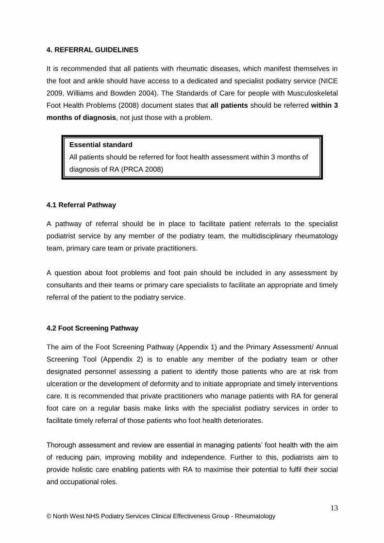
4. REFERRAL GUIDELINES
It is recommended that all patients with rheumatic diseases, which manifest themselves in
the foot and ankle should have access to a dedicated and specialist podiatry service (NICE
2009, Williams and Bowden 2004). The Standards of Care for people with Musculoskeletal
Foot Health Problems (2008) document states that all patients should be referred within 3
months of diagnosis, not just those with a problem.
4.1 Referral Pathway
A pathway of referral should be in place to facilitate patient referrals to the specialist
podiatrist service by any member of the podiatry team, the multidisciplinary rheumatology
team, primary care team or private practitioners.
A question about foot problems and foot pain should be included in any assessment by
consultants and their teams or primary care specialists to facilitate an appropriate and timely
referral of the patient to the podiatry service.
4.2 Foot Screening Pathway
The aim of the Foot Screening Pathway (Appendix 1) and the Primary Assessment/ Annual
Screening Tool (Appendix 2) is to enable any member of the podiatry team or other
designated personnel assessing a patient to identify those patients who are at risk from
ulceration or the development of deformity and to initiate appropriate and timely interventions
care. It is recommended that private practitioners who manage patients with RA for general
foot care on a regular basis make links with the specialist podiatry services in order to
facilitate timely referral of those patients who foot health deteriorates.
Thorough assessment and review are essential in managing patients’ foot health with the aim
of reducing pain, improving mobility and independence. Further to this, podiatrists aim to
provide holistic care enabling patients with RA to maximise their potential to fulfil their social
and occupational roles.
Essential standard
All patients should be referred for foot health assessment within 3 months of
diagnosis of RA (PRCA 2008)

14 © North West NHS Podiatry Services Clinical Effectiveness Group - Rheumatology
It has been shown that health and illness is mainly determined by lifestyle psychological
factors and socio-cultural environment rather than on biological status and conventional
health care (Micheal 2004). According to Waddell and Burton (2006) work is the most
effective way to improve well being of individuals therefore ignoring socio cultural factors
such as a patient’s ability to work could potentially lead to poorer health outcomes.
Individual podiatry services will have different clinical arrangements for new and existing
patients with RA in both primary and secondary services (and private practice) However, an
initial structured foot assessment and screening must be carried out for all patients with RA
at the first point of contact with any podiatrist and then referral on to the specialist podiatrist if
the management needs require specialist intervention or the input from the multidisciplinary
team. The assessment should include appropriate outcome measures and should be
repeated periodically to detect any changes in foot health status. The suggestion is that
those patients identified with foot problems should be reviewed on an annual basis as a
baseline minimum.
Essential Standards All people with RA and foot problems should have access to a podiatrist for assessment
and periodic review of their foot health needs. (NICE 2009)
All podiatry patients with RA should receive an initial structured foot assessment complete
with appropriate outcome measures with onward referral to more specialised colleagues as
required
Referral to a Podiatrist is an integral part of the early management of RA patients.
(ARMA 2004)

15 © North West NHS Podiatry Services Clinical Effectiveness Group - Rheumatology
5. PATIENT AND FOOT HEALTH ASSESSMENT
Clinical assessment should be systematic and thorough. The following components of a patient
assessment / screening process should be carried out as a minimum standard of care for all
new or existing patients presenting with new foot pathologies (see Appendix 1 and 2) . This
enables an individual tailored care plan to be produced. It is recommended that all existing
patient records are updated in line with these standards.
5.1 Essential Requirements for Assessment
Podiatry referral should be offered to all patients with RA and a Baseline Assessment should
include:-
Full Medical and surgical history (including disease duration).
Medication and pain management.
General health and systemic factors, examination for signs of extra-articular features of
disease- nodules, bursa, vasculitis, tendonitis, tenosynovitis.
Detailed assessment of foot and lower limb function and structure (both non weight-
bearing and weight-bearing).
Feel, look and move the foot assessing the foot position, deformities, range of
movement and location of painful, tender, swollen sites.
Assessment of foot pain using a scale of 0 (no pain) -10 (worst pain imaginable)
Assessment of patients’ main presenting problem, the pattern of distribution and
chronological development of symptoms. The impact of the problem, patients’
perceptions/knowledge and expectations also needs to be addressed.
Vascular assessment based on clinical signs and patients’ symptoms. Foot pulses
should be assessed using Doppler ultrasound which provides an objective
measurement of vascular status.
Sensation assessment with 10g monofilament as a minimum.
Assessment of nails, skin lesions and tissue viability also noting history of previous
ulceration.
Examination of the patients’ footwear and its suitability for both home and outdoor use
(Footwear Suitability Scale – Appendix 3).
Assess the need for pressure relief and foot orthoses.
Assess the need for referral for patients’ requiring a surgical opinion or to other
members of the MDT such as physiotherapy, occupational therapy or orthotist.
Lifestyle and social factors- ability to self care, neglect, smoking, alcohol, occupation
and activity/mobility.

16 © North West NHS Podiatry Services Clinical Effectiveness Group - Rheumatology
An annual review of foot health should be offered to those with identified foot problems.
Patients should be monitored and reassessed for changes in foot health and general
health status, allowing further outcomes to be predicted and patients’ treatment and
management plans to be changed accordingly
5.2 ‘Gold Standard’ Requirements for Assessment
In addition to the essential standards it is desirable that the following are also carried out:
Baseline measurements of foot pain, function and health status using measurement
tools such as the Foot Function Index (Budiman Mak et al 2006), Leeds Foot Impact
Scale (Helliwell et al 2005) or the Salford Rheumatoid Arthritis Foot Evaluation Index
(Walmsley et al 2012).
Assessment of the impact of foot problems on activities of daily living including a
patient’s ability to continue in or find employment
The use of tools such as DAS28 to evaluate disease activity
ABPI if further investigation of vascular status is required.
Assessment of tendon reflexes where indicated
All existing patient records are updated in line with these standards.
Direct referral for x-rays / ultrasound / MRI scans for detailed assessment and diagnosis
Annual review for patients with RA
5.3 Musculoskeletal Ultrasound for Foot and Ankle Pathology
The use of diagnostic ultrasound (US) by non-radiologists has increased in popularity,
particularly within rheumatology as technology has improved and equipment has become more
user-friendly (Brown, 2009, Taggart et al., 2011, Micu et al., 2012). US is painless, harmless
(no ionising radiation) and is readily accessible for use within the clinical environment.
Advances in Grey Scale (GS) and Power Doppler (PD) US imaging have enabled better and
timelier assessment of changes in joints and soft tissues due to inflammation associated with
rheumatoid arthritis (Schmidt, 2007, Balint et al., 2008).
For rheumatoid arthritis, the precise detection of synovitis has become fundamental to the
management of inflammatory disease (Brown, 2009,Kang et al., 2012). As such, there has
been an increase in focus on the use of US techniques in the assessment of rheumatoid foot
pathology (Bowen et al., 2013). Several authors have described the use of US to identify soft
tissue pathology within the foot (Riente et al., 2006, Joshua et al., 2007, McNally, 2008, Micu et

17 © North West NHS Podiatry Services Clinical Effectiveness Group - Rheumatology
al., 2012) especially pathology within the foot that is otherwise unseen by clinicians (Wakefield
et al., 2008, Bowen et al., 2010, Bowen et al., 2011, Hooper et al., 2012). Additional
advantages over other imaging techniques are that any area of the foot can be scanned rapidly
at one time-point and treatments such as guided steroid injections can be implemented
immediately (Bowen et al., 2013).
Summary of US detectable foot pathologies associated with rheumatoid arthritis
Effusions and impingements of the ankle joints.
Visualisation of synovial hypertrophy, especially within the metatarso-phalangeal joints
Tenosynovitis of extensor digitorum longus, extensor digitorum brevis, flexor digitorum
longus, flexor digitorum brevis, tibialis anterior, tibialis posterior, peroneus longus and
peroneus brevis tendons.
Visualisation of Achilles tendon in its full length - calcification, ruptures and retro-calcaneal
bursitis can be differentiated.
Diagnosis of synovitis, especially within the metatarso-phalangeal joints.
Diagnosis of Morton’s Neuroma.
Diagnosis of adventitial (within plantar fat pad) and anatomical (intermetatarsal) bursitis.
Screening rheumatoid patients for high metatarsal pressures.
Guidance of needle placement for steroid injections
Podiatrists’ scope
As clinical expertise in performing musculoskeletal US has advanced, there is a consequent
requirement for adequate training by non-radiologists to learn the techniques. Clinicians such as
podiatrists arguably have a discrete detailed anatomical knowledge of the foot and one study has
demonstrated good reliability of a podiatrist tested against a radiologist (kappa 0.702, p <0.01) in
the use of US for the evaluation of foot disease in RA (2008). US is compounded in that it is
highly operator dependent and there is a lengthy period required to develop the necessary skills
(Taggart et al., 2011,Marhadour et al., 2010). Some useful resources are available that aid
interpretation of US images. Riente et al. (2006) provide detailed documentation of a proposed
scanning protocol for the foot. Additionally, the US imaging characteristics of the normal
anatomical structures of the foot are well described (Micu et al., 2012) as well as techniques for
imaging the small joints of the forefoot (McNally, 2008) and the ankle and foot (Micu et al., 2012).

18 © North West NHS Podiatry Services Clinical Effectiveness Group - Rheumatology
6. MANAGEMENT OF FOOT PROBLEMS
6.1 Focus of Management
Treatment and care should take into account peoples’ needs and preferences. People with RA
should have the opportunity to make informed decisions about their care and treatment, in
partnership with their health professionals.
Good communication between healthcare professionals and patients is essential. It should be
supported by evidenced-based (where possible) information / care plan tailored to the individual
person’s needs. Treatment, care and information given should be appropriate to the individual
and take into account cultural, religious, language needs and be accessible to people with
physical, sensory or learning disabilities (NICE 2009).
Following a detailed assessment a management plan will be formulated between the podiatrist
and the patient. This may involve referring the patient to other members of the rheumatology
team for advice on interventions such as foot surgery, physiotherapy, specialist footwear or
steroid injections. It could also involve encouraging patients to seek help from other
organisations such as their employers and the job centre to ensure that the complications of
their disease are recognised and individual issues are addressed
Dependent on the presenting problems the podiatrist may offer the following interventions (each
is then covered in detail)
Patient education relating to all issues surrounding foot health – pages 19-20
Foot orthoses and Footwear (Advice / therapeutic) – pages 21-26
Management of plantar callus - pages 27-28
Conservative and surgical management of pathological nail conditions - pages 29-31
Management of Foot Ulceration – pages 32-33
Joint injections or referral to the member of the multidisciplinary team responsible for
this – pages 34-35
Referral for a surgical opinion - pages 36-37
Referral to other members of the rheumatology team i.e. physiotherapy, occupational
therapy, specialist nurses, orthotists and consultants
Providing supporting letters for employers to help patients access adaptations in their
work place to help with foot problems e.g. shorter hours on feet, chairs to sit as needed

19 © North West NHS Podiatry Services Clinical Effectiveness Group - Rheumatology
Regardless of the intervention/s, regular review appointments and open access to the
podiatry service for any developing acute problems
6.2 Patient Education Related to Foot Health
Patient Education (P.E) can be defined as: a set of planned educational activities designed to
improve patients’ health behaviours, health status and long term outcomes (Hill, 1997). Patient
education is considered to be an integral component in the armoury of R.A. management
strategies, to support and facilitate self-management of the disease (ARMA, 2004). Further to
this research has shown that individuals who are actively involved their own disease
management have better outcomes, improved self-efficacy, less pain and reduced incidence of
depression (Lorig et al, 2005; Kjeken et al, 2006).
The Standards of Care for People with Musculoskeletal Foot-health Problems (PRCA, 2008)
recommend specifically that patient-centred education should be provided to enable patients to
make informed choices about their foot care, and the role of the podiatrist as a vital member of
the Multidisciplinary team for the management of R.A. has been reinforced (NICE 2009).
There is a large body of evidence that supports the effectiveness of P.E. for patients with R.A.
that is delivered via a staged approach over the lifetime of the patient, with the content and
timing of education provision being driven by the needs of the individual (Barlow et al 2002;
Hammond 2003; Waxman et al 2003; Hennell et al 2004; Fautrel et al 2005; Koehn and Esdaile
2008). New podiatry-based research shows that identifying health education needs, and
provision of supportive verbal and written information can foster an effective therapeutic
relationship, supporting effective foot health education for people with RA (Graham et al 2012a;
Graham et al 2012b).
Using the recommendations and findings from the literature above as a guide, together with the
PRCA (2008) Foot Health standards, Podiatrists can embed the following key points into the
development of their Foot Health P.E. provision for individuals with R.A.
Education should be encouraged throughout the patients’ medical care with each
consultation becoming an opportunity for P.E and be based on an educational-
behavioural approach.

20 © North West NHS Podiatry Services Clinical Effectiveness Group - Rheumatology
The content of P.E. should be individualized and engaged, taking the individual
experiences as the point of departure according to the patients’ needs / wishes at the
point of contact and should reflect the fluctuating nature of the disease. Patients in
remission may sometimes prefer to focus on being well and avoid thinking about
possible side effects and chronic disease (Kristiansen et al 2012)
Patient Education should aim to include:
Disease specific information regarding; the causes & course of the disease and disease
management both verbal and written.
Direction on appropriate use of the internet for sourcing educational material.
Details regarding access to patient support groups
Advice regarding lifestyle choices (weight management, smoking cessation).
Advice regarding retail/therapeutic footwear/foot orthoses.
Maintenance of foot hygiene.
Aspects of self-care (including safe & unsafe practices.
Information regarding changes in foot health that should prompt further investigation.
Access to service / providers of podiatry care
Simple information giving only has short-term, limited effects upon health behaviour, but should
be used within a staged approach throughout the course of the disease. An opportune time for
general information giving is early in the diagnosis, based upon the patients’ own knowledge
requirements. To maintain the potential effects of P.E over the lifetime of the patient,
educational ‘booster’ sessions may be required.
Essential Standard
Patient education should include foot health self management advice and if
necessary demonstration, explanation of foot problems and their impact on the
individual, information on general disease management and sign posting for future
foot health needs

21 © North West NHS Podiatry Services Clinical Effectiveness Group - Rheumatology
6.3 Foot Orthoses and Footwear
The benefits of foot orthoses (insoles) and footwear have been recognised and
recommended “Functional insoles and therapeutic footwear should be available for all
people with RA if indicated” NICE (2009). For the purposes of clarity foot orthoses and
footwear options will be discussed separately. However, the practitioner should always
consider them together in relation to footwear suitability, choice of foot orthoses and the
potential mechanical effect of the footwear on not just the foot but the orthoses as well.
Foot orthoses
Foot orthoses are provided to two main groups of patients with RA; those with foot problems
associated with early disease and those with more established foot problems. The use of
appropriate footwear (Williams et al 2007) in conjunction with foot orthoses has been
recognised as minimising the pain and disability associated with RA Hodge 1999when there
is established foot deformity. The choice of foot orthoses in relation to design and function is
dependent on the amount of motion in the joint of the foot. This factor is not dependent on
disease duration as some patients with early disease have limited motion and some with
longer disease duration have good range of motion within the joints of the foot.
It is demonstrated that foot orthoses not only achieve pain reduction in the early RA foot but
have a sustained effect on the foot structure and hence achieve stability of the joints of the foot
and improve the patient’s mobility (Woodburn at al 2002(a)).
Therefore, there is the potential to prevent major functional and structural foot problems by
providing foot orthoses early on in the disease process if joint mobility is still good. However,
as foot changes have the potential to occur within 2 yrs of disease onset (Turner and
Woodburn, 2008) it is essential that patients are referred for assessment of foot function as
early as possible following diagnosis.
Essential Standards
Patients with a diagnosis of RA should be assessed as soon as possible following
diagnosis for structural problems with the lower limb and foot.
All patients with RA and foot pain should be considered for foot orthoses and /or
footwear advice, irrespective of disease duration.

22 © North West NHS Podiatry Services Clinical Effectiveness Group - Rheumatology
Once the structural problems are established and joint mobility is reduced, management
consists of reducing symptoms of pain and resultant mobility problems. Further to this,
redistributing foot pressures may contribute to the prevention of tissue breakdown and
ulceration over high pressure areas of the foot. RA subjects with metatarsal pain have 20 –
40 % lower pain pressure threshold (Hodge et al, 1999), these patients require more planter
pressure reduction to diminish their pain sensation.
There is a broad range of devices that employ a variety of different approaches to modify
foot and lower limb structure and function with general consensus within services providing
them that foot orthoses include these main groups:
Simple cushioning insoles
Insoles to which additional padding/additions can be applied
Contoured insoles intended to change the function of leg and foot joints, either:
o Custom made to a cast of the patient’s foot
o Supplied off the shelf +/- adaptations
However, the boundaries between the modes of action of the types are not always exact and
an individual device may include elements of more than one type or mode of action.
However, Clark et al., (2006), and Jackson et al., (2004) concluded that foot orthoses;
reduce pain and improve functional ability
Both hard and soft foot orthoses have the potential to reduce forefoot pain
Hard foot orthoses have the potential to reduce rearfoot pain in patients with early RA
Hard foot orthoses have the potential to reduce hallux abducto valgus
There is only anecdotal evidence for the use of simple cushioning insoles. Two small studies
indicate that prefabricated metatarsal padding (dome and bar shapes metatarsal pads)
reduces mean peak plantar foot pressure by up to 21% with bars and 12% with domes
(Jackson et al 2004) and both equally (Hodge et al 1999)
Essential Standard
Patients with established foot deformity should be assessed for accommodative foot
orthoses and footwear advice/ specialist footwear

23 © North West NHS Podiatry Services Clinical Effectiveness Group - Rheumatology
Hard Contoured foot orthoses are provided in order to improve the function of the foot and/or
lower limb. This assumes that there is some mobility in the joints of the foot in order to improve
function and realign the bony architecture. They are particularly useful for use in patients with
early diagnosis of RA. In this case there is an attempt to not only reduce pain but to maintain
good foot function and hence structure whilst the foot is vulnerable to deformity due to the
combination of the inflammatory process and abnormal mechanics.
Customised accommodative orthoses (total contact orthoses) are designed so that the material
follows closely the contours of the underside of the foot. The purpose is to redistribute the
pressures applied to the foot by standing and walking more evenly. This is particularly useful
where there are areas of increased pressure, for example, under the metatarsal heads. In this
instance the pressure is shifted to areas of the foot that do not normally bear weight such as the
arch area (Li et al 2000). They are particularly used where there is limited or no joint mobility
such as in the established RA foot and where tissue viability is poor. These orthoses are often
made from materials that also provide a cushioning effect, such as softer EVA or with additional
foam linings.
Dynamic impression insoles made by sequential foam padding & moulded under successive
walking compression have been demonstrated to reduce peak pressures & the VAS pain
score when compared to the moulded custom insole (Chang et al, 2012).
Footwear
Footwear
The choice of orthoses is governed by the suitability of the patient’s footwear, which may not
accommodate the ideal foot orthoses for their particular problem. All footwear is in itself
capable of modifying the structure and function of the body and therefore falls clearly within
the definition of orthoses and may the only thing that needs changing to solve functional
problems. Inappropriate footwear can be both a major contributing factor to foot impairment.
However, when it is right it has the potential to alleviate pain and increase mobility and
independence (with or without foot orthoses).
Essential Standards
Functional foot orthoses should be provided where the tarsal joints are unaffected.
Accommodative / cushioning orthoses should be provided for those patients with
structural foot deformity, painful symptoms and activity restriction

24 © North West NHS Podiatry Services Clinical Effectiveness Group - Rheumatology
In order for health professionals & researchers to accurately & efficiently critique an
individual’s footwear, a valid & reliable footwear assessment tool is required (Barton et al
2009, Nancarrow 1999)
Footwear can be sub divided into three main groups:
Standard retail footwear
Niche retail including comfort footwear as well as extra depth, extra width and odd-size
suppliers.
Specialist therapeutic footwear
Standard Retail footwear
There are now many manufacturers of retail footwear that are both appropriate for the foot
health of our patients The features of retail footwear that makes them ideal for the RA foot
would be -
Stable heel – broad enough for stability or elongated / flared to increase this effect
further
Extended heel counter
Padded topline – to reduce irritation to the retro-calcaneal area and the infra-malleolar
areas
No prominent internal seams
Winged toe puff
Increased toe spring or rocker sole – to reduce forefoot plantar pressures
Low laced – for ease of access
(Williams A and C 2010)
The suitability of retail footwear can be assessed using the Footwear suitability tool (Nancarrow
1999) see Appendix 3
In early disease many patients experience forefoot pain and changes to the shape of their
foot. Many patients recall that they had to increase their shoe size to accommodate a wider
forefoot. Specialist footwear manufacturers can be very helpful in offering advice and
providing wider-fitting shoes. The British Footwear Association provides detailed information
Essential Standard
Footwear assessment and advice should be given to all patients.

25 © North West NHS Podiatry Services Clinical Effectiveness Group - Rheumatology
about companies that make up the British footwear industry and consumer information about
hard-to-find footwear suitable for all foot sizes and shapes - see Appendix 4.
Specialist therapeutic footwear
Stock footwear is specialist footwear which is available in a variety of styles and fittings, for
example extra deep, and/ or extra wide and is generally suitable for mild to moderate deformity.
Bespoke footwear is an option when there is major deformity such as advanced rheumatoid
arthritis deformity or if there is a huge difference in symmetry, or if the foot dimensions are
outside the measurements for stock footwear.
Two systematic reviews (Egan et al 2003 and Farrow et al 2005) indicate that specialist
footwear is likely to be beneficial in patients with RA. Two RCT’s (Fransen and Edmonds 1997
and Williams et al 2007b) indicate that this footwear contributes to the reduction in pain and
increased mobility in patients with RA although the effect is improved when combined with
orthoses.
It is generally considered that the following patients could be considered for referral for
specialist footwear for the following reasons:
Failing to obtain retail footwear to fit the dimensions of the foot (including asymmetry)
Pressure symptoms such as skin lesions/sore areas on the feet
Increasing foot pain due to pressure from existing footwear
Excessive footwear ‘wear’ indicating that patients need more stability from increased
surface area of the plantar aspect of the footwear and increased rearfoot control from
the heel counter.
History of foot ulceration where footwear has been a contributory factor.
The Society of Chiropodists & Podiatrists (CPD update, May 2006, pS6) – ‘Who Should Be
Referred for Specialist Footwear?’ advises referring patients who have:
Problems associated with systemic disease (e.g. RA).
Functional/structural problems that impact on the foot.
Width, depth, length outside range of retail footwear (and asymmetry).
Provision of substantial foot orthoses that cannot be accommodated in retail footwear.

26 © North West NHS Podiatry Services Clinical Effectiveness Group - Rheumatology
It has been found that patients considered that it was important to receive information at the
point of referral so that they can make considered choices as to whether to be referred or not
(Williams et al 2007a). Without some knowledge of what is available the opportunity to
engage the patient in the decision making at this stage is lost and may be one of the reasons
patient expectations are not met. The option of referral for a surgical opinion should be
offered as an alternative to referral for footwear.
Stock footwear is specialist footwear which is available in a variety of styles and fittings, for
example extra deep, and/ or extra wide and is generally suitable for mild to moderate deformity.
This footwear is generally supplied with 3x3mm removable liners that provide the option for
being replaced with orthotic devices. Stock footwear with specific modifications is termed
‘modular’. Bespoke footwear is an option when there is major deformity such as advanced
rheumatoid arthritis deformity or if there is a huge difference in symmetry, or if the foot
dimensions are outside the measurements for stock footwear.
Essential standards –
Patients who are assessed as requiring therapeutic footwear should be informed of the
potential benefits and limitations of this footwear (in respect of cosmesis) and allowed to
decide on whether to be referred /provided with therapeutic footwear or not
Referral for surgical opinion should be offered as an alternative to referral for therapeutic
footwear

27 © North West NHS Podiatry Services Clinical Effectiveness Group - Rheumatology
6.4 Management of Plantar Callus
Persistent synovitis of the forefoot is associated with peri articular erosion, subluxation and
dislocation of the MTP joints which in term exposes the metatarsal heads to increased pressure
during gait. (Van der Leeden M, 2010). In response to increased focal stresses, the strateum
corneum thickens initially in a normal physiology response to chronic excessive pressure or
friction of the skin eventually however, pathological lesions (callosities) develop, which cause
pain and contribute to impairment of gait and related functional and health status in people with
RA.
Three studies have investigated callus reduction in RA.
Woodburn et al, (2000) concluded that a reduction in plantar callus with sharp debridement
reduced forefoot pain for approximately 7 days, but increased forefoot pressures in 10 out of 14
feet. This was not statistically significant but indicates that reduction of callus over prominent
metatarsal heads may lead to tissue damage. This would be of particular concern in patients
with the following factors
Foot deformity
Reduced tissue viability (long term steroid therapy, vasculitis, concurrent peripheral
vascular disease) and/or neuropathy.
Davys et al (2005) demonstrated a reduction in pain in 38 participants but concluded the effect
was no greater than sham treatment. Localised pressure or gait function did not significantly
improve following treatment, but they indicate what to include when managing plantar callus in
RA patients:
Pts need to be informed about causes and management of callus.
It is recommended that thick callus is debrided cautiously and frequently.
If infection is present, then overlying callus should be debrided to expose the
underlying infection. If ulceration is present, surrounding callus and necrotic tissue
should be appropriately debrided.
Removal of superficial callus over plantar bursae should be avoided altogether.
Advice about the use of emollients for dry plantar callus should be given. Patients
should be encouraged to self manage by applying emollient daily and to use a foot file
on these areas at least three times a week.
Adhesive plantar padding should not be used as a pressure relief mechanism
especially if there are tissue viability concerns. Instead a dry dressing secured with a
bandage can be used for localised protection.

28 © North West NHS Podiatry Services Clinical Effectiveness Group - Rheumatology
Pressure relieving and functional orthoses have been demonstrated in studies to reduce
forefoot pressures should be provided (McCormick et al 2013, Redmond et al 2009,
Van der Leeden et al 2006, Otter S et al 2004 and Woodburn et al 2002 (a))
Footwear advice should be provided with consideration given to footwear with the
necessary depth and width to accommodate the patients’ feet. (See Footwear and
Orthoses – pages 24-29).
Therapeutic footwear should be considered and when appropriate referred to an
Orthotist.
If the patient has severe pain in the forefoot and /or severely affected mobility, it may be
appropriate to consider a surgical referral and opinion.
Regardless of the interventions, regular review appointments and open access to the
Podiatry service for any developing acute problems.
A more recent study by (Siddle et al., 2013) further adds to the concern about routine sharp
debridement of callosities in people with RA. The study concluded that the long term effects of
sharp debridement of painful forefoot plantar callosities in people with RA when used in
conjunction with a combined therapeutic approach produced no additional benefit over a
combined therapeutic approach alone.
The authors suggest that the use of sharp debridement should be confined to the short term
alleviation of severe pain and address only high risk presentations such as extravasated blood
and suspected ulceration.
Essential Standard
Callus should be assessed in relation to symptoms and causative factors before
debridement is considered.
The focus of callus management should always be the reduction of foot pressures
with foot orthoses and suitable footwear first, before debridement is considered as a
safe or unsafe intervention

29 © North West NHS Podiatry Services Clinical Effectiveness Group - Rheumatology
6.5 Conservative and Surgical Management of Pathological Nail Conditions
Onychomychoses
Onychomycosis (OM) is an infection of the nail unit that can be caused by various species of
dermatophytes, yeasts, molds and even some bacteria. OM infects between 2% and 18% of
the population with increasing frequency as patient age increases to 20% and 30% for those
older than 60 years and 70 years respectively, (Derby et al 2011, Ameen 2010) and there is an
increased association with immune-compromised hosts (Bodman 2003). Bodman (2003 also
identified that if OM is left untreated, it can lead to subungual and skin ulceration, in patients
with RA.
The most sensitive diagnostic test is histopathological analysis of a nail clip biopsy. A Periodic
Acid-Schiff stain (PAS) test is commonly used as a quicker and more sensitive diagnostic
workup than traditional fungal cultures (Arca et al 2004). There are five types of OM:
Distal Subungual Onychomycosis (DSO)
Superficial White Onychomycosis (SWO)
Proximal Subungual Onychomycosis (PSO)
Total Dystrophic Onychomycosis(Primary) (TDO)
Total Dystrophic Onychomycosis (Secondary) (TDO)
Treatment of Onychomychoses
Regular Podiatry treatment. Thorough debridement of all dystrophic and hypertrophic
nail plates to relieve painful pressure and facilitate topical agent penetration to the nail
bed. This also allows the podiatrist to check for subungual ulceration.
Topical Therapy. Topical Lacquers such as Trocyl (Tioconazole), Loceryl (Amorofine)
and Lamisil (Terbinafine). These treatments can be effective in the treatment of early
infections with limited involvement, such as DSO and SWO. Occasional local irritation
and hypersensitivity reactions can occur, such as mild burning, erythema and itching.
BNF (2012) states systemic antifungal therapy is necessary if there is nail involvement
although antifungal treatment may not be necessary with asymptomatic Tinea infection
of the nails. Topical antifungals such as Amorolifine or Tioconazole may be useful for
treating early OM when there is mild DSO in up to 2 nails, SWO or where oral therapy is
contra-indicated. Oral antifungal therapies e.g. Sporanox (Itraconazole) and Lamisil
(Terbinifine hydrochloride) are frequently used as they have a broad spectrum of activity
and require a short duration of treatment. These treatments would be recommended for

30 © North West NHS Podiatry Services Clinical Effectiveness Group - Rheumatology
PSO and TDO. There are many possible contra indications which require caution when
prescribing.
o Hepatic and Renal Impairment
o Risk of exacerbation of Psoriasis
o Risk of Lupus-erythematosus like effect. (Autoimmune Disease)
o Pregnant and nursing mothers.
o Drug interactions
Patients with known or suspected immunodeficiency need to complete blood counts and
monitoring as the drug may induce a transient decrease in absolute lymphocyte counts which
may cause severe neutropenia. If clinical signs and symptoms are suggestive of a secondary
infection and full blood count shows a neutrophil count <1000 cells/mm treatment should be
discontinued.
Onychocryptosis
Onychocryptosis (O/C) is a common problem for which patients seek Podiatry treatment. The
nail may puncture the soft tissue and allow bacterial invasion resulting in paronychia and
infection, often accompanied by hypergranulation tissue.
Treatment of Onychocryptosis
In the first instance for mild O/C regular conservative podiatry treatment should be carried out in
an attempt to resolve the situation. If indicated an appropriate dressing regime and antibiotic
therapy should be arranged to assist management of localised infection. If the condition fails to
resolve or presents as gross O/C with pain, infection and / or hypergranulation tissue, partial or
total nail avulsion should be considered as first line treatment.
Essential Standards
Fungal infections (of the nail and skin) must be investigated and treated. If left
untreated they can lead to ulceration and secondary bacterial infection.
Discussion with the patients GP or consultant is advised before systemic
treatment is instigated
Essential Standard
Consultant advice should be taken on ingrown nails (O/C) if the patient is being
managed with a biologic therapy and where there are signs of clinical infection
and or the need for nail surgery

31 © North West NHS Podiatry Services Clinical Effectiveness Group - Rheumatology
Before undertaking nail surgery, a thorough assessment should be carried out (as per local
requirements) and informed consent obtained.
It is advised that all patients with RA undergoing nail surgery (regardless of their medical
management) should have a written agreement by their consultant or GP obtained by the
podiatrist planning to carry out the procedure.
The final decision to carry out nail surgery should take place on the day it is planned and
cancelled if there are any changes in general or foot health or medication that may have
implications to the procedure or post operative healing.
Prior to any decision regarding nail surgery it may be useful to consider the following:
ESR and CRP should be checked prior to surgery to check current disease activity
The trauma of a local anaesthetic and nail surgery on a patient with active disease can
increase the risk of vasculitis progressing to gangrene.
Raynauds phenomenon is characterised by an abnormal vasospastic response of the
digital arterioles to emotional or temperature changes. Nail surgery should never be
attempted during a vasospasm as the local anaesthetic stays in place longer acting as a
partial tourniquet. It may be advisable to carry out the surgery in the warmer summer
months
Prostocycline infusion may be necessary to maximise the circulation to the area
Patients taking immuno-suppressive drugs may require prophylactic antibiotics and
possibly suspension of their therapy. Consultant advice should be sort as necessary.
The patient’s medication may need to be increased in preparation of the trauma to
the body during the surgical procedure.
The optimum time for surgery may be after the patient has had a disease flare up
whereby close monitoring and altered medication has resulted in disease stability.
The use of a tourniquet may not be advised for the whole time during surgery. Some
consultants prefer that tourniquets are only used during phenolisation of the nail
matrix.
This guidance is not intended to replace any local trust nail surgery policy or protocol which
should be followed accordingly.

32 © North West NHS Podiatry Services Clinical Effectiveness Group - Rheumatology
6.6 Management of Foot Ulceration
It is likely that ulcers in Rheumatoid Arthritis (RA) are multifactorial in origin and these factors
may contribute to the poor rates of healing. Foot ulcers occur frequently on the dorsal aspect
of hammer toes and plantar aspect of the MTP joints (Firth 2008) Foot ulceration can be
recurrent, multiple sites are common with slow time to achieve healing which can pose risk
of infection (Siddle 2011)
Arterial disease as a factor contributing to foot ulceration has a higher incidence and
prevalence in RA (McEntegart et al 2001). Traumatic ulceration, secondary to foot or ankle
deformities may be made worse by poorly fitting shoes and/or sensory neuropathy which is
associated with RA. Immunosuppressive therapy (especially corticosteroids), poor nutrition
(common in long standing RA) and active RA (Siddle 2012a) may also contribute.
The role of cutaneous vasculitis in the aetiology of ulceration can be difficult to determine in
the feet. It is important to look for other clinical evidence of systemic vasculitis such as nail-
bed infarcts, splinter haemorrhages, mononeuritis multiplex. Systemic rheumatoid vasculitis
is a rare but serious extra – articular consequence usually occurring in longstanding RA
patients or patients with refractory RA (Murosaki 2012)
The aim of ulcer management is to create the best environment for healing to occur and to
minimise adverse factors that delay the healing process and patient comfort.
The factors are:-
Existing disease/medication
Poor nutrition
Poor patient compliance with treatments and advice
Inappropriate management of the ulcer.
The foot assessment should be structured and detailed including vascular, neurological and
foot structure/function assessments. Identification of risk factors such as poor nutrition,
smoking and contributory factors such as ill-fitting footwear is vital as these are potentially
Essential Standard
Optimum ulcer management can only be achieved by a holistic and integrated
multi-disciplinary team approach

33 © North West NHS Podiatry Services Clinical Effectiveness Group - Rheumatology
modifiable. Ideally ESR, platelet counts, blood glucose and FBC should be checked and X-
rays may also prove valuable in the management of foot ulceration.
Aims of Treatment:
Keep free from infection / relieve pain
Prevent deterioration / improve foot function
Promote healing / establish wound closure
Prevent reoccurrence / maintain tissue viability
Treatment
Assessment of the ulcer i.e. type, location, duration, size.
Debridement of the ulcer if necessary
Investigations as appropriate e.g. x-ray, wound swab if clinical infection is suspected.
Management of any infection according to local policies
Antibiotics via GP/consultant if required
Suitable dressings according to type of ulcer – see local Trust Protocols.
Pressure relief and/or provision of orthoses if indicated
Footwear assessment with appropriate action including advice, adaptation and referral
to orthotist if required.
Referral to consultant/GP/multi-disciplinary team member.
Patient education / involvement in the management of their condition.
Advise consultant / rheumatology team of ulcer / infection, particularly if the patient is
managed with a biologic therapy
Essential Standard
Contact the patient’s consultant / rheumatology nurse IMMEDIATELY if the
patient is being managed with Biologic therapy and develops an ulcer and/or
infection.

34 © North West NHS Podiatry Services Clinical Effectiveness Group - Rheumatology
6.7 Steroid Injection Therapy
The structures of the foot and ankle in RA are particularly susceptible to inflammation and are
amenable to both diagnostic and therapeutic injection of steroid. This therapy allows for specific
targeting of localised joints which may be symptomatic even though the general disease
process is controlled by oral medications. Therefore, the main indication for use of therapeutic
injection therapy is for active joint inflammation and pain relief but only in the absence of sepsis.
Hay et al (1999) found that the close proximity of joints in the foot can make accurate clinical
localisation difficult and guided injections using ultrasound are recommended where possible.
The use of injections can also be diagnostic if local anaesthetic is used allowing for
identification of problematic structures (Helliwell et al 2007).
Administering steroids via the intra articular or localised soft tissue approaches has advantages
over oral use of steroids. Typical systemic side effects seen with steroids are reduced and
improvement can be rapid. Ward et al (2008) found improvement following corticosteroid
injection up to and including 6 months post injection.
Common sites for injection include the ankle joint, subtalar joint, first metatarso-phalangeal
joint, interphalangeal joints, the plantar fascia, interdigital spaces, the tarsal tunnel, retro-
calcaneal bursae and tendon sheaths of the peroneal and posterior tibial tendons.
The choice of type of steroid used (long or short acting), +/- local anaesthetic is lacking and
depends on individual consultant choices, local policies and availability. Commonly used steroid
preparations include Methylprednisolone 10-60mg, Triamcinolone 10-40mg and Hydrocortisone
25-50mg depending on site of injection. Local anaesthetics used include Lidocane 1%, 2% or
Bupivacaine 0.25%, 0.5%.
The benefit gained from injection therapy depends on a number of factors:
Correct diagnosis of the presenting complaint
Essential standard
Consider steroid injection therapy for targeting localised, inflamed joints and soft
tissue structures when the general disease is controlled (but only in the absence of
sepsis).

35 © North West NHS Podiatry Services Clinical Effectiveness Group - Rheumatology
Appropriateness of injection therapy as treatment option
Degree of inflammation
Accurate placement of the injection
Type of steroid used
The amount of rest following the injection
Correction of any structural deformity using orthoses
All these factors contribute to both the benefit and duration of benefit from injection therapy.
Jones et al (1993) found that clinical response was closely associated with accuracy of injection
placement. In the foot, accurate placement is sometimes difficult and often injections are guided
using x-ray screening or ultrasound (U/S). Without guidance, accuracy of placement depends
purely on the skill of the practitioner. Using x-ray guidance often leads to delay in performing
the injection and exposes the patient to radiation. U/S guidance is seen as the way forward and
is likely to become more common as clinicians are trained in the modality and the technology
becomes cheaper and more readily available (Brown et al 2004).
As with any invasive procedure there are potential risks, which the referring practitioner needs
to be aware of and the administering practitioner needs to consider before injection is carried
out and discussed with the patient before informed consent is obtained.
There is believed to be a higher risk of post injection infection associated with injections in the
foot and ankle. However, anecdotally, this risk is reported to be low if good aseptic techniques
are adopted for any joint or soft tissue injection procedure. Soft tissue rupture, especially
related to injections of the plantar fascia is also more likely following steroid injection (Beales et
al 1999).
Essential standard
Injection therapy should be seen as an adjunct to conventional podiatric management
in combination with attempts to correct any structural deformity using orthoses
(Helliwell et al 2007)

36 © North West NHS Podiatry Services Clinical Effectiveness Group - Rheumatology
6.8 Foot Surgery
Whilst it is recognised that advances in the medical management of RA with biologic
therapies has seen a reduction in the requirement for orthopaedic surgery, many patients
with the disease will go on to develop problems with their feet and ankles that may require a
surgical opinion. People with RA should be referred for an early specialist surgical opinion if
any of the following do not respond to optimal non-surgical management
Persistent pain due to joint damage or other identifiable soft tissue cause
Worsening joint function
Progressive deformity
Persistent localised synovitis
(NICE guidelines 2009).
Reasons for surgical referral may include:
Persistent pain, stiffness, synovitis in the foot or ankle joints, tenosynovitis or tendon
ruptures, loss of function (Loveday et al 2012)
Foot deformities causing restriction in mobility due to pain, or recurrent ulceration.
Osteomyelitis / septic arthritis.
It is generally accepted that referrals for surgical opinion should be considered for
patients with RA when optimum conservative management has failed to bring their
symptoms to an acceptable level. A potential exceptions is early synovectomy in severe
disease, to prevent rapid joint destruction (Canseco K et al., 2011)
However, the patients reason for seeking surgery may be different with (Wilkinson and Maher,
2011) concluding that for the most part patients expect pain relief, improved mobility and
improved shoe fitting, but a small number of patients also expect a cosmetic improvement. The
potential outcomes of foot surgery need to be discussed prior to referral to orthopaedic services
to ensure that expectations are realistic and patients can make an informed decision.
Essential Standard
Red Flags requiring urgent referral include
Tendon rupture e.g. Tibialis posterior, Achilles Tendon
Septic arthritis
Suspicion of cancer affecting skin or bone

37 © North West NHS Podiatry Services Clinical Effectiveness Group - Rheumatology
It is important to provide effective care for foot and ankle problems that persist in spite of
improvements in disease management. Failure in non surgical care such as provision of
orthoses and specialist footwear is often followed by surgical intervention with orthopaedic foot
surgery accounting for one third of lower limb surgery in RA (Siddle et al 2011)
Conservative management (prior to surgical referral) should consider accommodative footwear,
orthoses, steroid injections and a comprehensive individualised package of podiatry care.
Backhouse et al (2011) in his study found that only 29% of the cohort had ever seen a podiatrist
and suggested a lack of integration between foot care providers and raises questions about the
timing of both conservative and surgical interventions. It is essential therefore that podiatrists
link with orthopaedic services as well as integrating into Rheumatology/MSK services
In relation to surgical outcome Loveday et al (2012) describes the aims of surgery to be control
pain, maintain foot and ankle function and prevent deformity, whilst Conaghan et al (1999) also
adds the restoration of function with the overall aim being to maintain independent mobility thus
improving quality of life. However, the aims of surgery need to be counterbalanced against the
potential risks of:
Infection ( hence the need for monitoring and immediate access for management of
the infection)
Recurrence of deformity (hence patients need re-assessment of foot orthoses
following surgery)
Non – union Hence patients should be reassessed for further surgical intervention
or continue to be managed conservatively)
Neuro – vascular damage (hence patients need monitoring after surgery)
Essential standard
Patients will need reassessing for their footwear needs and the need for footwear following
foot surgery

38 © North West NHS Podiatry Services Clinical Effectiveness Group - Rheumatology
6.9 Outcome Measures / Screening Tools
An outcome measure is an evaluation tool; the intention is to use the tool to obtain a baseline
measurement, before treatment and again after treatment, to ascertain how effective the
treatment has been. Outcome measures are used in conjunction with standard clinical
assessments and any more detailed investigations deemed necessary.
Traditional outcome measures/ evaluation tools, used in the management of the RA patient,
such as DAS28, HAQ, HAD and OSRA, whilst indicating the status and effects of RA in the
patient, do not include foot and ankle.
Commonly used foot specific tools such as the Foot Function Index (FFI) (Budiman-Mak et al.,
2006), the Manchester Foot Pain and Disability Questionnaire (MFPDQ) (Garrow et al., 2000)
and the Bristol Foot Score(BFS) (Barnett et al., 2005) are not RA specific.
As there is a high prevalence of significant foot and ankle problems in RA patients (Grondal et
al., 2008); outcome measures which are both foot and ankle specific, as well as RA specific,
are required to aid the management of the RA foot.
Darzi (2008) recommends the use of patient-reported outcome measures (PROMs), as a
means of ensuring that the patients’ view of their illness and treatments are central to our
interventions. This recommendation has been re-enforced by the expectation that PROMS will
become increasingly more important in decision making about NHS provision and funding
(Health., 2010) and the Kings Fund report (Devlin and Appleby, 2010)
The most commonly used PROM in clinical practice is the 10cm visual analogue scale (VAS).
This is a scale between 0= no pain, to 10= worst pain imaginable and is used to gauge a
patient’s perception of their pain at a given time. This can be applied to global pain and then
specifically to foot pain as a simple way of indentifying the impact of foot pain as part of the
overall picture.
However, it has been shown that the RA patient does not necessarily rate foot pain as the main
defining issue with their feet; they are more likely to identify quality of life, the ability to walk and
footwear choices as their biggest concerns, (Williams et al., 2007a, Williams et al., 2010, Otter
et al., 2012). Therefore more relevant PROMS are needed, in addition to VAS, to capture this
information.
A systematic review by (Walmsley et al., 2010) found only one RA disease specific PROM for
the foot and ankle; the Leeds Foot Impact Scale (LFIS) (Helliwell et al., 2005). LFIS is only
available via application to the author and is therefore not included in these guidelines.

39 © North West NHS Podiatry Services Clinical Effectiveness Group - Rheumatology
(Walmsley et al., 2012) developed a new RA foot specific, validated PROM; the Salford
Rheumatoid Arthritis Foot Evaluation (SAFE) Instrument. This contains 61 Questions in two
sections; with the aim of accurately reflecting the RA patients’ experiences to give a clinically
meaningful measurement.
The Swindon Foot and Ankle Questionnaire’ (SFAQ) (Waller et al., 2012) (Appendix 5) is a
patient reported foot symptom tool. It is a simple but rapid way of screening for foot pathology
rather than a tool to identify the physical and psychosocial impact of feet on the person
consisting of 10 questions plus foot diagrams. Though not as comprehensive as the SAFE
PROM, it is designed to be user friendly and the diagrams are a useful addition for the patient
to identify their areas of concern.
Both the SAFE and SFAQ have been designed for the patient to complete prior to their
appointment and the clinician to evaluate the score generated.
6.10 Audit
Clinical audit is a process which helps to ensure patients and service users receive the right
treatment from the right person in the right way. It does this by measuring the care and services
provided against evidence based standards and then narrowing the gap between existing
practice and what is known to be best practice. Clinical audit should reflect national and/or local
areas of concern and lead to improvements in the care provided. In the current climate audit
can also show clinical commissioning groups that standards are been met as well as help drive
business cases for development and investment in services.
The NWCEG – Rheumatology developed an audit tool in 2011 which can be used to audit
current rheumatology service provision in relation to the foot health management of people with
Rheumatoid Arthritis against available guidance and evidence standards at the time. The tool
covers 4 areas:
Service Provision
Assessment
Management
Professional Development
If you would like a copy of the audit tool, please contact:
Samantha Davies
[email protected] [email protected]
Anita Williams

40 © North West NHS Podiatry Services Clinical Effectiveness Group - Rheumatology
References
Ameen MD. Epidemiology of superficial fungal infections. Elsevier Clinics in Dermatology
(2010) 28,197-201
Arca E,M, Saracli A, Akar A.S,Yildiran T, Kurumlu Z, Gur A,R Polymerase chain reaction in the
diagnosis of Onychomycosis.' European Journal of Dermatology. 2004 Volume 14 No1, 52-5
Arthritis and Musculoskeletal Alliance - ARMA. (2004). Standards of Care for people with
Inflammatory Arthritis. www.arma.uk.net
Backhouse MR, Keenan AM, Hensor EMA, Young A, James D, Dixey J, Williams P, Prouse
P, Gough A, Helliwell PS, Redmond AC (2011) Use of conservative and surgical foot care in
an inception cohort of patients with rheumatoid arthritis. Rheumatology (Oxford) 2011
50:1586-1595
Balint, P, Mandl, P. & Kane D. 2008. "All that glistens is not gold"--Separating artefacts from
true Doppler signals in rheumatological ultrasound. Ann Rheum Dis, 67, 141-2
Barlow JH, Cullen LA, Rowe IF (2002); Educational preferences, psychological well-being and
self-efficacy among people with rheumatoid arthritis. Patient Education and Counselling.
2002;46; 11-19.
Barn, R., Turner, D. E., Rafferty, D., Sturrock, R. D. & Woodburn, J. 2013.
Tibialis posterior tenosynovitis and associated pes plano valgus in rheumatoid arthritis:
electromyography, multisegment foot kinematics, and ultrasound features. Arthritis Care Res
(Hoboken), 65, 495-502.
Barn, R., Turner, D. E., Rafferty, D., Sturrock, R. D. & Woodburn, J. 2012.
Tibialis posterior tenosynovitis and associated pes plano valgus in rheumatoid arthritis: EMG,
multi-segment foot kinematics and ultrasound features. Arthritis Care Res (Hoboken).
Barnett, S., Campbell R. & Harvey, I., (2005) ‘The Bristol Foot Score: developing a patient-
based foot- health measure.’ Journal of the American Podiatric Medical Association 95(3):264
72
Barrett EM, Scott DG, Wiles NJ, Symmons DP (2000). The impact of rheumatoid arthritis on
employment status in the early years of disease: a UK community-based study.
Rheumatology (Oxford) 2000; 39(12):1403-9.
Barton, C., Bonanno, D. & Menz, H. 2009. Development and evaluation of a tool for the
assessment of footwear characteristics. J Foot Ankle Res, 2, 10.
Beals TC, Pomeroy GC, Manoli A 2d. Posterior tendon insufficiency: diagnosis and treatment. J
Am Acad Orthop Surg 1999;7:112-8.
Blake A, Mandy PJ, Phil D, Stew G: Factors influencing the patient with rheumatoid arthritis in
their decision to seek podiatry. Musculoskeletal Care 2013

41 © North West NHS Podiatry Services Clinical Effectiveness Group - Rheumatology
Bodman, M.A, Feder, L., Nace, A.M.Topical treatments for Onychomychosis: A Historical
Perspective.
Journal of the American Podiatric Medical Association. 2003; 93; 136 - 141.
Bowen, C., Hooper, L, Edwards, C. & Arden, N. 2013. Using ultrasound to image the foot
in rheumatoid arthritis: current understanding, callenges and future scope. Imaging in
Medicine - Future Medicine, 5, In press
Bowen, C, Culliford, D, Allen, R., Beacroft, J., Gay, A., et al 2011. Forefoot pathology in
rheumatoid arthritis identified with ultrasound may not localise to areas of highest pressure:
cohort observations at baseline and twelve months. J Foot Ankle Res, 4, 25.
Bowen, C., Hooper, L, Edwards, C. & Arden, N. Dewsbury, K., Sampson, M, Sawyer S,
Burridge, J & Arden N 2010. Improvement in symptoms and signs in the forefoot of patients
with rheumatoid arthritis treated with anti-TNF therapy. J Foot Ankle Res, 3, 10
Bowen, C.J., Dewbury, K., Sampson, M., Sawyer, S., Burridge, J., Edwards, C.J. and Arden,
N.K. (2008) 'Musculoskeletal ultrasound imaging of the plantar forefoot in patients rheumatoid
arthritis: inter-observer agreement between a podiatrist and a radiologist, J Foot Ankle Res, Jul
28;1(1), p 5
Brophy, D. P., Cunnane, G., Fitzgerald, O. & Gibney, R. G. 1995. Technical report:
ultrasound guidance for injection of soft tissue lesions around the heel in chronic inflammatory
arthritis. Clin Radiol, 50, 120-2
Brown, A. K. 2009. Using ultrasonography to facilitate best practice in diagnosis and
Management of RA. Nat Rev Rheumatol, 5, 698-706
Brown A K, O’Connor P, Wakefield R, Roberts T, Karim Z, Emery P 2004. Practice, training
and assessment among experts performing musculoskeltal ultrasonography. Arthritis and
Rheumatism 2004; 51(6): 1018-1022
Budiman-Mak, E., Conrad, K., Stuck, R. and Matters, M. (2006). Theoretical model and
Rasch analysis to develop a revised Foot Function Index. Foot and Ankle International.
27(7): 519-27.
Budiman-Mak E, Conrad K, Roach K. (1991) The foot function index: A measure of foot pain
and disability. Journal of Clinical Epidemiology vol. 44, no.6, 561-570
Canesco K, Albert C, Long J, Khazzam M, Marks R & Harris G.F 2011. Postoperative Foot
and Ankle Kinematics in Rheumatoid Arthritis. Journal of Experimental & Clinical Medicine,
3, 233-238
Chang, B. C., Wang, J. Y., Huang, B. S., Lin, H. Y. & Lee, W. C. 2012. Dynamic impression
insole in rheumatoid foot with metatarsal pain. Clin Biomech (Bristol, Avon), 27, 196-201.

42 © North West NHS Podiatry Services Clinical Effectiveness Group - Rheumatology
Clark H, Rome K, Plant M, O'Hare K and Gray J A critical review of foot orthoses in the
rheumatoid arthritic foot Rheumatology. 2006; 45(2):139-145
Conaghan P, Green M, Emery P (1999).Established Rheumatoid Arthritis. Bailliere’s Clinical
Rheumatology 13: 561-75
Darzi A. 2008. High Quality Care for All: NHS Next Stage Review (Final Report). [Online].
Department of Health London.
Available: http://www.official-documents.gov.uk/document/cm74/7432/7432.pdf
Davys HJ, Turner DE, Helliwell PS, Conaghan PG, Emery P and Woodburn J (2005)
Debridement of plantar callosities in rheumatoid arthritis: a randomized controlled trial
Rheumatology ;44:207–210
Department of Health. ‘Equity and excellence: liberating the NHS’. London, UK: TSO; (2010)
https://www.gov.uk/government/uploads/system/uploads/attachment_data/file/213823/dh_11
7794.pdf
Derby R, Rohal P, Jackson C, Beutler A, Olsen C. Novel treatment of onychomycosis using the
over-the-counter mentholated ointment a clinical case series. 2011 Jan-Feb; 24 (1) : 69-74.doi:
10.3122/jabfm. 2011.01.100124
Devlin, N.J. & Appleby, J. (2010) ‘Getting the most out of PROMs; putting health outcomes
at the heart of NHS decision-making.’ Kings Fund London, UK
Dixon, W., Watson, K., Lunt, M., Hyrich, K.., Silman, A., Symmonds, D. & Register, BS.FR.
B. 2006. Rates of serious infection, including site-specific and bacterial intracellular infection,
in rheumatoid arthritis patients receiving anti-tumor necrosis factor therapy: results from the
British Society for Rheumatology Biologics Register. Arthritis Rheum, 54, 2368-76.
Egan M, Brosseau L, Farmer M, Ouimet MA, Rees S, Wells G, and Tugwell P. Splints and
Orthosis for treating rheumatoid arthritis (Review). Cochrane Database Syst Rev, CD004018.
The Cochrane Library 2003 (reprint 2005), Issue 3 John Wiley & Sons, Ltd
Emery, P., Breedveld, F., Dougados, M., Kalden, J. Schiff, M. & Smolen, J. 2002. Early referral
recommendation for newly diagnosed rheumatoid arthritis: evidence based development of a
clinical guide. Ann Rheum Dis, 61, 290-7.
Farrow, S. Kingsley, G. & Scott, D. 2005. Interventions for foot disease in rheumatoid arthritis: a
systematic review. Arthritis Rheum, 53, 593-602.
Fautrel B, Pham T, Gossec L, Combe B, Flipo RM, Goupille P, Le Loet X, Mariette X, Puechal
X, Wendling D, Schaeverbeke T, Sibilla J, Sany J, Dougados M (2005); Roles and modalities of
information and education in the management of patients with rheumatoid arthritis:
development of recommendations for clinical practice based on published evidence and expert
opinion. Joint Bone Spine. (2005); March;72(2): 163-70.

43 © North West NHS Podiatry Services Clinical Effectiveness Group - Rheumatology
Firth J, Hale C, Helliwell P, Hill J and Nelson A (2008); The prevalence of foot ulceration in
patients with rheumatoid arthritis. Arthritis and Rheumatism (Arthritis Care & Research) Vol.59,
No.2 , February 15, 2008, pp 200-205
Fransen M & Edmonds J. 1997 Off-the-shelf orthopaedic footwear for people with
rheumatoid arthritis. Arthritis Care and Research, 10, 250-256
Garrow AP, Papageorgiou AC, Silman AJ, Thomas E, Jayson MI, Macfarlane GJ.(2000)
Development and validation of a questionnaire to assess disabling foot pain.Pain.Mar;85(1-
2):107-13
Goksel Karatepe A, Gunaydin R, Adibelli ZH, Kaya T, Duruoz E: Foot deformities in patients
with rheumatoid arthritis: The relationship with foot functions. Int J Rheum Dis 2010,
13(2):158–163.
Graham, A.S., Hammond, A., Walmsley, S., Williams, AE. 2012a : Foot health education for
people with rheumatoid arthritis – some patient perspectives. Journal of Foot and Ankle
Research 2012, 5:23
Graham, A.S, Hammond, A., Williams, AE. 2012b. : Foot health education for people with
rheumatoid arthritis – the practitioner’s perspective. Journal of Foot and Ankle Research 2012,
5:2
Graham, A., Hammond, A. & Williams, A. 2011. Therapeutic Foot Health Education for
Patients with Rheumatoid Arthritis: A Narrative Review. Musculoskeletal Care.
Grondal L; Tengstrand B; Nordmark B; Wretenberg P; Stark A (2008)The foot: still the most
important reason for walking incapacity in rheumatoid arthritis: Distribution of symptomatic
joints in 1,000 RA patients Informa 2008, Vol. 79, No. 2 , Pages 257-261
Hammond A (2003); Patient Education in arthritis: Helping people change. Musculoskeletal
Care. (2003);Vol 1, No.2, pp 84-97.
Hay SM, Moore DJ, Cooper JR and Getty CJM. Diagnositic injections of the hindfoot joints in
patients with rheumatoid arthritis prior to surgical fusion, Foot 1999; 9(1): 40-43.
Helliwell P, Woodburn J, Redmond A, Turner D, Davys H. The Foot and Ankle in Rheumatoid
Arthritis. A comprehensive Guide. 2007 6;121. Churchill Livingstone.
Helliwell P, Reay N, Gilworth G, Redmond A, Slade A, Tennant A and Woodburn J (2005)
Development of a Foot Impact Scale for Rheumatoid Arthritis. Arthritis & Rheumatism
(Arthritis Care & Research) Vol. 53, No. 3, June 15, 2005, pp 418–422
Helliwell P (2003) Lessons to be learned: a review of a multidisciplinary foot clinic in
rheumatology. Rheumatology;42:1426-1427

44 © North West NHS Podiatry Services Clinical Effectiveness Group - Rheumatology
Hennell S.L., Brownsell C, Dawson J.K (2004); Development validation and use of a patient
knowledge questionnaire for patients with early rheumatoid arthritis. Rheumatology.
(2004);43;467-471.
Hennessy K, Woodburn J, Steultjens MP. Custom foot orthoses for rheumatoid arthritis: A
systematic review. Arthritis Care Res (Hoboken). 2012 Mar;64(3):311-20.
Hill J (1997); A practical guide to patient education and information giving. Bailliere’s Clinical
Rheumatology. Vol 11, No.1, Feb. 1997
Hodge M, Bach TM and Carter GM (1999) Orthotic management of plantar pressure and pain
in rheumatoid arthritis. Clinical Biomechanics 14 567-575
Hooper, L., Bowen, C., Gates, L., Culliford, D. J., Ball, Cet al: 2012. Prognostic indicators of foot
related disability in patients with rheumatoid arthritis: results of a prospective three-year
study. Arthritis Care Res (Hoboken), 64, 1116-24.
Jackson, L., Binning, J. & Potter, J. 2004. Plantar pressures in rheumatoid arthritis
using prefabricated metatarsal padding. J Am Podiatr Med Assoc, 94, 239-45.
Jones, A., Regan, M., Ledingham, J., Pattrick, M., Manhire, A. & Doherty, M. 1993.
Importance of placement of intra-articular steroid injections. BMJ, 307, 1329-30
Joshua, F., Lassere, M., Bruyn, G. A., Szkudlarek, M., Aredo, E., Schmidt, W. et al. 2007.
Summary findings of a systematic review of the ultrasound assessment of synovitis. J
Rheumatol, 34, 839-47
Kang, T., Lanni, S., Nam, J, Emery, P. & Wakefield, R. 2012. The evolution of ultrasound in
rheumatology. Ther Adv Musculoskelet Dis, 4, 399-411
Kitas, G. & Erb, N. 2003. Tackling ischaemic heart disease in rheumatoid arthritis.
Rheumatology (Oxford), 42, 607-13.
Kjeken, I et al (2006): Rheumatology Care: Involvement in medical decisions, received
information, satisfaction with care and unmet healthcare needs in patients with rheumatoid
arthritis and ankylosing spondylitis. Arthritis Care & Research. 2006; 55(3); 394-401.
Koehn C.L. and Esdaile J.M (2008): Patient Education and self-management of
musculoskeletal disease. Best Practice and Research Clinical Rheumatology. June 2008; Vol
22 No.3; 395-405
Korda J, Bálint GP (2004) When to Consult the Podiatrist Best Pract Res Clin Rheumatol.
2004 Aug;18(4):587-611.
Kristensen, T.M., Primdahl, J., Antoft, R., Horslev-Petersen, K. It Means Everything: Continuing
Normality of Everyday Life for People with Rheumatoid Arthritis in Early Remission.
Musculoskeletal Care 2012, 10:3

45 © North West NHS Podiatry Services Clinical Effectiveness Group - Rheumatology
Li, C., Imaishi, K., Shiba, N., Tagawat, Y., Maeda, T., Matsuo, S., Goto, T. & Yamanakay, K.
2000. Biomechanical evaluation of foot pressure and loading force during gait in rheumatoid
arthritic patients with and without foot orthosis. Kurume Med J, 47, 211-7.
Lorig K, Ritter PL, Plant K (2005); A disease specific self-help programme compared with a
generalised chronic disease self-help programme for arthritis patients. Arthritis and
Rheumatism. 2005, 53(6); 950-957.
Loveday DT, Jackson GE, Geary NPJ, (2012) The rheumatoid foot and ankle; Current
evidence. Foot Ankle Surg 18, 94-102
Luqmani, R., Hennell, S., Estrach, C. et al (2006). British Society for Rheumatology (BSR)
and British Health Professionals in Rheumatology (BHPR) Guidelines for the management of
RA - the first 2 years. Rheumatology (Oxford). 2006 Sep; 45(9): 1167-9. Epub 2006 Jul 13
McCormick CJ, Bonannod DB, Landorf KB. (2013) The effect of customized and sham foot
orthoses on plantar pressures. J Foot Ankle Res. 2103 May 17:6(1):19
McEntegart, A., Capell, H., Creran, D., Rumley, A., Woodward, M. & Lowe, G.. 2001.
Cardiovascular risk factors, including thrombotic variables, in a population with rheumatoid
arthritis. Rheumatology (Oxford), 40, 640-4.
McNally, E. G. 2008. Ultrasound of the small joints of the hands and feet: current status.
Skeletal Radiol, 37, 99-113
Magalhães E. de P. Davitt M , Filho D. J, Battistella L. R and Bértolo M. B. The effect of foot
orthoses in rheumatoid arthritis. Rheumatology 2006; 45(4):449-453
Marhadour, T., Jousse-Joulin, S., Chales G., Grange, L., Hacquard, C., et al; 2010.
Reproducibility of joint swelling assessments in long-lasting rheumatoid arthritis: influence on
Disease Activity Score-28 values (SEA-Repro study part I). J Rheumatol, 37, 932-7.
Micu, M.., Nestorova, R., Petranova, T., Porta, F., Radunovic, G., Vlad, V. & Iagnocco, A.
2012. Ultrasound of the ankle and foot in rheumatology. Med Ultrason, 14, 34-41
Nancarrow SA Footwear suitability scale:a measure of shoe fit for people with diabetes 1999
AJPM 33;2
Murosaki T, Nagashima T, Aoki Y, Imai Y, Iwamoto M, Minota S (2012) Foot ulcers caused by
rheumatoid vasculitis in a patient with rheumatoid arthritis undergoing Etanercept treatment.
Internal Medicine 51: 3181-3183
Nancarrow, S. 1999. 'Footwear suitability scale: a measure of shoe-fit for people with
disabilities'. Australasian Journal of Podiatric Medicine, 33, 57-63.

46 © North West NHS Podiatry Services Clinical Effectiveness Group - Rheumatology
National Institute for Clinical Excellence (NICE) 2009. Guidance for the management of
rheumatoid arthritis in adults. NICE February 2009. http://www.nice.org.uk/Guidance/CG79
National Rheumatoid Arthritis Society - NRAS (2010) What is Rheumatoid Arthritis?
http://nras.org.uk
Otter, S.J., Lucas, K., Springett, K., Moore, A., Davies, K., Young, A. & Walker-Bone, K.,
(2012) ‘Identifying patient-reported outcomes in rheumatoid arthritis: the impact of foot
symptoms on self-perceived quality of life.’ Musculoskeletal Care 10(2) pp.65-75
Otter SJ, Lucas K, Springett K, Moore A, Davies K, Cheek L, Young A and Walker-Bone K
(2010) Foot pain in rheumatoid arthritis prevalence, risk factors and management: an
epidemiological study: Clinical Rheumatology 29:255-271
Otter SJ, Bowen CJ, Young AK: Forefoot plantar pressures in rheumatoid arthritis. J Am
Podiatry Med Assoc 2004, 94:255-260.
Otter SJ, Young A, and Cryer JR, (2004) Biologic agents used to treat rheumatoid arthritis
and their relevance to podiatrists: A practice update Musculoskeletal Care Volume 2 Number
1 51-59
PRCA (2008) Standards of Care for people with Musculoskeletal Foot Health Problems.
www.prcassoc.org.uk/standards-project 2008.
Rat, A & Boissier M. 2004 Rheumatoid Arthritis: direct and indirect costs. Joint Bone Spine,
71, 518-24
Redmond AC, Landorf KB, Keenan AM. Contoured, prefabricated foot orthoses demonstrate
comparable mechanical properties to contoured, customised foot orthoses: a plantar
pressure study. J Foot Ankle Res. 2009 Jun 16;2:20.
Redmond, A., Waxman, R. & Helliwell, P.. 2006. Provision of foot health services in
rheumatology in the UK. Rheumatology (Oxford), 45, 571-6.
Riente, L., Delle Sedie, A., Iagnocco, A., Filuppucci, E., et al 2006. Ultrasound imaging for
the rheumatologist V. Ultrasonography of the ankle and foot. Clin Exp Rheumatol, 24, 493-8
Schmidt, W.A. 2007. Technology Insight: the role of color and power Doppler
ultrasonography in rheumatology. Nat Clin Pract Rheumatol, 3, 35-42; quiz 59
Scottish Intercollegiate Guidelines Network (SIGN 2000). Management of early rheumatoid
arthritis. SIGN Publication No. 48; 2000. Royal College of Physicians.
www.sign.ac.uk/pdf/qrg48.pdf
Shi K et al (2000). Foot deformities in rheumatoid arthritis and relevance of disease severity.
Journal of Rheumatology. 2000; 27(1): 84-89

47 © North West NHS Podiatry Services Clinical Effectiveness Group - Rheumatology
Siddle ,H.J; Redmond, A C ;Waxman R; Dagg A R; Alcacer-Potarch B; Wilkins R A; Helliwell P
S (2013) Debridement of painful forefoot plantar callosities in rheumatoid arthritis: the CARROT
randomised controlled trial. Clinical Rheumatology, vol 32, No 5, PP, 567-574.
Siddle H, Firth J, Waxman R, Nelson E, Helliwell P (2012a) A case series to describe the
clinical characteristics of foot ulceration in patients with rheumatoid arthritis. Clinical
Rheumatology 31:541-545
Siddle, H., Hodgson, R., O'Connor, P., Grainger, A., Redmond, A., Wakefield, R., Hensor, E.
& Helliwell, P. 2012b. Magnetic resonance arthrography of lesser metatarsophalangeal joints
in patients with rheumatoid arthritis: relationship to clinical, biomechanical, and radiographic
variables. J Rheumatol, 39, 1786-91.
Siddle HJ, Backhouse MR, Monkhouse R, Harris NJ, Helliwell PS (2011) Joint Orthopaedic and
Rheumatology Clinics: Evidence To Support The Guidelines. Musculoskelet Care 9 (2011) 180-
183
Strand, V., Kimberley, R. & Issacs, J. 2007. Biologic therapies in rheumatology: lessons
learned, future directions. Nat Rev Drug Discov, 6, 75-92.
Symmons D, Turner G, et al (2002). The prevalence of rheumatoid arthritis in the United
Kingdom: new estimates for a new century. Rheumatology 2002; 41(7):793-800.
Taggart, A., Benson, C. & Kane, D. 2011. Ultrasound in rheumatology. Topical Reviews.
Arthritis Research UK
Turner D and Woodburn J (2008) Characterising the clinical and biomechanical features of
severely deformed feet in rheumatoid arthritis. Europe PubMed Gait & Posture [2008,
28(4):574-580]
Turner, D., Helliwell, P., Siegel, K. & Woodburn, J. 2008. Biomechanics of the foot in
rheumatoid arthritis: identifying abnormal function and the factors associated with localised
disease 'impact'. Clin Biomech (Bristol, Avon), 23, 93-100.
Turner DE, Helliwell P.S, Emery P and Woodburn J (2006) The impact of rheumatoid
arthritis on foot function in the early stages of disease: a clinical case series BMC
Musculoskeletal Disorders, 7:102
Van der Leeden M, Steultjens MP, van Schaardenburj D, Dekker J (2010) Forefoot disease
activity in rheumatoid arthritis patients in remission: results of a cohort study. Arthritis Res Ther
12(1):R3
Van der Leeden M, Steultjens M, Ursum, J., Dahmen, R., Roorda, L.,
Schaardenburg, D. & Dekker, J. 2008. Prevalence and course of forefoot impairments and
walking disability in the first eight years of rheumatoid arthritis. Arthritis Rheum, 59, 1596-
602.

48 © North West NHS Podiatry Services Clinical Effectiveness Group - Rheumatology
Van der Leeden M, Steultjens M, Dekker JH, Prins AP, Dekker J: Forefoot joint damage,
pain and disability in rheumatoid arthritis patients with foot complaints: the role of plantar
pressure and gait characteristics. Rheumatology (Oxford) 2006, 45:465-469.
Waddell G, Burton K . Is work good for your health and well-being? TSO, London: 2006
Wakefield, R. J., Freeston, J. E., O'Connor, P., Reay, N., Budgen, A., et al. 2008. The optimal
assessment of the rheumatoid arthritis hindfoot: a comparative study of clinical examination,
ultrasound and high field MRI. Ann Rheum Dis, 67, 1678-82
Waller, R., Manuel, P. & Williamson, L., (2012) ‘The Swindon Foot and Ankle Questionnaire: Is
a Picture worth a thousand words?’ International Scholarly Research Network, ISRN
Rheumatology, Article ID 105479
Walmsley, S., Ravey, M., Graham, A., Lee, S., Teh, C. & Williams, A.E., (2012) ‘Development
of a patient-reported outcome measure for the foot affected by rheumatoid arthritis.’ Journal of
clinical epidemiology 65, pp.413-422. [online] Available at: http://ac.els-
cdn.com/S0895434511003647/1-s2.0-S089543561100
Walmsley, S., Williams, A.E., Ravey, M. & Graham, A. (2010) ‘The Rheumatoid foot: a
systematic literature review of patient-reported outcome measures.’ Journal of Foot and Ankle
Research, 3:12 [online] Available at: http://www.jfootankleres.com/content/3/1/12
Ward ST, Williams PL, Purkayastha S, 2008. Journal of Foot and Ankle Surgery, 2008, vol./is.
47/2(138-44), 1067-2516 (2008 Mar-Apr)
Waxman R, Woodburn H, Powell M, Woodburn J, Blackburn S, Helliwell P (2003); FOOTSTEP:
a randomized controlled trial investigating the clinical and costcost effectiveness of a patient
self-management program for basic foot care in the elderly. Journal of Clinical Epidemiology.
(2003); Nov; 56(11): 1092-9.
Wickman AM, Pinzur MS, Kadanoff R et al. Health-related quality of life for patients with
rheumatoid arthritis foot involvement. Foot & Ankle International. 2004; 25(1)
Wilkinson AN, Maher AJ (2011) Patient expectations of podiatric surgery in the United
Kingdom. Journal of Foot and Ankle Research2011,4:27
Williams, A. & Graham, A. 2012. 'My feet: visible, but ignored . . .' A qualitative study of foot
care for people with rheumatoid arthritis. Clin Rehabil, 26, 952-9.
Williams A, Nester C and Ravey M, Kottink A and Klapsing M. 2010. Women’s experiences of
wearing therapeutic footwear in Three European countries. J Foot Ankle Res, 3, 23.
Williams A. & Nester C. 2010. Foot Orthoses and Footwear Pocket
Guide, Oxford, Churchill Livingstone.
49 © North West NHS Podiatry Services Clinical Effectiveness Group - Rheumatology
Williams AE, Nester CJ and Ravey MI. A qualitative investigation of rheumatoid arthritis
patients’ experiences of therapeutic footwear- a qualitative investigation BMC Musculoskeletal
Disorders 2007a, 8:104
Williams A.E, Rome K and Nester C.J. A clinical trial of specialist footwear for patients with
rheumatoid arthritis. Rheumatology 2007b 46(2):302-307
Williams A.E Footwear assessment and management May 2006 Podiatry Now Supplement
Williams, A. & Bowden, A. 2004. Meeting the challenge for foot health in rheumatic diseases.
Foot, 14, 154-158.
Woodburn, J., Hennesey, K., Steultjens, M., McInnes, I. & Turner, D. 2010. Looking through
the 'window of opportunity': is there a new paradigm of podiatry care on the horizon in early
rheumatoid arthritis? J Foot Ankle Res, 3, 8.
Woodburn J, Barker S and Helliwell PS (2002 a ) A randomized controlled trial of Foot Orthoses
in Rheumatoid Arthritis. The Journal of Rheumatology. 29; 7:1377-1383
Woodburn J, Helliwell P.S and Barker S.(2002b) Three-dimensional kinematics at the ankle
joint complex in rheumatoid arthritis patients with painful valgus deformity of the rearfoot
Rheumatology;41:1406–1412
Woodburn J, Stableford Z. Helliwell P., (2000) Preliminary investigation of debridement of
plantar callosities in rheumatoid arthritis Rheumatology 2000@39;652-654
Woodburn J, Helliwell P. Foot problems in rheumatology. B J of Rheumatology 1997; 36:932–3
Woodburn J, Helliwell P (1996) Relation between heel position and the distribution of
forefoot plantar pressures and skin callosities in rheumatoid arthritis. Ann Rheum Dis
55(11):806-10.
Woolf AD. 2007. What healthcare services do people with musculoskeletal conditions need?
The role of rheumatology. Annals of the Rheumatic Diseases 2007 Mar;66(3):281-282
Woolf, A., ErwinN, & March, L. 2012. The need to address the burden of musculoskeletal
conditions. Best Pract Res Clin Rheumatol, 26, 183-224.
Young A, Dixey J, Kulinskaya E, Cox N, Davies P, Devlin J et al. Which patients stop working
because of rheumatoid arthritis? Results of five years’ follow up in 732 patients from the Early
RA Study (ERAS). Ann Rheum Dis 2002; 61(4):335-40.

50 © North West NHS Podiatry Services Clinical Effectiveness Group - Rheumatology
Foot Screening Pathway for People with RA
Does the patient complain of foot symptoms?
YES NOFOOT HEALTH EDUCATION ANNUAL REVIEW WITH
PODIATRIST
FOOT HEALTH ASSESSMENT
SKIN
Ulceration (NB
cellulitis may be
minimal –
indicator of
infection may be
pain)
Requirement for
antibiotics,
radiographs
NB Patients on
biologic therapy
require urgent
consultant
advice if feet
ulcerate
Evidence of
fungal infection
(diagnosed from
mycology results
SYMPTOMS
ASSOCIATED
WITH RA
Disease
flare.
Generally
unwell
Weight loss
Fatigue
Signs of
depression
VASCULAR
Claudication
Rest pain
Vasculitis
Absent pulses
(with hand
held Doppler)
NEUROLOGICAL
Sensory loss
Nerve
entrapment/
compression
FOOT
STRUCTURE
Pain and /
swelling
associated
with joints/
tendons
Inability to fit
into retail
shoes
Evidence of
pressure
related
lesions such
as callus
FOOT
FUNCTION
AND GAIT
Excessive
pronation
Lack of
stability
Falls
Poor
muscle
strength
Poor
posture
Reduced
range of
motion
Increasing
stiffness
LIFESTYLE
Unhealthy
lifestyle
habits e.g.
smoking
Poor diet
Weight loss
ACTIVITIES
OF DAILY
LIVING
Increasing
difficulty
with
everyday
tasks
Difficulty
coping with
fatigue
NAILS
Evidence of
bacterial
infection (need
for antibiotics
and/or advise
if patient is on
biologic
therapy)
Need for nail
surgery
Evidence of
fungal
infection
(diagnosed
from mycology
results
RHEUMATOLOGY
SPECIALIST NURSE
RHEUMATOLOGIST
SPECIALIST PODIATRIST
ORTHOTIST
PHYSIOTHERAPIST OCCUPATIONAL
THERAPIST
DIETICIAN
SMOKING
CESSATION
SURGICAL OPINION
NB IN THE ABSENCE OF A SPECIALIST PODIATRIST -
ADVISE SHOULD BE OBTAINED FROM THE PATIENTS CONSULTANT AS DENOTED BY A RED LINE
ADVISE SHOULD BE OBTAINED FROM AN ORTHOTIST AS DENOTED BY A BLUE LINE
BLACK LINES DENOTE DIRECT REFERRAL
GREEN LINES DENOTE MULTIDISCIPLINARY WORKING BETWEEN THE CORE RHEUMATOLOGY MULTIDISCIPLINARY TEAM
PAIN MANAGEMENT TEAM
Referrals via GP as
necessary
Excessive
pronation
GAIT
FOOT
STRUCTURE
AND
FUNCTION
Appendix 1

51 © North West NHS Podiatry Services Clinical Effectiveness Group - Rheumatology
Example of Primary Assessment / Annual Screening Tool
Name: NHS No: Address: Unit No: D.O.B. GP: Consultant: Diagnosis: Duration: Relevant Medical History: _______ Allergies:
1. Medication: NSAIDS: DMARDS: STEROIDS: BIOLOGIC: OTHER:
2. Vascular Assessment: Right Left
Palpation DP palpable / non palpable palpable / non palpable PT palpable / non palpable palpable / non palpable
Doppler Assessment: DP PT Intermittent Claudication: yes no yes no Rest Pain: yes no yes no Other relevant information
3. Neurological Assessment: Right Left 10g Monofilament: normal / abnormal normal / abnormal Symptoms: sharp pain burning dull ache numb tingling other
4. Foot Structure Assessment: Previous foot surgery / injury: Extra articular features: N/A Bursae sites Nodule sites Subluxed met heads Foot position: Range of joint movement - NWB: Right Left
Ankle – knee extended flexible / reduced / rigid flexible / reduced / rigid Ankle – knee flexed flexible / reduced / rigid flexible / reduced / rigid Subtalar flexible / reduced / rigid flexible / reduced / rigid Midtarsal flexible / reduced / rigid flexible / reduced / rigid 1
st Ray flexible / reduced / rigid flexible / reduced / rigid
HAV stage 1 / 2 / 3 / 4 / 5 1 / 2 / 3 / 4 / 5 Lesser toe involvement 2 / 3 / 4 / 5 2 / 3 / 4 / 5
Appendix 2

52 © North West NHS Podiatry Services Clinical Effectiveness Group - Rheumatology
5. Nail / skin problems Nail pathology Skin pathology Callus sites History of ulceration yes no site Current ulcer yes no site Cause arterial / vasculitis / small vessel disease / pressure Treatment details
6. Current pressure relief / orthotic management Type: simple insole pre-mould functional TCI NA Footwear: retail stock modular bespoke Appropriate yes / no Uses footwear sufficiently to benefit foot health yes / no If no why? Uncomfortable appearance weight other
7. Mobility: 8. Social factors: 9. Presenting complaint: 10. Any other relevant information (inc any treatment given):
11. Plan / Action / Collaboration: Routine podiatry treatment X-ray / MRI / US Annual recall / self referral Bloods Wound care management Injection clinic Orthotic intervention Education Consultant Rheumatologist Orthopaedic opinion Rheumatology Nurse Vascular investigation MDT. Specify.......................... Orthotist / Footwear Other
Clinician’s Signature: Date:

53 © North West NHS Podiatry Services Clinical Effectiveness Group - Rheumatology
Footwear Suitability Scale (Nancarrow 1999)
1. Is the heel of
your shoe less than
2.5cm (1”)?
As the height of your heel increases the pressure under the ball of your
foot becomes greater. Increased pressure can lead to callus and
ulceration
2. Does the shoe
have laces, buckles
or elastic to hold it
onto your foot?
If you wear slip on shoes with no restraining mechanism, your toes
must curl up to hold the shoes on. This can cause the tops of your toes
to rub on your shoes leading to corns and calluses. Secondly, the
muscles in your feet do not function as they should to help you walk,
instead they are being used less efficiently to hold your shoes on
3. Do you have 1cm
(approx thumb nail
length of space
between your
longest toe and the
end of your shoe
when standing?
This is the best guide for the length of the shoe, as different
manufacturers create shoes which are different sizes. Your toes should
not touch the end of the shoe as this is likely to cause injury to the toes
and place pressure on the toe nails
4. Do your shoes
have a well padded
sole?
Shoes should have supportive, but cushioned sole to absorb any shock
and reduce pressure under the feet
. Are your shoes
made from material
which breathes?
A warm, moist environment can harbour organisms such as those
which cause fungal infections
6. Do your shoes
protect your feet
from injury?
The main function of footwear is protection from the environment.
Ensure your shoes are able to prevent entry of foreign objects which
can injure the foot. If you have diabetes a closed toe is essential to
prevent injury to the foot.
7. Are your shoes
the same shape as
your feet?
Many shoes have pointed toes and cause friction over the tops of the
toes which can lead to corns, callus and ulceration. If you can see the
outline of your toes imprinted on your shoes, then the shoe is probably
the wrong shape for your foot
8. Is the heel
counter of your
shoe firm?
Hold the sides of the heel of your shoe between the thumb and
forefinger and try to push them together. If the heel compresses, it is too
soft to give your foot support. The heel counter provides much of the
support of the shoe and must be firm to press
If you have not put a tick in every box, your footwear is probably not protecting and supporting
your foot as it should be
Appendix 3

54 © North West NHS Podiatry Services Clinical Effectiveness Group - Rheumatology
List of Essential Standards
Page 8
Podiatrists are experts on foot disorders; both patients and rheumatologists can profit
from the involvement of a podiatrist
Page 13
All patients should be referred for foot health assessment within 3 months of diagnosis of
RA
Page 14
All people with RA and foot problems should have access to a podiatrist for assessment
and periodic review of their foot health needs.
All podiatry patients with RA should receive an initial structured foot assessment complete
with appropriate outcome measures with onward referral to more specialised colleagues as
required
Referral to a Podiatrist is an integral part of the early management of RA patients.
Page 20
Patient education should include foot health self management advice and if
necessary demonstration, explanation of foot problems and their impact on the
individual, information on general disease management and sign posting for future
foot health needs
Page 21
Patients with a diagnosis of RA should be assessed as soon as possible following
diagnosis for structural problems with the lower limb and foot.
All patients with RA and foot pain should be considered for foot orthoses and /or
footwear advice, irrespective of disease duration.
Page 22
Patients with established foot deformity should be assessed for accommodative foot
orthoses and footwear advice/ specialist footwear
Page 23
Functional foot orthoses should be provided where the tarsal joints are unaffected.
Accommodative / cushioning orthoses should be provided for those patients with
structural foot deformity, painful symptoms and activity restriction
Appendix 4

55 © North West NHS Podiatry Services Clinical Effectiveness Group - Rheumatology
Page 24
Footwear assessment and advice should be given to all patients
Page 26
Patients who are assessed as requiring therapeutic footwear should be informed of
the potential benefits and limitations of this footwear (in respect of cosmesis) and
allowed to decide on whether to be referred /provided with therapeutic footwear or
not
Referral for surgical opinion should be offered as an alternative to referral for
therapeutic footwear
Page 28
Callus should be assessed in relation to symptoms and causative factors before
debridement is considered.
The focus of callus management should always be the reduction of foot pressures
with foot orthoses and suitable footwear first, before debridement is considered as a
safe or unsafe intervention
Page 30
Fungal infections (of the nail and skin) must be investigated and treated. If left
untreated they can lead to ulceration and secondary bacterial infection.
Discussion with the patients GP or consultant is advised before systemic treatment is
instigated
Consultant advice should be taken on ingrown nails (O/C) if the patient is being
managed with a biologic therapy and where there are signs of clinical infection and or
the need for nail surgery
Page 32
Optimum ulcer management can only be achieved by a holistic and integrated multi-
disciplinary team approach
Page 33
Contact the patient’s consultant / rheumatology nurse IMMEDIATELY if the patient is
being managed with Biologic therapy and develops an ulcer and/or infection.
Page 34
Consider steroid injection therapy for targeting localised, inflamed joints and soft
tissue structures when the general disease is controlled (but only in the absence of
sepsis).

56 © North West NHS Podiatry Services Clinical Effectiveness Group - Rheumatology
Page 35
Injection therapy should be seen as an adjunct to conventional podiatric
management in combination with attempts to correct any structural deformity using
orthoses
Page 36
Red Flags requiring urgent referral include
o Tendon rupture e.g. Tibialis posterior, Achilles Tendon
o Septic arthritis
o Suspicion of cancer affecting skin or bone
Page 37
Patients will need reassessing for their footwear needs and the need for footwear
following foot surgery
Useful Website resources
Arthritis Research UK http://www.arthritisresearchuk.org
Arthritis Research UK information resources regarding arthritis medication
http://www.arthritisresearchuk.org/arthritis-information/drugs
Arthritis Research UK video resources: Musculoskeletal ultrasound: a beginner's guide to
normal peripheral joint anatomy. Issue 05-3.
http://www.arthritisresearchuk.org/health-professionals-and-students/video-
resources/msus/foot-scans.aspx
British National Formulary (BNF) http://www.bnf.org/bnf/org
British Society for Rheumatology http://www.rheumatology.org.uk/
British Society for Rheumatology / British Health Professionals in Rheumatology (BHPR)
http://www.rheumatology.org.uk/BHPR/
Geertsma, T. Ultrasound Cases: Musculoskeletal Joints and Tendons, 6.8 Foot. Hospital
Gelderse Vallei in Ede, The Netherlands and ALOKA Holding Europe AG in Zug,
Switzerland.
http://www.ultrasoundcases.info/category.aspx?cat=112
Grassi W: Advanced rheumatology sonography. Università Politecnica delle Marche,
Rome, Italy.
http://www.e-sonography.com/rheuma/
Healthy Footwear Group
http://www.healthy-footwear-guide.com/
National Rheumatoid Arthritis Society http://www.nras.org.uk/
The British Footwear Association http://britfoot.com

Swindon Foot and Ankle Questionnaire
Copyright © 2012 Rosemary Waller et al.
This is an open access article distributed under the Creative Commons Attribution License, which permits
unrestricted use, distribution, and reproduction in any medium, provided the original work is properly cited.
Appendix 5