gut microorganisms as promising targets for the …...review gut microorganisms as promising targets...
TRANSCRIPT
REVIEW
Gut microorganisms as promising targets for the managementof type 2 diabetes
Nathalie M. Delzenne1 & Patrice D. Cani1,2 & Amandine Everard1&
Audrey M. Neyrinck1& Laure B. Bindels1
Received: 15 April 2015 /Accepted: 7 July 2015 /Published online: 31 July 2015# Springer-Verlag Berlin Heidelberg 2015
Abstract Each human intestine harbours not only hundredsof trillions of bacteria but also bacteriophage particles,viruses, fungi and archaea, which constitute a complexand dynamic ecosystem referred to as the gut microbiota.An increasing number of data obtained during the last10 years have indicated changes in gut bacterial composi-tion or function in type 2 diabetic patients. Analysis of this‘dysbiosis’ enables the detection of alterations in specificbacteria, clusters of bacteria or bacterial functions associat-ed with the occurrence or evolution of type 2 diabetes; thesebacteria are predominantly involved in the control of in-flammation and energy homeostasis. Our review focuseson two key questions: does gut dysbiosis truly play a rolein the occurrence of type 2 diabetes, and will recent discov-eries linking the gut microbiota to host health be helpful forthe development of novel therapeutic approaches for type 2diabetes? Here we review how pharmacological, surgicaland nutritional interventions for type 2 diabetic patientsmay impact the gut microbiota. Experimental studies inanimals are identifying which bacterial metabolites andcomponents act on host immune homeostasis and glucosemetabolism, primarily by targeting intestinal cells involvedin endocrine and gut barrier functions. We discuss novelapproaches (e.g. probiotics, prebiotics and faecal transfer)
and the need for research and adequate intervention studiesto evaluate the feasibility and relevance of these new ther-apies for the management of type 2 diabetes.
Keywords Diabetes . Glycaemia . Gut microbiota . Obesity .
Prebiotic . Review
AbbreviationsAX ArabinoxylansAXOS Arabinoxylan oligosaccharidesBSH Bile salt hydrolaseDIO Diet-induced obesityFGF19 Fibroblast growth factor 19GLP Glucagon-like peptideGPR G protein-coupled receptorHFD High-fat dietITF Inulin-type fructansLPS LipopolysaccharidesPRR Pattern recognition receptorPYY Peptide YYRYGB Roux-en-Y gastric bypassSCFA Short-chain fatty acidTLR Toll-like receptorsTGR5 Transmembrane G protein-coupled receptor 5VSG Vertical sleeve gastrectomy
Introduction
The onset of type 2 diabetes is clearly associated with both hostgenetics and environmental factors (e.g. diet, physical activity).Emerging evidence indicates that the risk of developing type 2diabetes may involve a particular environmental factor, specifi-cally, the collection of microorganisms that inhabit our intestine.
* Nathalie M. [email protected]
1 Metabolism and Nutrition Research Group, Louvain Drug ResearchInstitute, Université catholique de Louvain, Avenue E. Mounier, 73,B1.73.11, 1200 Brussels, Belgium
2 Walloon Excellence in Life sciences and BIOtechnology (WELBIO),Louvain Drug Research Institute, Université catholique de Louvain,Brussels, Belgium
Diabetologia (2015) 58:2206–2217DOI 10.1007/s00125-015-3712-7

Each human intestine harbours not only hundreds of trillions ofbacteria, but also bacteriophage particles, viruses, fungi andarchaea, which constitute a complex and dynamic ecosystemwith which we live in symbiosis throughout our lifetime [1].Given that host genetics is thought to contribute to the profileof the gut microbiome, all living conditions, including dietaryhabits, exposure to xenobiotics (such as drugs, toxicants andadditives) or stresses (such as surgery and infections) will mod-ulate the gutmicrobiota, occasionally for a limited period of timedue to the resilience of this ecosystem [2]. This review startswith a description of the human studies relating the changes inthe gut microbiota to glycaemia in type 2 diabetic patients.
Dysbiosis related to type 2 diabetesand hyperglycaemia
Several clinical trials are ongoing to obtain more precise andmore reliable information about the changes in the composi-tion and function of the gut microbiota that may be specifical-ly associated with hyperglycaemia and type 2 diabetes, inde-pendently of other contributing factors (e.g. body weight)(for a recent review, see [3]).
Metagenomic data have revealed that patients with type 2diabetes exhibit a moderate degree of gut microbial dysbiosiscompared with patients with inflammatory bowel disease [4].The proportions of the phylum Firmicutes and the class Clos-tridia are significantly reduced, whereas the class of the gram-negative Betaproteobacteria is highly enriched in the faeces oftype 2 diabetic patients compared with non-diabetic individ-uals, and the proportion of Betaproteobacteria is positivelycorrelated with plasma glucose levels [5].
Interestingly, the microbiome of type 2 diabetic patients arecharacterised by the depletion of several butyrate-producingbacteria, including Clostridium species, Eubacterium rectale,Faecalibacterium prausnitzii, Roseburia intestinalis andRoseburia inulinivorans [4, 6, 7], and an enrichment of op-portunistic pathogens [4]. Bacteria increased in the gut of type2 diabetic patients also include the sulphate-reducing bacteriaDesulfovibrio, as well as Lactobacillus gasseri, Lactobacillusreuteri and Lactobacillus plantarum [6, 7]. Curiously, thetreatment of Japanese type 2 diabetic patients withα-glucosidase inhibitors has been shown to increaseLactobacillus spp. [7]. In accordance with these findings, anincreasing number of observational studies have reportedchanges in the gut microbiota associated with type 2 diabetes,but the outcomes are not always concordant. Zhang et al founda decreased abundance of Akkermansia muciniphila, a mucus-colonising bacterium that plays a role in gut barrier function,in diabetic and glucose-intolerant patients [8]; this observationhas been reported in several studies of obese individuals. Dataon a Chinese population indicated the opposite effect, specif-ically, an increase in A. muciniphila in type 2 diabetic patients
[4]. Thus, it appears that genetic background and/or medica-tion can influence the gut microbiota, which might explaindiscrepancies between studies.
Many articles have reported a correlation between changesin the gut microbiota and markers of type 2 diabetes.Lactobacillus species correlate positively with fasting glucoseand HbA1c levels whereas Clostridium species correlate neg-atively with fasting glucose, HbA1c and insulin levels [6]. Arecent study suggests that a higher blood glucose concentra-tion may be predicted by a reduction in the proportion ofanaerobes, particularly Bacteroides [9].
Importantly, different features of metabolic disorders, in-cluding markers of glucose metabolism disorders(i.e. insulinaemia and HOMA-IR), but not BMI or bodyweight, are significantly associated with the gene count ofthe microbiome, suggesting that individuals with a low genecount are characterised by metabolic disturbances known toincrease the risk of diabetes [10].
The functions of the microbiome are also affected in type 2diabetic patients, such as an increase in membrane transport ofsugars or branched amino acids, the activity of enzymes in-volved in xenobiotic or carbohydrate metabolism, or sulphatereduction [4, 6]. In contrast, functions involved in cell motility,butyrate synthesis and cofactor and vitamin metabolism aredecreased in type 2 diabetic patients [4, 6]. Importantly, markersrelated to oxidative stress resistance are also enriched in type 2diabetic patients, suggesting a type 2 diabetes-associated in-crease in defence mechanisms in the gut microbiota [4, 6].
Important questions remain unanswered regarding the long-term persistence of the changes specifically associated withdiabetes and the cause–effect relationship of dysbiosis withthe occurrence or progression of type 2 diabetes in humans.Clearly, because the alterations in glucose metabolism can betransmitted by gut microbiota transfer in germ-free mice [11],some gut microbial populations/functions may play an activerole in the pathogenesis of glucose metabolism disorders. Forevident ethical reasons, the ‘transfer’ of the diabetic phenotypevia the gut microbiota has never been tested in humans.
Bacterial components and metabolites proneto interact with glucose homeostasis: an overviewof the molecular mechanisms underlyingmicrobe–host interactions in the context of diabetes
A chronic low-grade inflammation in type 2 diabetes appearsto be a driver of metabolic alterations linked to obesity. Theinflammation in the different tissues contributes to insulin re-sistance. The triggers of the inflammatory response includeendoplasmic reticulum stress, inflammasome activation andToll-like receptors (TLRs). The involvement of TLRs impli-cates a response to bacterial elements present in the gutmicrobiota [12, 13].
Diabetologia (2015) 58:2206–2217 2207
Bacterial components involved in diabetes Gut microbesare able to communicate with the host via specific cellmembranes or related molecules that may activate patternrecognition receptors (PRRs). These PRRs are involved inthe recognition of molecular patterns (known as pathogen-associated molecular patterns or PAMPs) that are specificto bacteria and other microorganisms. The most studiedPRRs are the TLRs. It is understood that the stimulationof TLR-4 by bacterial lipopolysaccharides (LPS) results inan inflammatory response, cytokine production andchemokine-mediated recruitment of acute inflammatorycells [14]. In 2007, our laboratory first discovered thatthe gut microbiota also contributes to the onset of insulinresistance and type 2 diabetes via mechanisms associatedwith an increase in plasma LPS, defined as metabolicendotoxaemia [15]. In experimental obesity and type 2diabetes, metabolic endotoxaemia is associated with analtered composition of the gut microbiota and with in-creased intestinal permeability [15–17]. Several humanstudies also reported an increase in LPS or LPS-bindingprotein levels in association with type 2 diabetes [18].Taken together, these data highlight a strong relationshipbetween the gut microbiota, inflammation and metabolicperturbations, including hyperglycaemia. More recently,we discovered that specifically inactivating a protein ofthe innate immune system that is involved in the signal-ling of most TLRs (i.e. deleting the protein myeloid dif-ferentiation primary response gene 88 [MyD88]) in intes-tinal cells induces body weight loss and improves type 2diabetes associated with obesity in mice fed a high-fat diet(HFD). Importantly, this phenomenon is mediated by gutmicrobiota-dependent mechanisms, and these data clearlysuggest that intestinal cell walls play a crucial role in thesystemic metabolic response to bacterial elements [19].The efficacy of the gut barrier is controlled by numerouspathways and cell types, including mucus-producing gob-let cells, tight junction proteins, the endocannabinoid sys-tem and immune responses [20]. In addition, other bacte-rial components, such as peptidoglycans, which bindnucleotide-binding oligomerisation domain-containingprotein 2 (NOD2) receptors, are likely to play a protectiverole in the control of insulin resistance and obesity. In-deed, experimental data have recently shown that inhibi-tion of peptidoglycan signalling in Nod2−/− mice fed anHFD provokes dysbiosis and promotes bacterial adher-ence in the mucosae and bacterial accumulation in theliver, thereby contributing to systemic inflammation, in-sulin resistance and adiposity [21]. Similarly, TLR5-deficient mice, which lose their response to bacterial fla-gellin in the intestinal mucosa, show mild loss ofglycaemic control, which is likely to be driven by insulinresistance and partially compensated for by increased in-sulin production—conditions typically observed in
humans with the metabolic syndrome [11]. In humans, anonsense polymorphism (R392X) in TLR5 appears toprotect against obesity but, as consistent with findings inanimals, predisposes individuals to type 2 diabetes [22].
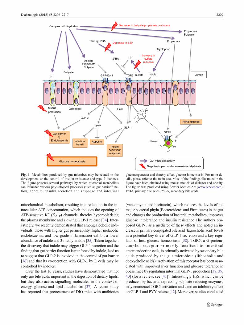
Bacterial metabolites and glucose homeostasisMetabolitesproduced by gut microbes may also be related to the develop-ment, or the control, of insulin resistance and type 2 diabetes.Most of the data illustrated in Fig. 1 have been obtained usingmouse models of diabetes and obesity. As explained below,several metabolites canmodulate the endocrine function of thegut, potentially affecting glucose homeostasis.
Short-chain fatty acids (SCFAs; e.g. butyrate, propionateand acetate) are among the most widely investigated metabo-lites produced by the gut microbiota that interfere with hostmetabolism. These molecules are produced by the microbialfermentation of specific oligo- or polysaccharides(i.e. non-digestible carbohydrates) via distinct metabolic path-ways [23]. The effect of SCFAs on insulin sensitivity andenergy metabolism is now widely accepted, although variousphysiological pathways have been suggested. Indeed, SCFAsare able to modify the levels of several gut peptides involvedin glucose metabolism, gut barrier function and energyhomeostasis [24–26]. For example, butyrate and propionatewere shown to suppress weight gain in mice with HFD-induced obesity (DIO), and acetate was shown to reduce foodintake in healthy mice [27, 28]. The majority of the pathwaysunderlying these effects remain unknown. Several studieshave suggested that the effects of SCFAs are mediated bythe members of a recently identified G protein-coupled recep-tor family that includes G protein-coupled receptors 43 and 41(GPR43 and GPR41, respectively) (for a review, see [29]).The binding of SCFAs to GPR43 and GPR41 increases theplasma levels of glucagon-like peptide-1 (GLP-1) and peptideYY (PYY), leading to improved glucose homeostasis andreduced appetite (for a review, see [30]). Interesting studiesin animals have shown that butyrate activates the expressionof genes involved in intestinal gluconeogenesis via acAMP-dependent mechanism, whereas propionate, alreadyknown as a substrate for gluconeogenesis, promotes intestinalgluconeogenic gene expression via a gut–brain neural circuitinvolving GPR41. The subsequent release of glucose into theportal vein contributes to the regulation of glycaemia and in-sulin sensitivity [31].
Recent data have indicated that the production of indole, ametabolite produced by gut bacteria from tryptophan, mayalso contribute to the secretion of GLP-1 by intestinalenteroendocrine cells [32, 33]. Chimerel et al discovered thatindole inhibits voltage-gated K+ channels, thereby changingthe action potential properties of L cells and leading to en-hanced Ca2+ entry, which acutely triggers GLP-1 secretion[34]. More importantly, it has been found that over a longerperiod of stimulation indole acts as an inhibitor of
2208 Diabetologia (2015) 58:2206–2217
mitochondrial metabolism, resulting in a reduction in the in-tracellular ATP concentration, which induces the opening ofATP-sensitive K+ (KATP) channels, thereby hyperpolarisingthe plasma membrane and slowing GLP-1 release [34]. Inter-estingly, we recently demonstrated that among alcoholic indi-viduals, those with higher gut permeability, higher metabolicendotoxaemia and low-grade inflammation exhibit a lowerabundance of indole and 3-methyl indole [35]. Taken together,the discovery that indole may trigger GLP-1 secretion and thefinding that gut barrier function is reinforced by indole, lead usto suggest that GLP-2 is involved in the control of gut barrier[36] and that its co-secretion with GLP-1 by L cells may becontrolled by indoles.
Over the last 10 years, studies have demonstrated that notonly are bile acids important in the digestion of dietary lipids,but they also act as signalling molecules in the context ofenergy, glucose and lipid metabolism [37]. A recent studyhas reported that pretreatment of DIO mice with antibiotics
(vancomycin and bacitracin), which reduces the levels of themajor bacterial phyla (Bacteroidetes and Firmicutes) in the gutand changes the production of bacterial metabolites, improvesglucose intolerance and insulin resistance The authors pro-posed GLP-1 as a mediator of these effects and noted an in-crease in primary conjugated bile acid (taurocholic acid) levelsas a potential key driver of GLP-1 secretion and a key regu-lator of host glucose homeostasis [38]. TGR5, a G protein-coupled receptor primarily localised to intestinalenteroendocrine cells, is primarily activated by secondary bileacids produced by the gut microbiota (lithocholic anddeoxycholic acids). Activation of this receptor has been asso-ciated with improved liver function and glucose tolerance inobese mice by regulating intestinal GLP-1 production [37, 39,40] (for a review, see [41]). Interestingly H2S, which can beproduced by bacteria expressing sulphate-reducing enzymes,may counteract TGR5 activation and exert an inhibitory effecton GLP-1 and PYY release [42]. Moreover, studies conducted
GLP-2
GLP-1
Insulinsecretion/response
Gut barrier
Endotoxaemia
PYY
AppetiteIntestinaltransit
GPR43/41 TGR5
AcetatePropionateButyrate
Indole
Tryptophan
GPR41
Mucus
productionL cell
Neuron
Glucose homeostasis
Portal glucose
PropionateButyrate
Complex carbohydrates
Butyrate
Tau/Gly-1°BA
2°BA
Decrease in BSH
Sulfate
H2SIncrease in
sulfatereducers
Decrease in butyrate/propionate producers
Gut microbial activity
Negative impact of diabetes-related dysbiosis
Propionate
? +
Goblet cell
Lumen
Fig. 1 Metabolites produced by gut microbes may be related to thedevelopment or the control of insulin resistance and type 2 diabetes.The figure presents several pathways by which microbial metabolitescan influence various physiological processes (such as gut barrier func-tion, appetite, insulin secretion and response and intestinal
gluconeogenesis) and thereby affect glucose homeostasis. For more de-tails, please refer to the main text. Most of the findings illustrated in thefigure have been obtained using mouse models of diabetes and obesity.The figure was produced using Servier MedicalArt (www.servier.com).1°BA, primary bile acids; 2°BA, secondary bile acids
Diabetologia (2015) 58:2206–2217 2209

in mice have demonstrated that the gut microbiota regulate theexpression of fibroblast growth factor 15 (for which theorthologous protein in humans is fibroblast growth factor 19[FGF19]) in the gut by activating the farnesoid X receptor—these hormones are responsible for transmitting bile acid-induced signals in targeted tissues to regulate weight gainand insulin resistance [40, 43, 44]. Joyce et al have shown thatpromoting the activity of bile salt hydrolase (BSH)—anenzyme distributed across the major bacterial divisions andarchaea that catalyses the deconjugation of bile acids to pro-duce secondary bile acids—in the gut microbiota may directlycontrol body weight, blood cholesterol levels, hepatic lipidlevels and fat mass gain [45]. Interestingly, a recent interven-tion study involving the administration of a BSH-activeL. reuteri strain to healthy volunteers led to an increase in totalplasma (conjugated and unconjugated) bile acid levels thatcorrelated with the serum FGF19 levels [46]. The impact ofchanging the availability and the profile of bile acids on hostglucose homeostasis remains to be clearly established inhumans, but these metabolites appear to function as importantmediators of host metabolism.
Thus, although the influence of the gut microbiota onenergy metabolism is multifactorial, different targets involv-ing immunity and/or specific metabolites have beenemphasised in recent studies, clearly demonstrating the ratio-nale for searching for novel therapeutic targets based on com-pounds derived from or produced by bacteria.
Potential contribution of the gut microbiotato the pharmacological or surgical treatment of type2 diabetes
The discovery of the gut microbiota as a metabolic partner inthe management of type 2 diabetes also led to the publicationof studies investigating whether gut microbes play a role in thebenefits of type 2 diabetes therapies.
Metformin is the most widely used glucose-lowering drug.However, its mechanism of action remains unclear [47]. Afirst clue regarding the involvement of the gastrointestinaltract in the benefits of metformin came from the observationthat intravenous administration of metformin was unable toreduce glycaemia [48]. A second clue came from the findingthat the improvement in glucose tolerance induced by metfor-min was abrogated in mice treated with broad-spectrum anti-biotics [49]. Strikingly, Shin et al reported that metformininduced a profound shift in the microbial ecosystem in favourof Akkermansia spp. and that oral administration ofA. muciniphila improved glucose tolerance [49], therebyconfirming the results obtained at our laboratory [50]. Theauthors thus suggested that a modulation of the gut microbiota(likely an increase in the Akkermansia spp. population) maycontribute to the glucose-lowering effects of metformin. A
few months later, Lee et al confirmed that metformin treat-ment induces an increase in the A. muciniphila population anddemonstrated a negative correlation between glycaemia andA. muciniphila abundance. Interestingly, co-incubation ofmetformin and mouse stool samples led to an enrichment inA. muciniphila [51], suggesting that metformin directly inter-acts with the gut microbiota to foster the growth ofA. muciniphila.
Acarbose, an α-glucosidase inhibitor that is almost exclu-sively used in Asia, is another type 2 diabetes drugwith effectsthat could be related to the gut microbiota. In Chinese patients,the inclusion of acarbose as part of their glucose-loweringmedicat ion has been reported to increase faecalBifidobacterium spp. and reduce LPS levels [52].
Interestingly, new therapeutic agents proposed for the treat-ment of type 2 diabetes (sitagliptin and exenatide) exploit theGLP-1 pathway. As mentioned earlier, GLP-1 secretion can alsobe stimulated by metabolites produced by the gut microbiota[25]. Reimer et al demonstrated that co-administration ofsitagliptin and a viscous fermentable fibre, which is brokendown into SCFA, more effectively reduced fasting glycaemiain obese Zucker rats than either treatment alone [53]. Similarresults were obtained in the same model when this fibre wascombinedwithmetformin orwithmetformin and sitagliptin [54].
Currently, the combination of medical therapy with bariat-ric surgery (vertical sleeve gastrectomy [VSG] or Roux-en-Ygastric bypass [RYGB]) appears to more effectively controlglycaemia than medical therapy alone in obese patients withuncontrolled diabetes [55]. In this context, studies have foundthat RYGB restructures the gut microbiota in humans and rats[56, 57]. Transfer of the gut microbiota of mice that underwentRYGB to non-operated germ-free mice resulted in weight lossand decreased fat mass but no change in fasting glycaemia,providing the first evidence that changes in the gut microbiotacontribute to the metabolic improvements conferred byRYGB [56]. As explained above, bile acids might link thegut microbiota to the host. Their levels are modified afterbariatric surgery, and VSG does not improve hyperglycaemiain mice carrying a targeted genetic deletion of the farnesoid Xreceptor, implicating bile acids as bacterial modulators of hosthomeostasis in this context [58]. Bile acids are without doubtvery interesting mediators. However, the differences in bileacid and cholesterol metabolism between mice and humansmake it difficult to translate the data from the animal models tothe human situation.
Novel therapeutic approaches of type 2 diabetesbased on the understanding of gut microbiota–hostinteractions
Aside from the classical treatments, the recently recognisedimplication of gut microbes in the physiopathology of type 2
2210 Diabetologia (2015) 58:2206–2217

diabetes opens a novel area of research for developing newstrategies to tackle this disease using gut microbes.
Microbiota transfer An original study recently investigatedthis approach using an infusion of faecal microbiota from leandonors to recipients with the metabolic syndrome [59]. Thetransfer of a microbiota sample from healthy patients was ableto increase the levels of butyrate-producing bacteria and insu-lin sensitivity in insulin-resistant recipients [59], thus suggest-ing that the isolation of the microbiota from faecal contentmight be developed as a therapeutic strategy to increase insu-lin sensitivity in humans. However, this type of experimentassessing the role of the gut microbiota in the control of dia-betes in humans is currently a proof-of-concept rather than apotential therapy. Additional studies are needed to confirm thelack of harmful effects linked to the transfer of faecalmicroorganisms, most of which are unidentified anduncharacterised at present.
Probiotic approach More specific approaches may also beconsidered for type 2 diabetic patients. Probiotics are livemicroorganisms that, when administered in adequateamounts, confer a health benefit to the host (i.e. humans)[60]. To date, the major probiotic strains that have shownbeneficial effects on glucose metabolism in humans belongto the Lactobacillus genus (i.e. L. plantarum 299v,Lactobacillus acidophilus NCFM and L. gasseri SBT2055)[61–63]. These observations may appear to be discordant, assome Lactobacillus species have been shown to be increasedin type 2 diabetic patients, as previously discussed. However,the increase in Lactobacillus species in type 2 diabetes hasnever been demonstrated to have a direct impact on the dis-ease. Moreover, the effects obtained using probiotics are prob-ably strain-specific; thus, different strains of the same speciesmay exert distinct effects. Importantly, it could be interestingto investigate other ‘beneficial’ microorganisms that are de-creased in diabetic patients.
Among the bacteria that could potentially be used for thetreatment of type 2 diabetes, A. muciniphila appears to be ofparticular interest. By administering A. muciniphila MucT
(ATTC BAA-835) in a diet-induced mouse model of type 2diabetes, we demonstrated the direct beneficial effects of thisbacterium on glucose metabolism [50]. First,A. muciniphila isable to counteract fasting hyperglycaemia in diet-inducedmouse model of type 2 diabetes by preventing the increasein G6pc (glucose-6-phosphatase) mRNA expression [50].This suggests that A. muciniphila thwarts the deleterious in-crease in gluconeogenesis in diabetic mice. Moreover, admin-istration of live A. muciniphila alleviates glucose intolerancein HFD-induced diabetic mice [49, 50]. However, additionalstudies are needed to establish whether A. muciniphila can beused as a probiotic for patients with type 2 diabetes, andof these, intervention studies in humans are of utmost
importance. Finally, A. muciniphila is probably not the solebacterium that could be beneficial for the treatment of thesepatients; other bacteria, such as F. prausnitzii, which playsan important role in the maintenance of the gut barrier andin the control of inflammation, could also be interesting toinvestigate (for a review, see [64]).
Non-bacterial ‘colonisers’ of the gut of potential interest Inaddition to the classical probiotic bacteria, several other typesof living organism might contribute to the therapeutic arsenalfor treating hyperglycaemia in the future. Here, we considerthe current knowledge on fungi, archaea and helminths re-garding their relationship with host glycaemia.
Our understanding of the contribution of the mycobiota(fungal community) to health and disease remains in its infan-cy [65]. Our laboratory recently provided the first evidencesupporting the hypothesis that fungi can influence host metab-olism. The yeast Saccharomyces boulardii changed the gutmicrobiota and reduced certain features of the metabolic syn-drome in genetically obese and diabetic mice. However, thisyeast did not change fasting glycaemia in these mice [66].Improving our understanding of the mycobiota and its rela-tionship with the host might lead in the future to the develop-ment of new therapies for the metabolic syndrome.
The predominant archaeon member in the human gut isMethanobrevibacter smithii. How this methanogenicarchaeon collaborates with saccharolytic bacteria such asBacteroides thetaiotaomicron to metabolise complex carbo-hydrates was elegantly established almost 10 years ago [67].This symbiotic association increases adiposity when inoculat-ed into germ-free mice [67]. In humans, methanogenicarchaea are increased in obese vs lean individuals [68], andintestinal methane production in obese individuals is associ-ated with a higher BMI [69]. However, this association cannotbe generalised to all archaea [70], and their relationship withglycaemia has not been reported.
Helminths are known to induce T helper type 2-orientedimmunity in association with eosinophilia. For this reason,Nippostrongylus brasiliensis has been used in a mouse modelof DIO to maintain eosinophil homeostasis in adipose tissue,and this intervention led to reduced adipose macrophagecounts and fasting glucose levels [71]. In accordance withthese results, metabonomic investigation of mice infectedwithSchistosoma mansoni suggested a stimulation of glycolysis,which might also contribute to the glucose-lowering effectassociated with helminth infection [72]. Moreover, as hel-minths influence the gut microbiota (e.g. increasedlactobacilli) [73, 74], we cannot exclude an indirect effect ofhelminths on host metabolism via modulation of the gut mi-crobiota. Voluntary infection with helminths might not consti-tute an appropriate therapeutic approach to reducing bloodglucose levels. However, unravelling the biological mecha-nisms underlying the beneficial effects of helminths on
Diabetologia (2015) 58:2206–2217 2211
glucose metabolism (such as the induction of eosinophilia orthe stimulation of the growth of lactobacilli) should revealnew therapeutic targets and would help to identify how thegut ecosystem plays a role in the control of host metabolism.
A place for nutrition in the managementof glycaemia-related dysbiosis
Inulin-type fructans Nutrition plays an important role in themanagement of diabetes. Indeed, some nutrients are able todecrease the postprandial glucose response. Cereals, legumes,fruits and spices are four important food groups that containactive ingredients (such as dietary fibre and polyphenols) thatare able to reduce glycaemia and insulin responses in humans[75]. The glucose-lowering effect of fibre intake may dependon the fibre type, amount and/or source. Dietary inulin-typefructans (ITF), which are present in various fruits and vegeta-bles, are fermentable carbohydrates that display prebioticproperties, as their metabolisation by gut microorganismsmodulates the composition and/or activity of the gut microbi-ota, thus conferring a beneficial physiological effect on thehost [76]. ITF increase the number of endocrine L cells inthe jejunum and colon of rodents and promote the productionand release of the active forms of GLP-1, thereby decreasingglycaemia [77–81]. A systematic review conducted to evalu-ate the effectiveness of dietary ITF on serum glucose inhumans revealed that four out of 13 eligible randomised con-trolled trials published from 1984 to 2009 reported a decreasein serum glucose concentrations [82]. Interestingly, in healthyvolunteers, 2 weeks of treatment with ITF (16 g per day)increased the postprandial release of gut peptides (specificallyGLP-1 and gastric inhibitory peptide), modified eating behav-iour (increased satiety and decreased energy intake) and de-creased postprandial glycaemia [83]. One study performed ona limited number of patients at risk for cardiovascular diseasedid not support the effect of ITF on insulin sensitivity [84].Short-chain-enriched inulin (10 g/day) caused a significantdecrease in the levels of fasting plasma glucose, HbA1c andinflammatory markers (IL-6, TNF-α and LPS) compared withmaltodextrin in a trial of 52 overweight type 2 diabetes wom-en over a period of 8 weeks [85]. In a study of the correlationsbetween glycaemic control by ITF in obese women and gutbacteria, changes in Clostridium cluster IV group (which wasincreased by ITF) were negatively correlated with fastingglycaemia, insulinaemia and HOMA-IR [86]. In contrast,changes in Propionibacterium spp., Bacteroides intestinalisand Bacteroides vulgatus, all three of which were significantlydecreased by prebiotic treatment, were positively correlatedwith the changes in glucose homeostasis. Serum LPS levelswere negatively correlated with several bacterial phyla andspecies , specif ical ly Firmicutes , Act inobacter ia ,Bifidobacterium and F. prausnitzii, all of which were
promoted by ITF. The promotion of Bifidobacterium by ITFis logical since these bacteria express β-fructosidase, but theother changes, such as the interesting increase in F. prausnitzii,remain unexplained.
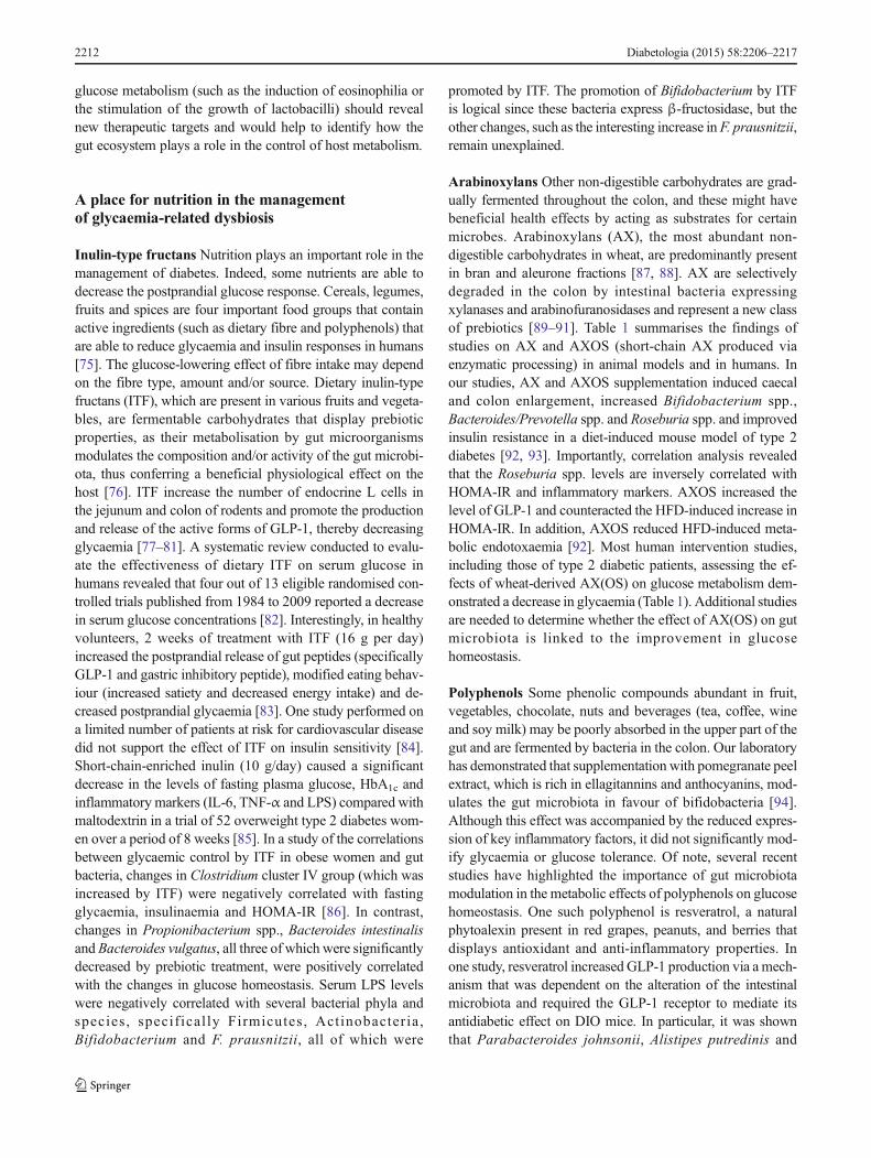
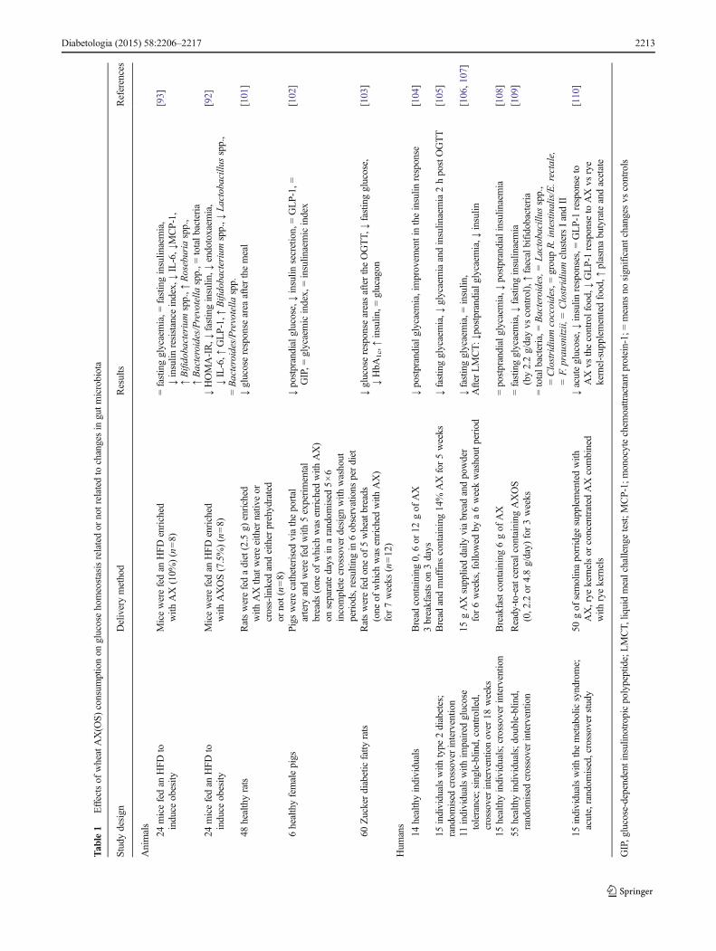
Arabinoxylans Other non-digestible carbohydrates are grad-ually fermented throughout the colon, and these might havebeneficial health effects by acting as substrates for certainmicrobes. Arabinoxylans (AX), the most abundant non-digestible carbohydrates in wheat, are predominantly presentin bran and aleurone fractions [87, 88]. AX are selectivelydegraded in the colon by intestinal bacteria expressingxylanases and arabinofuranosidases and represent a new classof prebiotics [89–91]. Table 1 summarises the findings ofstudies on AX and AXOS (short-chain AX produced viaenzymatic processing) in animal models and in humans. Inour studies, AX and AXOS supplementation induced caecaland colon enlargement, increased Bifidobacterium spp.,Bacteroides/Prevotella spp. and Roseburia spp. and improvedinsulin resistance in a diet-induced mouse model of type 2diabetes [92, 93]. Importantly, correlation analysis revealedthat the Roseburia spp. levels are inversely correlated withHOMA-IR and inflammatory markers. AXOS increased thelevel of GLP-1 and counteracted the HFD-induced increase inHOMA-IR. In addition, AXOS reduced HFD-induced meta-bolic endotoxaemia [92]. Most human intervention studies,including those of type 2 diabetic patients, assessing the ef-fects of wheat-derived AX(OS) on glucose metabolism dem-onstrated a decrease in glycaemia (Table 1). Additional studiesare needed to determine whether the effect of AX(OS) on gutmicrobiota is linked to the improvement in glucosehomeostasis.
Polyphenols Some phenolic compounds abundant in fruit,vegetables, chocolate, nuts and beverages (tea, coffee, wineand soy milk) may be poorly absorbed in the upper part of thegut and are fermented by bacteria in the colon. Our laboratoryhas demonstrated that supplementation with pomegranate peelextract, which is rich in ellagitannins and anthocyanins, mod-ulates the gut microbiota in favour of bifidobacteria [94].Although this effect was accompanied by the reduced expres-sion of key inflammatory factors, it did not significantly mod-ify glycaemia or glucose tolerance. Of note, several recentstudies have highlighted the importance of gut microbiotamodulation in the metabolic effects of polyphenols on glucosehomeostasis. One such polyphenol is resveratrol, a naturalphytoalexin present in red grapes, peanuts, and berries thatdisplays antioxidant and anti-inflammatory properties. Inone study, resveratrol increased GLP-1 production via a mech-anism that was dependent on the alteration of the intestinalmicrobiota and required the GLP-1 receptor to mediate itsantidiabetic effect on DIO mice. In particular, it was shownthat Parabacteroides johnsonii, Alistipes putredinis and
2212 Diabetologia (2015) 58:2206–2217
Tab
le1
Effectsof
wheatAX(O
S)consum
ptionon
glucosehomeostasisrelatedor
notrelated
tochangesin
gutm
icrobiota
Studydesign
Deliverymethod
Results
References
Animals
24micefedan
HFDto
induce
obesity
Micewerefedan
HFDenriched
with
AX(10%
)(n=8)
=fastingglycaemia,=
fastinginsulin
aemia,
↓insulin
resistance
index,↓IL-6,↓MCP-1,
↑Bifidobacteriumspp.,↑
Roseburia
spp.,
↑Bacteroides/Prevotella
spp.,=
totalb
acteria
[93]
24micefedan
HFDto
induce
obesity
Micewerefedan
HFDenriched
with
AXOS(7.5%)(n=8)
↓HOMA-IR,↓
fastinginsulin
,↓endotoxaem
ia,
↓IL-6,↑
GLP-1,↑
Bifidobacteriumspp.,↓
Lactobacillus
spp.,
=Bacteroides/Prevotella
spp.
[92]
48healthyrats
Ratswerefedadiet(2.5
g)enriched
with
AXthatwereeither
nativ
eor
cross-lin
kedandeither
prehydrated
ornot(n=8)
↓glucoseresponse
area
afterthemeal
[101]
6healthyfemalepigs
Pigs
werecatheterised
viatheportal
artery
andwerefedwith
5experimental
breads
(one
ofwhich
was
enriched
with
AX)
onseparatedays
inarandom
ised
5×6
incompletecrossoverdesign
with
washout
periods,resulting
in6observations
perdiet
↓postprandialglucose,↓insulin
secretion,=GLP-1,=
GIP,=
glycaemicindex,=insulin
aemicindex
[102]
60Zuckerdiabeticfatty
rats
Ratswerefedoneof
5wheatbreads
(one
ofwhich
was
enriched
with
AX)
for7weeks
(n=12)
↓glucoseresponse
areasaftertheOGTT,
↓fastingglucose,
↓HbA
1c,↑insulin
,=glucagon
[103]
Hum
ans
14healthyindividuals
Bread
containing
0,6or
12gof
AX
3breakfastson
3days
↓postprandialglycaemia,improvem
entintheinsulin
response
[104]
15individualswith
type
2diabetes;
random
ised
crossoverinterventio
nBread
andmuffins
containing
14%
AXfor5weeks
↓fastingglycaemia,↓
glycaemiaandinsulin
aemia2hpostOGTT
[105]
11individualswith
impaired
glucose
tolerance;single-blin
d,controlled,
crossoverinterventio
nover
18weeks
15gAXsupplieddaily
viabreadandpowder
for6weeks,followed
bya6weekwashout
period
↓fastingglycaemia,=
insulin
,After
LMCT:↓
postprandialglycaemia,↓
insulin
[106,107]
15healthyindividuals;crossoverinterventio
nBreakfastcontaining
6gof
AX
=postprandialglycaemia,↓
postprandialinsulin
aemia
[108]
55healthyindividuals;double-blin
d,random
ised
crossoverinterventio
nReady-to-eatcerealcontainingAXOS
(0,2.2or
4.8g/day)
for3weeks
=fastingglycaemia,↓
fastinginsulin
aemia
(by2.2g/dayvs
control),↑
faecalbifidobacteria
=totalb
acteria,=Bacteroides,=
Lactobacillu
sspp.,
=Clostridium
coccoides,=groupR.intestin
alis/E.rectale,
=F.prausnitzii,
=Clostridium
clustersIandII
[109]
15individualswith
themetabolicsyndrome;
acute,random
ised,crossover
study
50gof
semolinaporridge
supplementedwith
AX,rye
kernelsor
concentrated
AXcombined
with
ryekernels
↓acuteglucose,↓insulin
responses,=GLP-1response
toAXvs
thecontrolfood,↓GLP-1response
toAXvs
rye
kernel-supplem
entedfood,↑
plasmabutyrateandacetate
[110]
GIP,glucose-dependent
insulin
otropicpolypeptide;LMCT,
liquidmealchallengetest;M
CP-1;
monocytechem
oattractant
protein-1;
=means
nosignificantchanges
vscontrols
Diabetologia (2015) 58:2206–2217 2213

Bacteroides vulgatus, the levels of which were increasedby HFD treatment, disappeared 5 weeks after resveratrolsupplementation [95]. In another study, mice fed an HFDsupplemented with 4% green tea powder for 8 weeks had asignificantly increased insulin response compared with con-trol mice [96]. In addition, fasting plasma glucose, insulin andHOMA-IR levels were lower in mice fed the green tea sup-plement for 11 or 22weeks. In a third study, the administrationof cranberry extract, which is rich in proanthocyanidins, im-proved insulin sensitivity in high-fat/high-sucrose diet-fedmice. In this study, cranberry extract treatment markedly in-creased the proportion of Akkermansia and decreased intesti-nal inflammation [97]. Finally, a double-blind trial revealedthat changes in the gut microbiota are associated with theglucose-lowering effects of a traditional berberine-containingChinese herbal formula in type 2 diabetic patients [98]. In-deed, this decoction significantly increased F. prausnitzii,which was negatively correlated with fasting blood glucose,HbA1c and postprandial blood glucose levels and was posi-tively correlated with HOMA of beta cell function.
Importantly, energy-free artificial sweeteners were exten-sively introduced to our diets with the intention of reducingenergy intake and normalising blood glucose levels without‘sweet-toothed’ humans having to compromise. A recentstudy demonstrated that the consumption of commonly usedartificial sweetener formulations drives the development ofglucose intolerance via the induction of compositional andfunctional alterations to the intestinal microbiota [99]. Wheth-er the bacterial populations or metabolic pathways altered bythe consumption of artificial sweeteners are similar to thosedescribed in individuals with or developing diabetes remainsto be elucidated [99, 100].
Conclusions
Type 2 diabetes, a complex disease that is often associatedwith obesity, develops via the interaction between geneticand environmental factors. We believe that the gut microbiotarepresents an environmental factor of type 2 diabetes that wasneglected in the past due to the complexity of its analysis andto the lack of an understanding of the mechanisms underlyingthe interactions between gut microbes and host metabolism.The current interest in the gut microbiota as a potential targetfor the management of non-communicable diseases such astype 2 diabetes partially relies on the novel methodologiesavailable for analysing the composition and function of thegut microbiota, as well as on the recent discoveries of hostmolecular targets that are prone to ‘respond’ to bacterial me-tabolites/components. To those who might question the rele-vance of gut dysbiosis in the occurrence of type 2 diabetes, wewould say that all of the data supporting a causative role ofdysbiosis in type 2 diabetes have been obtained using germ-
free animals into which the intestinal content of diabetic micewas transferred. As far as the development of novel therapeu-tic approaches is concerned, intervention studies using probi-otic, prebiotic, or microbial transplantation have been success-ful in a very limited number of published reports. Nutritionaladvice is crucial in the management of diabetes. We believethat a better characterisation of the nutrients that are able tomodulate the gut microbiota in favour of anti-inflammatorybacteria or bacterial metabolites is needed to provide adequateadvice to patients who are at risk for type 2 diabetesdevelopment.
Funding NMD is a recipient of FRS-FNRS grants (CDR J.0122.15,1.5121.12F), of grants from the Walloon Region (FOOD4GUT project,convention 1318148; CAPPLE project , convention 6605;NUTRIGUTIOR project, convention 6918), of the European Union’sSeventh Framework Program community (KBBE.2013.2.2-02MyNewGut project) and of IWT subsidies (SBO-project). PDC, a re-search associate at the Fonds de la Recherche Scientifique (FRS-FNRS),Belgium, is a recipient of an ERC Starting Grant 2013 (European Re-search Council, Starting Grant 336452-ENIGMO), PDR subsidies (Projetde recherches T0.138.14; Fonds de la Recherche Scientifique, Belgium)and ARC subsidies (Concerted Research Activities-French Communityof Belgium convention: 12/17-047) and is supported by the FRS-FNRSvia the FRFS-WELBIO under Grant number WELBIO-CR-2012S-02R.LBB and AE are postdoctoral fellows at the FRS-FNRS.
Duality of interest The authors declare that there is no duality of inter-est associated with this manuscript.
Contribution statement All authors were responsible for drafting thearticle and revising it critically for important intellectual content. Allauthors approved the version to be published.
References
1. Hoffmann C, Dollive S, Grunberg S et al (2013) Archaea andfungi of the human gut microbiome: correlations with diet andbacterial residents. PLoS One 8, e66019
2. Goodrich JK, Waters JL, Poole AC et al (2014) Human geneticsshape the gut microbiome. Cell 159:789–799
3. Allin KH, Nielsen T, Pedersen O (2015) Mechanisms in endocri-nology: gut microbiota in patients with type 2 diabetes mellitus.Eur J Endocrinol 172:R167–R177
4. Qin J, Li Y, Cai Z et al (2012) A metagenome-wide associationstudy of gut microbiota in type 2 diabetes. Nature 490:55–60
5. Larsen N, Vogensen FK, van den Berg FW et al (2010) Gut mi-crobiota in human adults with type 2 diabetes differs from non-diabetic adults. PLoS One 5, e9085
6. Karlsson FH, Tremaroli V, Nookaew I et al (2013) Gutmetagenome in European women with normal, impaired and di-abetic glucose control. Nature 498:99–103
7. Sato J, Kanazawa A, Ikeda F et al (2014) Gut dysbiosis and de-tection of “live gut bacteria” in blood of Japanese patients withtype 2 diabetes. Diabetes Care 37:2343–2350
8. Zhang X, Shen D, Fang Z et al (2013) Human gut microbiotachanges reveal the progression of glucose intolerance. PLoS One8, e71108
2214 Diabetologia (2015) 58:2206–2217
9. Sepp E, Kolk H, Loivukene K, Mikelsaar M (2014) Higher bloodglucose level associated with body mass index and gut microbiotain elderly people.Microb Ecol Health Dis. doi:10.3402/mehd.v25.22857
10. Le Chatelier E, Nielsen T, Qin J et al (2013) Richness of humangut microbiome correlates with metabolic markers. Nature 500:541–546
11. Vijay-Kumar M, Aitken JD, Carvalho FA et al (2010) Metabolicsyndrome and altered gut microbiota in mice lacking Toll-likereceptor 5. Science 328:228–231
12. Hameed I,Masoodi SR,Mir SA, NabiM, Ghazanfar K, Ganai BA(2015) Type 2 diabetes mellitus: from a metabolic disorder to aninflammatory condition. World J Diabetes 6:598–612
13. Gregor MF, Hotamisligil GS (2011) Inflammatory mechanisms inobesity. Annu Rev Immunol 29:415–445
14. Beutler B (2004) Inferences, questions and possibilities in Toll-like receptor signalling. Nature 430:257–263
15. Cani PD, Amar J, Iglesias MA, et al (2007) Metabolicendotoxemia initiates obesity and insulin resistance. Diabetes1761-1772
16. Cani PD, Bibiloni R, Knauf C et al (2008) Changes in gut micro-biota control metabolic endotoxemia-induced inflammation inhigh-fat diet-induced obesity and diabetes in mice. Diabetes 57:1470–1481
17. Cani PD, Neyrinck AM, Fava F et al (2007) Selective increases ofbifidobacteria in gut microflora improve high-fat-diet-induced di-abetes in mice through a mechanism associated withendotoxaemia. Diabetologia 50:2374–2383
18. Sun L, Yu Z, Ye X et al (2010) A marker of endotoxemia isassociated with obesity and related metabolic disorders in appar-ently healthy Chinese. Diabetes Care 33:1925–1932
19. Everard A, Geurts L, Caesar R et al (2014) Intestinal epithelialMyD88 is a sensor switching host metabolism towards obesityaccording to nutritional status. Nat Commun 5:5648
20. Geurts L, Neyrinck AM, Delzenne NM, Knauf C, Cani PD (2014)Gut microbiota controls adipose tissue expansion, gut barrier andglucose metabolism: novel insights into molecular targets and in-terventions using prebiotics. Benefic Microbes 5:3–17
21. Denou E, Lolmede K, Garidou L et al (2015) Defective NOD2peptidoglycan sensing promotes diet-induced inflammation,dysbiosis, and insulin resistance. EMBO Mol Med 9:259–274
22. Al-Daghri NM, Clerici M, Al-Attas O et al (2013) A nonsensepolymorphism (R392X) in TLR5 protects from obesity but pre-disposes to diabetes. J Immunol 190:3716–3720
23. Reichardt N, Duncan SH, Young P et al (2014) Phylogenetic dis-tribution of three pathways for propionate production within thehuman gut microbiota. ISME J 8:1323–1335
24. Reimann F, Tolhurst G, Gribble FM (2012) G-protein-coupledreceptors in intestinal chemosensation. Cell Metab 15:421–431
25. Tolhurst G, Heffron H, Lam YS et al (2012) Short-chain fattyacids stimulate glucagon-like peptide-1 secretion via the G-protein-coupled receptor FFAR2. Diabetes 61:364–371
26. Plaisancie P, Dumoulin V, Chayvialle JA, Cuber JC (1995)Luminal glucagon-like peptide-1(7-36) amide-releasing factorsin the isolated vascularly perfused rat colon. J Endocrinol 145:521–526
27. Lin HV, Frassetto A, Kowalik EJ Jr et al (2012) Butyrate andpropionate protect against diet-induced obesity and regulate guthormones via free fatty acid receptor 3-independent mechanisms.PLoS One 7, e35240
28. Frost G, Sleeth ML, Sahuri-Arisoylu M et al (2014) The short-chain fatty acid acetate reduces appetite via a central homeostaticmechanism. Nat Commun 5:3611
29. Bindels LB, Dewulf EM, Delzenne NM (2013) GPR43/FFA2:physiopathological relevance and therapeutic prospects. TrendsPharmacol Sci 34:226–232
30. Everard A, Cani PD (2014) Gut microbiota and GLP-1. RevEndocr Metab Disord 15:189–196
31. De Vadder F, Kovatcheva-Datchary P, Goncalves D et al (2014)Microbiota-generated metabolites promote metabolic benefits viagut-brain neural circuits. Cell 156:84–96
32. Yokoyama MT, Carlson JR (1979) Microbial metabolites of tryp-tophan in the intestinal tract with special reference to skatole. Am JClin Nutr 32:173–178
33. DeMoss RD, Moser K (1969) Tryptophanase in diverse bacterialspecies. J Bacteriol 98:167–171
34. Chimerel C, Emery E, Summers DK, Keyser U, Gribble FM,Reimann F (2014) Bacterial metabolite indole modulates incretinsecretion from intestinal enteroendocrine L cells. Cell Rep 9:1202–1208
35. Leclercq S, Matamoros S, Cani PD et al (2014) Intestinal perme-ability, gut-bacterial dysbiosis, and behavioral markers of alcohol-dependence severity. Proc Natl Acad Sci U S A 111:E4485–E4493
36. Cani PD, Possemiers S, Van de WT et al (2009) Changes in gutmicrobiota control inflammation in obese mice through a mecha-nism involving GLP-2-driven improvement of gut permeability.Gut 58:1091–1103
37. Thomas C, Gioiello A, Noriega L et al (2009) TGR5-mediatedbile acid sensing controls glucose homeostasis. Cell Metab 10:167–177
38. Hwang I, Park YJ, Kim YR et al (2015) Alteration of gut micro-biota by vancomycin and bacitracin improves insulin resistancevia glucagon-like peptide 1 in diet-induced obesity. FASEB J 29:2397–2411
39. Claus SP, Tsang TM, Wang Y et al (2008) Systemicmulticompartmental effects of the gut microbiome on mousemet-abolic phenotypes. Mol Syst Biol 4:219
40. Sayin SI, Wahlstrom A, Felin J et al (2013) Gut microbiota regu-lates bile acid metabolism by reducing the levels of tauro-beta-muricholic acid, a naturally occurring FXR antagonist. Cell Metab17:225–235
41. Prawitt J, Caron S, Staels B (2011) Bile acid metabolism and thepathogenesis of type 2 diabetes. Curr Diab Rep 11:160–166
42. Bala V, Rajagopal S, Kumar DP et al (2014) Release of GLP-1 andPYY in response to the activation of G protein-coupled bile acidreceptor TGR5 is mediated by Epac/PLC-ε pathway and modu-lated by endogenous H2S. Front Physiol 5:420
43. Li F, Jiang C, Krausz KW et al (2013) Microbiome remodellingleads to inhibition of intestinal farnesoid X receptor signalling anddecreased obesity. Nat Commun 4:2384
44. Degirolamo C, Rainaldi S, Bovenga F, Murzilli S, Moschetta A(2014) Microbiota modification with probiotics induces hepaticbile acid synthesis via downregulation of the Fxr–Fgf15 axis inmice. Cell Rep 7:12–18
45. Joyce SA, MacSharry J, Casey PG et al (2014) Regulation of hostweight gain and lipid metabolism by bacterial bile acid modifica-tion in the gut. Proc Natl Acad Sci U S A 111:7421–7426
46. Martoni CJ, Labbe A, Ganopolsky JG, Prakash S, Jones ML(2015) Changes in bile acids, FGF-19 and sterol absorption inresponse to bile salt hydrolase active L. reuteri NCIMB 30242.Gut Microbes 6:57–65
47. Rena G, Pearson ER, Sakamoto K (2013) Molecular mechanism ofaction ofmetformin: old or new insights?Diabetologia 56:1898–1906
48. Bonora E, Cigolini M, Bosello O et al (1984) Lack of effect ofintravenous metformin on plasma concentrations of glucose, insu-lin, C-peptide, glucagon and growth hormone in non-diabetic sub-jects. Curr Med Res Opin 9:47–51
49. Shin NR, Lee JC, Lee HY et al (2014) An increase in theAkkermansia spp. population induced by metformin treatmentimproves glucose homeostasis in diet-induced obese mice. Gut63:727–735
Diabetologia (2015) 58:2206–2217 2215

50. Everard A, Belzer C, Geurts L et al (2013) Cross-talk betweenAkkermansia muciniphila and intestinal epithelium controls diet-induced obesity. Proc Natl Acad Sci U S A 110:9066–9071
51. Lee H, Ko G (2014) Effect of metformin on metabolic improve-ment and gut microbiota. Appl Environ Microbiol 80:5935–5943
52. Su B, Liu H, Li J et al (2014) Acarbose treatment affects the serumlevels of inflammatory cytokines and the gut content ofbifidobacteria in Chinese patients with type 2 diabetes mellitus. JDiabetes. doi:10.1111/1753-0407.12232
53. Reimer RA, Grover GJ, Koetzner L et al (2012) Sitagliptin re-duces hyperglycemia and increases satiety hormone secretionmore effectively when used with a novel polysaccharide in obeseZucker rats. J Nutr 142:1812–1820
54. Reimer RA, Grover GJ, Koetzner L, Gahler RJ, Lyon MR, WoodS (2014) Combining sitagliptin/metformin with a functional fiberdelays diabetes progression in Zucker rats. J Endocrinol 220:361–373
55. Schauer PR, Kashyap SR, Wolski K et al (2012) Bariatric surgeryversus intensive medical therapy in obese patients with diabetes. NEngl J Med 366:1567–1576
56. Liou AP, Paziuk M, Luevano JM Jr, Machineni S, Turnbaugh PJ,Kaplan LM (2013) Conserved shifts in the gut microbiota due togastric bypass reduce host weight and adiposity. Sci Transl Med 5:178ra41
57. Aron-Wisnewsky J, Dore J, Clement K (2012) The importance ofthe gut microbiota after bariatric surgery. Nat Rev GastroenterolHepatol 9:590–598
58. Ryan KK, Tremaroli V, Clemmensen C et al (2014) FXR is amolecular target for the effects of vertical sleeve gastrectomy.Nature 509:183–188
59. Vrieze A, van Nood E, Holleman F et al (2012) Transfer of intes-tinal microbiota from lean donors increases insulin sensitivity inindividuals with metabolic syndrome. Gastroenterology 143:913–916
60. Hill C, Guarner F, Reid G et al (2014) Expert consensus docu-ment: the International Scientific Association for Probiotics andPrebiotics consensus statement on the scope and appropriate useof the term probiotic. Nat Rev Gastroenterol Hepatol 11:506–514
61. Bukowska H, Pieczul-Mroz J, Jastrzebska M, Chelstowski K,Naruszewicz M (1998) Decrease in fibrinogen and LDL-cholesterol levels upon supplementation of diet withLactobacillus plantarum in subjects with moderately elevatedcholesterol. Atherosclerosis 137:437–438
62. Andreasen AS, Larsen N, Pedersen-Skovsgaard T et al (2010)Effects of Lactobacillus acidophilus NCFM on insulin sensitivityand the systemic inflammatory response in human subjects. Br JNutr 104:1831–1838
63. Ogawa A, Kadooka Y, Kato K, Shirouchi B, Sato M (2014)Lactobacillus gasseri SBT2055 reduces postprandial and fastingserum non-es ter i f ied fa t ty ac id levels in Japanesehypertriacylglycerolemic subjects. Lipids Health Dis 13:36
64. Miquel S, Martin R, Bridonneau C et al (2014) Ecology and me-tabolism of the beneficial intestinal commensal bacteriumFaecalibacterium prausnitzii. Gut Microbes 5:146–151
65. Mukherjee PK, Sendid B, Hoarau G, Colombel JF, Poulain D,Ghannoum MA (2015) Mycobiota in gastrointestinal diseases.Nat Rev Gastroenterol Hepatol 12:77–87
66. Everard A, Matamoros S, Geurts L, Delzenne NM, Cani PD(2014) Saccharomyces boulardii administration changes gut mi-crobiota and reduces hepatic steatosis, low-grade inflammation,and fat mass in obese and type 2 diabetic db/db mice. MBio 5:e01011–e01014
67. Samuel BS, Gordon JI (2006) A humanized gnotobiotic mousemodel of host-archaeal-bacterial mutualism. Proc Natl Acad Sci US A 103:10011–10016
68. Zhang H, DiBaise JK, Zuccolo A et al (2009) Human gut micro-biota in obesity and after gastric bypass. Proc Natl Acad Sci U S A106:2365–2370
69. Basseri RJ, Basseri B, Pimentel M et al (2012) Intestinal methaneproduction in obese individuals is associated with a higher bodymass index. Gastroenterol Hepatol (N Y) 8:22–28
70. Fernandes J, Wang A, SuWet al (2013) Age, dietary fiber, breathmethane, and fecal short chain fatty acids are interrelated inArchaea-positive humans. J Nutr 143:1269–1275
71. Wu D, Molofsky AB, Liang HE et al (2011) Eosinophils sustainadipose alternatively activated macrophages associated with glu-cose homeostasis. Science 332:243–247
72. Wang Y, Holmes E, Nicholson JK et al (2004) Metabonomic in-vestigations in mice infected with Schistosoma mansoni: an ap-proach for biomarker identification. Proc Natl Acad Sci U S A101:12676–12681
73. Walk ST, Blum AM, Ewing SA,Weinstock JV, Young VB (2010)Alteration of the murine gut microbiota during infection with theparasitic helminth Heligmosomoides polygyrus. Inflamm BowelDis 16:1841–1849
74. Reynolds LA, Smith KA, Filbey KJ et al (2014) Commensal-pathogen interactions in the intestinal tract: lactobacilli promoteinfection with, and are promoted by, helminth parasites. GutMicrobes 5:522–532
75. Thondre PS (2013) Food-based ingredients to modulate bloodglucose. Adv Food Nutr Res 70:181–227
76. Bindels LB, Delzenne NM, Cani PD, Walter J (2015) Towards amore comprehensive concept for prebiotics. Nat RevGastroenterol Hepatol 12:303–310
77. Cani PD, Dewever C, Delzenne NM (2004) Inulin-type fructansmodulate gastrointestinal peptides involved in appetite regulation(glucagon-like peptide-1 and ghrelin) in rats. Br J Nutr 92:521–526
78. Delzenne NM, Cani PD, Daubioul C, Neyrinck AM (2005)Impact of inulin and oligofructose on gastrointestinal peptides.Br J Nutr 93(Suppl 1):S157–S161
79. Cani PD, Daubioul CA, Reusens B, Remacle C, Catillon G,Delzenne NM (2005) Involvement of endogenous glucagon-likepeptide-1(7-36) amide on glycaemia-lowering effect ofoligofructose in streptozotocin-treated rats. J Endocrinol 185:457–465
80. Cani PD, Neyrinck AM, Maton N, Delzenne NM (2005)Oligofructose promotes satiety in rats fed a high-fat diet: involve-ment of glucagon-like peptide-1. Obes Res 13:1000–1007
81. Cani PD, Knauf C, Iglesias MA, Drucker DJ, Delzenne NM,Burcelin R (2006) Improvement of glucose tolerance and hepaticinsulin sensitivity by oligofructose requires a functional glucagon-like peptide 1 receptor. Diabetes 55:1484–1490
82. Bonsu NK, Johnson CS,McLeod KM (2011) Can dietary fructanslower serum glucose? J Diabetes 3:58–66
83. Cani PD, Lecourt E, Dewulf EM et al (2009) Gut microbiotafermentation of prebiotics increases satietogenic and incretin gutpeptide production with consequences for appetite sensation andglucose response after a meal. Am J Clin Nutr 90:1236–1243
84. Tripkovic L, Muirhead NC, Hart KH, Frost GS, Lodge JK (2014)The effects of a diet rich in inulin or wheat fibre on markers ofcardiovascular disease in overweight male subjects. J Hum NutrDiet. doi:10.1111/jhn.12251
85. Dehghan P, Pourghassem Gargari B, Asghari Jafar-abadi M(2014) Oligofructose-enriched inulin improves some inflammato-ry markers and metabolic endotoxemia in women with type 2diabetes mellitus: a randomized controlled clinical trial.Nutrition 30:418–423
86. Dewulf EM, Cani PD, Claus SP et al (2013) Insight into theprebiotic concept: lessons from an exploratory, double blind inter-vention study with inulin-type fructans in obese women. Gut 62:1112–1121
2216 Diabetologia (2015) 58:2206–2217

87. Neyrinck AM, Delzenne NM (2010) Potential interest of gut mi-crobial changes induced by non-digestible carbohydrates of wheatin the management of obesity and related disorders. Curr OpinClin Nutr Metab Care 13:722–728
88. Andersson AA, Andersson R, Piironen Vet al (2013) Contents ofdietary fibre components and their relation to associated bioactivecomponents in whole grain wheat samples from theHEALTHGRAIN diversity screen. Food Chem 136:1243–1248
89. Grootaert C, Van den Abbeele P, Marzorati M et al (2009)Comparison of prebiotic effects of arabinoxylan oligosaccharidesand inulin in a simulator of the human intestinal microbial eco-system. FEMS Microbiol Ecol 69:231–242
90. Vardakou M, Palop CN, Christakopoulos P, Faulds CB, GassonMA, Narbad A (2008) Evaluation of the prebiotic properties ofwheat arabinoxylan fractions and induction of hydrolase activityin gut microflora. Int J Food Microbiol 123:166–170
91. Hughes SA, Shewry PR, Li L, Gibson GR, Sanz ML, Rastall RA(2007) In vitro fermentation by human fecal microflora of wheatarabinoxylans. J Agric Food Chem 55:4589–4595
92. Neyrinck AM, Van Hee VF, Piront N et al (2012) Wheat-derivedarabinoxylan oligosaccharides with prebiotic effect increasesatietogenic gut peptides and reduce metabolic endotoxemia indiet-induced obese mice. Nutr Diabetes 2, e28
93. Neyrinck AM, Possemiers S, Druart C et al (2011) Prebiotic ef-fects of wheat arabinoxylan related to the increase inbifidobacteria, Roseburia and Bacteroides/Prevotella in diet-induced obese mice. PLoS One 6, e20944
94. Neyrinck AM, Van Hee VF, Bindels LB, De BF, Cani PD,Delzenne NM (2013) Polyphenol-rich extract of pomegranatepeel alleviates tissue inflammation and hypercholesterolaemia inhigh-fat diet-induced obese mice: potential implication of the gutmicrobiota. Br J Nutr 109:802–809
95. Dao TM, Waget A, Klopp P et al (2011) Resveratrol increasesglucose induced GLP-1 secretion in mice: a mechanism whichcontributes to the glycemic control. PLoS One 6, e20700
96. Axling U, Olsson C, Xu J et al (2012) Green tea powder andLactobacillus plantarum affect gut microbiota, lipid metabolismand inflammation in high-fat fed C57BL/6J mice. Nutr Metab(Lond) 9:105
97. Anhe FF, Roy D, PilonG et al (2014) A polyphenol-rich cranberryextract protects from diet-induced obesity, insulin resistance andintestinal inflammation in association with increased Akkermansiaspp. population in the gut microbiota of mice. Gut 64:872–883
98. Xu J, Lian F, Zhao L et al (2015) Structural modulation of gutmicrobiota during alleviation of type 2 diabetes with a Chineseherbal formula. ISME J 9:552–562
99. Suez J, Korem T, Zeevi D et al (2014) Artificial sweeteners induceglucose intolerance by altering the gut microbiota. Nature 514:181–186
100. Feehley T, Nagler CR (2014) Health: the weighty costs of non-caloric sweeteners. Nature 514:176–177
101. Vogel B, Gallaher DD, Bunzel M (2012) Influence of cross-linkedarabinoxylans on the postprandial blood glucose response in rats. JAgric Food Chem 60:3847–3852
102. Christensen KL, Hedemann MS, Laerke HN et al (2013)Concentrated arabinoxylan but not concentrated beta-glucan inwheat bread has similar effects on postprandial insulin as whole-grain rye in porto-arterial catheterized pigs. J Agric Food Chem61:7760–7768
103. Hartvigsen ML, Jeppesen PB, Laerke HN, Njabe EN, KnudsenKE, Hermansen K (2013) Concentrated arabinoxylan in wheatbread has beneficial effects as rye breads on glucose and changesin gene expressions in insulin-sensitive tissues of Zucker diabeticfatty (ZDF) rats. J Agric Food Chem 61:5054–5063
104. Lu ZX, Gibson PR, Muir JG, Fielding M, O’Dea K (2000)Arabinoxylan fiber from a by-product of wheat flour processingbehaves physiologically like a soluble, fermentable fiber in thelarge bowel of rats. J Nutr 130:1984–1990
105. Lu ZX, Walker KZ, Muir JG, O'Dea K (2004) Arabinoxylan fibreimproves metabolic control in people with type II diabetes. Eur JClin Nutr 58:621–628
106. Garcia AL, Otto B, Reich SC et al (2007) Arabinoxylan consump-tion decreases postprandial serum glucose, serum insulin and plas-ma total ghrelin response in subjects with impaired glucose toler-ance. Eur J Clin Nutr 61:334–341
107. Garcia AL, Steiniger J, Reich SC et al (2006) Arabinoxylan fibreconsumption improved glucose metabolism, but did not affectserum adipokines in subjects with impaired glucose tolerance.Horm Metab Res 38:761–766
108. Mohlig M, Koebnick C, Weickert MO et al (2005) Arabinoxylan-enriched meal increases serum ghrelin levels in healthy humans.Horm Metab Res 37:303–308
109. Maki KC, Gibson GR, Dickmann RS et al (2012) Digestive andphysiologic effects of a wheat bran extract, arabino-xylan-oligo-saccharide, in breakfast cereal. Nutrition 28:1115–1121
110. Hartvigsen ML, Laerke HN, Overgaard A, Holst JJ, BachKnudsen KE, Hermansen K (2014) Postprandial effects of testmeals including concentrated arabinoxylan and whole grain ryein subjects with the metabolic syndrome: a randomised study. EurJ Clin Nutr 68:567–574
Diabetologia (2015) 58:2206–2217 2217