gut microbiota and colonization resistance against …...microbes, collectively termed gut...
TRANSCRIPT

Gut Microbiota and Colonization Resistance against BacterialEnteric Infection
Q. R. Ducarmon,a,b R. D. Zwittink,a,b B. V. H. Hornung,a,b W. van Schaik,c V. B. Young,d,e E. J. Kuijpera,b,f,g
aCenter for Microbiome Analyses and Therapeutics, Leiden University Medical Center, Leiden, NetherlandsbExperimental Bacteriology, Department of Medical Microbiology, Leiden University Medical Center, Leiden, NetherlandscInstitute of Microbiology and Infection, University of Birmingham, Birmingham, United KingdomdDepartment of Microbiology and Immunology, University of Michigan, Ann Arbor, Michigan, USAeDepartment of Internal Medicine/Infectious Diseases Division, University of Michigan Medical Center, Ann Arbor, Michigan, USAfClinical Microbiology Laboratory, Department of Medical Microbiology, Leiden University Medical Center, Leiden, NetherlandsgNetherlands Donor Feces Bank, Leiden, Netherlands
SUMMARY . . . . . . . . . . . . . . . . . . . . . . . . . . . . . . . . . . . . . . . . . . . . . . . . . . . . . . . . . . . . . . . . . . . . . . . . . . . . . . . . . . . . . . . . 1INTRODUCTION . . . . . . . . . . . . . . . . . . . . . . . . . . . . . . . . . . . . . . . . . . . . . . . . . . . . . . . . . . . . . . . . . . . . . . . . . . . . . . . . . . 2MECHANISMS PROVIDING COLONIZATION RESISTANCE . . . . . . . . . . . . . . . . . . . . . . . . . . . . . . . . . . 2
Short-Chain Fatty Acids . . . . . . . . . . . . . . . . . . . . . . . . . . . . . . . . . . . . . . . . . . . . . . . . . . . . . . . . . . . . . . . . . . . . . . . 2Bile Acids . . . . . . . . . . . . . . . . . . . . . . . . . . . . . . . . . . . . . . . . . . . . . . . . . . . . . . . . . . . . . . . . . . . . . . . . . . . . . . . . . . . . . . . 3Bacteriocins . . . . . . . . . . . . . . . . . . . . . . . . . . . . . . . . . . . . . . . . . . . . . . . . . . . . . . . . . . . . . . . . . . . . . . . . . . . . . . . . . . . . 4Nutrient Competition . . . . . . . . . . . . . . . . . . . . . . . . . . . . . . . . . . . . . . . . . . . . . . . . . . . . . . . . . . . . . . . . . . . . . . . . . . 5Mucus Layers . . . . . . . . . . . . . . . . . . . . . . . . . . . . . . . . . . . . . . . . . . . . . . . . . . . . . . . . . . . . . . . . . . . . . . . . . . . . . . . . . . . 5Bacteriophages . . . . . . . . . . . . . . . . . . . . . . . . . . . . . . . . . . . . . . . . . . . . . . . . . . . . . . . . . . . . . . . . . . . . . . . . . . . . . . . . . 6
EFFECTS OF VARIOUS NONANTIBIOTIC DRUGS ON GUT COLONIZATIONRESISTANCE . . . . . . . . . . . . . . . . . . . . . . . . . . . . . . . . . . . . . . . . . . . . . . . . . . . . . . . . . . . . . . . . . . . . . . . . . . . . . . . . . 7
Proton Pump Inhibitors . . . . . . . . . . . . . . . . . . . . . . . . . . . . . . . . . . . . . . . . . . . . . . . . . . . . . . . . . . . . . . . . . . . . . . . 7Antidiabetics . . . . . . . . . . . . . . . . . . . . . . . . . . . . . . . . . . . . . . . . . . . . . . . . . . . . . . . . . . . . . . . . . . . . . . . . . . . . . . . . . . . 7Antipsychotics . . . . . . . . . . . . . . . . . . . . . . . . . . . . . . . . . . . . . . . . . . . . . . . . . . . . . . . . . . . . . . . . . . . . . . . . . . . . . . . . . . 8
COLONIZATION RESISTANCE AGAINST SPECIFIC BACTERIAL ENTERIC PATHOGENS . . 8C. difficile . . . . . . . . . . . . . . . . . . . . . . . . . . . . . . . . . . . . . . . . . . . . . . . . . . . . . . . . . . . . . . . . . . . . . . . . . . . . . . . . . . . . . . . 9S. Typhimurium . . . . . . . . . . . . . . . . . . . . . . . . . . . . . . . . . . . . . . . . . . . . . . . . . . . . . . . . . . . . . . . . . . . . . . . . . . . . . . . 10Enterohemorrhagic E. coli . . . . . . . . . . . . . . . . . . . . . . . . . . . . . . . . . . . . . . . . . . . . . . . . . . . . . . . . . . . . . . . . . . . 11S. flexneri . . . . . . . . . . . . . . . . . . . . . . . . . . . . . . . . . . . . . . . . . . . . . . . . . . . . . . . . . . . . . . . . . . . . . . . . . . . . . . . . . . . . . . 13C. jejuni . . . . . . . . . . . . . . . . . . . . . . . . . . . . . . . . . . . . . . . . . . . . . . . . . . . . . . . . . . . . . . . . . . . . . . . . . . . . . . . . . . . . . . . . 14V. cholerae . . . . . . . . . . . . . . . . . . . . . . . . . . . . . . . . . . . . . . . . . . . . . . . . . . . . . . . . . . . . . . . . . . . . . . . . . . . . . . . . . . . . 15Y. enterocolitica . . . . . . . . . . . . . . . . . . . . . . . . . . . . . . . . . . . . . . . . . . . . . . . . . . . . . . . . . . . . . . . . . . . . . . . . . . . . . . . 16L. monocytogenes . . . . . . . . . . . . . . . . . . . . . . . . . . . . . . . . . . . . . . . . . . . . . . . . . . . . . . . . . . . . . . . . . . . . . . . . . . . . . 18
BACTERIAL DEFENSE MECHANISMS AGAINST BACTERIOPHAGES . . . . . . . . . . . . . . . . . . . . . . 19CONCLUDING REMARKS . . . . . . . . . . . . . . . . . . . . . . . . . . . . . . . . . . . . . . . . . . . . . . . . . . . . . . . . . . . . . . . . . . . . . . 21ACKNOWLEDGMENTS . . . . . . . . . . . . . . . . . . . . . . . . . . . . . . . . . . . . . . . . . . . . . . . . . . . . . . . . . . . . . . . . . . . . . . . . . 22REFERENCES . . . . . . . . . . . . . . . . . . . . . . . . . . . . . . . . . . . . . . . . . . . . . . . . . . . . . . . . . . . . . . . . . . . . . . . . . . . . . . . . . . . . . 22
SUMMARY The gut microbiome is critical in providing resistance against coloniza-tion by exogenous microorganisms. The mechanisms via which the gut microbiotaprovide colonization resistance (CR) have not been fully elucidated, but they includesecretion of antimicrobial products, nutrient competition, support of gut barrier in-tegrity, and bacteriophage deployment. However, bacterial enteric infections are animportant cause of disease globally, indicating that microbiota-mediated CR can bedisturbed and become ineffective. Changes in microbiota composition, and potentialsubsequent disruption of CR, can be caused by various drugs, such as antibiotics,proton pump inhibitors, antidiabetics, and antipsychotics, thereby providing oppor-tunities for exogenous pathogens to colonize the gut and ultimately cause infection.In addition, the most prevalent bacterial enteropathogens, including Clostridioidesdifficile, Salmonella enterica serovar Typhimurium, enterohemorrhagic Escherichia coli,
Citation Ducarmon QR, Zwittink RD, HornungBVH, van Schaik W, Young VB, Kuijper EJ. 2019.Gut microbiota and colonization resistanceagainst bacterial enteric infection. MicrobiolMol Biol Rev 83:e00007-19. https://doi.org/10.1128/MMBR.00007-19.
Copyright © 2019 American Society forMicrobiology. All Rights Reserved.
Address correspondence to Q. R. Ducarmon,[email protected].
Published
REVIEW
September 2019 Volume 83 Issue 3 e00007-19 mmbr.asm.org 1Microbiology and Molecular Biology Reviews
5 June 2019
on July 7, 2020 by guesthttp://m
mbr.asm
.org/D
ownloaded from

Shigella flexneri, Campylobacter jejuni, Vibrio cholerae, Yersinia enterocolitica, and Liste-ria monocytogenes, can employ a wide array of mechanisms to overcome coloniza-tion resistance. This review aims to summarize current knowledge on how the gutmicrobiota can mediate colonization resistance against bacterial enteric infectionand on how bacterial enteropathogens can overcome this resistance.
KEYWORDS bacterial enteric infection, bacteriocins, bile acids, colonizationresistance, enteric pathogens, gut microbiota, microbiome, mucus layer, nutrientcompetition, short-chain fatty acids, bacteriophages
INTRODUCTION
The human gastrointestinal (GI) tract is colonized by an enormous number ofmicrobes, collectively termed gut microbiota, including bacteria, viruses, fungi,
archaea, and protozoa. Bacteria achieve the highest cell density, estimated to beapproximately 1011 bacteria/ml in the colon (1). Research has long focused on thepathogenicity of microbes and not on their potential beneficial roles in human health.These beneficial roles include aiding immune system maturation, production of short-chain fatty acids (SCFAs), vitamin synthesis, and providing a barrier against colonizationwith potential pathogens (2). Additionally, the gut microbiota have extensive interac-tions with our immune system, and they have been associated with many immune-mediated diseases both in and outside the gut (3–5). Over the last 10 years, there hasbeen increased interest in elucidating the bidirectional relationship between the gutmicrobiota and human health and disease. This has been partly propelled by improvedsequencing technologies, allowing the profiling of entire microbial communities athigh efficiency and low cost (6).
Hundreds of different bacterial species inhabiting the healthy human gut have beenidentified (7, 8). Initial studies seeking to elucidate the relationship between the humanmicrobiota and health and disease were largely observational; the gut microbiotacompositions of diseased and healthy groups would be compared and subsequentlyassociated with clinical markers (9). Currently, the field is moving toward more func-tional and mechanistic studies by including other omics techniques.
In healthy individuals, the gut microbiota provide protection against infection bydeploying multiple mechanisms, including secretion of antimicrobial products, nutrientcompetition, support of epithelial barrier integrity, bacteriophage deployment, andimmune activation. Together, these mechanisms contribute to resistance against col-onization by exogenous microorganisms (colonization resistance [CR]) (10). Also, how-ever, in the absence of a fully functional immune system, the gut microbiota canprovide crucial and nonredundant protection against a potentially lethal pathogen (11).This review discusses the mechanisms used by the gut microbiota to provide CR, theimpacts of various drugs on the gut microbiota and thereby on CR, and the strategiesof specific bacterial pathogens to overcome CR and ultimately cause enteric infection.
MECHANISMS PROVIDING COLONIZATION RESISTANCE
The gut microbiota produce various products with antimicrobial effects, includingSCFAs, secondary bile acids and bacteriocins. Each of these contributes to CR in aproduct-specific manner. Their general mechanisms of action are described below. Thecontribution of the immune system in conferring CR has been extensively reviewedpreviously and is outside the scope of this review (12, 13).
Short-Chain Fatty Acids
SCFAs are mainly produced by bacteria through fermentation of nondigestiblecarbohydrates (Fig. 1) (14). The three main SCFAs are acetate, propionate, and butyrate,constituting 90% to 95% of the total SCFA pool (15). Under homeostatic conditions,butyrate is the main nutrient for enterocytes and is metabolized through �-oxidation.Thereby, an anaerobic milieu inside the gut can be maintained (16). SCFAs can impairbacterial growth by affecting intracellular pH and metabolic functioning. SCFA concen-trations have been shown to be inversely related to pH throughout different regions of
Ducarmon et al. Microbiology and Molecular Biology Reviews
September 2019 Volume 83 Issue 3 e00007-19 mmbr.asm.org 2
on July 7, 2020 by guesthttp://m
mbr.asm
.org/D
ownloaded from

the gut (17). At lower pH, SCFAs are more prevalent in their nonionized forms, andthese nonionized acids can diffuse across the bacterial membrane into the cytoplasm.Within the cytoplasm, they dissociate, resulting in a buildup of anions and protons,leading to a lower intracellular pH (18).
In the presence of acetate, metabolic functioning of Escherichia coli could beimpaired by preventing the biosynthesis of methionine, leading to accumulation oftoxic homocysteine and growth inhibition. Growth inhibition was partly relieved bysupplementing the growth medium with methionine, showing that this metabolicdysfunction is one of the factors by which SCFAs impair bacterial growth (19).
Bile Acids
Bile acids, which possess antimicrobial properties, are produced by the liver andexcreted in the intestinal tract to aid in the digestion of dietary lipids. After productionof primary bile acids in the liver, they are subsequently conjugated with glycine ortaurine to increase solubility (20). They are then stored in the gallbladder and, uponfood intake, are released into the duodenum to increase solubilization of ingestedlipids. A large part of the conjugated primary bile acids (50% to 90%) is reabsorbed inthe distal ileum, while the remainder can be subjected to bacterial metabolism in thecolon (20). There, conjugated bile acids can be deconjugated by bile salt hydrolases(BSH), which are abundantly present in the gut microbiome (21). Deconjugated primarybile acids can subsequently be converted into the two main secondary bile acids,deoxycholic acid and lithocholic acid, by a few bacteria, mostly Clostridium species, via7�-dehydroxylation through a complex biochemical pathway (21–23) (Fig. 1). A crucialstep during the conversion is encoded by the baiCD genes, which are found in severalClostridium strains, including Clostridium scindens (24). Deoxycholic acid is bactericidalto many bacteria, including Staphylococcus aureus, Bacteroides thetaiotaomicron, Clos-tridioides difficile, bifidobacteria, and lactobacilli, by membrane disruption and subse-quent leakage of cellular content (25–28).
The importance of bacteria for conversion of primary bile acids was demonstratedby investigating bile acid profiles in germ-free mice, where no secondary bile acids
FIG 1 Outline of gut microbiota-mediated colonization resistance mechanisms. Fiber obtained from the diet isfermented by the gut microbiota into SCFAs. Bacteriocin producers produce bacteriocins capable of targeting aspecific pathogen. Primary bile acids can be converted by a very select group of gut microbiota into secondary bileacids, which generally have properties antagonistic to pathogens. Nutrient competition of native microbiota canlimit access to nutrients for a pathogen. Specific organisms can use SCFAs, bacteriocins, and primary bile acids toincrease their virulence, as discussed in the text.
Gut Microbiota, Colonization Resistance, and Infection Microbiology and Molecular Biology Reviews
September 2019 Volume 83 Issue 3 e00007-19 mmbr.asm.org 3
on July 7, 2020 by guesthttp://m
mbr.asm
.org/D
ownloaded from

could be measured (29). Very few colonic bacteria, less than 0.025% of the total gutmicrobiota, are capable of performing 7�-dehydroxylation (23, 30). One of thesebacteria, C. scindens, is associated with colonization resistance against C. difficilethrough secondary-bile-acid production (22, 31). A follow-up in vivo study demon-strated that C. scindens provided CR in the first day postinfection (p.i.), but protectionand secondary-bile-acid production were lost at 72 days p.i. (32). C. scindens on its ownwas also not sufficient to inhibit C. difficile outgrowth in humans (33). Together, thesestudies suggest either that C. scindens requires cooperation with other secondary-bile-acid-producing bacteria or that other mechanisms were involved in providing CR. Thesecondary bile acid lithocholic acid may exert its antimicrobial effects, and potentiallyits effects on CR, in an indirect manner. Lithocholic acid has been shown to enhancetranscription for the antimicrobial peptide LL-37 in gut epithelium using an HT-29 cellline (34). However, no increased mRNA transcription or protein translation of LL-37 wasobserved in another study using a Caco2 cell line (35).
Bacteriocins
Bacteriocins are short, toxic peptides produced by specific bacterial species that caninhibit the colonization and growth of other species (36) (Fig. 1). Their mechanisms ofaction are multiple and include disturbing RNA and DNA metabolism and killing cellsthrough pore formation in the cell membrane (37–40). Bacteriocins can be divided intothose produced by Gram-positive bacteria and those produced by Gram-negativebacteria. Further classification of bacteriocins has been extensively discussed previously(41, 42). The bacteriocins of Gram-positive bacteria are mostly produced by lactic acidbacteria (e.g., Lactococcus and Lactobacillus) and some Streptococcus species and arefurther subdivided into three major classes on the basis of the molecular weight of thebacteriocins and the presence of posttranslational modifications (42). Bacteriocinsproduced by Gram-negative bacteria, mostly by Enterobacteriaceae, can be broadlydivided into high-molecular-weight proteins (colicins) and lower-molecular-weightpeptides (microcins) (41).
The lantibiotic nisin is the best-studied bacteriocin and is produced by Lactococcuslactis strains. It has potent activity against many Gram-positive bacteria but has muchless intrinsic activity against Gram-negative organisms (43–45). By itself, nisin does notinduce growth inhibition of Gram-negative bacteria, since binding to lipid II—the maintarget—is prevented by the outer bacterial membrane (46). Therefore, studies haveused different methods to overcome this problem by combining nisin with chelatingagents, like EDTA, antibiotics, and engineered nisin peptides (47–52). These compoundscan destabilize the outer membrane, allowing nisin to exert its damaging effect (53, 54).
Several in vivo models have confirmed the potency of bacteriocins in providing CR.Lactobacillus salivarius UCC 118, which produces the bacteriocin Abp118, was able
to significantly protect mice from infection by direct killing of Listeria monocytogenes,while a UCC 118 mutant could not, confirming the protective role of Abp118 againstthe foodborne pathogen (55).
Another example is Bacillus thuringiensis DPC 6431, which produces the bacteriocinthuricin (36). Thuricin targets several C. difficile strains, including the highly virulent PCRribotype 027. In vitro, its activity was more potent than that of metronidazole, thecommon treatment for C. difficile infection (56). In a colon model system, metronida-zole, vancomycin, and thuricin all effectively reduced C. difficile levels. However, thuricinhas the advantage of conserving the gut microbiota composition. This is highlyrelevant, as a disturbed microbiota is associated with increased susceptibility to infec-tion (57, 58).
Enterobacteriaceae members can produce specific bacteriocins called colicins, andone example, colicin FY, is encoded by the Yersinia frederiksenii Y27601 plasmid.Recombinant E. coli strains capable of producing colicin FY were shown to be highlyeffective against Yersinia enterocolitica in vitro (59). In vivo experiments were performedby first administering the recombinant E. coli strains, after which mice were infectedwith Y. enterocolitica. In mice with normal gut microbiota, the recombinant strains did
Ducarmon et al. Microbiology and Molecular Biology Reviews
September 2019 Volume 83 Issue 3 e00007-19 mmbr.asm.org 4
on July 7, 2020 by guesthttp://m
mbr.asm
.org/D
ownloaded from

not inhibit Y. enterocolitica infection, while infection was effectively reduced in micepretreated with streptomycin (59). This was most probably the result of increasedcolonization capacity of recombinant E. coli in the inflamed gut, while the normal gutmicrobiota provided sufficient CR to prevent E. coli colonization (59).
Microcins are also produced by Enterobacteriaceae but differ from colicins in severalways (60). For example, microcins are much smaller (�10 kDa), and microcin productionis not lethal to the producing bacterium, in contrast to colicin production (60). E. coliNissle 1917, capable of producing microcin M and microcin H47, could significantlyinhibit Salmonella enterica serovar Typhimurium in vitro and in vivo (61). This inhibition,however, was seen only during intestinal inflammation, when S. Typhimurium ex-presses siderophores to scavenge iron from an iron-depleted environment. As micro-cins are able to conjugate to siderophores and S. Typhimurium takes up the sidero-phore during iron scavenging, microcins are introduced into the bacterial cell in aTrojan-horse-like manner (62).
In silico identification of bacteriocin gene clusters shows that much remains to bediscovered in this area, as 74 clusters were identified in the gut microbiota (63). Not allof these clusters may be active in vivo, but it illustrates the potential relevance ofbacteriocin production by the gut microbiota to providing colonization resistance.
Nutrient Competition
Bacteria have to compete for nutrients present in the gut. This is especially relevantfor bacterial strains belonging to the same species, as they often require similarnutrients. The importance of nutrient competition in providing CR has been shown inmultiple studies using multiple E. coli strains (64–67). Indigenous E. coli strains competewith pathogenic E. coli O157:H7 for the amino acid proline (64). In fecal suspensions,depletion of the proline pool by high-proline-utilizing E. coli strains inhibited growth ofpathogenic E. coli. This inhibition could be reversed by adding proline to the medium,thereby confirming nutrient competition between the strains (64). In addition to aminoacids, different E. coli strains use distinct sugars present in the intestinal mucus (65).When two commensal E. coli strains that together utilize the same sugars as E. coliO157:H7 were present in the mouse gut, E. coli O157:H7 was unable to colonize afterit was administered to the mice. However, E. coli O157:H7 successfully colonized whenonly one of the commensals was present. This indicated that the two commensalscomplement each other to sufficiently deplete all the sugars used by the pathogenic E.coli strain (66). Nutrient competition is not limited to macronutrients but can extend tomicronutrients, such as iron. S. Typhimurium is known to take up large amounts of ironfrom the inflamed gut during infection (67). Upon a single administration of theprobiotic E. coli Nissle 1917, which was proposed to scavenge iron very efficiently, S.Typhimurium levels were reduced by more than 2 log units during infection via thelimitation of iron availability. Administration of E. coli Nissle 1917 prior to infection withS. Typhimurium led to 445-fold lower colonization (67).
Finally, genome scale metabolic models have been used to reconstruct microbiome-wide metabolic networks, which could partly predict which species utilize specificcompounds from their environment (68). These models have been used to studynutrient utilization by C. difficile, which is described below.
Together, these studies show that colonization resistance through nutrient compe-tition is most effective when the microbiota take up key nutrients that are required bythe pathogen (Fig. 1). Future strategies, therefore, could aim at administering probioticstrains that are able to outcompete pathogens for specific nutrients. This is especiallyrelevant at times of gut microbiota disturbance, e.g., during and following antibiotictreatment, as this is the time window in which it is easiest for exogenous bacteria tocolonize the GI tract.
Mucus Layers
The gut barrier consists of the inner and outer mucus layers, the epithelial barrier,and its related immune barrier. It is beyond the scope of this review to discuss the full
Gut Microbiota, Colonization Resistance, and Infection Microbiology and Molecular Biology Reviews
September 2019 Volume 83 Issue 3 e00007-19 mmbr.asm.org 5
on July 7, 2020 by guesthttp://m
mbr.asm
.org/D
ownloaded from

immunological characteristics of the epithelial barrier, the highly complex host-microbeinteractions occurring at the mucus layer, and host-associated genetic polymorphismsassociated with mucus layer composition, as these have been extensively describedpreviously (12, 13, 69, 70). Instead, a general description with various examples of howthe mucus layer provides CR is given.
The inner mucus layer is impenetrable and firmly attached to the epithelium,forming a physical barrier for bacteria, thereby preventing direct interaction with theepithelial layer and a potential inflammatory response (71, 72). Commensal gut mi-crobes reside and metabolize nutrients in the nonattached outer mucus layer. Thinningof the mucus layer leads to increased susceptibility to pathogen colonization, whichcan result from a Western-style diet deficient in microbiota-accessible carbohydrates(MACs) (58). When MACs were scarce, mucus-degrading bacteria (Akkermansia mucini-phila and Bacteroides caccae) fed on the outer mucus layer in a gnotobiotic-mousemodel, resulting in closer proximity of bacteria to the epithelial layer (58). The hostadapted by increasing muc2 expression, the main producer of intestinal mucin glycans,but failed to do so sufficiently. Inner-mucus-layer damage, however, could be reversedby administration of Bifidobacterium longum, perhaps due to stimulation of mucusgeneration (73).
The composition of the microbiota is thus a contributing factor to the integrity ofthe mucus barrier. Genetically identical mice housed in different rooms at the samefacility showed distinct microbiota compositions, with one group of mice showing amore penetrable barrier (74). When fecal-microbiota transplant (FMT) was performedon germ-free mice, they displayed the same barrier function as their respective donors.No specific microbes were identified as responsible for the change in observed barrierfunction (74).
In conclusion, the mucus layers provide a first barrier of defense against colonizationby exogenous microorganisms. Diet has been shown to be an important factor in theproper functioning of the layers, suggesting that dietary intervention, or specific pro-and prebiotics, may be a future therapeutic option.
Bacteriophages
Bacteriophages are the most abundant microorganisms on our planet and are alsopresent in high numbers in the human gut (75, 76). Bacteriophages have beenproposed as potential alternatives to antibiotics, as they are highly specific, targetingonly a single or a few bacterial strains, thereby minimizing the impact on commensalmembers of the microbiota (75, 77) (Fig. 1). Their complex interactions in the intestinewith both host immunity and bacterial inhabitants are starting to be explored, butmuch remains to be elucidated (76). Here, we focus on their relationship with bacterialenteropathogens.
Vibrio cholerae infection could be controlled using a prophylactic phage cocktail inmice and rabbits (78). The prophylactic cocktail killed V. cholerae in vitro, reducedcolonization by V. cholerae in the mouse gut, and prevented cholera-like diarrhea inrabbits. Importantly, the authors suggest that the concentration of phages in the gutis an important criterion for successful prevention of infection, as the time betweenphage cocktail administration and V. cholerae inoculation was associated with treat-ment outcome (78). Similar findings have been demonstrated for Campylobacter jejunicolonization in chickens, where a phage cocktail reduced C. jejuni levels by severalorders of magnitude (79).
Bacteriophages can also confer a competitive advantage on commensals. Entero-coccus faecalis V583 harbors phages that infect and kill other E. faecalis strains, therebycreating a niche for E. faecalis V583 (80).
Phages play an important role in excluding specific gut bacteria and can therebycontribute to CR. Therapeutic use in humans is not yet performed on a wide scale in theWestern world, as sufficient evidence for their safety and efficacy is still lacking (81).However, recent case reports indicate that bacteriophage treatment has definite futurepotential for treating multidrug-resistant bacteria (82, 83).
Ducarmon et al. Microbiology and Molecular Biology Reviews
September 2019 Volume 83 Issue 3 e00007-19 mmbr.asm.org 6
on July 7, 2020 by guesthttp://m
mbr.asm
.org/D
ownloaded from

EFFECTS OF VARIOUS NONANTIBIOTIC DRUGS ON GUT COLONIZATIONRESISTANCE
Antibiotics have long been known for their deleterious effect on the gut microbiota.Recently, various other drugs have come to our attention due to their impact on ourmicrobial ecosystem. As the effects of antibiotics have been extensively reviewedpreviously (84, 85), the focus in the current review is on nonantibiotic drugs, namely,proton pump inhibitors (PPIs), antidiabetics, and antipsychotics.
Proton Pump Inhibitors
PPIs inhibit gastric acid production and are among the most prescribed drugs inWestern countries (86). A significant association between long-term use of PPIs and therisk of several bacterial enteric infections has been demonstrated in multiple systematicreviews (87–90).
Several studies have associated PPI use with microbiota alterations that may spe-cifically predispose to C. difficile infection and to small-intestinal bacterial outgrowth(91–95). Especially taxa prevalent in the oral microbiota (e.g., Streptococcus) wereassociated with PPI use, likely resulting from increased gastric pH, allowing colonizationby the bacteria further down the gastrointestinal tract (91–94). Administering PPIs to 12healthy volunteers for 4 weeks did not result in changes in diversity or in the overallmicrobiota composition. However, the abundances of specific taxa associated with C.difficile infection and gastrointestinal bacterial overgrowth increased, thereby poten-tially lowering colonization resistance against C. difficile (91).
The results of two mouse studies suggest that reduced bactericidal effect, due toincreased stomach pH, may be the most important factor for increased enteric infectionrisk. Mice received PPIs 7 days prior to infection with the murine pathogen Citrobacterrodentium, which resulted in increased numbers of C. rodentium organisms in thececum 1 h postinoculation compared to control mice (96). Similar results were observedin another study, where treatment of mice with PPIs led to increased colonization byvancomycin-resistant enterococci and Klebsiella pneumoniae (97). In spite of theirgeneral acceptance as a model for gut disturbances, it is important to note that themice were pretreated with clindamycin, which may limit generalizability (97). This is animportant issue when studying the effects of PPIs, as the combined use of medicationsin the human population complicates the study of the effects of PPIs on the microbiotaand CR. Even though large-scale studies have adjusted for confounders to filter out theeffect of PPIs on the gut microbiota, this does not represent a mechanistic study, whereonly PPIs would be administered (92, 98).
Therefore, more mechanistic studies investigating how PPIs increase the risk forenteric infection are required. These studies should exclusively administer PPIs tohealthy human volunteers or animals.
Antidiabetics
Metformin is the primary drug prescribed for treatment of type II diabetes mellitus(T2DM) and mainly acts by reducing hepatic glucose production, thereby loweringblood glucose levels (99). The current increase in the number of T2DM patients isunprecedented, and it is therefore crucial to evaluate metformin’s effect on the gutmicrobiota and colonization resistance (100).
The microbiota of T2DM patients are, among other changes, characterized bydepletion of butyrate-producing bacteria (101, 102). Metformin administration in-creases both the abundance of butyrate and other SCFA-producing bacteria and fecalSCFA levels and may thus contribute to colonization resistance. The underlying mech-anisms remain unknown (101, 103).
Another effect of metformin has been studied in an in vitro model, where it wasfound to reduce tight-junction dysfunction of the gut barrier by preventing tumornecrosis factor alpha (TNF-�)-induced damage to tight junctions (104). Similar findingsof improvement of tight-junction dysfunction were demonstrated using two in vivomodels, one using interleukin-10-deficient mice and one using a mouse colitis model
Gut Microbiota, Colonization Resistance, and Infection Microbiology and Molecular Biology Reviews
September 2019 Volume 83 Issue 3 e00007-19 mmbr.asm.org 7
on July 7, 2020 by guesthttp://m
mbr.asm
.org/D
ownloaded from

(105, 106). As tight junctions are a critical part of epithelial barrier integrity, alleviatingtheir impaired functioning likely improves CR.
In conclusion, metformin may have beneficial effects on CR, as its ability to raiseSCFA concentrations and improve tight-junction function suggests. The effects ofmetformin on the gut microbiota and CR in healthy organisms need further evaluation.
Antipsychotics
The interest in whether antipsychotics affect gut microbiota composition andcolonization resistance may surge after a recent publication demonstrating that anti-psychotics target microbes based on their structural composition (107). This led to thesuggestion that antibacterial activity may not simply be a side effect of antipsychoticsbut can be part of their mechanism of action (107). Various antipsychotics have beeninvestigated for their antibacterial effects, several of which are highlighted here.
In an in vitro model, olanzapine has been demonstrated to completely inhibit thegrowth of two potentially pathogenic bacteria, E. coli and E. faecalis (108). Pimozide hasbeen shown to inhibit the internalization of several bacteria, including L. monocyto-genes (109). An in vitro screening test evaluated the effects of fluphenazine on 482bacterial strains belonging to 10 different genera. Growth inhibition was demonstratedin multiple species, including five out of six Bacillus spp., 95 out of 164 staphylococci,138 out of 153 V. cholerae strains, and Salmonella serovars Typhi and Typhimurium.Significant protection by administering fluphenazine was shown in a mouse modelinfected with S. Typhimurium, as the number of viable cells in several organs was lowerand overall survival was higher than for controls (110).
Antipsychotics can also be used in combination with antibiotics to exert a syner-gistic antibacterial effect. Flupenthixol dihydrochloride (FD) was demonstrated to haveantibacterial activity both in vitro and in vivo (111). Coadministration of FD andpenicillin yielded extra protection against S. Typhimurium compared to singular ad-ministration of either drug (111). As antipsychotics have only recently been recognizedto have potential antimicrobial effects, studies have looked only at the effects onpathogens. It is likely that gut commensals are also affected by these drugs, but futurestudies will have to confirm this hypothesis.
Apart from their potential antibacterial effects, several antipsychotics were shown toincrease intestinal permeability in the distal ileum in rats and therefore showed apossible detrimental effect on CR (112). Curiously enough, use of antidepressants wasassociated with increased risk of C. difficile infection development, although no under-lying mechanism has yet been elucidated (113).
In conclusion, antipsychotics have definite antibacterial effects, but to our knowl-edge, no studies have yet been performed regarding their effects on colonizationresistance and bacterial enteric infection in vivo.
COLONIZATION RESISTANCE AGAINST SPECIFIC BACTERIAL ENTERICPATHOGENS
Other than antibiotic resistance acquisition, enteric pathogens possess multiplevirulence factors to overcome CR and cause infection. Some of these factors arecommon and apply to many bacterial species, while others are organism specific.Mechanisms implicated in antibiotic resistance development include horizontal genetransfer, mutational resistance, and altering the structure and thereby the efficacy ofthe antibiotic molecule. Full reviews describing these mechanisms in depth can befound elsewhere (114, 115). Here, the main focus is on how several of the mostprevalent and dangerous bacterial enteropathogens overcome the mechanisms pro-viding CR described here, namely, secretion of antimicrobial products, nutrient com-petition, mucus barrier integrity, and bacteriophage deployment. As insufficient knowl-edge is available on how each specific enteropathogen overcomes CR by renderingbacteriophages ineffective, apart from the well-known and conserved CRISPR (clusteredregularly interspaced short palindromic repeat)-Cas, an overview of the currentlyknown bacterial defense mechanisms is provided at the end of this review.
Ducarmon et al. Microbiology and Molecular Biology Reviews
September 2019 Volume 83 Issue 3 e00007-19 mmbr.asm.org 8
on July 7, 2020 by guesthttp://m
mbr.asm
.org/D
ownloaded from

C. difficile
C. difficile-associated diarrhea is the most common hospital-acquired infection,causing more than 450,000 diarrheal cases per year in the United States alone (116).Clinical symptoms can range from self-limiting diarrhea to bloody diarrhea, pseu-domembranous colitis, and ultimately death (117). However, in healthy individuals aswell, CR is not always successful against this opportunistic pathogen, resulting inasymptomatic colonization in 2% to 15% of the healthy population (118). The reasonwhy some asymptomatically colonized patients do not develop infection while othersdo may well be found in the gut microbiome, although no mechanisms have yet beenelucidated. C. difficile contains a pathogenicity locus with the information to produce itstwo major toxins, TcdA and TcdB. The significance of a third toxin, called binary toxin,is less clear. Toxin production in the colon is facilitated by disruption of the native gutmicrobiota, for instance, through antibiotic use (119).
The effects of SCFAs on C. difficile throughout its life cycle are currently unclear(120–122). In an antibiotic-treated-mouse model, decreased SCFA levels were associ-ated with impaired CR against C. difficile (120). CR was subsequently restored 6 weeksafter ending antibiotic treatment, with a concomitant increase in SCFAs, probablyresulting from restoration of the fermentative activity of the microbiota (120). Resto-ration of SCFA levels is also seen as an effect after fecal-microbiota transplantations inhumans (122). However, SCFA supplementation could not induce a significant decreasein C. difficile shedding levels up to 6 weeks postinfection (121). No study has yetinvestigated whether C. difficile possesses any mechanisms by which it becomesresistant to the effects of SCFAs, which warrants further research.
Compared to the effects of SCFAs, there is more clarity on the effects of bile acidson C. difficile. Secondary bile acids are toxic to both C. difficile spores and vegetativecells, while primary bile acids generally stimulate growth and spore germination(123–125). During antibiotic treatment, conversion of primary into secondary bile acidsis suppressed, and the reduction of secondary bile acids leads to a more favorableenvironment for C. difficile (120). In addition, C. difficile isolates causing the most severedisease in mice were also the isolates that showed the highest resistance to lithocholicacid in vitro (126). A relationship between the disease score and deoxycholic acid couldnot be shown (126). Secondary bile acid resistance may be strain dependent, butfurther research is warranted to draw this conclusion with certainty.
Intrinsic antibacteriocin properties have been described for C. difficile (127, 128).Nisin can inhibit the growth of vegetative cells and prevent C. difficile spore germina-tion in vitro (44). However, this does not hold for all C. difficile strains, as the mutantstrain MC119 had normal growth in sublethal concentrations. It was demonstrated thatthis resistance was at least partly due to export of nisin by an ABC transporter (127).Another identified mechanism was a net positive charge on the bacterial cell surface,resulting in lower efficacy of nisin, since nisin is attracted to a low negative charge onthe cell surface (128).
Using genome scale metabolic models in antibiotic-treated mice, it was demon-strated that C. difficile does not necessarily compete for specific nutrients againstspecialized bacteria but that it adapts to utilize a wide array of nutrients. This allowscolonization of diverse microbiomes where C. difficile is not limited to a specific nutrientniche (129). A follow-up study, also using a multiomics approach, showed that C. difficilealters the transcriptional activity of low-abundance taxa especially. The main genesshowing decreased transcription in these low-abundance taxa during infection com-pared to mock-infected mice were carbohydrate acquisition and utilization genes. Apossible reason for this could be that C. difficile attempts to create its own nutrientniche to facilitate colonization (130).
However, others have found specific nutrients that may be important for C. difficilecolonization and/or outgrowth. Three highly virulent ribotypes (RT), RT017, RT027, andRT078, have recently been demonstrated to utilize trehalose as a nutrient source (131,132). This was confirmed in a mouse model, where mice were challenged with spores
Gut Microbiota, Colonization Resistance, and Infection Microbiology and Molecular Biology Reviews
September 2019 Volume 83 Issue 3 e00007-19 mmbr.asm.org 9
on July 7, 2020 by guesthttp://m
mbr.asm
.org/D
ownloaded from

of either RT027 or a non-trehalose-metabolizing ribotype. After trehalose administra-tion, RT027 mice showed higher mortality in a dose-dependent manner (131).
C. difficile postantibiotic outgrowth depends partly on the production of succinateand sialic acid by commensals. B. thetaiotaomicron is capable of metabolizing polysac-charides and thereby produces sialic acid. Upon inoculation with C. difficile, micemonocolonized by B. thetaiotaomicron had an approximately five times higher densityof C. difficile in their feces than germ-free mice (133). Expression levels of genesinvolved in sialic acid metabolism were increased in the B. thetaiotaomicron model, andas expected, a sialidase-deficient B. thetaiotaomicron mutant led to highly reducedproduction of sialic acid and lower C. difficile density (133). The density of C. difficile washigher in B. thetaiotaomicron-infected mice fed a polysaccharide-rich diet than in micefed a chow diet (134). The succinate-to-butyrate pathway was crucial for C. difficileexpansion in B. thetaiotaomicron-infected mice, as wild- type (WT) C. difficile was moreeffective in establishing infection than a succinate-transporter-deficient C. difficile strain(134).
Micronutrient availability can affect the virulence of C. difficile. High zinc levels havebeen demonstrated to exacerbate C. difficile infection in mouse models (135). Mice feda high-zinc diet had higher toxin levels, higher proinflammatory cytokine levels, andincreased loss of barrier function. Furthermore, it was shown that calprotectin, azinc-binding protein, was important for limiting zinc availability to C. difficile duringinfection (135).
Together, these studies demonstrate the importance of specific nutrients used by C.difficile to establish colonization and infection.
Efficient colonization of the epithelial barrier is made possible by flagella and pili(136, 137). When mice were inoculated with flagellated or nonflagellated C. difficilestrains, higher levels of flagellated C. difficile were found in mouse ceca (136). The exactdestination of nonflagellated C. difficile remained unknown, as levels were not mea-sured in feces or in sections of the small intestine. Regarding pili, it has been shown thattype IV pili did not play a role in initial colonization but were crucial for epithelialadherence and long-lasting infection (137).
S. Typhimurium
S. Typhimurium is a nontyphoidal Salmonella serovar and an important cause ofgastroenteritis in humans. It has been estimated that globally 3.4 million invasivenontyphoidal Salmonella infections occur each year, 65.2% of which are attributable toserovar Typhimurium (138). It mostly causes self-limiting, nonbloody diarrhea in oth-erwise healthy individuals. However, it can lead to bloodstream infections and meta-static spread with eventual death, especially in infants and immunocompromisedindividuals (138, 139). S. Typhimurium contains two pathogenicity islands, SPI1 andSPI2. SPI1 contains mostly information for causing intestinal disease and cell invasion,while SPI2 is necessary for intracellular survival (140).
The effects of SCFAs on S. Typhimurium are not yet well defined. Butyrate andpropionate have been demonstrated to reduce the expression of invasion genes, whileacetate increased their expression in S. Typhimurium (141, 142). However, conflictingresults exist. An S. Typhimurium knockout mutant unable to metabolize butyratecaused less inflammation than a WT S. Typhimurium strain, suggesting that butyrate iscrucial for S. Typhimurium virulence (143). Furthermore, the study demonstrated thatbutyrate was necessary for the expression of invasion genes in mouse models. Incontrast, propionate inhibited S. Typhimurium in a dose-dependent manner in vitro,probably due to disturbance of intracellular pH (144). In an in vivo setting, it wasdemonstrated that a cocktail of propionate-producing Bacteroides species was suffi-cient to mediate CR against S. Typhimurium (144).
S. Typhimurium has developed mechanisms to overcome bile acids encountered inthe gut. When exposed to individual bile acids at sublethal levels in vitro, it can becomeresistant to originally lethal levels by changing the gene and protein expression ofseveral virulence regulators (145, 146). In addition, it has been demonstrated that a
Ducarmon et al. Microbiology and Molecular Biology Reviews
September 2019 Volume 83 Issue 3 e00007-19 mmbr.asm.org 10
on July 7, 2020 by guesthttp://m
mbr.asm
.org/D
ownloaded from

mixture of cholate and deoxycholate confers synergistic inhibition on invasion geneexpression in S. Typhimurium (147).
Innate resistance of S. Typhimurium to bacteriocins produced by Gram-positivebacteria is naturally conferred through its Gram-negative outer membrane (148).
Usage of nutrients produced by gut microbiota is believed to facilitate S. Typhimu-rium outgrowth. By causing inflammation and thereby altering the microbiota compo-sition, S. Typhimurium provides itself with a competitive advantage (149, 150).
Metabolic profiling in mice showed increased luminal lactate levels in the inflamedgut during S. Typhimurium infection, which could result from depletion of butyrate-producing bacteria (149). When butyrate is scarce, enterocytes switch to glycolysis, withlactate as the end product. Lactate is an important nutrient for S. Typhimurium, asindicated by decreased colonization of cecal and colonic lumen by an S. Typhimuriummutant lacking two lactate dehydrogenases (149). As explained in the introduction, ananaerobic milieu is maintained in the gut under homeostatic conditions. However,diffusion of oxygen from the tissue to the lumen is enabled by inflammation caused byS. Typhimurium, which alters enterocyte metabolism (151). Oxygen can then be used byS. Typhimurium to ferment several carbohydrates through respiration (152–155). Inconclusion, these findings suggest that S. Typhimurium creates its own niche in the gutby causing inflammation, subsequently shifting the microbiota composition andthereby nutrient availability so that it can optimally colonize and expand.
An intact and well-functioning mucus layer is crucial for protection against S.Typhimurium infection. WT mice infected with the attenuated ΔaroA strain, whichcauses severe colitis, showed increased muc2 gene expression and MUC2 production(156). Mortality and morbidity were high in Δmuc2 mice, and higher numbers of thepathogen were found in their livers and ceca and close to the epithelial layer (156).
S. Typhimurium may profit from mucin-degrading commensal microbiota. In agnotobiotic mouse model, complementation with mucin-degrading A. muciniphiladuring S. Typhimurium infection allowed S. Typhimurium to dominate the bacterialcommunity 5 days p.i. (157). This was not caused by an absolute increase in cellnumbers but by a decrease in other microbiota members. In addition, the complemen-tation with A. muciniphila led to increased inflammation, as indicated by increasedhistopathology scores and protein and mRNA levels of proinflammatory cytokines.Although generally considered a beneficial bacterium, A. muciniphila exacerbated S.Typhimurium infection by thinning the mucus layer, thereby promoting translocationof the pathogen to the epithelial layer (157).
Enterohemorrhagic E. coli
Shiga-toxin-producing E. coli (STEC) comprises a group of E. coli strains capable ofproducing Shiga toxins. Enterohemorrhagic E. coli (EHEC) is a subgroup of STEC whosemembers cause more severe disease, often with complications. Each year, approximately100,000 people are infected by the most common EHEC serotype, O157:H7 (158). Clinicalpresentation includes abdominal pain and bloody diarrhea, which can progress to toxin-mediated hemolytic uremic syndrome (159). The virulence of EHEC strains is mostlyencoded by Shiga toxin genes, stx1 and stx2, and by locus of enterocyte effacement (lee)genes, which are imperative for initial attachment to epithelial cells (160).
At present, outcomes regarding the effects of SCFAs on EHEC are mixed (161–165).LEE protein and gene expression was already enhanced at 1.25 mM butyrate, while foracetate and propionate, only minor changes were detected at 20 mM, with acetateproducing a repressive effect. In a separate growth experiment, acetate was moreefficient in inhibiting growth of EHEC than butyrate and propionate (162). Acetate wasobserved to have small repressive effects on EHEC in a study by Nakanishi et al., and thiswas also found by Fukuda et al. (162, 165). Mice fed acetylated starch prior to infectionshowed higher fecal acetate levels and improved survival rates compared to starch-fedmice (165). Acetate also prevented gut barrier dysfunction as measured by transepi-thelial electrical resistance and prevented translocation of the Shiga toxin to thebasolateral side of the epithelial cells (165). In Caco2 cells, EHEC epithelial adherence
Gut Microbiota, Colonization Resistance, and Infection Microbiology and Molecular Biology Reviews
September 2019 Volume 83 Issue 3 e00007-19 mmbr.asm.org 11
on July 7, 2020 by guesthttp://m
mbr.asm
.org/D
ownloaded from

was 10-fold higher when grown on butyrate than when grown on acetate or propi-onate (162). These results indicate that butyrate may be less effective in inhibiting EHECgrowth, and potentially colonization, than acetate and propionate, for which the exactpathways and genes involved have been elucidated (162, 163). In contrast, butyratewas found to be effective against EHEC in a pig model (161). Piglets given sodiumbutyrate 2 days prior to being infected with EHEC showed no symptoms 24 h p.i., whilethe control group developed multiple signs of disease, e.g., histopathological signs ofkidney damage. The sodium butyrate group did not show any signs of inflammationand shed fewer viable cells than the control group within 48 h (161). In vitro assaysdemonstrated that butyrate enhanced bacterial clearance, ultimately leading the au-thors to suggest that butyrate could be developed as a new drug to treat EHECinfection (161).
EHEC has multiple traits to fight against the potentially deleterious effects of bileacids. Bile acid mixtures upregulated gene expression of the AcrAB efflux pump anddownregulated ompF, a gene encoding an outer membrane porin (166). In addition,other genes responsible for limiting penetration of bile acids through the membrane(basR and basS) were upregulated, and this effect was concentration dependent.Interestingly, the bile acid mixtures did slightly downregulate stx2 subunit genes,encoding Shiga toxin production (166).
EHEC possesses natural resistance to bacteriocins, especially nisin, through itsGram-negative outer membrane, as described above. Three EHEC strains were screenedfor, among others, potential resistance to several colicinogenic E. coli strains (167). Invitro, resistance to E. coli strains producing a single colicin was observed, but resistanceto multiple colicins was rarely observed and could never be linked to acquiring aspecific plasmid (167).
Nutrient competition for proline and several sugars between EHEC and commensalE. coli strains is described in the introduction. In addition, ethanolamine (EA), a sourceof carbon, nitrogen, and energy for EHEC, has been investigated. It was demonstratedthat EA could diffuse across the bacterial membrane and that the eut genes were crucialfor metabolizing EA. eut sequences were absent in native bacterial genomes in thebovine gut, apart from commensal E. coli, indicating that EA provides a nutrient nichefor E. coli. When the eutB gene was knocked out in E. coli strain EDL933, it wasoutcompeted by commensal E. coli due to its inability to utilize EA, indicating its criticalimportance for colonization (168). During further transcriptomic investigations of EAutilization, it was noticed that genes involved in gluconeogenesis were upregulated ifno glucose was supplemented. Knockout of two genes within the gluconeogenesispathway led to a growth defect in a coculture with the wild type (169). This is in linewith a previous finding that optimal usage of gluconeogenic substrates by EDL933 isimportant for colonization (170). Since this effect was seen in medium consisting ofbovine small-intestinal contents, the relevance for the human gut remains unclear(169).
Coculturing of EHEC with B. thetaiotaomicron led to upregulation of genes involvedin nutrient competition in EHEC compared to culturing EHEC alone (171). In addition,the presence of B. thetaiotaomicron resulted in upregulation of multiple virulencegenes, including lee, likely due to regulation of a transcription factor involved in sensingcarbon metabolite concentrations in the environment (171). Using a combination of invitro and in vivo methods, Pacheco et al. showed that fucose cleaved from mucins byB. thetaiotaomicron could be an important nutrient for upregulating virulence andintestinal colonization by EHEC (172). Interestingly, fucose sensing and subsequentregulation of virulence genes were more important for successful colonization thanutilization of fucose for energy. This example indicates not only that nutrients can beutilized for energy, but that they can be important environmental signals for properlyregulating the timing of virulence (172).
Human colonoid monolayers were used to study the initial colonization mechanismsof EHEC (173). This study showed that EHEC disturbs the tight junctions, preferentiallyattaches to mucus-producing cells, and subsequently impairs the mucus layer (173). In
Ducarmon et al. Microbiology and Molecular Biology Reviews
September 2019 Volume 83 Issue 3 e00007-19 mmbr.asm.org 12
on July 7, 2020 by guesthttp://m
mbr.asm
.org/D
ownloaded from

addition, by using various in vitro models, it was demonstrated that the metallopro-tease StcE, produced by EHEC, enables degradation of MUC2 in the inner mucus layer,which may pave the way to the epithelial surface (174).
S. flexneri
Shigella infections mostly occur in developing countries, with S. flexneri the speciesmost frequently found (175). Annually, an estimated 164,000 people die from shigel-losis worldwide (176). Clinical presentation includes a wide variety of symptoms,including severe diarrhea, possibly containing blood and mucus, and abdominal pain(160). S. flexneri contains a virulence plasmid (pINV) that is necessary for invasion ofepithelial cells and intracellular survival (160).
No studies seem to have investigated the resistance mechanisms of S. flexneriagainst SCFAs. Butyrate has been investigated as a potential therapeutic agent, as itcounteracts a putative virulence mechanism of S. flexneri, namely, decreasing LL-37expression in the gut (177, 178). By suppressing LL-37 expression, S. flexneri is able tocolonize more deeply into intestinal crypts (178). Butyrate was able to increase rectalLL-37 expression in a subgroup of patients, which was associated with less inflamma-tion in rectal mucosa and lower levels of proinflammatory cytokines (177). However,butyrate treatment did not seem to impact clinical recovery (177).
The type 3 secretion system (T3SS), which is able to directly inject bacterial proteininto host cells and cause infection, is considered a key virulence factor. The S. flexneriT3SS can sense and bind the secondary bile acid deoxycholate, which leads to colo-calization of protein translocators at the needle tip (179, 180). In S. flexneri mutantslacking the needle structure, the deoxycholate-associated adhesion to and invasion ofhost epithelial cells by S. flexneri were diminished (181). At physiological levels of bilesalts, S. flexneri is able to grow normally in vitro, but at increased concentrations, growthis significantly reduced (182). Transcriptomics showed that during exposure to physi-ological bile salt levels, genes involved in drug resistance and virulence were upregu-lated, which was subsequently confirmed using reverse transcription-quantitative PCR(RT-qPCR). Deletion of a multidrug efflux pump led to sensitivity to bile salts andgrowth inability, confirming the importance of the pump to bile salt resistance (182).
Bacteriocin resistance has not been well studied in S. flexneri, but downregulatingantimicrobial peptide production in the gut has been suggested to be an importantvirulence mechanism (183). The downregulation of LL-37 early in infection was dem-onstrated both in gut biopsy specimens from patients and in cell lines (183). Sinceprotein and gene expression were not downregulated to the same degree, the authorsspeculated that there is an interference mechanism during active transcription of LL-37.Transcription of other antimicrobial peptides was also downregulated, especially in thehuman �-defensin hBD family (178, 183). It was demonstrated that S. flexneri showshigh sensitivity to LL-37 and hBD-3 peptides in vitro (178). This suggests that bydownregulating expression of antimicrobial peptides, S. flexneri creates an environmentin which it can survive and ultimately cause severe disease.
It is unknown how S. flexneri competes and utilizes nutrients in the luminal side ofthe gut. Therefore, a short description of how the bacterium rewires host cell metab-olism to support its survival after entering host cells is provided. These findings mightbe translatable and can at least provide insight into potential nutrient usage by S.flexneri in the lumen. Using a combination of metabolomics and proteomics, it wasdemonstrated that S. flexneri does not alter host cell metabolism in HeLa cells but thatit captures the majority of the pyruvate output (184). Pyruvate was demonstrated to bea crucial carbon source for S. flexneri cultured on a HeLa cell derivative, using metabo-lomics, transcriptomics, and bacterial mutants (185). S. flexneri converts pyruvate intoacetate via a very quick but energy-inefficient pathway, allowing rapid expansion of thebacterium intracellularly without rapid destruction of the host cell (184).
S. flexneri possesses special systems to alter mucus composition. Human colonoidmonolayers infected with S. flexneri showed increased extracellular release of mucins(186). The increased extracellular mucins were trapped at the cell surface, which
Gut Microbiota, Colonization Resistance, and Infection Microbiology and Molecular Biology Reviews
September 2019 Volume 83 Issue 3 e00007-19 mmbr.asm.org 13
on July 7, 2020 by guesthttp://m
mbr.asm
.org/D
ownloaded from

surprisingly favored access of S. flexneri to the apical surface, subsequently promotingcell invasion and cell-to-cell spread (186). Furthermore, expression of several genesencoding production of mucins and mucin glycosylation patterns were altered (186).Together, these results suggest that S. flexneri can alter the mucus environment so thatit can promote its own virulence.
C. jejuni
C. jejuni is associated with foodborne gastroenteritis and is estimated to cause morethan 800,000 infections annually in the United States alone (187). Major clinical symp-toms include diarrhea (both with and without blood), fever, and abdominal cramping(160). In rare cases, it can give rise to Guillain-Barré syndrome and reactive arthritis(187). It is a commensal bacterium in avian species, and it is not yet well understoodwhy it causes disease in humans (188).
There is a distinct lack of research on the resistance mechanisms of C. jejuni againstSCFAs, but one study found that SCFAs are important for colonization in chickens (189).Acetinogenesis, the conversion of pyruvate to acetate, is a crucial metabolic pathwayfor optimal colonization by C. jejuni. Mutants unable to use this pathway show impairedcolonization and decreased expression of acetinogenesis genes. Upon encountering amixture of SCFAs at physiological levels, the mutant was surprisingly able to restoreacetinogenesis gene expression to WT levels. Therefore, it was investigated whetherexpression of acetinogenic genes differs throughout the intestinal tract, as SCFAs aremost abundant in distal parts of the intestine. It was observed that both geneexpression and C. jejuni levels were highest in the cecum. The authors suggested thatC. jejuni can monitor SCFA levels in the gut so that, in response, it can expresscolonization factors (189). As this is the only study suggesting this hypothesis, furtherresearch is required for validation.
Results regarding bile acid resistance in C. jejuni are mixed, which may stem fromusing different animal models or bile acids. A specific multidrug efflux pump, CmeABC,was important for bile resistance in chickens (190). ΔcmeABC mutants showed impairedgrowth in vitro and unsuccessful colonization in chickens upon cholate administration,while cholate did not affect growth and colonization by the WT (190). This suggests thatthe efflux pump is critical for proper colonization by C. jejuni by mediating bile acidresistance. Another study elucidated the effects of secondary bile acids on C. jejuni(191). Upon administration of deoxycholate prior to and during infection, mice showeddecreased colitis. Unexpectedly, C. jejuni luminal colonization levels were not affected(191). In conclusion, C. jejuni colonization seems not to be affected by bile acids, butthey may be important in limiting disease progression.
Bacteriocin resistance is not common in C. jejuni. Multiple C. jejuni isolates (n � 137)were screened for resistance to two anti-Campylobacter bacteriocins, OR-7 and E-760,produced by the gut inhabitants L. salivarius and Enterococcus faecium. However, noisolates were found to harbor resistance (192). In a follow-up study, chickens weresuccessfully colonized with a C. jejuni strain prior to bacteriocin treatment, with the aimof studying bacteriocin resistance. Resistance developed in most chickens but was lostupon ending bacteriocin administration, suggesting resistance instability in vivo (193).
In contrast to most other enteric pathogens, C. jejuni does not metabolize carbohydratesas its main energy source. It is unable to oxidize glucose, fructose, galactose, and severaldisaccharides, including lactose, maltose, and trehalose, resulting from the absence of6-phosphofructokinase (194–197). Fucose could be metabolized by some C. jejuni strainsdue to the occurrence of an extra genomic island (197). The main energy sources for C.jejuni are organic acids, including acetate, and a limited number of amino acids (198–200).It is currently unclear what these metabolic adaptations mean for its colonization potential,but it is possible that C. jejuni occupies a unique macronutrient niche.
Iron regulation systems are critical for colonization by and persistence of C. jejuni. Inthe presence of sufficient iron, transporter and acquisition genes are downregulated(201). Mutants lacking genes involved in either iron acquisition or transport wereseverely impaired in colonizing the chick gut (201). Free-iron concentrations are
Ducarmon et al. Microbiology and Molecular Biology Reviews
September 2019 Volume 83 Issue 3 e00007-19 mmbr.asm.org 14
on July 7, 2020 by guesthttp://m
mbr.asm
.org/D
ownloaded from

extremely low in the gut, which forces C. jejuni to utilize other iron sources. It wasdemonstrated that lactoferrin and transferrin can also be used for this purpose, andmolecular pathways have been described (202). In short, transferrin-bound iron can beutilized only if it is in close proximity to the bacterial cell surface. Thereafter, it is mostlikely that iron is freed from the bacterial cell surface proteins, transported across theouter membrane, and subsequently internalized by an ABC transporter (202). Addition-ally, both in an in vitro setting and in a controlled human infection model with C. jejuni,the most upregulated genes were involved in iron acquisition (188, 203). These resultssuggest that iron regulation is maintained extremely well and that C. jejuni can obtainsufficient iron even in a harsh environment, such as the gut.
C. jejuni resides in the mucus layer prior to invading the epithelial cell. It can crossand reside there because of its powerful flagellum, which can change in conformationor rotation upon being challenged by higher viscosity (204, 205). C. jejuni can thus crossthe mucus layer at speeds that cannot be met by other enteric pathogens, and theflagellum can subsequently be used as an adhesin (205, 206).
Another characteristic important for C. jejuni’s success in crossing the mucus layer isits helix shape. In a mouse model, a WT strain or either of two rod-shaped C. jejunibacteria, a Δpgp1 or Δpgp2 mutant, was administered to cause infection (207). Therod-shaped mutants were demonstrated to be mostly nonpathogenic, whereas the WTstrain caused severe inflammation. The mutants were to some extent able to colonizethe mucus layer but could not cross it, explaining their nonpathogenicity (207).
V. cholerae
V. cholerae is one of the first bacterial pathogens for which the microbiota has beenconsidered to play an important role against infection (208). It is mainly prevalent incontaminated brackish or salt water and can cause outbreaks, particularly during warsand after natural disasters. In the first 2 years following the 2010 earthquake in Haiti,more than 600,000 people were infected with V. cholerae serogroup O1 biotype Ogawa,resulting in more than 7,000 deaths (209). The clinical course is characterized by waterydiarrhea, which can be so severe that it can result in dehydration, hypovolemic shock,and death (210). V. cholerae colonizes the small intestine by employing the toxin-coregulated pilus, after which it can cause severe infection and clinical symptomsthrough cholera enterotoxin production (210).
V. cholerae is able to utilize its acetate switch, the shift from elimination to assimi-lation of acetate, to increase its own virulence (211). In a Drosophila model, it wasdemonstrated that crbRS controlled the acetate switch, while acs1 was required foracetate assimilation (211). When either of the genes was knocked out, mortalitydecreased. Competition experiments demonstrated that WT V. cholerae had a growthadvantage over the ΔcrbS strain when the ΔcrbS and WT V. cholerae strains wereadministered together in a 9:1 ratio. This led the authors to suggest that acetateutilization may be important early in infection, when low levels of V. cholerae cells arepresent (211). Furthermore, acetate consumption led to dysregulation of host insulinsignaling pathways, ultimately leading to intestinal steatosis and increased mortality.Dysregulation of host insulin signaling was not observed in the ΔcrbS or Δacs1 strain,further confirming the role of acetate in V. cholerae virulence (211).
V. cholerae has a master regulator, toxT, which can directly activate several virulencefactors, including toxin production. Cholera toxin production was reduced by 97%when V. cholerae was grown in the presence of bile, which could be reversed aftergrowing the same cells in bile-free medium for a few hours (212). ctx and tcpA,encoding cholera toxin and the major structural unit of the toxin-coregulated pilus(respectively) and regulated by toxR and toxT, were highly repressed during bileexposure (212). Additionally, motility was increased approximately 1.6-fold in thepresence of bile (212). To elucidate which exact components of bile acids wereresponsible for the repression of these virulence genes, bile was fractionated. It wasfound that several unsaturated fatty acids strongly repressed ctx and tcpA and that theyupregulated expression of flrA, leading to increased motility (213). The reason for the
Gut Microbiota, Colonization Resistance, and Infection Microbiology and Molecular Biology Reviews
September 2019 Volume 83 Issue 3 e00007-19 mmbr.asm.org 15
on July 7, 2020 by guesthttp://m
mbr.asm
.org/D
ownloaded from

upregulation of flrA and downregulation of tcpA could be that the flagellum increasesthe speed of passing through the mucus layer, while the pilus would only slow it down.When lower concentrations of bile at the epithelial surface are encountered, expressioncan be reversed (214).
Two outer membrane porins, OmpU and OmpT, are directly regulated by the masterregulator toxR. Upon encountering bile acids, ompU and ompT are regulated in such away that bile acid entrance is prevented (215, 216). Furthermore, ΔtoxR mutants aremore sensitive to bile acids due to changed outer membrane composition (215).Recently, it was shown that toxR also regulates leuO (217). leuO was demonstrated toconfer bile resistance independently of the two porins, although its exact resistancemechanism is not yet elucidated (217).
Bacteriocin resistance in V. cholerae, to our knowledge, has not been studied, andfuture studies will have to reveal whether any resistance is present.
An important nutrient through which V. cholerae gains a competitive advantage issialic acid, a component of the mucus layer. Using streptomycin-pretreated mice thatwere given a mutant strain defective in sialic acid transport (ΔsiaM), it was shown thatsialic acid is not required for initial colonization but that it is important for persistentcolonization (218). Competition assays of the two mutant strains in the mouse intestine(small intestine, cecum, and large intestine) showed that the ΔsiaM strain was less fit tocompete in each environment, further indicating the necessity of sialic acid utilizationfor niche expansion of V. cholerae (218).
The El Tor strain may have a competitive advantage over “classical” strains due to itsdifferential carbohydrate metabolism (219). When grown in a glucose-rich medium, classi-cal strains display a growth defect compared to El Tor. It was observed that this was dueto production of organic acids through glucose metabolism, leading to acidification of themedium. El Tor biotypes were found to produce acetoin, a neutral compound, and todecrease organic acid production. This prevented acidification of the medium, leading tobetter growth. El Tor strains were also more successful in colonizing mice, especially whenextra glucose was administered. The classical types were shown to be able to produceacetoin, but glucose led to only a minor increase in the transcription of genes necessary foracetoin production (219). These studies have shown that specific metabolic pathways areused by V. cholerae to successfully colonize the gut.
One of the first studies on how the mucus layer can potentially be crossed by V.cholerae was reported almost 50 years ago (220). There, motile and nonmotile strainswere compared for pathogenicity after administration to mice. It was observed thatmotile strains were almost always deadly 36 h p.i., while most nonmotile strains had amortality rate of under 35% (220). One hypothesis offered by the authors was that,together with mucinase, the flagellum could effectively pass the mucus barrier (220).Specific mucin degradation mechanisms employed by V. cholerae have been identifiedsince, with hemagglutinin/protease (Hap), and TagA being the major ones (221–225).The presence of mucins and limitation of carbon sources and bile acids maximizedproduction of Hap, while glucose could partly reverse this effect (221). This may indicatethat under the conditions encountered in the gut, V. cholerae quickly aims to cross themucus layer and be in close contact with the epithelial cells. TagA, which is similar to StcEas described for EHEC, is also capable of degrading mucin (222). In conclusion, V. choleraehas developed a way of sensing environmental conditions and, in response to them, is ableto upregulate virulence factors that can degrade mucins. A simplified overview of V.cholerae virulence factors opposing CR can be found in Fig. 2.
Y. enterocolitica
Yersiniosis is mostly contracted through food or water contaminated with Y. enteroco-litica, and its prevalence is much higher in developing countries than in high-incomenations (160, 226). It is characterized by mild gastroenteritis and abdominal pain and isusually self-limiting, though pseudoappendicitis illnesses can occur (160). Virulence ismostly conferred through the presence of a 64- to 75-kb plasmid on which several virulencegenes are present, including yadA, which is crucial for epithelial adherence (227).
Ducarmon et al. Microbiology and Molecular Biology Reviews
September 2019 Volume 83 Issue 3 e00007-19 mmbr.asm.org 16
on July 7, 2020 by guesthttp://m
mbr.asm
.org/D
ownloaded from

The resistance of Y. enterocolitica to antibacterial compounds has not been muchstudied. One study investigated the effects of SCFAs, including acetic acid and propi-onic acid, on Y. enterocolitica at 4°C. Y. enterocolitica was less sensitive to acetic acidwhen cultured anaerobically than under aerobic culturing. Propionic acid was similarlyeffective in inhibiting growth with both culture methods (228). Even though conditionslike 4°C are not representative of the intestinal environment, this study might providesome initial clues to the effects of SCFAs on Y. enterocolitica. It is clear that moreresearch is required to further elucidate potential resistance mechanisms.
ompR, a transcriptional regulator in Y. enterocolitica, is probably able to upregulateexpression of the AcrAB-TolC efflux pump, which in turn is regulated by two compo-nents of the efflux pump, acrR and acrAB (229). A mixture of bile acids, but not thesecondary bile acid deoxycholate, was found to be the strongest inducer of acR andacrAB (229). Whether the upregulation of these efflux pump components contributes tobile acid resistance remains to be elucidated.
Bacteriocin resistance is so far mostly unknown in Y. enterocolitica. WA-314 and 8081are both 1B:O8 strains that are highly infective in murine models (230). WA-314possesses a putative colicin cluster for colicin production, but no expression wasobserved in a spot-on-lawn assay with 8081 and the colicin-sensitive E. coli K12 (230).It is likely that no specific resistance to colicin is present, as colicin has been shown toeffectively inhibit Y. enterocolitica infections in vivo (59).
Like most other enteric pathogens, Y. enterocolitica has sophisticated systems toacquire sufficient iron. Using these systems, Y. enterocolitica may be more efficient atscavenging iron than commensal members, thereby providing itself with a competitiveadvantage. Y. enterocolitica expresses yersiniabactin, encoded by ybt, a highly efficientsiderophore and a crucial component for lethality in mouse models (231, 232). Theexact mechanisms of iron uptake and transport have been extensively reviewedpreviously (233). Proteomics analysis revealed that Y. enterocolitica serovar 1A, whosepathogenic role is unclear, uses different proteins to successfully scavenge iron, as itlacks the Ybt protein (234).
Y. enterocolitica is the only pathogenic Yersinia species that can metabolize sucrose,cellobiose, indole, sorbose, and inositol (235). Additionally, it can degrade EA and1,2-propanediol (1,2-PD) by using tetrathionate as a terminal electron acceptor (235).
Mucus layer invasion and adherence of Y. enterocolitica were elucidated in greatdetail several decades ago (236–240). The YadA protein is used for initial attachment to
FIG 2 V. cholerae uses a wide array of mechanisms to overcome CR. First, it employs its acetate switch to useacetate to upregulate its own virulence. Nothing about potential bacteriocin resistance is presently known, and thissubject remains to be studied. To protect itself from bacteriophages, V. cholerae produces outer membrane vesicles(OMVs) that act as a decoy binding site for the attacking phages (see Bacterial Defense Mechanisms againstBacteriophages). Regulation of outer membrane porins allows them to prevent entry of bile acids when they areencountered. By employing specific mucin-degrading enzymes, V. cholerae releases sialic acid and subsequentlymetabolizes it.
Gut Microbiota, Colonization Resistance, and Infection Microbiology and Molecular Biology Reviews
September 2019 Volume 83 Issue 3 e00007-19 mmbr.asm.org 17
on July 7, 2020 by guesthttp://m
mbr.asm
.org/D
ownloaded from

the mucus (240). The preferential binding site on mucins is their carbohydrate moiety,but binding to mucin proteins is also possible under specific conditions (238). Y.enterocolitica uses a plasmid, pYV, with mucin degradation enzymes to thin the mucuslayer, facilitating crossing of the mucus layer (237, 240). Y. enterocolitica containing thepYV plasmid is not only able to successfully invade and degrade the mucus layer, butis also highly efficient in multiplying in this environment (240). After interacting withthe mucus layer, its bacterial cell surface was altered so that Y. enterocolitica becameless efficient in colonizing the brush border (240). This may be a host responsemechanism to prevent Y. enterocolitica invasion into deeper tissues. In a rabbit infectionmodel, persistent goblet cell hyperplasia and increased mucin secretion were observedthroughout the small intestine over 14 days (236). The extent of hyperplasia wasassociated with the severity of mucosal damage, indicating a compensatory mecha-nism. The mucin composition changed in infected rabbits, with a decrease in sialic acidand an increase in sulfate (236).
L. monocytogenes
L. monocytogenes causes listeriosis, a foodborne disease. Listeriosis is not highlyprevalent, with an estimated 23,150 people infected in 2010 worldwide, but it has ahigh mortality rate of 20% to 30% (241). The most common syndrome is febrilegastroenteritis, but complications can develop, such as bacterial sepsis and meningitis(241). This is especially relevant for vulnerable patient groups, such as immunocom-promised individuals, neonates, and fetuses (242). Virulence genes are present on an8.2-kb pathogenicity island, which includes internalin genes necessary for invading hostcells (243).
Culturing L. monocytogenes in the presence of high levels of butyrate leads toincorporation of more straight-chain fatty acids in the membrane (244, 245). This is nota natural state for L. monocytogenes, as normally its membrane consists of a very highpercentage of branched-chain fatty acids. When subsequently exposed to LL-37, itdisplays a survival defect compared to bacteria not grown in the presence of butyrate(244). It was not elucidated whether this survival defect was due to increased stress,altered membrane composition, or differentially regulated virulence factors. The effectsof propionate on L. monocytogenes growth, metabolism, and virulence factor expres-sion are dependent on temperature, oxygen availability, and pH (246). Therefore, it isnot possible to ascribe a general function to propionate in relation to L. monocytogenes.
L. monocytogenes possesses several bile acid resistance mechanisms, and in vitrotranscriptome and proteome analyses have provided insight into them. Transcriptomicsanalysis revealed that in response to cholic acid, among others, two efflux pumps wereupregulated, mdrM and mdrT (247). BrtA was shown to regulate the expression of theefflux pumps and to be able to sense bile acid levels. Bacterial abundance wasdetermined in multiple organs of mice infected with knockout strains of either effluxpump, but not in the intestine (247). Proteomic analyses found many changes inresponse to bile salts and included proteins associated with efflux pumps, metabolism,and DNA repair (248).
Bile salt hydrolases (BSH) are another way of combatting encountered bile acids. Itwas demonstrated that all Listeria species that infect mammals showed BSH enzymeactivity. BSH was crucial during infection of guinea pigs, as demonstrated by thedecreased ability of a Δbsh strain to cause a persistent infection (249). At decreased pHlevels, e.g., in the duodenum, bile salts are more acidic and show higher toxicity (250).However, this toxicity seems to be strain dependent (251). The strain responsible for a2011 outbreak even displayed higher bile resistance at pH 5.5 than at pH 7.0, furtherindicating that bile susceptibility may be strain dependent (251).
As discussed in Bacteriocins above, the Abp118 bacteriocin produced by L. salivariusprotected mice from L. monocytogenes infection (55).
However, several bacteriocins have been shown to be ineffective against L. mono-cytogenes, and the responsible mechanisms have been partly elucidated. Innate nisinresistance has been associated with multiple loci (252). One crucial gene was anrB,
Ducarmon et al. Microbiology and Molecular Biology Reviews
September 2019 Volume 83 Issue 3 e00007-19 mmbr.asm.org 18
on July 7, 2020 by guesthttp://m
mbr.asm
.org/D
ownloaded from

encoding a permease in an ABC transporter. Loss of the gene resulted in highsensitivity, not only to nisin, but also to several other bacteriocins (252). The mannosephosphotransferase system (Man-PTS), encoded by mptACD, is a main sugar uptakesystem, and two of its outer membrane proteins, IIC and IID, can serve as class IIbacteriocin receptors (253). In naturally resistant and spontaneously resistant strains,reduced expression of mptC and mptD was observed, although this could not be linkedto receptor mutations (254). The mpt operon is partly regulated by manR, and a manRmutant did not show any activation of the mpt operon (255). Development of bacte-riocin resistance was to some extent dependent on available carbohydrates (256).Several sugar sources impaired growth of L. monocytogenes when exposed to thebacteriocin leucocin A. Increased sensitivity to leucocin A was hypothesized to berelated to sugar uptake by the Man-PTS. When specific sugars are present, cells may notdownregulate the system even in the presence of bacteriocins, which possibly allowsleucocin A to use the Man-PTS as a docking molecule (256). Not only does L. mono-cytogenes display bacteriocin resistance, it also produces a bacteriocin, listeriolysin S,that modifies the gut microbiota so that intestinal colonization is promoted (257).Allobaculum and Alloprevotella, genera known to contain SCFA-producing strains, weresignificantly decreased in mice treated with listeriolysin S. L. monocytogenes strainsunable to produce listeriolysin S were impaired in competing with the native gutmicrobiota and colonized less efficiently (257).
Most reports about metabolic adaptations of L. monocytogenes have logically describedintracytosolic adaptations, as L. monocytogenes replicates intracellularly (258). Limitedinformation is available on nutrient competition of L. monocytogenes inside the lumen.Comparison of genome sequences between colonizing Listeria and noncolonizing Listerialed to identification of, among others, a vitamin B12-dependent 1,2-PD degradation path-way in colonizing Listeria, dependent on the pduD gene (259). Mice were coinfected witha ΔpduD strain and a WT strain. Within 3 h after feeding, a large number of the ΔpduDbacteria were shed in feces, and 21 h later, the number of viable cells had decreasedsignificantly. At 10 days p.i., the ΔpduD strain was completely cleared, while the WT strainwas shed for up to 4 more days. This indicates that the ability to degrade 1,2-PD offers L.monocytogenes a distinct competitive advantage (259).
Multiple adhesins and internalins that facilitate L. monocytogenes retention in themucus layer have been characterized (260–263). InlB, InlC, InlL, and InlJ were demon-strated to bind to MUC2, but not to epithelial cell surface MUC1 (262, 263). Histopatho-logical analysis of a listeriosis rat model revealed that L. monocytogenes was present inthe mucus layer less than 3 h p.i. (261). At that time point, very few L. monocytogenesbacteria were present on the epithelial cells (261).
BACTERIAL DEFENSE MECHANISMS AGAINST BACTERIOPHAGES
As research investigating how each enteric pathogen overcomes CR by renderingbacteriophages ineffective is still in its infancy, here, we describe the most widelyemployed resistance mechanisms. The bacteriophage infectious cycle involves a lyticcycle and a lysogenic cycle. Phages have to bind to a receptor on the bacterial surfaceto be able to insert their genomic material, usually DNA, into the bacterial cytoplasmand subsequently circularize their DNA (264). Here, lysogenic and lytic bacteriophagemechanisms start to branch (Fig. 3). Lytic phages start DNA replication, assemble theirproteins, and pack their DNA into the typical bacteriophage shape with a capsid headand tail. After sufficient replication, the phages use lytic enzymes to form holes in thebacterial cell membrane, eventually leading to lysis of the cell and phage spreading.Lysogenic phages integrate their DNA in the bacterial chromosome and becomeprophages. Reproduction is then ensured through vertical transmission, and uponinduction, prophages can also enter the lytic cycle (265) (Fig. 3). In general, factors thatinduce the lytic phase are compounds or conditions with bactericidal effects, e.g., aDNA-damaging agent (266).
The first step in preventing bacteriophage infection is to prevent surface receptorrecognition. Outer membrane vesicles are produced by Gram-negative bacteria and
Gut Microbiota, Colonization Resistance, and Infection Microbiology and Molecular Biology Reviews
September 2019 Volume 83 Issue 3 e00007-19 mmbr.asm.org 19
on July 7, 2020 by guesthttp://m
mbr.asm
.org/D
ownloaded from
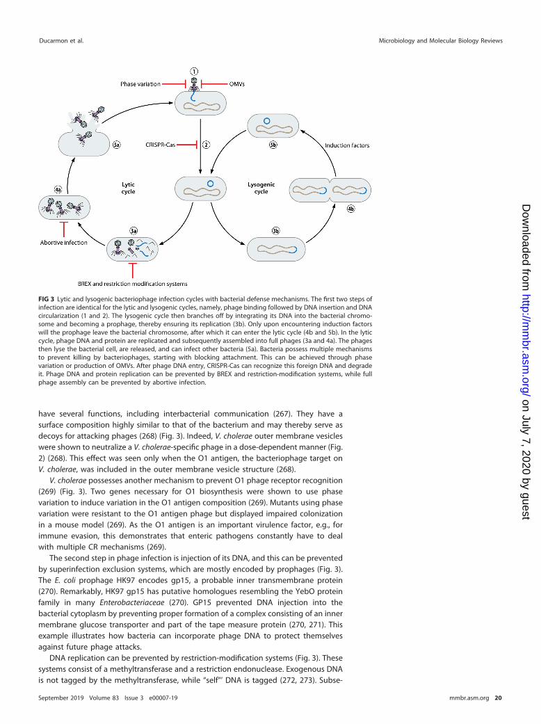
have several functions, including interbacterial communication (267). They have asurface composition highly similar to that of the bacterium and may thereby serve asdecoys for attacking phages (268) (Fig. 3). Indeed, V. cholerae outer membrane vesicleswere shown to neutralize a V. cholerae-specific phage in a dose-dependent manner (Fig.2) (268). This effect was seen only when the O1 antigen, the bacteriophage target onV. cholerae, was included in the outer membrane vesicle structure (268).
V. cholerae possesses another mechanism to prevent O1 phage receptor recognition(269) (Fig. 3). Two genes necessary for O1 biosynthesis were shown to use phasevariation to induce variation in the O1 antigen composition (269). Mutants using phasevariation were resistant to the O1 antigen phage but displayed impaired colonizationin a mouse model (269). As the O1 antigen is an important virulence factor, e.g., forimmune evasion, this demonstrates that enteric pathogens constantly have to dealwith multiple CR mechanisms (269).
The second step in phage infection is injection of its DNA, and this can be preventedby superinfection exclusion systems, which are mostly encoded by prophages (Fig. 3).The E. coli prophage HK97 encodes gp15, a probable inner transmembrane protein(270). Remarkably, HK97 gp15 has putative homologues resembling the YebO proteinfamily in many Enterobacteriaceae (270). GP15 prevented DNA injection into thebacterial cytoplasm by preventing proper formation of a complex consisting of an innermembrane glucose transporter and part of the tape measure protein (270, 271). Thisexample illustrates how bacteria can incorporate phage DNA to protect themselvesagainst future phage attacks.
DNA replication can be prevented by restriction-modification systems (Fig. 3). Thesesystems consist of a methyltransferase and a restriction endonuclease. Exogenous DNAis not tagged by the methyltransferase, while “self”’ DNA is tagged (272, 273). Subse-
FIG 3 Lytic and lysogenic bacteriophage infection cycles with bacterial defense mechanisms. The first two steps ofinfection are identical for the lytic and lysogenic cycles, namely, phage binding followed by DNA insertion and DNAcircularization (1 and 2). The lysogenic cycle then branches off by integrating its DNA into the bacterial chromo-some and becoming a prophage, thereby ensuring its replication (3b). Only upon encountering induction factorswill the prophage leave the bacterial chromosome, after which it can enter the lytic cycle (4b and 5b). In the lyticcycle, phage DNA and protein are replicated and subsequently assembled into full phages (3a and 4a). The phagesthen lyse the bacterial cell, are released, and can infect other bacteria (5a). Bacteria possess multiple mechanismsto prevent killing by bacteriophages, starting with blocking attachment. This can be achieved through phasevariation or production of OMVs. After phage DNA entry, CRISPR-Cas can recognize this foreign DNA and degradeit. Phage DNA and protein replication can be prevented by BREX and restriction-modification systems, while fullphage assembly can be prevented by abortive infection.
Ducarmon et al. Microbiology and Molecular Biology Reviews
September 2019 Volume 83 Issue 3 e00007-19 mmbr.asm.org 20
on July 7, 2020 by guesthttp://m
mbr.asm
.org/D
ownloaded from

quently, nontagged DNA can be cleaved. This system is viewed as a primitive innatebacterial defense system. However, it was found that the system is not perfect, asrestriction-modification systems can also attack self-DNA (274).
Currently, many groups are actively investigating the adaptive bacterial immunesystem CRISPR-Cas, and this has been extensively reviewed previously (275, 276).CRISPR-Cas is present in about 45% of sequenced bacterial genomes, although it isunknown if its prevalence is similar in gut bacteria (277, 278). In short, it consists ofCRISPR arrays (sets of short repetitive DNA elements with variable DNA sequences[spacers] separating the repetitive DNA sets) and an operon of CRISPR-associated genes(Cas). The spacers are pieces of foreign DNA derived from bacteriophage DNA or othermobile genetic elements, such as plasmids. The defense mechanism consists of adap-tation followed by expression and interference. During adaptation, Cas proteins canrecognize foreign phage DNA and integrate a piece of this DNA as a new spacer intothe CRISPR array. This allows the bacterium to build an immunological memory of allthe phages it previously encountered. The expression response entails transcriptionof the CRISPR array, followed by processing into smaller RNA pieces (crRNAs). crRNAsconsist of two outer parts of repeated DNA sequences with a spacer in between. Toform the eventual Cas-crRNA complex, crRNAs are combined with at least one Casprotein. This complex then travels through the bacterial cell, and when it identifies acDNA sequence representative of the previously encountered bacteriophage, it cleavesand degrades the foreign DNA.
In 2015, a novel phage resistance system, called bacteriophage exclusion (BREX),was discovered (279). BREX is able to block DNA replication but does not preventbacteriophage attachment to the bacterium (Fig. 3). It also uses methylation as guid-ance to identify self and exogenous DNA but is different from restriction-modificationsystems, as it does not cleave the exogenous DNA (279). Almost 10% of all bacterialgenomes sequenced were found to have BREX, suggesting that it is a well-conserveddefense mechanism against bacteriophages (279). In spite of this promise as a defensemechanism, no further papers have been published regarding BREX functioning in, e.g.,pathogenic bacteria.
Bacterial cells can perform an apoptosis-like action called abortive infection, result-ing in death of the infected cell and thereby protecting surrounding bacterial cells (280)(Fig. 3). These systems have not been much elucidated for enteric pathogens at amolecular level, though the relevance of the system has been shown for the gutbacteria Shigella dysenteriae and E. coli (281, 282). The abortive infection systems arebest studied in L. lactis, a bacterium widely used in production of fermented foods(283).
CONCLUDING REMARKS
Currently, bacterial enteric infections still cause a heavy disease burden worldwide.For many bacterial pathogens, the virulence factors involved in infection are under-stood, but less is known concerning the failure of the gut microbiota to providecolonization resistance against these enteropathogens. A more comprehensive under-standing of why the microbiota fail to confer sufficient CR could lead to developmentof specific therapies aiming to restore CR. It is likely that no single bacterium will beused as the “holy grail” to restore CR but that a bacterial consortium with complemen-tary functions will be used instead. This would be preferable to the currently often usedFMT, where it is not well known what exact components are transferred to the patient.One could imagine not only that these consortia could be used to treat existinginfections, but that they could also be administered prophylactically in susceptiblepatient groups. In addition, more attention has recently been given to several drugsthat were previously not linked to gut health due to their potentially disturbing effecton the gut microbiota and perhaps CR. In conclusion, we reviewed many of the latestinsights in the rapidly evolving fields of gut microbiota, colonization resistance, andbacterial enteric infection. We are looking forward to the coming years, when more
Gut Microbiota, Colonization Resistance, and Infection Microbiology and Molecular Biology Reviews
September 2019 Volume 83 Issue 3 e00007-19 mmbr.asm.org 21
on July 7, 2020 by guesthttp://m
mbr.asm
.org/D
ownloaded from

knowledge on gut microbiota and CR will undoubtedly be gained, ultimately leading tomore microbiota-based therapies.
ACKNOWLEDGMENTSThis research received no specific grant from any funding agency in the public,
commercial, or not-for-profit sectors. B.V.H.H. and E.J.K. are supported by an unre-stricted grant from Vedanta Biosciences Inc. W.V.S. is supported by a Royal SocietyWolfson Research Merit Award (WM160092). V.B.Y. is supported by grant AI124255 fromthe National Institutes of Health (United States) and is a consultant for VedantaBiosciences Inc.
Q.R.D., R.D.Z., B.V.H.H., and E.J.K. designed the structure and content of the reviewand performed literature research. Q.R.D. wrote the manuscript with guidance fromR.D.Z. and B.V.H.H. We all critically reviewed and revised the manuscript and read andapproved the final manuscript.
REFERENCES1. Sender R, Fuchs S, Milo R. 2016. Revised estimates for the number of
human and bacteria cells in the body. PLoS Biol 14:e1002533. https://doi.org/10.1371/journal.pbio.1002533.
2. Sommer F, Backhed F. 2013. The gut microbiota—masters of hostdevelopment and physiology. Nat Rev Microbiol 11:227–238. https://doi.org/10.1038/nrmicro2974.
3. Round JL, Palm NW. 2018. Causal effects of the microbiota on immune-mediated diseases. Sci Immunol 3:eaao1603. https://doi.org/10.1126/sciimmunol.aao1603.
4. Opazo MC, Ortega-Rocha EM, Coronado-Arrazola I, Bonifaz LC, BoudinH, Neunlist M, Bueno SM, Kalergis AM, Riedel CA. 2018. Intestinalmicrobiota influences non-intestinal related autoimmune diseases.Front Microbiol 9:432. https://doi.org/10.3389/fmicb.2018.00432.
5. Li Q, Gao Z, Wang H, Wu H, Liu Y, Yang Y, Han L, Wang X, Zhao L, TongX. 2018. Intestinal immunomodulatory cells (T lymphocytes): a bridgebetween gut microbiota and diabetes. Mediators Inflamm 2018:9830939. https://doi.org/10.1155/2018/9830939.
6. Sadowsky MJ, Staley C, Heiner C, Hall R, Kelly CR, Brandt L, Horuts KA.2017. Analysis of gut microbiota—an ever changing landscape. GutMicrobes 8:268 –275. https://doi.org/10.1080/19490976.2016.1277313.
7. Eckburg PB, Bik EM, Bernstein CN, Purdom E, Dethlefsen L, Sargent M,Gill SR, Nelson KE, Relman DA. 2005. Diversity of the human intestinalflora. Science 308:1635–1638. https://doi.org/10.1126/science.1110591.
8. Almeida A, Mitchell AL, Boland M, Forster SC, Gloor GB, Tarkowska A,Lawley TD, Finn RD. 2019. A new genomic blueprint of the human gutmicrobiota. Nature 568:499 –504. https://doi.org/10.1038/s41586-019-0965.
9. Turnbaugh PJ, Hamady M, Yatsunenko T, Cantarel BL, Duncan A, Ley RE,Sogin ML, Jones WJ, Roe BA, Affourtit JP, Egholm M, Henrissat B, HeathAC, Knight R, Gordon JI. 2009. A core gut microbiome in obese and leantwins. Nature 457:480 – 484. https://doi.org/10.1038/nature07540.
10. Vollaard EJ, Clasener HA. 1994. Colonization resistance. AntimicrobAgents Chemother 38:409 – 414. https://doi.org/10.1128/AAC.38.3.409.
11. Becattini S, Littmann ER, Carter RA, Kim SG, Morjaria SM, Ling L,Gyaltshen Y, Fontana E, Taur Y, Leiner IM, Pamer EG. 2017. Commensalmicrobes provide first line defense against Listeria monocytogenesinfection. J Exp Med 214:1973–1989. https://doi.org/10.1084/jem.20170495.
12. Martens EC, Neumann M, Desai MS. 2018. Interactions of commensaland pathogenic microorganisms with the intestinal mucosal barrier.Nat Rev Microbiol 16:457– 470. https://doi.org/10.1038/s41579-018-0036-x.
13. Pickard JM, Zeng MY, Caruso R, Nunez G. 2017. Gut microbiota: role inpathogen colonization, immune responses, and inflammatory disease.Immunol Rev 279:70 – 89. https://doi.org/10.1111/imr.12567.
14. Louis P, Flint HJ. 2017. Formation of propionate and butyrate by thehuman colonic microbiota. Environ Microbiol 19:29 – 41. https://doi.org/10.1111/1462-2920.13589.
15. Ríos-Covián D, Ruas-Madiedo P, Margolles A, Gueimonde M, de LosReyes-Gavilán CG, Salazar N. 2016. Intestinal short chain fatty acids andtheir link with diet and human health. Front Microbiol 7:185. https://doi.org/10.3389/fmicb.2016.00185.
16. Litvak Y, Byndloss MX, Baumler AJ. 2018. Colonocyte metabolismshapes the gut microbiota. Science 362:eaat9076. https://doi.org/10.1126/science.aat9076.
17. Cummings JH, Pomare EW, Branch WJ, Naylor CPE, Macfarlane GT.1987. Short chain fatty acids in human large intestine, portal, hepaticand venous blood. Gut 28:1221–1227. https://doi.org/10.1136/gut.28.10.1221.
18. Repaske DR, Adler J. 1981. Change in intracellular pH of Escherichia colimediates the chemotactic response to certain attractants and repel-lents. J Bacteriol 145:1198 –1208.
19. O’Byrne C, Booth IR, Roe AJ, McLaggan D. 2002. Inhibition of Escherichiacoli growth by acetic acid a problem with methione byosynthesis andhomocysteine toxicity. Microbiology 148:2215–2222. https://doi.org/10.1099/00221287-148-7-2215.
20. Hofmann AF. 1999. The continuing importance of bile acids in liver andintestinal disease. Arch Intern Med 159:2647–2658. https://doi.org/10.1001/archinte.159.22.2647.
21. Ridlon JM, Kang DJ, Hylemon PB. 2006. Bile salt biotransformations byhuman intestinal bacteria. J Lipid Res 47:241–259. https://doi.org/10.1194/jlr.R500013-JLR200.
22. Buffie CG, Bucci V, Stein RR, McKenney PT, Ling L, Gobourne A, No D,Liu H, Kinnebrew M, Viale A, Littmann E, van den Brink MR, Jenq RR,Taur Y, Sander C, Cross JR, Toussaint NC, Xavier JB, Pamer EG. 2015.Precision microbiome reconstitution restores bile acid mediated resis-tance to Clostridium difficile. Nature 517:205–208. https://doi.org/10.1038/nature13828.
23. Hirano S, Nakam R, Tamaki M, Masuda N, Oda H. 1981. Isolation andcharacterization of thirteen intestinal microorganisms capable of 7alpha-dehydroxylating bile acids. Appl Environ Microbiol 41:737–745.
24. Kang DJ, Ridlon JM, Moore DR II, Barnes S, Hylemon PB. 2008. Clostrid-ium scindens baiCD and baiH genes encode stereo-specific 7alpha/7beta-hydroxy-3-oxo-delta4-cholenoic acid oxidoreductases. BiochimBiophys Acta 1781:16 –25. https://doi.org/10.1016/j.bbalip.2007.10.008.
25. Sannasiddappa TH, Lund PA, Clarke SR. 2017. In vitro antibacterialactivity of unconjugated and conjugated bile salts on Staphylococcusaureus. Front Microbiol 8:1581. https://doi.org/10.3389/fmicb.2017.01581.
26. Kurdi P, Kawanishi K, Mizutani K, Yokota A. 2006. Mechanism of growthinhibition by free bile acids in lactobacilli and bifidobacteria. J Bacteriol188:1979 –1986. https://doi.org/10.1128/JB.188.5.1979-1986.2006.
27. Watanabe M, Fukiya S, Yokota A. 2017. Comprehensive evaluation ofthe bactericidal activities of free bile acids in the large intestine ofhumans and rodents. J Lipid Res 58:1143–1152. https://doi.org/10.1194/jlr.M075143.
28. Thanissery R, Winston JA, Theriot CM. 2017. Inhibition of spore germi-nation, growth, and toxin activity of clinically relevant C. difficile strainsby gut microbiota derived secondary bile acids. Anaerobe 45:86 –100.https://doi.org/10.1016/j.anaerobe.2017.03.004.
29. Sayin SI, Wahlstrom A, Felin J, Jantti S, Marschall HU, Bamberg K,Angelin B, Hyotylainen T, Oresic M, Backhed F. 2013. Gut microbiotaregulates bile acid metabolism by reducing the levels of tauro-beta-
Ducarmon et al. Microbiology and Molecular Biology Reviews
September 2019 Volume 83 Issue 3 e00007-19 mmbr.asm.org 22
on July 7, 2020 by guesthttp://m
mbr.asm
.org/D
ownloaded from

muricholic acid, a naturally occurring FXR antagonist. Cell Metab 17:225–235. https://doi.org/10.1016/j.cmet.2013.01.003.
30. Wells JE, Berr F, Thomas LA, Dowling RH, Hylemon PB. 2000. Isolationand characterization of cholic acid 7�-dehydroxylating fecal bacteriafrom cholesterol gallstone patients. J Hepatol 32:4 –10. https://doi.org/10.1016/S0168-8278(00)80183-X.
31. Sorg JA, Sonenshein AL. 2010. Inhibiting the initiation of Clostridiumdifficile spore germination using analogs of chenodeoxycholic acid, abile acid. J Bacteriol 192:4983– 4990. https://doi.org/10.1128/JB.00610-10.
32. Studer N, Desharnais L, Beutler M, Brugiroux S, Terrazos MA, Menin L,Schurch CM, McCoy KD, Kuehne SA, Minton NP, Stecher B, Bernier-Latmani R, Hapfelmeier S. 2016. Functional intestinal bile acid 7�-dehydroxylation by Clostridium scindens associated with protectionfrom Clostridium difficile infection in a gnotobiotic mouse model. FrontCell Infect Microbiol 6:191. https://doi.org/10.3389/fcimb.2016.00191.
33. Amrane S, Bachar D, Lagier JC, Raoult D. 2018. Clostridium scindens ispresent in the gut microbiota during Clostridium difficile infection: ametagenomic and culturomic analysis. J Clin Microbiol 56:e01663-17.https://doi.org/10.1128/JCM.01663-17.
34. Termen S, Tollin M, Rodriguez E, Sveinsdottir SH, Johannesson B,Cederlund A, Sjovall J, Agerberth B, Gudmundsson GH. 2008. PU.1 andbacterial metabolites regulate the human gene CAMP encoding anti-microbial peptide LL-37 in colon epithelial cells. Mol Immunol 45:3947–3955. https://doi.org/10.1016/j.molimm.2008.06.020.
35. Peric M, Koglin S, Dombrowski Y, Gross K, Bradac E, Ruzicka T, SchauberJ. 2009. VDR and MEK-ERK dependent induction of the antimicrobialpeptide cathelicidin in keratinocytes by lithocholic acid. Mol Immunol46:3183–3187. https://doi.org/10.1016/j.molimm.2009.08.010.
36. Rea MC, Sit CS, Clayton E, O’Connor PM, Whittal RM, Zheng J, VederasJC, Ross RP, Hill C. 2010. Thuricin CD, a posttranslationally modifiedbacteriocin with a narrow spectrum of activity against Clostridiumdifficile. Proc Natl Acad Sci U S A 107:9352–9357. https://doi.org/10.1073/pnas.0913554107.
37. Parks WM, Bottrill AR, Pierrat OA, Durrant MC, Maxwell A. 2007. Theaction of the bacterial toxin, microcin B17, on DNA gyrase. Biochimie89:500 –507. https://doi.org/10.1016/j.biochi.2006.12.005.
38. Destoumieux-Garzon D, Peduzzi J, Thomas X, Djediat C, Rebuffat S.2006. Parasitism of iron-siderophore receptors of Escherichia coli by thesiderophore-peptide microcin E492m and its unmodified counterpart.Biometals 19:181–191. https://doi.org/10.1007/s10534-005-4452-9.
39. Mukhopadhyay J, Sineva E, Knight J, Levy RM, Ebright RH. 2004. Anti-bacterial peptide microcin J25 inhibits transcription by binding withinand obstructing the RNA polymerase secondary channel. Mol Cell14:739 –751. https://doi.org/10.1016/j.molcel.2004.06.010.
40. Cotter PD, Ross RP, Hill C. 2013. Bacteriocins—a viable alternative toantibiotics? Nat Rev Microbiol 11:95–105. https://doi.org/10.1038/nrmicro2937.
41. Rebuffat S. 2011. Bacteriocins from Gram-negative bacteria: a classifi-cation?, p 55–72. In Drider D, Rebuffat S (ed), Prokaryotic antimicrobialpeptides: from genes to applications. Springer, New York, NY.
42. Rea MC, Ross RP, Cotter PD, Hill C. 2011. Classification of bacteriocinsfrom Gram-positive bacteria, p 29 –53. In Drider D, Rebuffat S (ed),Prokaryotic antimicrobial peptides: from genes to applications.Springer, New York, New York, NY.
43. Severina E, Severin A, Tomasz A. 1998. Antibacterial efficacy of nisinagainst multidrug-resistant Gram-positive pathogens. J AntimicrobChemother 41:341–347. https://doi.org/10.1093/jac/41.3.341.
44. Le Lay C, Dridi L, Bergeron MG, Ouellette M, Fliss IL. 2016. Nisin is aneffective inhibitor of Clostridium difficile vegetative cells and sporegermination. J Med Microbiol 65:169 –175. https://doi.org/10.1099/jmm.0.000202.
45. Benkerroum N, Sandine WE. 1988. Inhibitory action of nisin againstListeria monocytogenes. J Dairy Sci 71:3237–3245. https://doi.org/10.3168/jds.S0022-0302(88)79929-4.
46. Stevens KA, Sheldon BW, Klapes NA, Klaenhammer TR. 1991. Nisintreatment for inactivation of Salmonella species and other gram-negative bacteria. Appl Environ Microbiol 57:3613–3615.
47. Prudencio CV, Mantovani HC, Cecon PR, Vanetti MC. 2015. Differencesin the antibacterial activity of nisin and bovicin HC5 against SalmonellaTyphimurium under different temperature and pH conditions. J ApplMicrobiol 118:18 –26. https://doi.org/10.1111/jam.12680.
48. Singh AP, Prabha V, Rishi P. 2013. Value addition in the efficacy of
conventional antibiotics by nisin against Salmonella. PLoS One8:e76844. https://doi.org/10.1371/journal.pone.0076844.
49. Singh AP, Preet S, Rishi P. 2014. Nisin/beta–lactam adjunct therapyagainst Salmonella enterica serovar Typhimurium: a mechanistic ap-proach. J Antimicrob Chemother 69:1877–1887. https://doi.org/10.1093/jac/dku049.
50. Li Q, Montalban-Lopez M, Kuipers OP. 2018. Increasing the antimicro-bial activity of nisin-based lantibiotics against gram-negative patho-gens. Appl Environ Microbiol 84:e00052-18. https://doi.org/10.1128/AEM.00052-18.
51. Ritchie JM, Greenwich JL, Davis BM, Bronson RT, Gebhart D, Williams SR,Martin D, Scholl D, Waldor MK. 2011. An Escherichia coli O157-specificengineered pyocin prevents and ameliorates infection by E. coliO157:H7 in an animal model of diarrheal disease. Antimicrob AgentsChemother 55:5469 –5474. https://doi.org/10.1128/AAC.05031-11.
52. Gillor O, Nigro LM, Riley MA. 2005. Genetically engineered bacteriocinsand their potential as the next generation of antimicrobials. Curr PharmDes 11:1067–1075. https://doi.org/10.2174/1381612053381666.
53. Hasper HE, Kramer NE, Smith JL, Hillman JD, Zachariah C, Kuipers OP, deKruijff B, Breukink E. 2006. An alternative bactericidal mechanism ofaction for lantibiotic peptides that target lipid II. Science 313:1636 –1637. https://doi.org/10.1126/science.1129818.
54. Wiedemann I, Breukink E, van Kraaij C, Kuipers OP, Bierbaum G, deKruijff B, Sahl HG. 2001. Specific binding of nisin to the peptidoglycanprecursor lipid II combines pore formation and inhibition of cell wallbiosynthesis for potent antibiotic activity. J Biol Chem 276:1772–1779.https://doi.org/10.1074/jbc.M006770200.
55. Corr SC, Li Y, Riedel CU, O’Toole PW, Hill C, Gahan CGM. 2007. Bacte-riocin production as a mechanism for the antiinfective activity ofLactobacillus salivarius UCC118. Proc Natl Acad Sci U S A 104:7617–7621. https://doi.org/10.1073/pnas.0700440104.
56. Rea MC, Dobson A, O’Sullivan O, Crispie F, Fouhy F, Cotter PD, Shana-han F, Kiely B, Hill C, Ross RP. 2011. Effect of broad- and narrow-spectrum antimicrobials on Clostridium difficile and microbial diversityin a model of the distal colon. Proc Natl Acad Sci U S A 108:4639 – 4644.https://doi.org/10.1073/pnas.1001224107.
57. Rea MC, Alemayehu D, Casey PG, O’Connor PM, Lawlor PG, Walsh M,Shanahan F, Kiely B, Ross RP, Hill C. 2014. Bioavailability of the anti-clostridial bacteriocin thuricin CD in gastrointestinal tract. Microbiology160:439 – 445. https://doi.org/10.1099/mic.0.068767-0.
58. Desai MS, Seekatz AM, Koropatkin NM, Kamada N, Hickey CA, Wolter M,Pudlo NA, Kitamoto S, Terrapon N, Muller A, Young VB, Henrissat B,Wilmes P, Stappenbeck TS, Nunez G, Martens EC. 2016. A dietaryfiber-deprived gut microbiota degrades the colonic mucus barrier andenhances pathogen susceptibility. Cell 167:1339 –1353.e21. https://doi.org/10.1016/j.cell.2016.10.043.
59. Bosák J, Micenková L, Hrala M, Pomorská K, Kunova Bosakova M, KrejciP, Göpfert E, Faldyna M, Šmajs D. 2018. Colicin FY inhibits pathogenicYersinia enterocolitica in mice. Sci Rep 8:12242. https://doi.org/10.1038/s41598-018-30729-7.
60. Gillor O, Kirkup BC, Riley MA. 2004. Colicins and microcins: the nextgeneration antimicrobials. Adv Appl Microbiol 54:129 –146. https://doi.org/10.1016/S0065-2164(04)54005-4.
61. Sassone-Corsi M, Nuccio SP, Liu H, Hernandez D, Vu CT, Takahashi AA,Edwards RA, Raffatellu M. 2016. Microcins mediate competition amongEnterobacteriaceae in the inflamed gut. Nature 540:280 –283. https://doi.org/10.1038/nature20557.
62. Rebuffat S. 2012. Microcins in action: amazing defence strategies ofEnterobacteria. Biochem Soc Trans 40:1456 –1462. https://doi.org/10.1042/BST20120183.
63. Walsh CJ, Guinane CM, Hill C, Ross RP, O’Toole PW, Cotter PD. 2015. Insilico identification of bacteriocin gene clusters in the gastrointestinaltract, based on the Human Microbiome Project’s reference genomedatabase. BMC Microbiol 15:183. https://doi.org/10.1186/s12866-015-0515-4.
64. Momose Y, Hirayama K, Itoh K. 2008. Competition for proline betweenindigenous Escherichia coli and E. coli O157:H7 in gnotobiotic miceassociated with infant intestinal microbiota and its contribution to thecolonization resistance against E. coli O157:H7. Antonie Van Leeuwen-hoek 94:165–171. https://doi.org/10.1007/s10482-008-9222-6.
65. Fabich AJ, Jones SA, Chowdhury FZ, Cernosek A, Anderson A, SmalleyD, McHargue JW, Hightower GA, Smith JT, Autieri SM, Leatham MP, LinsJJ, Allen RL, Laux DC, Cohen PS, Conway T. 2008. Comparison of carbonnutrition for pathogenic and commensal Escherichia coli strains in the
Gut Microbiota, Colonization Resistance, and Infection Microbiology and Molecular Biology Reviews
September 2019 Volume 83 Issue 3 e00007-19 mmbr.asm.org 23
on July 7, 2020 by guesthttp://m
mbr.asm
.org/D
ownloaded from

mouse intestine. Infect Immun 76:1143–1152. https://doi.org/10.1128/IAI.01386-07.
66. Maltby R, Leatham-Jensen MP, Gibson T, Cohen PS, Conway T. 2013.Nutritional basis for colonization resistance by human commensalEscherichia coli strains HS and Nissle 1917 against E. coli O157:H7 in themouse intestine. PLoS One 8:e53957. https://doi.org/10.1371/journal.pone.0053957.
67. Deriu E, Liu JZ, Pezeshki M, Edwards RA, Ochoa RJ, Contreras H, LibbySJ, Fang FC, Raffatellu M. 2013. Probiotic bacteria reduce SalmonellaTyphimurium intestinal colonization by competing for iron. Cell HostMicrobe 14:26 –37. https://doi.org/10.1016/j.chom.2013.06.007.
68. Borenstein E, Kupiec M, Feldman MW, Ruppin E. 2008. Large-scalereconstruction and phylogenetic analysis of metabolic environments.Proc Natl Acad Sci U S A 105:14482–14487. https://doi.org/10.1073/pnas.0806162105.
69. Peterson LW, Artis D. 2014. Intestinal epithelial cells: regulators ofbarrier function and immune homeostasis. Nat Rev Immunol 14:141–153. https://doi.org/10.1038/nri3608.
70. Kashyap PC, Marcobal A, Ursell LK, Smits SA, Sonnenburg ED, CostelloEK, Higginbottom SK, Domino SE, Holmes SP, Relman DA, Knight R,Gordon JI, Sonnenburg JL. 2013. Genetically dictated change in hostmucus carbohydrate landscape exerts a diet-dependent effect on thegut microbiota. Proc Natl Acad Sci U S A 110:17059 –17064. https://doi.org/10.1073/pnas.1306070110.
71. Johansson ME, Larsson JM, Hansson GC. 2011. The two mucus layers ofcolon are organized by the MUC2 mucin, whereas the outer layer is alegislator of host-microbial interactions. Proc Natl Acad Sci U S A108:4659 – 4665. https://doi.org/10.1073/pnas.1006451107.
72. Atuma C, Strugala V, Allen A, Holm L. 2001. The adherent gastrointes-tinal mucus gel layer thickness and physical state in vivo. Am J PhysiolGastrointest Liver Physiol 280:G922–G929. https://doi.org/10.1152/ajpgi.2001.280.5.G922.
73. Schroeder BO, Birchenough GMH, Stahlman M, Arike L, Johansson MEV,Hansson GC, Backhed F. 2018. Bifidobacteria or fiber protects againstdiet-induced microbiota-mediated colonic mucus deterioration. CellHost Microbe 23:27– 40.e7. https://doi.org/10.1016/j.chom.2017.11.004.
74. Jakobsson HE, Rodriguez-Pineiro AM, Schutte A, Ermund A, Boysen P,Bemark M, Sommer F, Backhed F, Hansson GC, Johansson ME. 2015.The composition of the gut microbiota shapes the colon mucus barrier.EMBO Rep 16:164 –177. https://doi.org/10.15252/embr.201439263.
75. Keen EC. 2015. A century of phage research: bacteriophages and theshaping of modern biology. Bioessays 37:6 –9. https://doi.org/10.1002/bies.201400152.
76. Mirzaei MK, Maurice CF. 2017. Ménage à trois in the human gut:interactions between host, bacteria and phages. Nat Rev Microbiol15:397– 408. https://doi.org/10.1038/nrmicro.2017.30.
77. Wittebole X, De Roock S, Opal SM. 2014. A historical overview ofbacteriophage therapy as an alternative to antibiotics for the treatmentof bacterial pathogens. Virulence 5:226 –235. https://doi.org/10.4161/viru.25991.
78. Yen M, Cairns LS, Camilli A. 2017. A cocktail of three virulent bacterio-phages prevents Vibrio cholerae infection in animal models. Nat Com-mun 8:14187. https://doi.org/10.1038/ncomms14187.
79. Fischer S, Kittler S, Klein G, Glunder G. 2013. Impact of a single phageand a phage cocktail application in broilers on reduction of Campylo-bacter jejuni and development of resistance. PLoS One 8:e78543.https://doi.org/10.1371/journal.pone.0078543.
80. Duerkop BA, Clements CV, Rollins D, Rodrigues JL, Hooper LV. 2012. Acomposite bacteriophage alters colonization by an intestinal commen-sal bacterium. Proc Natl Acad Sci U S A 109:17621–17626. https://doi.org/10.1073/pnas.1206136109.
81. Henein A. 2013. What are the limitations on the wider therapeuticuse of phage? Bacteriophage 3:e24872. https://doi.org/10.4161/bact.24872.
82. Schooley RT, Biswas B, Gill JJ, Hernandez-Morales A, Lancaster J, LessorL, Barr JJ, Reed SL, Rohwer F, Benler S, Segall AM, Taplitz R, Smith DM,Kerr K, Kumaraswamy M, Nizet V, Lin L, McCauley MD, Strathdee SA,Benson CA, Pope RK, Leroux BM, Picel AC, Mateczun AJ, Cilwa KE,Regeimbal JM, Estrella LA, Wolfe DM, Henry MS, Quinones J, Salka S,Bishop-Lilly KA, Young R, Hamilton T. 2017. Development and use ofpersonalized bacteriophage-based therapeutic cocktails to treat a pa-tient with a disseminated resistant Acinetobacter baumannii infection.Antimicrob Agents Chemother 61:e00954-17. https://doi.org/10.1128/AAC.00954-17.
83. Chan BK, Turner PE, Kim S, Mojibian HR, Elefteriades JA, Narayan D.2018. Phage treatment of an aortic graft infected with Pseudomonasaeruginosa. Evol Med Public Health 2018:60 – 66. https://doi.org/10.1093/emph/eoy005.
84. Keeney KM, Yurist-Doutsch S, Arrieta MC, Finlay BB. 2014. Effects ofantibiotics on human microbiota and subsequent disease. Annu RevMicrobiol 68:217–235. https://doi.org/10.1146/annurev-micro-091313-103456.
85. Becattini S, Taur Y, Pamer EG. 2016. Antibiotic-induced changes in theintestinal microbiota and disease. Trends Mol Med 22:458 – 478. https://doi.org/10.1016/j.molmed.2016.04.003.
86. Forgacs I, Loganayagam A. 2008. Overprescribing proton pump inhib-itors. BMJ 336:2–3. https://doi.org/10.1136/bmj.39406.449456.BE.
87. Leonard J, Marshall JK, Moayyedi P. 2007. Systematic review of therisk of enteric infection in patients taking acid suppression. Am JGastroenterol 102:2047–2056. https://doi.org/10.1111/j.1572-0241.2007.01275.x.
88. Wei L, Ratnayake L, Phillips G, McGuigan CC, Morant SV, Flynn RW,Mackenzie IS, MacDonald TM. 2017. Acid-suppression medications andbacterial gastroenteritis: a population-based cohort study. Br J ClinPharmacol 83:1298 –1308. https://doi.org/10.1111/bcp.13205.
89. Hafiz RA, Wong C, Paynter S, David M, Peeters G. 2018. The risk ofcommunity-acquired enteric infection in proton pump inhibitortherapy: systematic review and meta-analysis. Ann Pharmacother 52:613– 622. https://doi.org/10.1177/1060028018760569.
90. Bavishi C, Dupont HL. 2011. Systematic review: the use of protonpump inhibitors and increased susceptibility to enteric infection.Aliment Pharmacol Ther 34:1269 –1281. https://doi.org/10.1111/j.1365-2036.2011.04874.x.
91. Freedberg DE, Toussaint NC, Chen SP, Ratner AJ, Whittier S, Wang TC,Wang HH, Abrams JA. 2015. Proton pump inhibitors alter specific taxain the human gastrointestinal microbiome: a crossover trial. Gastroen-terology 149:883– 885.e9. https://doi.org/10.1053/j.gastro.2015.06.043.
92. Imhann F, Bonder MJ, Vich Vila A, Fu J, Mujagic Z, Vork L, Tigchelaar EF,Jankipersadsing SA, Cenit MC, Harmsen HJ, Dijkstra G, Franke L, XavierRJ, Jonkers D, Wijmenga C, Weersma RK, Zhernakova A. 2016. Protonpump inhibitors affect the gut microbiome. Gut 65:740 –748. https://doi.org/10.1136/gutjnl-2015-310376.
93. Jackson MA, Goodrich JK, Maxan M-E, Freedberg DE, Abrams JA, PooleAC, Sutter JL, Welter D, Ley RE, Bell JT, Spector TD, Steves CJ. 2016.Proton pump inhibitors alter the composition of the gut microbiota.Gut 65:749 –756. https://doi.org/10.1136/gutjnl-2015-310861.
94. Takagi T, Naito Y, Inoue R, Kashiwagi S, Uchiyama K, Mizushima K,Tsuchiya S, Okayama T, Dohi O, Yoshida N, Kamada K, Ishikawa T,Handa O, Konishi H, Okuda K, Tsujimoto Y, Ohnogi H, Itoh Y. 2018. Theinfluence of long-term use of proton pump inhibitors on the gutmicrobiota: an age-sex-matched case-control study. J Clin BiochemNutr 62:100 –105. https://doi.org/10.3164/jcbn.17-78.
95. Sieczkowska A, Landowski P, Gibas A, Kaminska B, Lifschitz C. 2018.Long-term proton pump inhibitor therapy leads to small bowel bacte-rial overgrowth as determined by breath hydrogen and methane ex-cretion. J Breath Res 12:036006. https://doi.org/10.1088/1752-7163/aa9dcf.
96. Yasutomi E, Hoshi N, Adachi S, Otsuka T, Kong L, Ku Y, Yamairi H, InoueJ, Ishida T, Watanabe D, Ooi M, Yoshida M, Tsukimi T, Fukuda S, AzumaT. 2018. Proton pump inhibitors increase the susceptibility of mice tooral infection with enteropathogenic bacteria. Dig Dis Sci 63:881– 889.https://doi.org/10.1007/s10620-017-4905-3.
97. Stiefel U, Rao A, Pultz MJ, Jump RL, Aron DC, Donskey CJ. 2006.Suppression of gastric acid production by proton pump inhibitor treat-ment facilitates colonization of the large intestine by vancomycin-resistant Enterococcus spp. and Klebsiella pneumoniae in clindamycin-treated mice. Antimicrob Agents Chemother 50:3905–3907. https://doi.org/10.1128/AAC.00522-06.
98. Imhann F, Vich Vila A, Bonder MJ, Lopez Manosalva AG, Koonen DPY,Fu J, Wijmenga C, Zhernakova A, Weersma RK. 2017. The influence ofproton pump inhibitors and other commonly used medication on thegut microbiota. Gut Microbes 8:351–358. https://doi.org/10.1080/19490976.2017.1284732.
99. Foretz M, Guigas B, Bertrand L, Pollak M, Viollet B. 2014. Metformin:from mechanisms of action to therapies. Cell Metab 20:953–966.https://doi.org/10.1016/j.cmet.2014.09.018.
100. Smyth S, Heron A. 2006. Diabetes and obesity: the twin epidemics. NatMed 12:75– 80. https://doi.org/10.1038/nm0106-75.
Ducarmon et al. Microbiology and Molecular Biology Reviews
September 2019 Volume 83 Issue 3 e00007-19 mmbr.asm.org 24
on July 7, 2020 by guesthttp://m
mbr.asm
.org/D
ownloaded from

101. Forslund K, Hildebrand F, Nielsen T, Falony G, Le Chatelier E, SunagawaS, Prifti E, Vieira-Silva S, Gudmundsdottir V, Pedersen HK, Arumugam M,Kristiansen K, Voigt AY, Vestergaard H, Hercog R, Costea PI, Kultima JR,Li J, Jorgensen T, Levenez F, Dore J, Meta HITC, Nielsen HB, Brunak S,Raes J, Hansen T, Wang J, Ehrlich SD, Bork P, Pedersen O. 2015.Disentangling type 2 diabetes and metformin treatment signatures inthe human gut microbiota. Nature 528:262–266. https://doi.org/10.1038/nature15766.
102. Qin J, Li Y, Cai Z, Li S, Zhu J, Zhang F, Liang S, Zhang W, Guan Y, ShenD, Peng Y, Zhang D, Jie Z, Wu W, Qin Y, Xue W, Li J, Han L, Lu D, WuP, Dai Y, Sun X, Li Z, Tang A, Zhong S, Li X, Chen W, Xu R, Wang M, FengQ, Gong M, Yu J, Zhang Y, Zhang M, Hansen T, Sanchez G, Raes J,Falony G, Okuda S, Almeida M, LeChatelier E, Renault P, Pons N, BattoJ-M, Zhang Z, Chen H, Yang R, Zheng W, Li S, Yang H, Wang J, EhrlichSD, Nielsen R, Pedersen O, Kristiansen K, Wang J. 2012. A metagenome-wide association study of gut microbiota in type 2 diabetes. Nature490:55– 60. https://doi.org/10.1038/nature11450.
103. Wu H, Esteve E, Tremaroli V, Khan MT, Caesar R, Mannerås-Holm L,Ståhlman M, Olsson LM, Serino M, Planas-Fèlix M, Xifra G, Mercader JM,Torrents D, Burcelin R, Ricart W, Perkins R, Fernàndez-Real JM, BäckhedF. 2017. Metformin alters the gut microbiome of individuals withtreatment-naive type 2 diabetes, contributing to the therapeutic ef-fects of the drug. Nat Med 23:850 – 858. https://doi.org/10.1038/nm.4345.
104. Zhou HY, Zhu H, Yao XM, Qian JP, Yang J, Pan XD, Chen XD. 2017.Metformin regulates tight junction of intestinal epithelial cells viaMLCK-MLC. Eur Rev Med Pharmacol Sci 21:5239 –5246. https://doi.org/10.26355/eurrev_201711_13847.
105. Deng J, Zeng L, Lai X, Li J, Liu L, Lin Q, Chen Y. 2018. Metformin protectsagainst intestinal barrier dysfunction via AMPKalpha1-dependent inhi-bition of JNK signalling activation. J Cell Mol Med 22:546 –557. https://doi.org/10.1111/jcmm.13342.
106. Xue Y, Zhang H, Sun X, Zhu MJ. 2016. Metformin improves ilealepithelial barrier function in interleukin-10 deficient mice. PLoS One11:e0168670. https://doi.org/10.1371/journal.pone.0168670.
107. Maier L, Pruteanu M, Kuhn M, Zeller G, Telzerow A, Anderson EE,Brochado AR, Fernandez KC, Dose H, Mori H, Patil KR, Bork P, Typas A.2018. Extensive impact of non-antibiotic drugs on human gut bacteria.Nature 555:623– 628. https://doi.org/10.1038/nature25979.
108. Morgan AP, Crowley JJ, Nonneman RJ, Quackenbush CR, Miller CN,Ryan AK, Bogue MA, Paredes SH, Yourstone S, Carroll IM, Kawula TH,Bower MA, Sartor RB, Sullivan PF. 2014. The antipsychotic olanzapineinteracts with the gut microbiome to cause weight gain in mouse. PLoSOne 9:e115225. https://doi.org/10.1371/journal.pone.0115225.
109. Lieberman LA, Higgins DE. 2009. A small–molecule screen identifies theantipsychotic drug pimozide as an inhibitor of Listeria monocytogenesinfection. Antimicrob Agents Chemother 53:756 –764. https://doi.org/10.1128/AAC.00607-08.
110. Dastidar SG, Chaudhury A, Annadurai S, Roy S, Mookerjee M,Chakrabarty AN. 1995. In vitro and in vivo antimicrobial action offluphenazine. J Chemother 7:201–206. https://doi.org/10.1179/joc.1995.7.3.201.
111. Jeyaseeli L, Dasgupta A, Dastidar SG, Molnar J, Amaral L. 2012. Evidenceof significant synergism between antibiotics and the antipsychotic,antimicrobial drug flupenthixol. Eur J Clin Microbiol Infect Dis 31:1243–1250. https://doi.org/10.1007/s10096-011-1435-3.
112. Cussotto S, Strain CR, Fouhy F, Strain RG, Peterson VL, Clarke G, StantonC, Dinan TG, Cryan JF. 2018. Differential effects of psychotropic drugson microbiome composition and gastrointestinal function. Psychophar-macology 235:1–15. https://doi.org/10.1007/s00213-018-5006-5.
113. Rogers MA, Greene MT, Young VB, Saint S, Langa KM, Kao JY, AronoffDM. 2013. Depression, antidepressant medications, and risk of Clostrid-ium difficile infection. BMC Med 11:121. https://doi.org/10.1186/1741-7015-11-121.
114. Munita JM, Arias CA. 2016. Mechanisms of antibiotic resistance. Micro-biol Spectr 4:VMBF-0016-2015. https://doi.org/10.1128/microbiolspec.VMBF-0016-2015.
115. Davies J, Davies D. 2010. Origins and evolution of antibiotic resistance.Microbiol Mol Biol Rev 74:417– 433. https://doi.org/10.1128/MMBR.00016-10.
116. Lessa FC, Mu Y, Bamberg WM, Beldavs ZG, Dumyati GK, Dunn JR, FarleyMM, Holzbauer SM, Meek JI, Phipps EC, Wilson LE, Winston LG, CohenJA, Limbago BM, Fridkin SK, Gerding DN, McDonald LC. 2015. Burden of
Clostridium difficile infection in the United States. N Engl J Med 372:825– 834. https://doi.org/10.1056/NEJMoa1408913.
117. Smits WK, Lyras D, Lacy DB, Wilcox MH, Kuijper EJ. 2016. Clostridiumdifficile infection. Nat Rev Dis Primers 2:16020. https://doi.org/10.1038/nrdp.2016.20.
118. Crobach MJT, Vernon JJ, Loo VG, Kong LY, Pechine S, Wilcox MH,Kuijper EJ. 2018. Understanding Clostridium difficile colonization. ClinMicrobiol Rev 31:e00021-17. https://doi.org/10.1128/CMR.00021-17.
119. Keller JJ, Kuijper EJ. 2015. Treatment of recurrent and severe Clostrid-ium difficile infection. Annu Rev Med 66:373–386. https://doi.org/10.1146/annurev-med-070813-114317.
120. Theriot CM, Koenigsknecht MJ, Carlson PE, Jr, Hatton GE, Nelson AM, LiB, Huffnagle GB, Z Li J, Young VB. 2014. Antibiotic-induced shifts in themouse gut microbiome and metabolome increase susceptibility toClostridium difficile infection. Nat Commun 5:3114. https://doi.org/10.1038/ncomms4114.
121. Lawley TD, Clare S, Walker AW, Stares MD, Connor TR, Raisen C,Goulding D, Rad R, Schreiber F, Brandt C, Deakin LJ, Pickard DJ, DuncanSH, Flint HJ, Clark TG, Parkhill J, Dougan G. 2012. Targeted restorationof the intestinal microbiota with a simple, defined bacteriotherapyresolves relapsing Clostridium difficile disease in mice. PLoS Pathog8:e1002995. https://doi.org/10.1371/journal.ppat.1002995.
122. Seekatz AM, Theriot CM, Rao K, Chang YM, Freeman AE, Kao JY, YoungVB. 2018. Restoration of short chain fatty acid and bile acid metabolismfollowing fecal microbiota transplantation in patients with recurrentClostridium difficile infection. Anaerobe 53:64 –73. https://doi.org/10.1016/j.anaerobe.2018.04.001.
123. Sorg JA, Sonenshein AL. 2008. Bile salts and glycine as cogerminants forClostridium difficile spores. J Bacteriol 190:2505–2512. https://doi.org/10.1128/JB.01765-07.
124. Sorg JA, Sonenshein AL. 2009. Chenodeoxycholate is an inhibitor ofClostridium difficile spore germination. J Bacteriol 191:1115–1117.https://doi.org/10.1128/JB.01260-08.
125. Wilson KH. 1983. Efficiency of various bile salt preparations for stimu-lation of Clostridium difficile spore germination. J Clin Microbiol 18:1017–1019.
126. Lewis BB, Carter RA, Pamer EG. 2016. Bile acid sensitivity and in vivovirulence of clinical Clostridium difficile isolates. Anaerobe 41:32–36.https://doi.org/10.1016/j.anaerobe.2016.05.010.
127. McBride SM, Sonenshein AL. 2011. Identification of a genetic locusresponsible for antimicrobial peptide resistance in Clostridium difficile.Infect Immun 79:167–176. https://doi.org/10.1128/IAI.00731-10.
128. McBride SM, Sonenshein AL. 2011. The dlt operon confers resistance tocationic antimicrobial peptides in Clostridium difficile. Microbiology157:1457–1465. https://doi.org/10.1099/mic.0.045997-0.
129. Jenior ML, Leslie JL, Young VB, Schloss PD. 2017. Clostridium difficilecolonizes alternative nutrient niches during infection across distinctmurine gut microbiomes. mSystems 2:e00063-17. https://doi.org/10.1128/mSystems.00063-17.
130. Jenior ML, Leslie JL, Young VB, Schloss PD. 2018. Clostridium difficilealters the structure and metabolism of distinct cecal microbiomesduring initial infection to promote sustained colonization. mSphere3:e00261-18. https://doi.org/10.1128/mSphere.00261-18.
131. Collins J, Robinson C, Danhof H, Knetsch CW, van Leeuwen HC, LawleyTD, Auchtung JM, Britton RA. 2018. Dietary trehalose enhances viru-lence of epidemic Clostridium difficile. Nature 553:291–294. https://doi.org/10.1038/nature25178.
132. Collins J, Danhof H, Britton RA. 2018. The role of trehalose in the globalspread of epidemic Clostridium difficile. Gut Microbes :1– 6. https://doi.org/10.1080/19490976.2018.1491266.
133. Ng KM, Ferreyra JA, Higginbottom SK, Lynch JB, Kashyap PC, GopinathS, Naidu N, Choudhury B, Weimer BC, Monack DM, Sonnenburg JL.2013. Microbiota–liberated host sugars facilitate post-antibiotic expan-sion of enteric pathogens. Nature 502:96 –99. https://doi.org/10.1038/nature12503.
134. Ferreyra JA, Wu KJ, Hryckowian AJ, Bouley DM, Weimer BC, SonnenburgJL. 2014. Gut microbiota-produced succinate promotes C. difficile in-fection after antibiotic treatment or motility disturbance. Cell HostMicrobe 16:770 –777. https://doi.org/10.1016/j.chom.2014.11.003.
135. Zackular JP, Moore JL, Jordan AT, Juttukonda LJ, Noto MJ, NicholsonMR, Crews JD, Semler MW, Zhang Y, Ware LB, Washington MK, ChazinWJ, Caprioli RM, Skaar EP. 2016. Dietary zinc alters the microbiota anddecreases resistance to Clostridium difficile infection. Nat Med 22:1330 –1334. https://doi.org/10.1038/nm.4174.
Gut Microbiota, Colonization Resistance, and Infection Microbiology and Molecular Biology Reviews
September 2019 Volume 83 Issue 3 e00007-19 mmbr.asm.org 25
on July 7, 2020 by guesthttp://m
mbr.asm
.org/D
ownloaded from

136. Tasteyre A, Barc MC, Collignon A, Boureau H, Karjalainen T. 2001. Roleof FliC and FliD flagellar proteins of Clostridium difficile in adherenceand gut colonization. Infect Immun 69:7937–7940. https://doi.org/10.1128/IAI.69.12.7937-7940.2001.
137. McKee RW, Aleksanyan N, Garret EM, Tamayo R. 2018. Type IV pilipromote Clostridium difficile adherence and persistence in a mousemodel of infection. Infect Immun 86:e00943-17. https://doi.org/10.1128/IAI.00943-17.
138. Ao TT, Feasey NA, Gordon MA, Keddy KH, Angulo FJ, Crump JA. 2015.Global burden of invasive nontyphoidal Salmonella disease, 2010.Emerg Infect Dis 21:941–949. https://doi.org/10.3201/eid2106.140999.
139. Feasey NA, Dougan G, Kingsley RA, Heyderman RS, Gordon MA. 2012.Invasive non-typhoidal salmonella disease: an emerging and neglectedtropical disease in Africa. Lancet 379:2489 –2499. https://doi.org/10.1016/S0140-6736(11)61752-2.
140. Coburn B, Li Y, Owen D, Vallance BA, Finlay BB. 2005. Salmonellaenterica serovar Typhimurium pathogenicity island 2 is necessary forcomplete virulence in a mouse model of infectious enterocolitis.Infect Immun 73:3219 –3227. https://doi.org/10.1128/IAI.73.6.3219-3227.2005.
141. Kwon YM, Ricke SC. 1998. Induction of acid resistance of SalmonellaTyphimurium by exposure to short-chain fatty acids. Appl EnvironMicrobiol 64:3458 –3463.
142. Lawhon S, Maurer R, Suyemoto M, Altier C. 2002. Intestinal short�chainfatty acids alter Salmonella Typhimurium invasion gene expression andvirulence through BarASirA. Mol Microbiol 46:1451–1464. https://doi.org/10.1046/j.1365-2958.2002.03268.x.
143. Bronner DN, Faber F, Olsan EE, Byndloss MX, Sayed NA, Xu G, Yoo W,Kim D, Ryu S, Lebrilla CB, Baumler AJ. 2018. Genetic ablation of butyrateutilization attenuates gastrointestinal Salmonella disease. Cell HostMicrobe 23:266 –273.e4. https://doi.org/10.1016/j.chom.2018.01.004.
144. Jacobson A, Lam L, Rajendram M, Tamburini F, Honeycutt J, Pham T,Van Treuren W, Pruss K, Stabler SR, Lugo K, Bouley DM, Vilches-MoureJG, Smith M, Sonnenburg JL, Bhatt AS, Huang KC, Monack D. 2018. Agut commensal-produced metabolite mediates colonization resistanceto Salmonella infection. Cell Host Microbe 24:296 –307.e7. https://doi.org/10.1016/j.chom.2018.07.002.
145. van Velkinburgh JC, Gunn JS. 1999. PhoP-PhoQ-regulated loci arerequired for enhanced bile resistance. Infect Immun 67:1614 –1622.
146. Hernandez SB, Cota I, Ducret A, Aussel L, Casadesus J. 2012. Adaptationand preadaptation of Salmonella enterica to bile. PLoS Genet8:e1002459. https://doi.org/10.1371/journal.pgen.1002459.
147. Eade CR, Hung CC, Bullard B, Gonzalez-Escobedo G, Gunn JS, Altier C.2016. Bile acids function synergistically to repress invasion gene ex-pression in Salmonella by destabilizing the invasion regulator hilD.Infect Immun 84:2198 –2208. https://doi.org/10.1128/IAI.00177-16.
148. Gänzle MG, Hertel C, Hammes WP. 1999. Resistance of Escherichia coliand Salmonella against nisin and curvacin A. Int J Food Microbiol48:37–50. https://doi.org/10.1016/S0168-1605(99)00026-4.
149. Gillis CC, Hughes ER, Spiga L, Winter MG, Zhu W, Furtado de CarvalhoT, Chanin RB, Behrendt CL, Hooper LV, Santos RL, Winter SE. 2018.Dysbiosis-associated change in host metabolism generates lactate tosupport Salmonella growth. Cell Host Microbe 23:54 – 64.e6. https://doi.org/10.1016/j.chom.2017.11.006.
150. Stecher B, Robbiani R, Walker AW, Westendorf AM, Barthel M, KremerM, Chaffron S, Macpherson AJ, Buer J, Parkhill J, Dougan G, von MeringC, Hardt WD. 2007. Salmonella enterica serovar Typhimurium exploitsinflammation to compete with the intestinal microbiota. PLoS Biol5:2177–2189. https://doi.org/10.1371/journal.pbio.0050244.
151. Rivera-Chavez F, Zhang LF, Faber F, Lopez CA, Byndloss MX, Olsan EE,Xu G, Velazquez EM, Lebrilla CB, Winter SE, Baumler AJ. 2016. Depletionof butyrate-producing Clostridia from the gut microbiota drives anaerobic luminal expansion of Salmonella. Cell Host Microbe 19:443– 454. https://doi.org/10.1016/j.chom.2016.03.004.
152. Spiga L, Winter MG, Furtado de Carvalho T, Zhu W, Hughes ER, Gillis CC,Behrendt CL, Kim J, Chessa D, Andrews-Polymenis HL, Beiting DP,Santos RL, Hooper LV, Winter SE. 2017. An oxidative central metabolismenables Salmonella to utilize microbiota-derived succinate. Cell HostMicrobe 22:291–301.e6. https://doi.org/10.1016/j.chom.2017.07.018.
153. Thiennimitr P, Winter SE, Winter MG, Xavier MN, Tolstikov V, Huseby DL,Sterzenbach T, Tsolis RM, Roth JR, Baumler AJ. 2011. Intestinal inflam-mation allows Salmonella to use ethanolamine to compete with themicrobiota. Proc Natl Acad Sci U S A 108:17480 –17485. https://doi.org/10.1073/pnas.1107857108.
154. Faber F, Thiennimitr P, Spiga L, Byndloss MX, Litvak Y, Lawhon S,Andrews-Polymenis HL, Winter SE, Bäumler AJ. 2017. Respiration ofmicrobiota-derived 1,2-propanediol drives Salmonella expansion dur-ing colitis. PLoS Pathog 13:e1006129. https://doi.org/10.1371/journal.ppat.1006129.
155. Faber F, Tran L, Byndloss MX, Lopez CA, Velazquez EM, Kerrinnes T,Nuccio SP, Wangdi T, Fiehn O, Tsolis RM, Baumler AJ. 2016. Host-mediated sugar oxidation promotes post-antibiotic pathogen expan-sion. Nature 534:697– 699. https://doi.org/10.1038/nature18597.
156. Zarepour M, Bhullar K, Montero M, Ma C, Huang T, Velcich A, Xia L,Vallance BA. 2013. The mucin Muc2 limits pathogen burdens andepithelial barrier dysfunction during Salmonella enterica serovar Typhi-murium colitis. Infect Immun 81:3672–3683. https://doi.org/10.1128/IAI.00854-13.
157. Ganesh BP, Klopfleisch R, Loh G, Blaut M. 2013. Commensal Akkerman-sia muciniphila exacerbates gut inflammation in SalmonellaTyphimurium-infected gnotobiotic mice. PLoS One 8:e74963. https://doi.org/10.1371/journal.pone.0074963.
158. Eppinger M, Cebula TA. 2015. Future perspectives, applications andchallenges of genomic epidemiology studies for food-bornepathogens: a case study of enterohemorrhagic Escherichia coli (EHEC)of the O157:H7 serotype. Gut Microbes 6:194 –201. https://doi.org/10.4161/19490976.2014.969979.
159. Pai CH, Ahmed N, Lior H, Johnson WM, Sims HV, Woods DE. 1988.Epidemiology of sporadic diarrhea due to verocytotoxin-producingEscherichia coli: a two-year prospective study. J Infect Dis 157:1054 –1057. https://doi.org/10.1093/infdis/157.5.1054.
160. Carroll KC, Pfaller MA, Landry ML, McAdam AJ, Patel R, Richter SS,Warnock DW. 2019. Manual of clinical microbiology, 12th ed, vol 1. ASMPress, Washington, DC.
161. Xiong H, Guo B, Gan Z, Song D, Lu Z, Yi H, Wu Y, Wang Y, Du H. 2016.Butyrate upregulates endogenous host defense peptides to enhancedisease resistance in piglets via histone deacetylase inhibition. Sci Rep6:27070. https://doi.org/10.1038/srep27070.
162. Nakanishi N, Tashiro K, Kuhara S, Hayashi T, Sugimoto N, Tobe T. 2009.Regulation of virulence by butyrate sensing in enterohaemorrhagicEscherichia coli. Microbiology 155:521–530. https://doi.org/10.1099/mic.0.023499-0.
163. Takao M, Yen H, Tobe T. 2014. LeuO enhances butyrate-induced viru-lence expression through a positive regulatory loop in enterohaemor-rhagic Escherichia coli. Mol Microbiol 93:1302–1313. https://doi.org/10.1111/mmi.12737.
164. Zumbrun SD, Melton-Celsa AR, Smith MA, Gilbreath JJ, Merrell DS,O’Brien AD. 2013. Dietary choice affects Shiga toxin-producing Esche-richia coli (STEC) O157:H7 colonization and disease. Proc Natl Acad SciUSA 110:E2126 –2133. https://doi.org/10.1073/pnas.1222014110.
165. Fukuda S, Toh H, Hase K, Oshima K, Nakanishi Y, Yoshimura K, Tobe T,Clarke JM, Topping DL, Suzuki T, Taylor TD, Itoh K, Kikuchi J, Morita H,Hattori M, Ohno H. 2011. Bifidobacteria can protect from enteropatho-genic infection through production of acetate. Nature 469:543–547.https://doi.org/10.1038/nature09646.
166. Kus JV, Gebremedhin A, Dang V, Tran SL, Serbanescu A, Barnett FosterD. 2011. Bile salts induce resistance to polymyxin in enterohemorrhagicEscherichia coli O157:H7. J Bacteriol 193:4509 – 4515. https://doi.org/10.1128/JB.00200-11.
167. Schamberger GP, Diez-Gonzalez F. 2005. Assessment of resistance tocolicinogenic Escherichia coli by E. coli O157:H7 strains. J Appl Micro-biol 98:245–252. https://doi.org/10.1111/j.1365-2672.2004.02452.x.
168. Bertin Y, Girardeau JP, Chaucheyras-Durand F, Lyan B, Pujos-Guillot E,Harel J, Martin C. 2011. Enterohaemorrhagic Escherichia coli gains acompetitive advantage by using ethanolamine as a nitrogen source inthe bovine intestinal content. Environ Microbiol 13:365–377. https://doi.org/10.1111/j.1462-2920.2010.02334.x.
169. Bertin Y, Deval C, de la Foye A, Masson L, Gannon V, Harel J, Martin C,Desvaux M, Forano E. 2014. The gluconeogenesis pathway is involvedin maintenance of enterohaemorrhagic Escherichia coli O157:H7 inbovine intestinal content. PLoS One 9:e98367. https://doi.org/10.1371/journal.pone.0098367.
170. Miranda RL, Conway T, Leatham MP, Chang DE, Norris WE, Allen JH,Stevenson SJ, Laux DC, Cohen PS. 2004. Glycolytic and gluconeogenicgrowth of Escherichia coli O157:H7 (EDL933) and E. coli K–12 (MG1655)in the mouse intestine. Infect Immun 72:1666 –1676. https://doi.org/10.1128/IAI.72.3.1666-1676.2004.
171. Curtis MM, Hu Z, Klimko C, Narayanan S, Deberardinis R, Sperandio V.
Ducarmon et al. Microbiology and Molecular Biology Reviews
September 2019 Volume 83 Issue 3 e00007-19 mmbr.asm.org 26
on July 7, 2020 by guesthttp://m
mbr.asm
.org/D
ownloaded from

2014. The gut commensal Bacteroides thetaiotaomicron exacerbatesenteric infection through modification of the metabolic landscape. CellHost Microbe 16:759 –769. https://doi.org/10.1016/j.chom.2014.11.005.
172. Pacheco AR, Curtis MM, Ritchie JM, Munera D, Waldor MK, Moreira CG,Sperandio V. 2012. Fucose sensing regulates bacterial intestinal colo-nization. Nature 492:113–117. https://doi.org/10.1038/nature11623.
173. In J, Foulke-Abel J, Zachos NC, Hansen AM, Kaper JB, Bernstein HD,Halushka M, Blutt S, Estes MK, Donowitz M, Kovbasnjuk O. 2016.Enterohemorrhagic Escherichia coli reduce mucus and intermicrovillarbridges in human stem cell-derived colonoids. Cell Mol GastroenterolHepatol 2:48 – 62.e3. https://doi.org/10.1016/j.jcmgh.2015.10.001.
174. Hews CL, Tran SL, Wegmann U, Brett B, Walsham ADS, Kavanaugh D,Ward NJ, Juge N, Schuller S. 2017. The StcE metalloprotease of entero-haemorrhagic Escherichia coli reduces the inner mucus layer and pro-motes adherence to human colonic epithelium ex vivo. Cell Microbiol19:e12717. https://doi.org/10.1111/cmi.12717.
175. Kotloff KL, Winickoff JP, Ivanoff B, Clemens JD, Swerdlow DL, SansonettiPJ, Adak GK, Levine MM. 1999. Global burden of Shigella infectionsimplications for vaccine development. Bull World Health Organ 77:651– 666.
176. Kotloff KL, Riddle MS, Platts-Mills JA, Pavlinac P, Zaidi A. 2018. Shigel-losis. Lancet 391:801– 812. https://doi.org/10.1016/S0140-6736(17)33296-8.
177. Raqib R, Sarker P, Mily A, Alam NH, Arifuzzaman ASM, Rekha RS,Andersson J, Gudmundsson GH, Cravioto A, Agerberth B. 2012. Efficacyof sodium butyrate adjunct therapy in shigellosis a randomized,double-blind, placebo-controlled clinical trial. BMC Infect Dis 12:111.https://doi.org/10.1186/1471-2334-12-111.
178. Sperandio B, Regnault B, Guo J, Zhang Z, Stanley SL, Jr, Sansonetti PJ,Pedron T. 2008. Virulent Shigella flexneri subverts the host innateimmune response through manipulation of antimicrobial peptide geneexpression. J Exp Med 205:1121–1132. https://doi.org/10.1084/jem.20071698.
179. Olive AJ, Kenjale R, Espina M, Moore DS, Picking WL, Picking WD. 2007.Bile salts stimulate recruitment of IpaB to the Shigella flexneri surface,where it colocalizes with IpaD at the tip of the type III secretion needle.Infect Immun 75:2626 –2629. https://doi.org/10.1128/IAI.01599-06.
180. Stensrud KF, Adam PR, La Mar CD, Olive AJ, Lushington GH, SudharsanR, Shelton NL, Givens RS, Picking WL, Picking WD. 2008. Deoxycholateinteracts with IpaD of Shigella flexneri in inducing the recruitment ofIpaB to the type III secretion apparatus needle tip. J Biol Chem 283:18646 –18654. https://doi.org/10.1074/jbc.M802799200.
181. Brotcke Zumsteg A, Goosmann C, Brinkmann V, Morona R, ZychlinskyA. 2014. IcsA is a Shigella flexneri adhesin regulated by the type IIIsecretion system and required for pathogenesis. Cell Host Microbe15:435– 445. https://doi.org/10.1016/j.chom.2014.03.001.
182. Nickerson KP, Chanin RB, Sistrunk JR, Rasko DA, Fink PJ, Barry EM,Nataro JP, Faherty CS. 2017. Analysis of Shigella flexneri resistance,biofilm formation, and transcriptional profile in response to bile salts.Infect Immun 85:e01067-16. https://doi.org/10.1128/IAI.01067-16.
183. Islam D, Bandholtz L, Nilsson J, Wigzell H, Christensson B, Agerberth B,Gudmundsson GH. 2001. Downregulation of bactericidal peptides inenteric infections a novel immune escape mechanism. Nat Med7:180 –185. https://doi.org/10.1038/84627.
184. Kentner D, Martano G, Callon M, Chiquet P, Brodmann M, Burton O,Wahlander A, Nanni P, Delmotte N, Grossmann J, Limenitakis J,Schlapbach R, Kiefer P, Vorholt JA, Hiller S, Bumann D. 2014. Shigellareroutes host cell central metabolism to obtain high-flux nutrientsupply for vigorous intracellular growth. Proc Natl Acad Sci U S A111:9929 –9934. https://doi.org/10.1073/pnas.1406694111.
185. Waligora EA, Fisher CR, Hanovice NJ, Rodou A, Wyckoff EE, Payne SM.2014. Role of intracellular carbon metabolism pathways in Shigellaflexneri virulence. Infect Immun 82:2746 –2755. https://doi.org/10.1128/IAI.01575-13.
186. Sperandio B, Fischer N, Joncquel Chevalier-Curt M, Rossez Y, Roux P,Robbe Masselot C, Sansonetti PA. 2013. Virulent Shigella flexneri affectssecretion, expression, and glycosylation of gel-forming mucins inmucus-producing cells. Infect Immun 81:3632–3643. https://doi.org/10.1128/IAI.00551-13.
187. Kaakoush NO, Castano-Rodriguez N, Mitchell HM, Man SM. 2015. Globalepidemiology of Campylobacter infection. Clin Microbiol Rev 28:687–720. https://doi.org/10.1128/CMR.00006-15.
188. Crofts AA, Poly FM, Ewing CP, Kuroiwa JM, Rimmer JE, Harro C, Sack D,Talaat KR, Porter CK, Gutierrez RL, DeNearing B, Brubaker J, Laird RM,
Maue AC, Jaep K, Alcala A, Tribble DR, Riddle MS, Ramakrishnan A,McCoy AJ, Davies BW, Guerry P, Trent MS. 2018. Campylobacter jejunitranscriptional and genetic adaptation during human infection. NatMicrobiol 3:494 –502. https://doi.org/10.1038/s41564-018-0133-7.
189. Luethy PM, Huynh S, Ribardo DA, Winter SE, Parker CT, Hendrixson DR.2017. Microbiota-derived short-chain fatty acids modulate expressionof Campylobacter jejuni determinants required for commensalism andvirulence. mBio 8:e00407-17. https://doi.org/10.1128/mBio.00407-17.
190. Lin J, Sahin O, Michel LO, Zhang Q. 2003. Critical role of multidrug effluxpump CmeABC in bile resistance and in vivo colonization of Campylo-bacter jejuni. Infect Immun 71:4250 – 4259. https://doi.org/10.1128/IAI.71.8.4250-4259.2003.
191. Sun X, Winglee K, Gharaibeh RZ, Gauthier J, He Z, Tripathi P, Avram D,Bruner S, Fodor A, Jobin C. 2018. Microbiota-derived metabolic factorsreduce campylobacteriosis in mice. Gastroenterology 154:1751–1763.e2.https://doi.org/10.1053/j.gastro.2018.01.042.
192. Hoang KV, Stern NJ, Saxton AM, Xu F, Zeng X, Lin J. 2011. Prevalence,development, and molecular mechanisms of bacteriocin resistance inCampylobacter. Appl Environ Microbiol 77:2309 –2316. https://doi.org/10.1128/AEM.02094-10.
193. Hoang KV, Stern NJ, Lin J. 2011. Development and stability of bacteri-ocin resistance in Campylobacter spp. J Appl Microbiol 111:1544 –1550.https://doi.org/10.1111/j.1365-2672.2011.05163.x.
194. Velayudhan J, Kelly DJ. 2002. Analysis of gluconeogenic and anaple-rotic enzymes in Campylobacter jejuni an essential role for phosphoe-nolpyruvate carboxykinase. Microbiology 148:685– 694. https://doi.org/10.1099/00221287-148-3-685.
195. Line JE, Hiett KL, Guard-Bouldin J, Seal BS. 2010. Differential carbonsource utilization by Campylobacter jejuni 11168 in response to growthtemperature variation. J Microbiol Methods 80:198 –202. https://doi.org/10.1016/j.mimet.2009.12.011.
196. Gripp E, Hlahla D, Didelot X, Kops F, Maurischat S, Tedin K, Alter T,Ellerbroek L, Schreiber K, Schomburg D, Janssen T, Bartholomäus P,Hofreuter D, Woltemate S, Uhr M, Brenneke B, Grüning P, Gerlach G,Wieler L, Suerbaum S, Josenhans C. 2011. Closely related Campylobac-ter jejuni strains from different sources reveal a generalist rather than aspecialist lifestyle. BMC Genomics 12:584. https://doi.org/10.1186/1471-2164-12-584.
197. Muraoka WT, Zhang Q. 2011. Phenotypic and genotypic evidence forL-fucose utilization by Campylobacter jejuni. J Bacteriol 193:1065–1075.https://doi.org/10.1128/JB.01252-10.
198. Velayudhan J, Jones MA, Barrow PA, Kelly DJ. 2004. L-serine catabolismvia an oxygen-labile L-serine dehydratase is essential for colonization ofthe avian gut by Campylobacter jejuni. Infect Immun 72:260 –268.https://doi.org/10.1128/IAI.72.1.260-268.2004.
199. Wright JA, Grant AJ, Hurd D, Harrison M, Guccione EJ, Kelly DJ, MaskellDJ. 2009. Metabolite and transcriptome analysis of Campylobacter jejuniin vitro growth reveals a stationary-phase physiological switch. Micro-biology 155:80 –94. https://doi.org/10.1099/mic.0.021790-0.
200. Leach S, Harvey P, Wait R. 1997. Changes with growth rate in themembrane lipid composition of and amino acid utilization by contin-uous cultures of Campylobacter jejuni. J Appl Microbiol 82:631– 640.https://doi.org/10.1111/j.1365-2672.1997.tb02873.x.
201. Palyada K, Threadgill D, Stintzi A. 2004. Iron acquisition and regulationin Campylobacter jejuni. J Bacteriol 186:4714 – 4729. https://doi.org/10.1128/JB.186.14.4714-4729.2004.
202. Miller CE, Rock JD, Ridley KA, Williams PH, Ketley JM. 2008. Utilizationof lactoferrin-bound and transferrin-bound iron by Campylobacter je-juni. J Bacteriol 190:1900 –1911. https://doi.org/10.1128/JB.01761-07.
203. Liu MM, Boinett CJ, Chan ACK, Parkhill J, Murphy MEP, Gaynor EC. 2018.Investigating the Campylobacter jejuni transcriptional response to hostintestinal extracts reveals the involvement of a widely conserved ironuptake system. mBio 9:e01347-18. https://doi.org/10.1128/mBio.01347-18.
204. Ferrero R, Lee A. 1988. Motility of Campylobacter jejuni in a viscousenvironment comparison with conventional rod-shaped bacteria. J GenMicrobiol 134:53–59. https://doi.org/10.1099/00221287-134-1-53.
205. Szymanski CM, King M, Haardt M, Armstrong GD. 1995. Campylo-bacter jejuni motility and invasion of Caco-2 cells. Infect Immun63:4295– 4300.
206. McSweegan E, Walker RI. 1986. Identification and characterization oftwo Campylobacter jejuni adhesins for cellular and mucous substrates.Infect Immun 53:141–148.
207. Stahl M, Frirdich E, Vermeulen J, Badayeva Y, Li X, Vallance BA, Gaynor
Gut Microbiota, Colonization Resistance, and Infection Microbiology and Molecular Biology Reviews
September 2019 Volume 83 Issue 3 e00007-19 mmbr.asm.org 27
on July 7, 2020 by guesthttp://m
mbr.asm
.org/D
ownloaded from

EC. 2016. The helical shape of Campylobacter jejuni promotes in vivopathogenesis by aiding transit through intestinal mucus and coloniza-tion of crypts. Infect Immun 84:3399 –3407. https://doi.org/10.1128/IAI.00751-16.
208. Freter R. 1955. The fatal enteric cholera infection in the guinea pig,achieved by inhibition of normal enteric flora. J Infect Dis 97:57– 65.https://doi.org/10.1093/infdis/97.1.57.
209. Barzilay EJ, Schaad N, Magloire R, Mung KS, Boncy J, Dahourou GA,Mintz ED, Steenland MW, Vertefeuille JF, Tappero JW. 2013. Cholerasurveillance during the Haiti epidemic—the first 2 years. N Engl J Med368:599 – 609. https://doi.org/10.1056/NEJMoa1204927.
210. Kaper JB, Morris JG, Jr, Levine MM. 1995. Cholera. Clin Microbiol Rev8:48 – 86. https://doi.org/10.1128/CMR.8.1.48.
211. Hang S, Purdy AE, Robins WP, Wang Z, Mandal M, Chang S, MekalanosJJ, Watnick PI. 2014. The acetate switch of an intestinal pathogendisrupts host insulin signaling and lipid metabolism. Cell Host Microbe16:592– 604. https://doi.org/10.1016/j.chom.2014.10.006.
212. Gupta S, Chowdhury R. 1997. Bile affects production of virulencefactors and motility of Vibrio cholerae. Infect Immun 65:1131–1134.
213. Chatterjee A, Dutta PK, Chowdhury R. 2007. Effect of fatty acids andcholesterol present in bile on expression of virulence factors andmotility of Vibrio cholerae. Infect Immun 75:1946 –1953. https://doi.org/10.1128/IAI.01435-06.
214. Sengupta C, Ray S, Chowdhury R. 2014. Fine tuning of virulenceregulatory pathways in enteric bacteria in response to varying bile andoxygen concentrations in the gastrointestinal tract. Gut Pathog 6:38.https://doi.org/10.1186/s13099-014-0038-9.
215. Provenzano D, Schuhmacher DA, Barker JL, Klose KE. 2000. The viru-lence regulatory protein ToxR mediates enhanced bile resistance inVibrio cholerae and other pathogenic Vibrio species. Infect Immun68:1491–1497. https://doi.org/10.1128/IAI.68.3.1491-1497.2000.
216. Duret G, Delcour AH. 2010. Size and dynamics of the Vibrio choleraeporins OmpU and OmpT probed by polymer exclusion. Biophys J98:1820 –1829. https://doi.org/10.1016/j.bpj.2010.01.010.
217. Ante VM, Bina XR, Howard MF, Sayeed S, Taylor DL, Bina JE. 2015. Vibriocholerae leuO transcription is positively regulated by toxR and contrib-utes to bile resistance. J Bacteriol 197:3499 –3510. https://doi.org/10.1128/JB.00419-15.
218. McDonald ND, Lubin JB, Chowdhury N, Boyd EF. 2016. Host-derivedsialic acids are an important nutrient source required for optimalbacterial fitness in vivo. mBio 7:e02237-15. https://doi.org/10.1128/mBio.02237-15.
219. Yoon SS, Mekalanos JJ. 2006. 2,3-butanediol synthesis and the emer-gence of the Vibrio cholerae El Tor biotype. Infect Immun 74:6547– 6556. https://doi.org/10.1128/IAI.00695-06.
220. Guentzel MN, Berry LJ. 1975. Motility as a virulence factor for Vibriocholerae. Infect Immun 11:890 – 897.
221. Silva AJ, Pham K, Benitez JA. 2003. Haemagglutinin/protease expres-sion and mucin gel penetration in El Tor biotype Vibrio cholerae.Microbiology 149:1883–1891. https://doi.org/10.1099/mic.0.26086-0.
222. Szabady RL, Yanta JH, Halladin DK, Schofield MJ, Welch RA. 2011. TagAis a secreted protease of Vibrio cholerae that specifically cleaves mucinglycoproteins. Microbiology 157:516 –525. https://doi.org/10.1099/mic.0.044529-0.
223. Benitez JA, Spelbrink RG, Silva A, Philips TE, Stanley CM, Boesman-Finkelstein M, Finkelstein RA. 1997. Adherence of Vibrio cholerae tocultured differentiated human intestinal cells an in vitro colonizationmodel. Infect Immun 65:3474 –3477.
224. Booth BA, Boesman–Finkelstein M, Finkelstein RA. 1984. Vibrio choleraehemagglutinin protease nicks cholera enterotoxin. Infect Immun 45:558 –560.
225. Finkelstein RA, Hanne LF. 1982. Purification and characterization of thesoluble hemagglutinin (cholera lectin) (produced by Vibrio cholerae.Infect Immun 36:1199 –1208.
226. Rahman A, Bonny TS, Stonsaovapak S, Ananchaipattana C. 2011. Yer-sinia enterocolitica: Epidemiological studies and outbreaks. J Pathog2011:239391. https://doi.org/10.4061/2011/239391.
227. Bancerz-Kisiel A, Pieczywek M, Łada P, Szweda W. 2018. The mostimportant virulence markers of Yersinia enterocolitica and their roleduring infection. Genes 9:235. https://doi.org/10.3390/genes9050235.
228. El-Ziney MG, De MH, Debeve Re JM. 1997. Growth and survival kineticsof Yersinia enterocolitica IP 383 O:9 as affected by equimolar concen-trations of undissociated short-chain organic acids. Int J Food Microbiol34:233–247. https://doi.org/10.1016/S0168-1605(96)01194-4.
229. Raczkowska A, Trzos J, Lewandowska O, Nieckarz M, Brzostek K. 2015.Expression of the AcrAB components of the AcrAB-TolC multidrugefflux pump of Yersinia enterocolitica is subject to dual regulation byOmpR. PLoS One 10:e0124248. https://doi.org/10.1371/journal.pone.0124248.
230. Garzetti D, Bouabe H, Heesemann J, Rakin A. 2012. Tracing genomicvariations in two highly virulent Y. enterocolitica strains with unequalability. BMC Genomics 13:467. https://doi.org/10.1186/1471-2164-13-467.
231. Carniel E, Mazigh D, Mollaret HH. 1987. Expression of iron-regulatedproteins in Yersinia species and their relation to virulence. Infect Immun55:277–280.
232. Heesemann J, Hantke K, Vocke T, Saken E, Rakin A, Stojiljkovic I, BernerR. 1993. Virulence of Yersinia enterocolitica is closely associated withsiderophore production, expression of an iron-repressible outer mem-brane polypeptide of 65 000 Da and pesiticin sensitivity. Mol Microbiol8:397– 408. https://doi.org/10.1111/j.1365-2958.1993.tb01583.x.
233. Perry RD, Fetherston JD. 2011. Yersiniabactin iron uptake: mechanismsand role in Yersinia pestis pathogenesis. Microbes Infect 13:808 – 817.https://doi.org/10.1016/j.micinf.2011.04.008.
234. Kanaujia PK, Bajaj P, Kumar S, Singhal N, Virdi JS. 2015. Proteomicanalysis of Yersinia enterocolitica biovar 1A under iron-rich and iron-poor conditions indicate existence of efficiently regulated mechanismsof iron homeostasis. J Proteomics 124:39 – 49. https://doi.org/10.1016/j.jprot.2015.04.015.
235. Reuter S, Connor TR, Barquist L, Walker D, Feltwell T, Harris SR, FookesM, Hall ME, Petty NK, Fuchs TM, Corander J, Dufour M, Ringwood T,Savin C, Bouchier C, Martin L, Miettinen M, Shubin M, Riehm JM,Laukkanen-Ninios R, Sihvonen LM, Siitonen A, Skurnik M, Falcao JP,Fukushima H, Scholz HC, Prentice MB, Wren BW, Parkhill J, Carniel E,Achtman M, McNally A, Thomson NR. 2014. Parallel independent evo-lution of pathogenicity within the genus Yersinia. Proc Natl Acad SciU S A 111:6768 – 6773. https://doi.org/10.1073/pnas.1317161111.
236. Mantle M, Atkins E, Kelly J, Thakore E, Buret A, Gall DG. 1991. Effects ofYersinia enterocolitica infection on rabbit intestinal and colonic gobletcells and mucin morphometrics, histochemistry, and biochemistry. Gut32:1131–1138. https://doi.org/10.1136/gut.32.10.1131.
237. Mantle M, Basaraba L, Peacock SC, Gall DG. 1989. Binding of Yersiniaenterocolitica to rabbit intestinal brush border membranes, mucus, andmucin. Infect Immun 57:3292–3299.
238. Mantle M, Husar SD. 1994. Binding of Yersinia enterocolitica topurified, native small intestinal mucins from rabbits and humansinvolves interactions with the mucin carbohydrate moiety. InfectImmun 62:1219 –1227.
239. Mantle M, Rombough C. 1993. Growth in and breakdown of purifiedrabbit small intestinal mucin by Yersinia enterocolitica. Infect Immun61:4131– 4138.
240. Paerregaard A, Espersen F, Jensen OM, Skurnik M. 1991. Interactionsbetween Yersinia enterocolitica and rabbit ileal mucus growth, adhe-sion, penetration, and subsequent changes in surface hydrophobicity.Infect Immun 59:253–260.
241. de Noordhout CM, Devleesschauwer B, Angulo FJ, Verbeke G, HaagsmaJ, Kirk M, Havelaar A, Speybroeck N. 2014. The global burden oflisteriosis: a systematic review and meta-analysis. Lancet Infect Dis14:1073–1082. https://doi.org/10.1016/S1473-3099(14)70870-9.
242. McLauchlin J. 1990. Human listeriosis in Britain, 1967-85, a summaryof 722 cases. Epidemiol Infect 104:181–189. https://doi.org/10.1017/S0950268800059343.
243. Roberts AJ, Wiedmann M. 2003. Pathogen, host and environmentalfactors contributing to the pathogenesis of listeriosis. Cell Mol Life Sci60:904 –918. https://doi.org/10.1007/s00018-003-2225-6.
244. Sun Y, Wilkinson BJ, Standiford TJ, Akinbi HT, O’Riordan MX. 2012. Fattyacids regulate stress resistance and virulence factor production forListeria monocytogenes. J Bacteriol 194:5274 –5284. https://doi.org/10.1128/JB.00045-12.
245. Julotok M, Singh AK, Gatto C, Wilkinson BJ. 2010. Influence of fatty acidprecursors, including food preservatives, on the growth and fatty acidcomposition of Listeria monocytogenes at 37 and 10°C. Appl EnvironMicrobiol 76:1423–1432. https://doi.org/10.1128/AEM.01592-09.
246. Rinehart E, Newton E, Marasco MA, Beemiller K, Zani A, Muratore MK,Weis J, Steinbicker N, Wallace N, Sun Y. 2018. Listeria monocytogenesresponse to propionate is differentially modulated by anaerobicity.Pathogens 7:60. https://doi.org/10.3390/pathogens7030060.
247. Quillin SJ, Schwartz KT, Leber JH. 2011. The novel Listeria monocyto-
Ducarmon et al. Microbiology and Molecular Biology Reviews
September 2019 Volume 83 Issue 3 e00007-19 mmbr.asm.org 28
on July 7, 2020 by guesthttp://m
mbr.asm
.org/D
ownloaded from

genes bile sensor BrtA controls expression of the cholic acid effluxpump MdrT. Mol Microbiol 81:129 –142. https://doi.org/10.1111/j.1365-2958.2011.07683.x.
248. Payne A, Schmidt TB, Nanduri B, Pendarvis K, Pittman JR, Thornton JA,Grissett J, Donaldson JR. 2013. Proteomic analysis of the response ofListeria monocytogenes to bile salts under anaerobic conditions. J MedMicrobiol 62:25–35. https://doi.org/10.1099/jmm.0.049742-0.
249. Dussurget O, Cabanes D, Dehoux P, Lecuit M, European Listeria Ge-nome Consortium, Buchrieser C, Glaser P, Cossart P. 2002. Listeriamonocytogenes bile salt hydrolase is a PrfA-regulated virulence factorinvolved in the intestinal and hepatic phases of listeriosis. Mol Micro-biol 45:1095–1106. https://doi.org/10.1046/j.1365-2958.2002.03080.x.
250. Begley M, Gahan CGM, Hill C. 2002. Bile stress response in Listeriamonocytogenes LO28: adaptation, cross-protection, and identificationof genetic loci involved in bile resistance. Appl Environ Microbiol68:6005– 6012. https://doi.org/10.1128/AEM.68.12.6005-6012.2002.
251. White SJ, McClung DM, Wilson JG, Roberts BN, Donaldson JR. 2015.Influence of pH on bile sensitivity amongst various strains of Listeriamonocytogenes under aerobic and anaerobic conditions. J Med Micro-biol 64:1287–1296. https://doi.org/10.1099/jmm.0.000160.
252. Collins B, Curtis N, Cotter PD, Hill C, Ross RP. 2010. The ABC transporterAnrAB contributes to the innate resistance of Listeria monocytogenes tonisin, bacitracin, and various lactam antibiotics. Antimicrob AgentsChemother 54:4416 – 4423. https://doi.org/10.1128/AAC.00503-10.
253. Diep DB, Skaugen M, Salehian Z, Holo H, Nes IF. 2007. Commonmechanisms of target cell recognition and immunity for class II bacte-riocins. Proc Natl Acad Sci U S A 104:2384 –2389. https://doi.org/10.1073/pnas.0608775104.
254. Kjos M, Nes IF, Diep DB. 2011. Mechanisms of resistance to bacteriocinstargeting the mannose phosphotransferase system. Appl Environ Mi-crobiol 77:3335–3342. https://doi.org/10.1128/AEM.02602-10.
255. Dalet K, Cenatiempo Y, Cossart P, European Listeria Genome Consor-tium, Héchard Y. 2001. A sigma(54)-dependent PTS permease of themannose family is responsible for sensitivity of Listeria monocytogenesto mesentericin Y105. Microbiology 147:3263–3269. https://doi.org/10.1099/00221287-147-12-3263.
256. Balay DR, Ganzle MG, McMullen LM. 2018. The effect of carbohydratesand bacteriocins on the growth kinetics and resistance of Listeriamonocytogenes. Front Microbiol 9:347. https://doi.org/10.3389/fmicb.2018.00347.
257. Quereda JJ, Dussurget O, Nahori MA, Ghozlane A, Volant S, Dillies MA,Regnault B, Kennedy S, Mondot S, Villoing B, Cossart P, Pizarro–CerdaJ. 2016. Bacteriocin from epidemic Listeria strains alters the host intes-tinal microbiota to favor infection. Proc Natl Acad Sci U S A 113:5706 –5711. https://doi.org/10.1073/pnas.1523899113.
258. Chen GY, Pensinger DA, Sauer JD. 2017. Listeria monocytogenes cyto-solic metabolism promotes replication, survival, and evasion of innateimmunity. Cell Microbiol 19:e12762. https://doi.org/10.1111/cmi.12762.
259. Schardt J, Jones G, Muller-Herbst S, Schauer K, D’Orazio SEF, Fuchs TM.2017. Comparison between Listeria sensu stricto and Listeria sensu latostrains identifies novel determinants involved in infection. Sci Rep7:17821. https://doi.org/10.1038/s41598-017-17570-0.
260. Mariscotti JF, Quereda JJ, Garcia-Del Portillo F, Pucciarelli MG. 2014. TheListeria monocytogenes LPXTG surface protein Lmo1413 is an invasinwith capacity to bind mucin. Int J Med Microbiol 304:393– 404. https://doi.org/10.1016/j.ijmm.2014.01.003.
261. Pron B, Boumaila C, Jaubert F, Sarnacki S, Monnet J, Berche P, GaillardJ. 1998. Comprehensive study of the intestinal stage of listeriosis in arat ligated ileal loop system. Infect Immun 66:747–755.
262. Linden SK, Bierne H, Sabet C, Png CW, Florin TH, McGuckin MA, CossartP. 2008. Listeria monocytogenes internalins bind to the human intestinalmucin MUC2. Arch Microbiol 190:101–104. https://doi.org/10.1007/s00203-008-0358-6.
263. Popowska M, Krawczyk-Balska A, Ostrowski R, Desvaux M. 2017. InlLfrom Listeria monocytogenes is involved in biofilm formation and ad-hesion to mucin. Front Microbiol 8:660. https://doi.org/10.3389/fmicb.2017.00660.
264. Segev N, Laub A, Cohen G. 1980. A circular form of bacteriophage P1
DNA made in lytically infected cells of Escherichia coli. Virology 101:261–271. https://doi.org/10.1016/0042-6822(80)90501-2.
265. Davies EV, Winstanley C, Fothergill JL, James CE. 2016. The role oftemperate bacteriophages in bacterial infection. FEMS Microbiol Lett363:fnw015. https://doi.org/10.1093/femsle/fnw015.
266. Refardt D, Rainey PB. 2010. Tuning a genetic switch: experimentalevolution and natural variation of prophage induction. Evolution 64:1086 –1097. https://doi.org/10.1111/j.1558-5646.2009.00882.x.
267. Schwechheimer C, Kuehn MJ. 2015. Outer-membrane vesicles fromgram-negative bacteria: biogenesis and functions. Nat Rev Microbiol13:605– 619. https://doi.org/10.1038/nrmicro3525.
268. Reyes-Robles T, Dillard RS, Cairns LS, Silva-Valenzuela CA, Housman M,Ali A, Wright ER, Camilli A. 2018. Vibrio cholerae outer membranevesicles inhibit bacteriophage infection. J Bacteriol 200:e00792-17.https://doi.org/10.1128/JB.00792-17.
269. Seed KD, Faruque SM, Mekalanos JJ, Calderwood SB, Qadri F, Camilli A.2012. Phase variable O antigen biosynthetic genes control expressionof the major protective antigen and bacteriophage receptor in Vibriocholerae O1. PLoS Pathog 8:e1002917. https://doi.org/10.1371/journal.ppat.1002917.
270. Cumby N, Edwards AM, Davidson AR, Maxwell KL. 2012. The bacterio-phage HK97 gp15 moron element encodes a novel superinfectionexclusion protein. J Bacteriol 194:5012–5019. https://doi.org/10.1128/JB.00843-12.
271. Cumby N, Reimer K, Mengin-Lecreulx D, Davidson AR, Maxwell KL.2015. The phage tail tape measure protein, an inner membrane proteinand a periplasmic chaperone play connected roles in the genomeinjection process of E. coli phage HK97. Mol Microbiol 96:437– 447.https://doi.org/10.1111/mmi.12918.
272. Haberman A, Heywood J, Meselson M. 1972. DNA modification meth-ylase activity of Escherichia coli restriction endonucleases K and P. ProcNatl Acad Sci U S A 69:3138 –3141. https://doi.org/10.1073/pnas.69.11.3138.
273. Brockes JP, Brown PR, Murray K. 1972. The deoxyribonucleic acidmodification enzyme of bacteriophage P1. Biochem J 127:1–10. https://doi.org/10.1042/bj1270001.
274. Pleska M, Qian L, Okura R, Bergmiller T, Wakamoto Y, Kussell E, Guet CC.2016. Bacterial autoimmunity due to a restriction-modification system.Curr Biol 26:404 – 409. https://doi.org/10.1016/j.cub.2015.12.041.
275. Mohanraju P, Makarova KS, Zetsche B, Zhang F, Koonin EV, van derOost J. 2016. Diverse evolutionary roots and mechanistic variations ofthe CRISPR-Cas systems. Science 353:aad5147. https://doi.org/10.1126/science.aad5147.
276. Amitai G, Sorek R. 2016. CRISPR-Cas adaptation: insights into themechanism of action. Nat Rev Microbiol 14:67–76. https://doi.org/10.1038/nrmicro.2015.14.
277. Touchon M, Bernheim A, Rocha EP. 2016. Genetic and life-history traitsassociated with the distribution of prophages in bacteria. ISME J 10:2744 –2754. https://doi.org/10.1038/ismej.2016.47.
278. Grissa I, Vergnaud G, Pourcel C. 2007. CRISPRFinder: a Web tool toidentify clustered regularly interspaced short palindromic repeats. Nu-cleic Acids Res 35:W52–W57. https://doi.org/10.1093/nar/gkm360.
279. Goldfarb T, Sberro H, Weinstock E, Cohen O, Doron S, Charpak-AmikamY, Afik S, Ofir G, Sorek R. 2015. BREX is a novel phage resistance systemwidespread in microbial genomes. EMBO J 34:169 –183. https://doi.org/10.15252/embj.201489455.
280. Gross SR. 1954. Abortive Infection of a strain of Escherichia coli bycolipphage T2. J Bacteriol 68:36 – 42.
281. Fields KL. 1969. Comparison of the action of colicins El and K onEscherichia coli with the effects of abortive infection by virulent bacte-riophages. J Bacteriol 97:78 – 82.
282. Smith HS, Pizer LI, Pylkas L, Lederberg S. 1969. Abortive infection ofShigella dysenteriae P2 by T2 bacteriophage. J Virol 4:162–168.
283. Ainsworth S, Stockdale S, Bottacini F, Mahony J, van Sinderen D. 2014.The Lactococcus lactis plasmidome: much learnt, yet still lots to dis-cover. FEMS Microbiol Rev 38:1066 –1088. https://doi.org/10.1111/1574-6976.12074.
Gut Microbiota, Colonization Resistance, and Infection Microbiology and Molecular Biology Reviews
September 2019 Volume 83 Issue 3 e00007-19 mmbr.asm.org 29
on July 7, 2020 by guesthttp://m
mbr.asm
.org/D
ownloaded from