guidor® product portfolio marketing strategy & tactical plan
TRANSCRIPT

GUIDOR® Bioresorbable Matrix Barrier
Case: Guided Bone Regeneration Case with GUIDOR® Matrix Barrier left exposed during healing
Indications – Precautions – Adverse Reactions
Indications • To aid in bone regeneration and augmentation in oral surgery for:
– Extraction socket site preservation – Immediate implant placement at time of extraction or delayed placement when additional
bone regeneration is desired – Ridge augmentation – Sinus elevation – Stable barrier for the containment of bone grafting materials
Precautions • GUIDOR Matrix Barrier has not been clinically tested in children, pregnant or
nursing women, immuno-compromised patients (diabetes, chemotherapy, irradiation, infection with HIV) or in patients with extra large defects, for extensive bone augmentation or for use in treatment of failing implants.
Adverse Reactions • Possible complications following any oral surgery include thermal sensitivity, flap
sloughing, some loss of crestal bone height, abscess formation, infection, pain, and complications associated with the use of anesthesia. As with any type of surgical therapy, the patient may experience minor discomfort for a few days.
Source: Instructions for Use – GBR Configuration , GDR14009 03/2014
Case Background
• 63 year old male. • Mandibular right first molar (tooth #30) deemed
hopeless due to a combined endodontic-periodontal lesion.
Decision: • Extract tooth and place an implant.

Courtesy of Paul Rosen, DMD, MS, PC (Yardley, PA)
Figure 1: Pre-op Clinical View (Tooth #30)
Pretreatment clinical view of mandibular right first molar (tooth # 30) in a 63 year old male with an unremarkable medical history. The tooth is hopeless due to a combined endodontic-periodontal lesion. The tissue bleeds rather easily due to the infection.

Figure 2: Pre-op Radiograph (Tooth #30)
Courtesy of Paul Rosen, DMD, MS, PC (Yardley, PA)
Radiograph of tooth #30 suggests severe bone loss in the furcation that approaches the apices of the tooth.

Figure 3: Extraction Site (Tooth #30)
Courtesy of Paul Rosen, DMD, MS, PC (Yardley, PA)
Extraction of the tooth and minimal flap reflection has been performed. The buccal and lingual plates are thinner but intact and there is an absence of the furcal bone.

Figure 4: Placement of Cortical FDBA
Courtesy of Paul Rosen, DMD, MS, PC (Yardley, PA)
After thorough debridement of the socket, cortical freeze-dried bone allograft has been placed into the socket with slight overfill to reduce the amount of resorption.

Figure 5: Placement of GUIDOR Matrix Barrier
Courtesy of Paul Rosen, DMD, MS, PC (Yardley, PA)
A GUIDOR Matrix Barrier has been trimmed to fit over the grafted bone. The barrier membrane extends approximately 3 mm beyond the socket opening and is tucked under the flap to secure it.

Figure 6: Suturing Defect Site
Courtesy of Paul Rosen, DMD, MS, PC (Yardley, PA)
Suturing of the site involves a combination of interrupted and horizontal mattress techniques using 6-0 expanded polytetrafluoroethylene sutures. GUIDOR Matrix Barrier is left exposed since primary closure is not achieved.
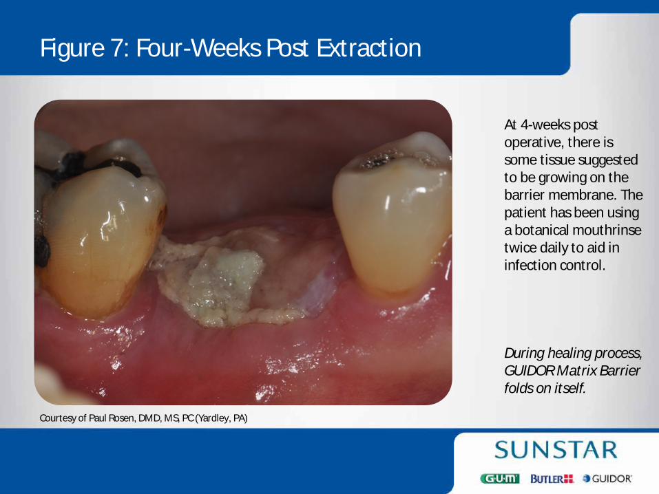
Figure 7: Four-Weeks Post Extraction
Courtesy of Paul Rosen, DMD, MS, PC (Yardley, PA)
At 4-weeks post operative, there is some tissue suggested to be growing on the barrier membrane. The patient has been using a botanical mouthrinse twice daily to aid in infection control. During healing process, GUIDOR Matrix Barrier folds on itself.

Figure 8: Radiograph at Four-Months Post Extraction
Courtesy of Paul Rosen, DMD, MS, PC (Yardley, PA)
Radiograph of the site exposed at 4 months following the extraction. There has been good containment and consolidation of the bone graft material.

Figure 9: Occlusal View at Reentry for Implant Placement
Courtesy of Paul Rosen, DMD, MS, PC (Yardley, PA)
Occlusal view following flap reflection at 4 months reveals good fill of the socket. More coronally, there is some loose connective tissue covering the regenerated bone.

Figure 10: Facial View at Reentry for Implant Placement
Courtesy of Paul Rosen, DMD, MS, PC (Yardley, PA)
Facial view of the site demonstrates the maintenance of good bone height.

Figure 11: Implant Placed in New Bone
Courtesy of Paul Rosen, DMD, MS, PC (Yardley, PA)
This electowetted surfaced implant is placed into favorable prosthetic position for restoration. A 4 mm high healing abutment of polyethylether ketone (PEEK) has been placed on the implant to allow for transgingival healing.
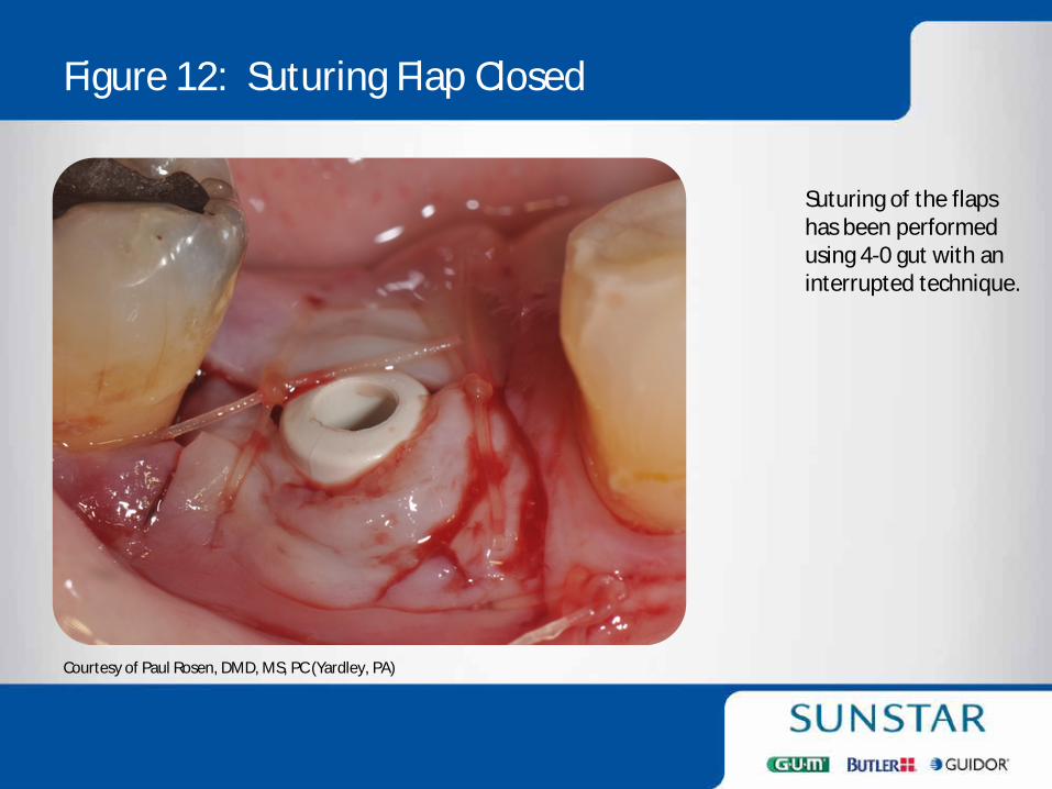
Figure 12: Suturing Flap Closed
Courtesy of Paul Rosen, DMD, MS, PC (Yardley, PA)
Suturing of the flaps has been performed using 4-0 gut with an interrupted technique.

Figure 13: Radiograph Immediately after Implant Placement
Courtesy of Paul Rosen, DMD, MS, PC (Yardley, PA)
Radiograph exposed of the implant on the day of placement demonstrating good bone height surrounding the implant.

Figure 14: Radiograph after Crown Placement
Courtesy of Paul Rosen, DMD, MS, PC (Yardley, PA)
Final crown has been completed 3 months following implant placement. The final restoration demonstrates a platform-switched restoration.
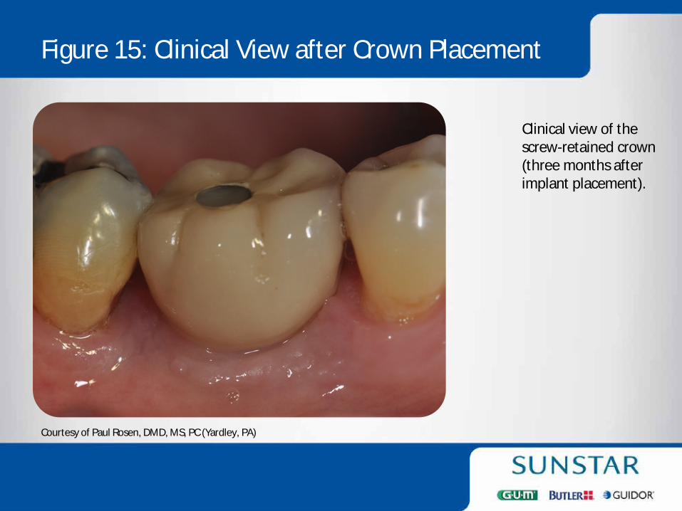
Figure 15: Clinical View after Crown Placement
Courtesy of Paul Rosen, DMD, MS, PC (Yardley, PA)
Clinical view of the screw-retained crown (three months after implant placement).

GUIDOR® Bioresorbable Matrix Barrier
GUIDOR Matrix Barrier is available in various shapes and sizes, with and without ligatures.
Please visit www.GUIDOR.com for latest product information and Instructions for Use.
GUIDOR is a registered trademark of Sunstar Suisse SA. ©2015 Sunstar Americas, Inc. All Rights Reserved. GDR15002

Sunstar has a wide variety of dental solutions to meet your office needs.