guidelines for management of patients with orthopaedic ... · 4 | guidelines for management of...
TRANSCRIPT

West Suffolk Hospital Department of Trauma and Orthopaedic Surgery
Guidelines for management of patients with orthopaedic conditions
THE BEST OF HEALTH FOR WEST SUFFOLK
integrated working
compiled by
| Guidelines for management of patients with orthopaedic conditions2
West SuffolkNHS Foundation Trust
WEST SUFFOLK HOSPITAL
Department of Trauma and Orthopaedic Surgery
Ideal referrals for common orthopaedic conditions
Consultants at West Suffolk Hospital have prepared this set of guidelines for common orthopaedic conditions to indicate circumstances where, ideally, the patient should be referred to the Trauma and Orthopaedics (T&O) department, and situations where it would be more appropriate to offer treatment in a primary care or community setting.
Although the T&O team are happy to see any referral from primary care, the team do recognise that, for some patients referred to secondary care, the T&O team cannot offer much more than could be provided in primary care or the community.
This booklet sets out to help GPs and other members of primary care teams to signpost patients suffering from joint pain to the most appropriate treatment pathway for their condition.
Martin Wood
January 2012
Updated to reflect new pathways, January 2013
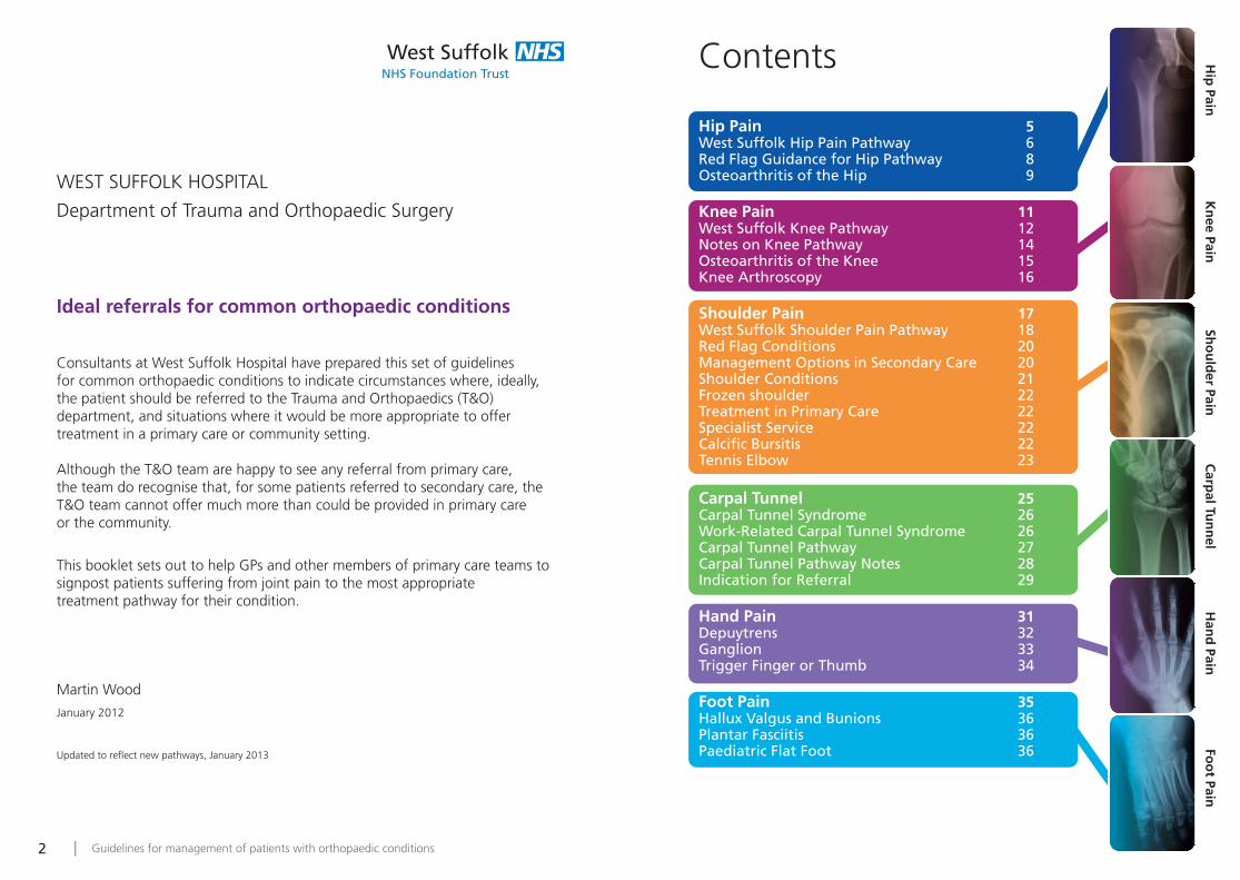
Contents
Hip Pain 5West Suffolk Hip Pain Pathway 6Red Flag Guidance for Hip Pathway 8Osteoarthritis of the Hip 9
Knee Pain 11West Suffolk Knee Pathway 12Notes on Knee Pathway 14Osteoarthritis of the Knee 15Knee Arthroscopy 16
Shoulder Pain 17West Suffolk Shoulder Pain Pathway 18Red Flag Conditions 20Management Options in Secondary Care 20Shoulder Conditions 21Frozen shoulder 22Treatment in Primary Care 22Specialist Service 22Calcific Bursitis 22Tennis Elbow 23
Carpal Tunnel 25Carpal Tunnel Syndrome 26Work-Related Carpal Tunnel Syndrome 26Carpal Tunnel Pathway 27Carpal Tunnel Pathway Notes 28Indication for Referral 29
Hand Pain 31Depuytrens 32Ganglion 33Trigger Finger or Thumb 34
Foot Pain 35Hallux Valgus and Bunions 36Plantar Fasciitis 36Paediatric Flat Foot 36
Hip
Pain K
nee Pain
Sho
uld
er Pain C
arpal Tu
nn
el Han
d Pain
Foo
t Pain
| Guidelines for management of patients with orthopaedic conditions4
West Suffolk Hip Pain Pathway
Red Flag Guidance for Hip Pathway
Osteoarthritis of the Hip
Hip
Pain
Guidelines for management of patients with orthopaedic conditions | 7| Guidelines for management of patients with orthopaedic conditions6
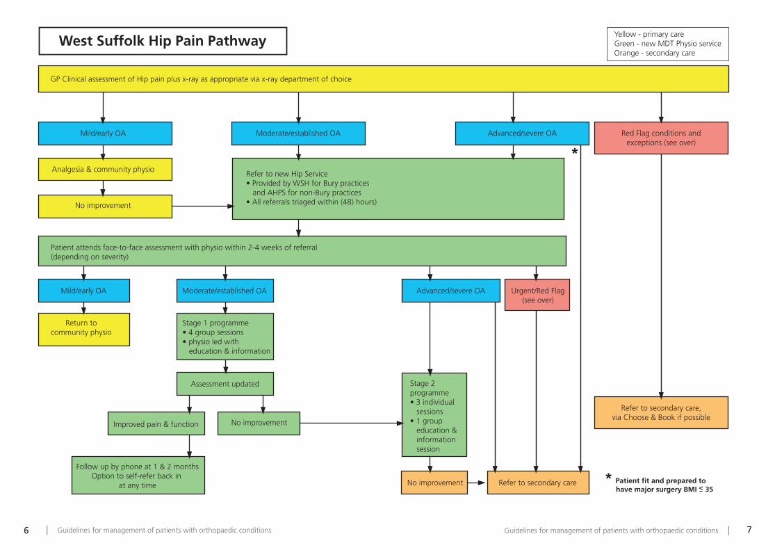
West Suffolk Hip Pain Pathway
*
* Patient fit and prepared to have major surgery BMI < 35
Refer to secondary care
Refer to secondary care,via Choose & Book if possible
No improvement
GP Clinical assessment of Hip pain plus x-ray as appropriate via x-ray department of choice
Mild/early OA
Mild/early OA Moderate/established OA
Return tocommunity physio
Stage 1 programme• 4 group sessions• physio led with education & information
Assessment updated
Improved pain & function No improvement
Follow up by phone at 1 & 2 monthsOption to self-refer back in
at any time
Stage 2 programme• 3 individual sessions• 1 group education & information session
Urgent/Red Flag(see over)
Advanced/severe OA
Patient attends face-to-face assessment with physio within 2-4 weeks of referral (depending on severity)
Refer to new Hip Service• Provided by WSH for Bury practices and AHPS for non-Bury practices• All referrals triaged within (48) hours)
Yellow - primary careGreen - new MDT Physio serviceOrange - secondary care
Analgesia & community physio
No improvement
Moderate/established OA Advanced/severe OA Red Flag conditions andexceptions (see over)
Guidelines for management of patients with orthopaedic conditions | 9| Guidelines for management of patients with orthopaedic conditions8
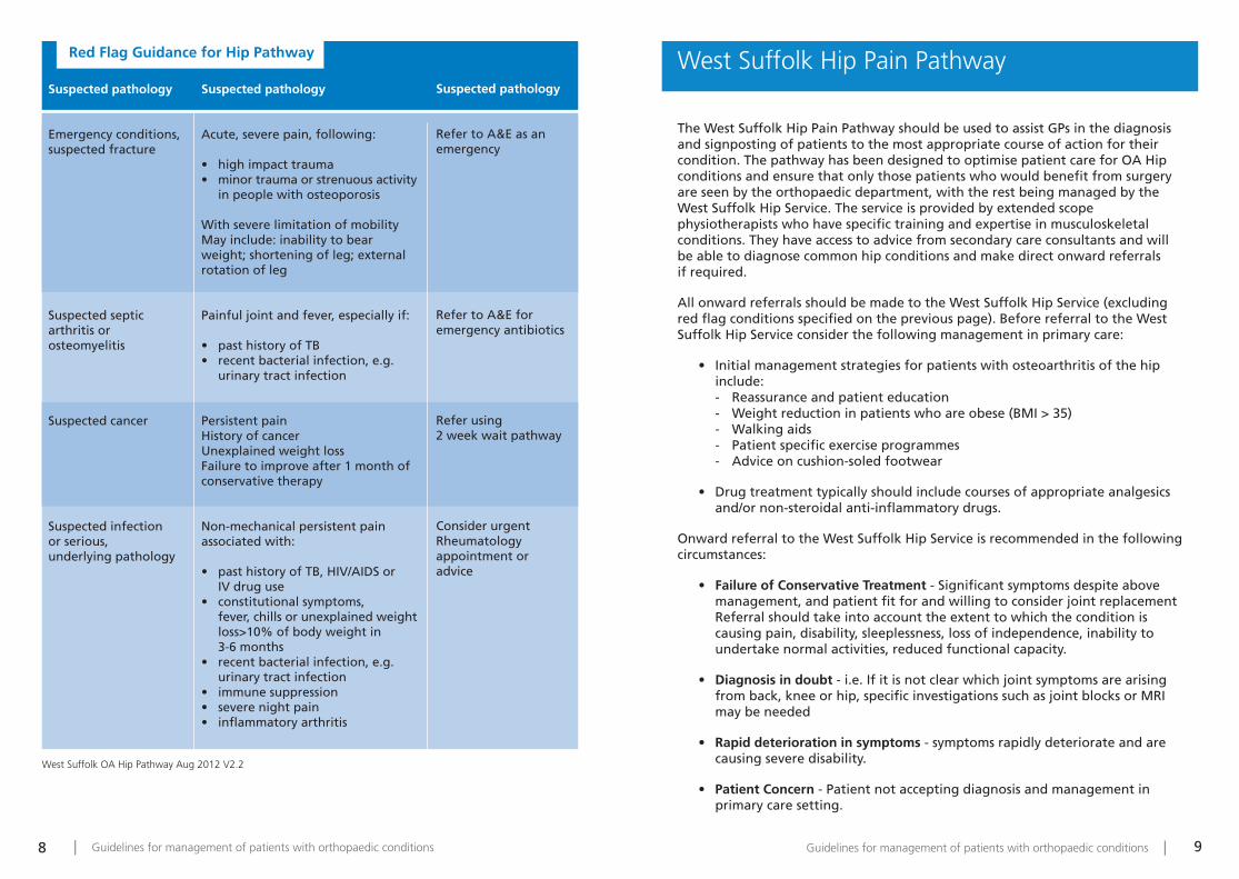
Suspected pathology
Emergency conditions, suspected fracture
Suspected septic arthritis or osteomyelitis
Suspected cancer
Suspected infection or serious, underlying pathology
Suspected pathology
Refer to A&E as anemergency
Refer to A&E for emergency antibiotics
Refer using 2 week wait pathway
Consider urgentRheumatology appointment or advice
Suspected pathology
Acute, severe pain, following:
• high impact trauma• minor trauma or strenuous activity
in people with osteoporosis
With severe limitation of mobilityMay include: inability to bearweight; shortening of leg; externalrotation of leg
Painful joint and fever, especially if:
• past history of TB• recent bacterial infection, e.g.
urinary tract infection
Persistent painHistory of cancerUnexplained weight lossFailure to improve after 1 month ofconservative therapy
Non-mechanical persistent painassociated with:
• past history of TB, HIV/AIDS or IV drug use
• constitutional symptoms, fever, chills or unexplained weight loss>10% of body weight in 3-6 months
• recent bacterial infection, e.g. urinary tract infection
• immune suppression• severe night pain• inflammatory arthritis
West Suffolk OA Hip Pathway Aug 2012 V2.2
West Suffolk Hip Pain Pathway
The West Suffolk Hip Pain Pathway should be used to assist GPs in the diagnosisand signposting of patients to the most appropriate course of action for theircondition. The pathway has been designed to optimise patient care for OA Hipconditions and ensure that only those patients who would benefit from surgeryare seen by the orthopaedic department, with the rest being managed by theWest Suffolk Hip Service. The service is provided by extended scopephysiotherapists who have specific training and expertise in musculoskeletalconditions. They have access to advice from secondary care consultants and willbe able to diagnose common hip conditions and make direct onward referrals if required.
All onward referrals should be made to the West Suffolk Hip Service (excludingred flag conditions specified on the previous page). Before referral to the WestSuffolk Hip Service consider the following management in primary care:
• Initial management strategies for patients with osteoarthritis of the hip include: - Reassurance and patient education - Weight reduction in patients who are obese (BMI > 35)- Walking aids- Patient specific exercise programmes- Advice on cushion-soled footwear
• Drug treatment typically should include courses of appropriate analgesics and/or non-steroidal anti-inflammatory drugs.
Onward referral to the West Suffolk Hip Service is recommended in the followingcircumstances:
• Failure of Conservative Treatment - Significant symptoms despite above management, and patient fit for and willing to consider joint replacementReferral should take into account the extent to which the condition is causing pain, disability, sleeplessness, loss of independence, inability to undertake normal activities, reduced functional capacity.
• Diagnosis in doubt - i.e. If it is not clear which joint symptoms are arising from back, knee or hip, specific investigations such as joint blocks or MRI may be needed
• Rapid deterioration in symptoms - symptoms rapidly deteriorate and are causing severe disability.
• Patient Concern - Patient not accepting diagnosis and management in primary care setting.
Red Flag Guidance for Hip Pathway
| Guidelines for management of patients with orthopaedic conditions10
West Suffolk Knee Pathway
Notes on Knee Pathway
Osteoarthritis of the Knee
Kn
ee Pain

Guidelines for management of patients with orthopaedic conditions | 13| Guidelines for management of patients with orthopaedic conditions12
West Suffolk Knee Pathway
*
*
Knee problem
Not responding to usual primary care
Referral triaged by Senior Physiotherapist (< 24 hours)
Refer to Community MSK service
Assessment by MSK physiotherapist - diagnosis made
Inflammatory
Mechanicalsymptoms
Degenerativearthritis
Early AO Anteriorknee pain
Acute KneeInjury
Not improved Improved
Septic arthritis/crystal arthropathy
Knee fracture/dislocation/tendon rupture
Secondary care(On call orthopaedic team)
Accident & Emergency Department West Suffolk HospitalA&E – Clinical Specialist Physiotherapist
Secondary care
(Elective orthopaedic/- Clinic - A&E referrals)
Refer to Knee ServiceWeight loss/lifestyle advice/
Analgesia advice/exercise advice
Physiotherapy for 3 months
Accident& Emergency
Secondary care(Elective rheumatology)
Severe
Degenerative Anterior knee pain
Appointment within 2-4 weeks Appointment within 72 hours
Acute knee injury GP tests and treatment
Improved Not improvedMild -Moderate
Yellow - primary careGreen - new MDT physio service
Orange - secondarycare
* Patient fit and prepared to have major surgery - BMI < 35–
Early OA, Anterior Knee Pain or Acute Knee Injury patients referred using the West Suffolk Knee Pathway paperwork may be referred direct to the MSK if their condition is identified at the triage stage
West Suffolk Knee Pathway Aug 2012 V2.2

Guidelines for management of patients with orthopaedic conditions | 15| Guidelines for management of patients with orthopaedic conditions14
West Suffolk Knee Pathway
The West Suffolk Knee Pathway should be used to assist GPs in the diagnosis andsignposting of patients to the most appropriate course of action for theircondition. The pathway has been designed to optimise patient care for OA Kneeconditions and ensure that only those patients who would benefit from surgeryare seen by the orthopaedic department, with the rest being managed by the WestSuffolk Knee Service. The service is provided by extended scope physiotherapistswho have specific training and expertise in musculoskeletal conditions. They haveaccess to advice from secondary care consultants and will be able to diagnosecommon knee conditions and make direct onward referrals if required.
All onward referrals should be made to the West Suffolk Knee Service (excludingred flag conditions). Before referral to the West Suffolk Knee Service consider thefollowing management in primary care:
• Initial management strategies for patients with osteoarthritis of the knee include:
- Reassurance and patient education - Weight reduction in patients who are obese (BMI > 35)- Walking aids- Patient specific exercise programmes- Advice on cushion-soled footwear
• Drug treatment typically should include courses of appropriate analgesics and/or non-steroidal anti-inflammatory drugs.
Onward referral to the West Suffolk Knee Service is recommended in thefollowing circumstances:
• Failure of Conservative Treatment - Significant symptoms despite above management, and patient fit for and willing to consider joint replacementReferral should take into account the extent to which the condition is causing pain, disability, sleeplessness, loss of independence, inability to undertake normal activities, reduced functional capacity.
• Diagnosis in doubt - i.e. If it is not clear which joint symptoms are arising from back, knee or hip, specific investigations such as joint blocks or MRI may be needed
• Rapid deterioration in symptoms - symptoms rapidly deteriorate and are causing severe disability.
• Patient Concern - Patient not accepting diagnosis and management in primary care setting.
Term
Degenerative
Traumatic
Inflammatory
Septic arthritis/crystalarthropathy
Knee dislocation
X-rays
Knee score
Severe arthritis
Assessment within 72 hours for traumaticconditions
Mechanical symptoms
Anterior knee pain
Gout etc
Weight loss
Notes
Chronic history, pain is the main symptom, particularly onweight bearing
Acute history usually less than 6 weeks, acute pain with orwithout mechanical symptoms
Swelling and aching are the main symptoms
Clinically difficult to differentiate, both need investigations(FBC, ESR,CRP) and knee aspiration
High risk of neurovascular injury
Standing AnteroPosterior, lateral and skyline views in all cases
New Zealand score or West Suffolk Knee Score
Complete loss of joint space on x-rays; extremely unlikely tobenefit from physiotherapy
Early diagnosis and treatment of ligamentous injuriesprotects other knee structures (menisci, articular cartilage)and leads to better functional results (outcomes); someperipheral meniscal tears can be repaired
Giving way/instability (sudden loss of knee muscle control),locking (inability to fully extend – meniscal tear), pseudo-locking (usually associated with with acute knee pain,inability to flex or extend)
Patellofemoral degeneration, patellar tendinopathy (non-insertional, insertional), Osgood-Schlatter, Hoffa’s fat padimpingement, plica syndrome, biomechanical patellartracking problem
Inflammatory arthropathies (osteoarthritis, rheumatoidarthritis, gout, pseudo-gout, psoriatic arthropathy, reactivearthropathy)
Advisable for any knee condition if BMI>30 (knee is loadedwith 3 times body weight during walking)
West Suffolk Knee Pathway Aug 2012 V2.2
Notes on Knee Pathway
| Guidelines for management of patients with orthopaedic conditions16
Knee Arthroscopy
Various management pathways for both traumatic and non-traumatic knee painhave been produced recently. It is beyond the scope of this document to includethe details of these.
Knee Arthroscopy is indicated for the treatment of:
• Acute Medial and Lateral Meniscal Tears, either meniscectomy or repair
• Removal of loose bodies• Diagnostic evaluation of suspected intra articular lesions if MRI
findings equivocal• Osteoarthritis associated with meniscal or chondral lesions
i.e. with mechanical symptoms of locking or giving way• Repair excision or grafting of articular cartilage lesions• Septic Arthritis• Synovitis due to Rheumatoid Arthritis• Synovial Tumour or PVNS• Patellofemoral pain / plica Syndrome / Hoffa Lesion• Pseudogout / chondrocalcinosis
Knee Arthroscopy is rarely used as a primary diagnostic procedure. West Suffolk Shoulder Pain Pathway
Red Flag Conditions
Management Optionsin Secondary Care
Shoulder Conditions
Frozen Shoulder
Treatment in Primary Care
Specialist Service
Sho
uld
er Pain

Guidelines for management of patients with orthopaedic conditions | 19| Guidelines for management of patients with orthopaedic conditions18
West Suffolk Shoulder Pain Pathway
Shoulder Pain
X-ray – AP and axillary to eliminate dislocation
If no dislocation: Use NSAIDs, steroid injections* as required to manage pain
Consider referral to PHYSIO
Posterior injection
If no improvement after 3 months, consider referral to T&O
Local injection
Weak/painful abductionSignificantly reduced rotation
Instability
Considerreferral to
T&O
Considerreferral toPHYSIO
PossibleDiagnosis
Symptom
NoNo Yes
ImpingementChronically dislocated
If dislocation,REFER TO T&O
Glenohumeralarthritis
Frozenshoulder
AcromioclavicularJoint (ACJ)
arthritis
Pain onACJ
palpationand/or
cross-bodyabduction
Anteriorshoulder
tendernesscorrespondingto long head
of bicep
Use NSAIDs,steroid injections* -
subacromial tomanage pain;discourage use
of slings;Consider referral to
PHYSIO
If no improvementafter 3 months,
refer to T&O
Bicepstendonitis
No historyof trauma
Historyof trauma
REFER TOPHYSIO
REFER TOT&O
Eliminate Red Flag conditions (see notes)
Eliminate Calcific BursitisAcute, severe pain on palpation beneath the acromion.
Treat with heat/cold, NSAIDs, steroid injections*Consider x-ray – refer to T&O after 3 months.
* Steroid injections: 2 injections 6 wks apart
West Suffolk Shoulder Pathway 2012 V5

Guidelines for management of patients with orthopaedic conditions | 21| Guidelines for management of patients with orthopaedic conditions20
Suspectedpathology
Recent fracturesor dislocations
Infection
Malignancy
Neurologicallesion or cervicalpathology
Polymyalgiarheumatica
Clinical features
History of recent trauma. Unusualdeformity, swelling or joint effusion
Symptoms suggestive of septicarthritis e.g. fever or chills; hot,swollen joint
Previous history of cancer orsuspected malignancy, unexplaineddeformity, lymphadenopathy, weightloss, night pain
Unexplained wasting, significantsensory or motor deficits,neurovascular compromise, painassociated with neck movements
Age over 50 yrs, symptoms for over 2 wks, bilateral shoulder and /orpelvic girdle aching, morningstiffness lasting over 45 minutes,evidence of an acute phase response
Referral route
Refer to A&E
Refer to A&E
Consider investigations andreferral on 2 Week Waitpathway if appropriate
Depending on severity, refer to:MSK community clinicNeurologyA&E (if suspecting stroke)
Treat in primary care (if nosymptoms of giant cell arteritis)orRefer to Rheumatology if notconfident to treat in primary care
Red flag conditions
Management options in secondary care (if conservative treatment, where appropriate, fails)
Frozen shoulder
Glenohumeral arthritis
Calcific tendinopathy
Biceps tendinopathy
Atraumatic instability
Acromioclavicular Joint (ACJ) arthritis
Impingement, rotator cuff tear
Manipulation under anaesthesia or capsular release
Arthroscopic debridement and steroid injection, hemi or totalarthroplasty
Arthroscopic removal of calcium deposits followed bydebridement may be beneficial
Arthroscopic debridement, tenotomy
Surgery is rarely required – symptoms usually improve withphysiotherapy, very rarely, capsulorrhaphy may be required
Excision of distal end of clavicle
Impingement – 80% improve within 6 months. If cuff tear ispresent, repair if possible. Chronic tears in elderly patients –consider shoulder replacement.NB: Ultrasound scans should not alter GP management andshould only be ordered in a T&O setting
Shoulder Conditions
The West Suffolk Shoulder Pain Pathway should be used to assist GPs in thediagnosis and signposting of patients to the most appropriate course of actionfor their condition. The pathway has been designed to optimise patient care forshoulder conditions and ensure that only those patients who would benefit fromsurgery are seen by the orthopaedic department, with the rest being managedby primary care intervention or physiotherapy. The physiotherapists have specifictraining and expertise in musculoskeletal conditions. They have access to advicefrom secondary care consultants and will be able to diagnose common shoulderconditions and make direct onward referrals if required.
For all specialist referrals please record the side of the problem as some of thesepatients may be referred for further investigations before they are seen in thespecialist clinic.
Painful shoulder (Impingement pain)
Characterised by pain on abduction, on reaching behind the body or on thethrowing motion. Rarely seen in individuals below 40 years of age.
Treatment in primary care: • Explain aetiology and advise on avoidance of precipitating movements or
activities. NSAIDS, physiotherapy and consider steroid injection. Mostpatients experience relief within a 2-3 month period.
• If improvement is not seen within 3 months or if atypical symptoms,investigate with x-rays (AP, axillary and outlet views) and refer for specialistopinion.
Specialist services:• Confirm or exclude the diagnosis.• Provide non-operative or operative management depending on the
patient’s needs and symptoms.
Guidelines for management of patients with orthopaedic conditions | 23| Guidelines for management of patients with orthopaedic conditions22
Frozen shoulder
Painful stiff joint usually not following any trauma. May be seen in any agegroup. Characteristically the passive and the active movements are similarlyrestricted. This diagnosis cannot be given unless x-rays have confirmed absenceof arthritis or bony injury.
Treatment in primary care:• Refer for x-ray AP, outlet and axillary views. If x-rays are normal explain
aetiology and long recovery time (1-2 years). NSAIDS and painkillers,physiotherapy.
• If no improvement in 6 weeks or if significant pain unresponsive to non-operative management refer for specialist opinion.
Specialist service:• Investigate as appropriate, confirm the diagnosis.• Consider intra-articular steroid injection or manipulation under anaesthesia
or other surgical intervention.
Calcific Bursitis
Significant shoulder pain often associated with local signs of inflammation. May be seen in any age group. Not normally related to trauma.
Treatment in Primary care: • Treat with anti-inflammatory medication and painkillers. Consider x-ray
(AP, outlet and axillary views). If infection is ruled out (blood test andclinical findings) consider steroid and local anaesthetic injection.
• If improvement is not seen within 6 weeks consider specialist referral.
Specialist services:• Establish the diagnosis and consider treatment either with steroid and local
anaesthetic injections, aspiration or surgical intervention.
Acromioclavicular joint arthritis
Seen either late following trauma or as a result of degenerative changes in theelderly population. Point tenderness over the acromioclavicular joint withoutany referral.
Treatment in primary care: • Anti-inflammatory medication, physiotherapy, explain aetiology.
Consider local anaesthetic and steroid injection into the acromioclavicularjoint.
• If no improvement after three months consider specialist referral.
Specialist services:• To establish the diagnosis and provide management either via injections or
surgical excision of the joint.
Shoulder instability:• This may either be traumatic or atraumatic. If the shoulder joint is proven
unstable (documented anterior or posterior dislocations of theglenohumeral joint) consider specialist referral. In general atraumaticinstability is treated with physiotherapy, traumatic instability with surgery.
Tennis Elbow
Pain localised to the outer aspect of the elbow joint exacerbated by wristextension against resistance. Not necessarily associated with participation in sports.
Treatment in Primary care: • Confirm diagnosis and arrange treatment with physiotherapy, elbow brace,
consider steroid injection (not more than two injections as risk of skin orsubcutaneous fat necrosis exist).
• If no improvement with non-operative treatment after 6 months, considerreferral for specialist opinion.
Specialist services: • Establish the diagnosis.

| Guidelines for management of patients with orthopaedic conditions24
Carpal Tunnel Syndrome
Work-Related Carpal Tunnel Syndrome
Carpal Tunnel Pathway
Carpal Tunnel Pathway Notes
Indication for Referral
Carp
al Tun
nel
Guidelines for management of patients with orthopaedic conditions | 27| Guidelines for management of patients with orthopaedic conditions26
Carpal Tunnel Syndrome
Mild and Moderate Carpal Tunnel Syndrome can be managed in primary careusing the Carpal Tunnel Pathway.
Work-Related Carpal Tunnel Syndrome
No clear association between work activities and development of “de novo”Carpal Tunnel Syndrome. Work activities may aggravate pre-existing CarpalTunnel.
i.e.• Symptoms for less than three months • Intermittent symptoms, with periods of complete resolution• No fixed sensory or motor symptoms or signs • Treatable or self limiting cause of carpal tunnel syndrome
Exclude:pregnancy, hypothyroidism and diabetes clinically and/or by investigation.
Consider following management in primary care:• Nocturnal, neutral wrist splint • Activity/work-place modification (if clear association apparent) and referral
to hand therapy service• Steroid injection around median nerve if trained injector available
y
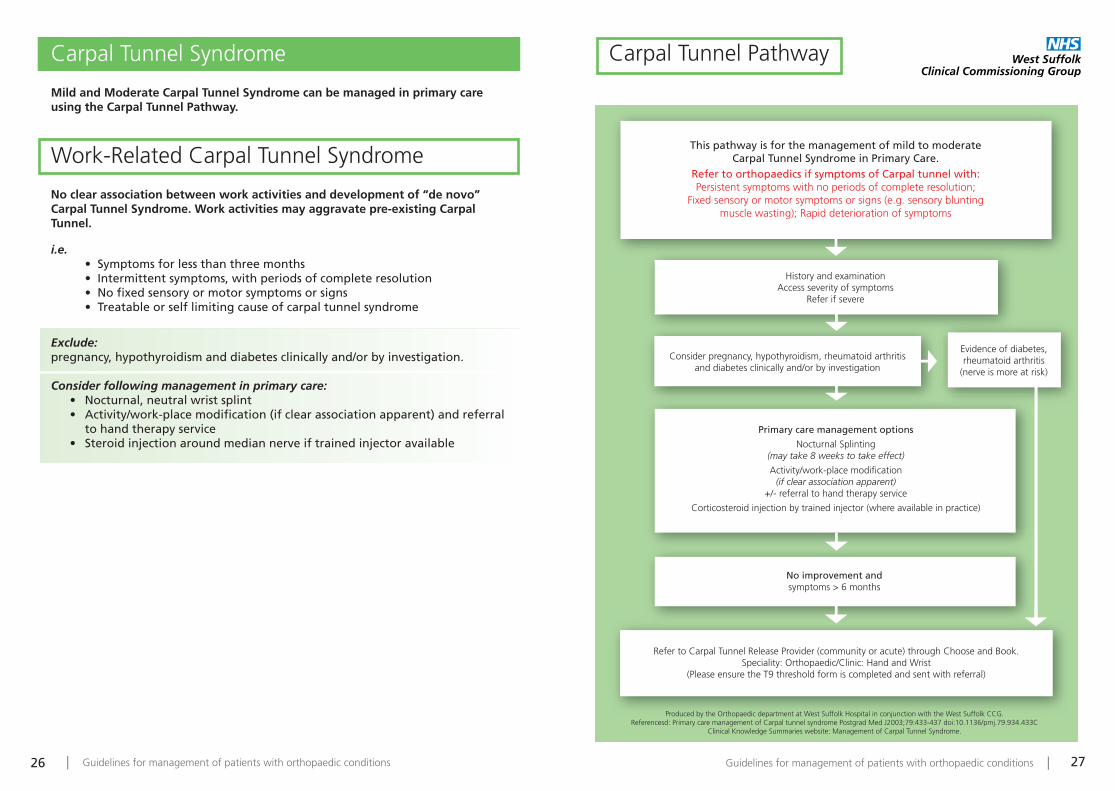
This pathway is for the management of mild to moderate Carpal Tunnel Syndrome in Primary Care.
Refer to orthopaedics if symptoms of Carpal tunnel with:Persistent symptoms with no periods of complete resolution;
Fixed sensory or motor symptoms or signs (e.g. sensory blunting muscle wasting); Rapid deterioration of symptoms
History and examinationAccess severity of symptoms
Refer if severe
Evidence of diabetes,rheumatoid arthritis
(nerve is more at risk)
Consider pregnancy, hypothyroidism, rheumatoid arthritis and diabetes clinically and/or by investigation
Primary care management options
Nocturnal Splinting(may take 8 weeks to take effect)
Activity/work-place modification(if clear association apparent)
+/- referral to hand therapy service
Corticosteroid injection by trained injector (where available in practice)
No improvement andsymptoms > 6 months
Refer to Carpal Tunnel Release Provider (community or acute) through Choose and Book. Speciality: Orthopaedic/Clinic: Hand and Wrist
(Please ensure the T9 threshold form is completed and sent with referral)
Produced by the Orthopaedic department at West Suffolk Hospital in conjunction with the West Suffolk CCG.Referencesd: Primary care management of Carpal tunnel syndrome Postgrad Med J2003;79:433-437 doi:10.1136/pmj.79.934.433C
Clinical Knowledge Summaries website: Management of Carpal Tunnel Syndrome.
Carpal Tunnel Pathway
Guidelines for management of patients with orthopaedic conditions | 29| Guidelines for management of patients with orthopaedic conditions28
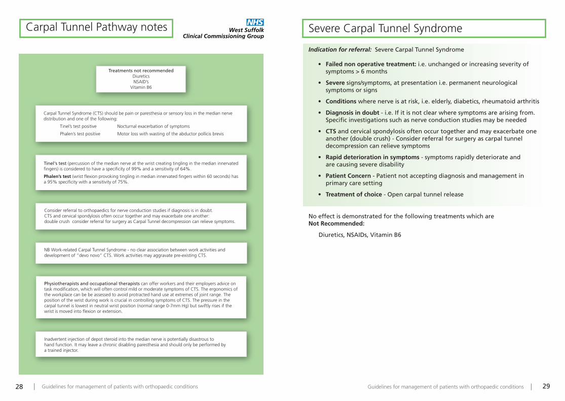
Treatments not recommendedDiureticsNSAID’s
Vitamin B6
Carpal Tunnel Syndrome (CTS) should be pain or paresthesia or sensory loss in the median nerve distribution and one of the following:
Tinel’s test positive Nocturnal exacerbation of symptoms
Phalen’s test positive Motor loss with wasting of the abductor pollicis brevis
Tinel's test (percussion of the median nerve at the wrist creating tingling in the median innervated fingers) is considered to have a specificity of 99% and a sensitivity of 64%.
Phalen’s test (wrist flexion provoking tingling in median innervated fingers within 60 seconds) hasa 95% specificity with a sensitivity of 75%.
Consider referral to orthopaedics for nerve conduction studies if diagnosis is in doubt.CTS and cervical spondylosis often occur together and may exacerbate one another: double crush consider referral for surgery as Carpal Tunnel decompression can relieve symptoms.
NB Work-related Carpal Tunnel Syndrome - no clear association between work activities and development of “devo novo” CTS. Work activities may aggravate pre-existing CTS.
Physiotherapists and occupational therapists can offer workers and their employers advice on task modification, which will often control mild or moderate symptoms of CTS. The ergonomics of the workplace can be be assessed to avoid protracted hand use at extremes of joint range. The position of the wrist during work is crucial in controlling symptoms of CTS. The pressure in the carpal tunnel is lowest in neutral wrist position (normal range 0-7mm Hg) but swiftly rises if the wrist is moved into flexion or extension.
Inadvertent injection of depot steroid into the median nerve is potentially disastrous to hand function. It may leave a chronic disabling paresthesia and should only be performed by a trained injector.
Carpal Tunnel Pathway notes Severe Carpal Tunnel Syndrome
Indication for referral: Severe Carpal Tunnel Syndrome
• Failed non operative treatment: i.e. unchanged or increasing severity of symptoms > 6 months
• Severe signs/symptoms, at presentation i.e. permanent neurological symptoms or signs
• Conditions where nerve is at risk, i.e. elderly, diabetics, rheumatoid arthritis
• Diagnosis in doubt - i.e. If it is not clear where symptoms are arising from. Specific investigations such as nerve conduction studies may be needed
• CTS and cervical spondylosis often occur together and may exacerbate one another (double crush) - Consider referral for surgery as carpal tunneldecompression can relieve symptoms
• Rapid deterioration in symptoms - symptoms rapidly deteriorate and are causing severe disability
• Patient Concern - Patient not accepting diagnosis and management in primary care setting
• Treatment of choice - Open carpal tunnel release
No effect is demonstrated for the following treatments which are Not Recommended:
Diuretics, NSAIDs, Vitamin B6
| Guidelines for management of patients with orthopaedic conditions30
Dupuytrens
Ganglion
Trigger Finger
or Thumb
Han
d Pain
Guidelines for management of patients with orthopaedic conditions | 33| Guidelines for management of patients with orthopaedic conditions32
Dupuytren's
Classification and referral of Dupuytren's
Mild Dupuytren's disease does not require surgical treatment.Patients with Moderate disease should be referred for possible surgery, preferably before disease becomes severe.
Mild: • No functional problems• No contracture• Mild metacarpophalangeal joint contracture only (<30 degrees)
Treatment:• Reassure• Observe
Moderate: • Notable functional problems (gloves, can’t get hand in pocket)• Moderate metacarpophalangeal joint contracture (30 – 60 degrees)• Moderate proximal interphalangeal joint contracture (<30 degrees)• First web contracture
“Heuston’s tabletop test”:Patient can’t get hand flat on table without seeing daylight underneath, or can get a finger underneath = moderate or severe disease – requires referral for surgery.
Treatment:Refer for surgery: • Limited fasciectomy
Severe • Severe contracture of both metacarpophalangeal (>60) joint and proximal
interphalangeal joint (>30).
Treatment:Refer for surgery:• Limited fasciectomy• Dermofasciectomy + skin graft• Fusion• Amputation
Ganglion
Most ganglia do not justify surgical treatment on the NHS.
Over 50% of ganglia will spontaneously resolve if left long enough (up to 10 years). Pain associated with a ganglion may persist after surgical excision. (? due to defect in wrist capsule which caused ganglion). Up to 40%recurrence rate after surgical excision.
Classification and referral of Ganglia
Mild: • Asymptomatic lump, transilluminates.
Treatment:• Reassure • Observe
Moderate: • Symptomatic lump; long duration of symptoms• Occult ganglia• Cancer- phobia
Treatment: • Reassure / Observe• Aspiration for cancer reassurance • Refer for ultrasound if concerns re diagnosis
Severe: • Severe pain with restriction of activities of daily living; concern re
diagnosis
Treatment:• Refer for specialist opinion and possible surgery

| Guidelines for management of patients with orthopaedic conditions34
Trigger Finger or Thumb
Classification and referral
Mild or moderate trigger finger should initially be managed in primary care.Resistant or recurrent disease should be referred for possible surgical treatment.
Note - if a patient has triggering caused by an underlying condition such asDiabetes or Rheumatoid Arthritis, or if they have required surgery in the past,they are unlikely to be cured by steroid injections.
Mild: (“pre-triggering”) • History of pain, catching or “click” around finger or thumb• Tender A1 pulley; but fully mobile finger
Treatment:• Analgesia• Topical NSAID, Massage
Moderate: Triggering with:
• Difficulty actively extending finger • Need for passive finger extension • Loss of complete active flexion
Treatment:• Night Splinting• Steroid injection to flexor sheath up to x2• If no improvement or recurrence within 3 months refer for surgical release
Severe: • Fixed contracture
Treatment:• Urgent Referral for Surgical trigger release Hallux Valgus
and Bunions
Plantar Fasciitis
Paediatric Flat Foot
Hallux Valgus and Bunions
Plantar Fasciitis
Paediatric Flat Foot
Foo
t Pain

Guidelines for management of patients with orthopaedic conditions | 37| Guidelines for management of patients with orthopaedic conditions36
Hallux Valgus and BunionsHallux valgus is defined as an angle of greater than 15 degrees at the firstmetatarsophalangeal joint in the AP plain. A bunion is the formation of dorsomedialosteophyte at the first metatarsophalangeal joint. There are many surgical optionswhich achieve mixed clinical results and have a multitude of complications.Conservative measures should be tried before referral for surgical treatment.
Primary treatment:• Advice on low heeled, wide forefoot shoes with soft leather uppers • Referral to chiropodist • Referral to orthotics (e.g. comfort shoes)
Refer when:• There is severe deformity (overriding toes) • There is severe pain from the metatarsophalangeal joint or bunion • Conservative methods have failed
Plantar FasciitisPlantar fasciitis is a benign, usually self-limiting condition which ultimately respondsto conservative treatment and even in the presence of a calcaneal spur on an x-ray isnot usually treated surgically. A calcaneal spur is not indicative of any disorder.
Primary treatment:• NSAIDs • Silicone heel pad • Steroid injection under the trigger point • Physiotherapy for stretch exercises of plantar fascia and tendo-achilles
Refer when:• There is doubt about the diagnosis• Patient not accepting diagnosis and management in primary care setting
Paediatric Flat FootFlat foot can either be flexible or fixed. A flexible flat foot is flat when weight-bearing but forms a normal arch when non-weight-bearing or when standing on tiptoe. Flexible flat foot is non-pathologic and requires no treatment. Rigid flat footmay be caused by tarsal coalition or neuromuscular conditions and is pathological.
Primary treatment:• Flexible flat foot requires no treatment
Refer when:• Flat foot is rigid • Other pathology is suspected

Guidelines for management of patients with orthopaedic conditions | 39| Guidelines for management of patients with orthopaedic conditions38
Notes
For more information contact:Department of Trauma and OrthopaedicsWest Suffolk HospitalHardwick LaneBury St EdmundsSuffolk IP33 2QZ
Telephone: 01284 713713
©2013. WSCCG. GFX: 2938