guideline pediatric congenital heart disease · guideline pediatric congenital heart disease ......
TRANSCRIPT

Guideline pediatric congenital heart disease
www.kinderkardiologie.org/dgpkLeitlinien.shtm

Guideline primary cardiomyopathies
Content
Definition of primary cardiomyopathies
Dilated cardiomyopathy
Hypertrophic cardiomyopathy
Genetic testing and prevention
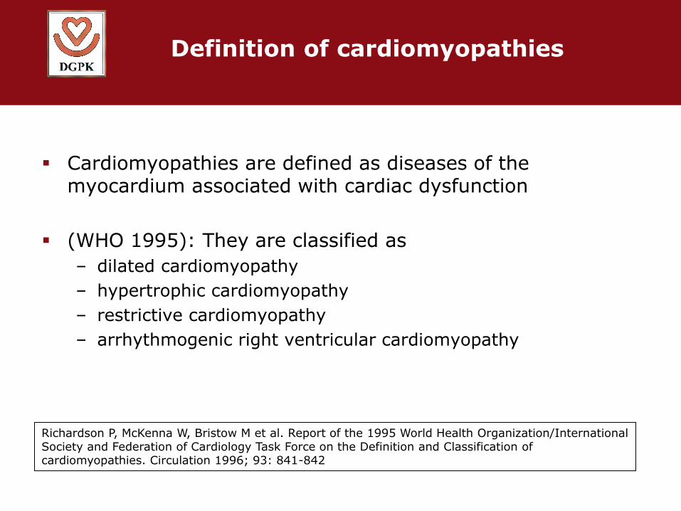
Definition of cardiomyopathies
Cardiomyopathies are defined as diseases of the myocardium associated with cardiac dysfunction
(WHO 1995): They are classified as
– dilated cardiomyopathy
– hypertrophic cardiomyopathy
– restrictive cardiomyopathy
– arrhythmogenic right ventricular cardiomyopathy
Richardson P, McKenna W, Bristow M et al. Report of the 1995 World Health Organization/International Society and Federation of Cardiology Task Force on the Definition and Classification of cardiomyopathies. Circulation 1996; 93: 841-842

Classification of primary cardiomyopathies AHA statement 2006

Secondary cardiomyopathies

Incidence of cardiomyopathies in patients <19 yrs.: 1.13 on 100.000 children
(8.34 on 100.000 in infants)
6
dilativehypertrophicrestrictive and other

dilative cardiomyopathy definition and clinical symptoms
Dilation of left and later right ventricle
severe systolic dysfunction
Congestive heart failure
Cold sweating
Dyspnoea
Lung odema
Peripheral cyanosis
Hepatomegaly
Failure to thrive
Clinical differential diagnosis:
Pneumonia
Sepsis

dilative cardiomyopathy primary diagnostics
Echocardiography Left venticular
diameter/function
Mitral regurgitation
Coronary morphology
Pulmonary artery pressure (tricuspid/pulmonary regurgitation)
ECG (not specific)
left heart strain
Arrhythmias
Thoracic X-ray (not specific)
Cardiomegaly
Pulmonary congestion
Cardiac catheterization (necessary after primary stabilization)
ALCAPA
Pulmonary artery pressure
Myocardial biopsy DD: myocarditis
Molecular pathology/ immunohistochemistry
Cardiac MRI DD: myocarditis (late enhancement)
Biomarkers NT-pro/BNP
Genetic testing Not recommended in isolated CMP
Meaningful in patients with combination of AV-block or myopathy

dilative cardiomyopathy - possible causes
9
Hereditary/gentically caused disease
Post-myocarditis
Metabolic disease
Other secondary cardiomyopathy
Coronary artery disease (ALCAPA)
Tachycardia induced cardiomyopathy
Goodwin, Muntoni, Muscle Nerve 2005;32:577-88

dilative cardiomyopathy - differential diagnoses
Nikolaus A. Haas; Ulrich Kleideiter - Kinderkardiologie: Klinik und Praxis der Herzerkrankungen bei Kindern, Jugendlichen und jungen Erwachsenen, Thieme 2011

dilative cardiomyopathy - differential diagnoses
Nikolaus A. Haas; Ulrich Kleideiter - Kinderkardiologie: Klinik und Praxis der Herzerkrankungen bei Kindern, Jugendlichen und jungen Erwachsenen, Thieme 2011

dilative cardiomyopathy - therapeutic options
Drug therapie for congestive heart failure (look at guideline) ACE-inhibitor
ß-blocker
Aldosteron-antagonists
Diuretics
Anti-coagulants
Anti-arrhythmics (guideline)
ICD-implantation (guideline)
Cardiac resynchronisation therapy
Cardiac-assist-system
Heart transplantation (HTX)
Short axis echocardiography of a 5 month old male

dilative cardiomyopathy follow-up examinations
Echocardiography Left ventricle
Mitral valve
Pulmonary artery hypertension (PAH)
Biomarkers (1x/y)
Holter-ECG (every 2 yrs.)
Repeated catheterizations if signs of PAH are existent
Spiroergometry (every 3 yrs.)
Involvement of HTX-center

hypertrophic cardiomyopathy definition and clinical symptoms
Asymmetric hypertrophy of the left ventricle
+/- obstruction of left ventricular outflow tract
Disease of the sacomer
90% hereditary disease (autosomal dominant)
Systolic heart murmur
palpitations
Fatigue
Dyspnoea (on exertion)
Syncope (on exertion)

hypertrophic cardiomyopathy primary diagnostics
Echocardiography Left ventricular measurements
Thickness of the septum and different wall segments
Distribution of hypertrophy (asymmetric/concentric)
Assessment of left ventricular apex
Systolic anterior movement of mitral valve (SAM phenomenon)
Thickness of right ventricular wall
PW-/CW-Doppler: left ventricular obstruction (subaortal, mid-ventricular, apical)
Stress echocardiography Recommended in symptomatic patients
with LVOTO-rest-gradient < 30 mmHg
Valsalva´s manoeuvre

hypertrophic cardiomyopathy primary diagnostics
Cardiac MRI
Recommended in patients with insufficient echo-window
Verification of fibrosis (late gadolinium enhancement)
ECG (left ventricular hypertrophy, repolarisation patterns)
Holter-ECG, Ergometry (arrhythmias)
Thoracic X-ray (not necessary for primary diagnostics)
Cardiac catherization
LV-Angiography/coronary imaging recommended before surgery in patients with LVOTO
Endomyocardial biopsy
uncertain phenotype/etiology
Elimination of secondary hypertrophy in cases of uncertain concentric myocardial hypertrophy
Genetic testing
Limited indication in patients <18 yrs. of age
Only for clarification of differential diagnoses in uncertain phenotypes
17yrs. old male, asymmetric HCM

hypertrophic cardiomyopathy differential diagnoses
Differential diagnoses
Storage diseases (e.g. Pompe disease)
Genetic disorders (e.g. Noonan diseases)
neuromuscular diseases
PRKAG2-syndrom (cardiac glycogenosis associated with Wolff-Parkinson-White Syndrom)
Danon disease (X-chromosomal inherited lysosomal storage disease)
SYMPTOMS IN CHILDREN with Noonan disease

Hypertrophic cardiomyopathy (HCM) - risk factors
Main risk factors for suddencardiac death (according to adult patients)
Resuitation for cardiac arrest or non-sustained ventricular tachycardia
Sudden cardiac death in familiy history
History of non-sustained ventricular tachycardia (>3 Schläge < 30 sec, HF >120/min.)
Profound thickness of the septum >30 mm (controversially discussed)
Insufficient rise of systolic blood pressure under exertion (syst. ≤ 20 mmHg)
Rapid progression of the disease
1. Maron BJ, McKenna WJ, Danielson GK et al. American College of Cardiology/European Society of Cardiology Clinical
Expert Consensus Document on Hypertrophic Cardiomyopathy. A report of the American College of Cardiology Foundation Task Force on Clinical Expert Consensus Documents and the European Society of Cardiology Committee for Practice Guidelines. European heart journal 2003; 24: 1965-1991.
2. Monserrat L, Elliott PM, Gimeno JR, Sharma S, Penas-Lado M and McKenna WJ. Non-sustained ventricular tachycardia in hypertrophic cardiomyopathy: an independent marker of sudden death risk in young patients. Journal of the American College of Cardiology 2003; 42: 873-879

hypertrophic cardiomyopathy follow-up
Follow-up-visits At least annually
(ECG, echocardiography)
High risk patients LVOT-peak echo-gradient >30 mmHg
moderate or profound LV-wall hypertrophy
rapid progress of the disease
unfavorable patient or family history
Holter-ECG and ergometry annually
Maron BJ, McKenna WJ, Danielson GK et al. American College of Cardiology/European Society of Cardiology Clinical Expert Consensus Document on Hypertrophic Cardiomyopathy. A report of the American College of Cardiology Foundation Task Force on Clinical Expert Consensus Documents and the European Society of Cardiology Committee for Practice Guidelines. European heart journal 2003; 24: 1965-1991

Hypertrophic cardiomyopathy (HCM) – sports and primary prevention
No competitive sport *1
No isometric muscular exertion *1
For detailed recommendations look at the current guideline of the European
Society of Cardiology *2
Early detection and medical screenig before athletic activities
HCM, ARVC and ion-channel disorders are mean reasons for sudden cardiac death in athlets <35 yrs. of age
Preliminary medical examination before athletic sports should include patients medical and family history, physical examination, ECG
Guideline committee recommends a medical screening examination (including ECG) for all adolescents at the age of 12-14 yrs. and in the „Jugendarbeitsschutzuntersuchung“ (strong agreement of the committee)
1. Maron BJ, Chaitman BR, Ackerman MJ et al. Recommendations for physical activity and recreational sports participation for young patients with genetic cardiovascular diseases. Circulation 2004; 109: 2807-2816.
2. Pelliccia A, Corrado D, Bjornstad HH et al. Recommendations for participation in competitive sport and leisure-time physical activity in individuals with cardiomyopathies, myocarditis and pericarditis. European journal of cardiovascular prevention and rehabilitation : official journal of the European Society of Cardiology, Working Groups on Epidemiology & Prevention and Cardiac Rehabilitation and Exercise Physiology 2006; 13: 876-885

Hypertrophic cardiomyopathy (HCM) – therapeutic options
lipophilic ß-blockers (e.g. Propranolol, Metoprolol or
Bisoprolol): recommended in symptomatic patients
CA-antagonists (e.g. Verapamil): increased risk for
deterioration of congestive heart failure/death in
patients with severe hypertrophy;
contraindication for use in infants
Amiodaron: protection against sudden cardiac death
not shown; side effects of permanent application are
more serious in children/adolescents
ICD-Implantation: only reliable primary and
secondary prevention for sudden cardiac death (look
at guideline)
Septal myectomy to treat LVOTO
Transcatheter intervention in individual
cases/controversialy discussed
Rarely heart transplantation
1. Ostman-Smith I. Hypertrophic cardiomyopathy in childhood and adolescence - strategies to prevent sudden death. Fundamental & clinical pharmacology 2010; 24: 637-652.
2. Ostman-Smith I, Wettrell G and Riesenfeld T. A cohort study of childhood hypertrophic cardiomyopathy - Improved survival following high-dose beta-adrenoceptor antagonist treatment. Journal of the American College of Cardiology 1999; 34: 1813-1822.
3. Maron BJ, Yacoub M and Dearani JA. Controversies in cardiovascular medicine. Benefits of surgery in obstructive hypertrophic cardiomyopathy: bring septal myectomy back for European patients. European heart journal 2011; 32: 1055-1058

Primary cardiomyopathies – family members and prevention
Family history (3 generations)
Medical examination of first-degree relatives (incl. children)
Conspicuous medical finding/carrier of mutation: annually controls
Time interval for controls in inconspicuous siblings during
childhood:
Every 3 yrs.
in hypertrophic cardiomyopathy annually from the age of 12. yrs.
1. Beudt U, Heidemann S, Henn W et al. S2-Leitlinie Humangenetische Diagnostik und genetische Beratung. Medgen 2011; 23: 281-323. 2. Ackerman MJ, Priori SG, Willems S et al. HRS/EHRA expert consensus statement on the state of genetic testing for the channelopathies
and cardiomyopathies. Europace 2011; 13: 1077-1109

Primary cardiomyopathies – family members and prevention
Genetic testing
Counseling (institut for human genetics) recommended for patients and families
Children and adolscents show larger number of
Inherited/gentically caused cardiomyopathies
Mitochondrial or metabolically caused cardiomyopathies
Chromosomal defects, dysmorphic syndroms
Familiy-screening and -management (adjustment of risk in asymptomatic carriers of mutation)
Predictive genetic testing in children/adolescents is rated very controversially
Predictive genetic testing is legal exclusively after qualified genetic counseling
1. Beudt U, Heidemann S, Henn W et al. S2-Leitlinie Humangenetische Diagnostik und genetische Beratung. Medgen 2011; 23: 281-323. 2. Ackerman MJ, Priori SG, Willems S et al. HRS/EHRA expert consensus statement on the state of genetic testing for the channelopathies
and cardiomyopathies. Europace 2011; 13: 1077-1109