growth characteristics and tracing antagonistic properties
TRANSCRIPT

Available online at www.worldscientificnews.com
( Received 09 May 2021; Accepted 28 May 2021; Date of Publication 29 May 2021 )
WSN 158 (2021) 91-104 EISSN 2392-2192
Growth Characteristics and Tracing Antagonistic Properties of Bacillus flexus and Bacillus subtilis as Antibacterials to Overcome the Attack of Aeromonas
hydrophila Bacteria on Fish
Zulfahana Amatulloh*, Yuniar Mulyani, Lantun Paradhita Dewanti, Kiki Haetami
Fisheries and Marine Sciences, University of Padjadjaran, Bandung - Sumedang KM. 21 Jatinangor 45363, Indonesia
*E-mail address: [email protected]
ABSTRACT
This research was conducted to assess the antibacterial potential of Bacillus flexus and Bacillus
subtilis against Aeromonas hydrophila. Bacterial samples came from the stock of the FPIK Unpad
Biotechnology Laboratory. This research was conducted in July 2020 - May 2021 in Laboratorium
Biotechnology FPIK Padjadjaran University. This research was conducted using qualitative and
quantitative descriptive analysis. Bacteria were collected using the purposive sampling method,
Observation of the growth curve using the turbidimetric method, and the disc diffusion method for the
inhibition zone test with bacterial supernatant treatment with a concentration of 50%, a concentration of
100%, bacterial culture, chloramphenicol antibiotics with a concentration of 4000 ppm as a positive
control. The results showed that B. flexus bacteria experienced an exponential phase at 6 to 12 hours,
after which it entered a stationary phase at 14 to 72 hours. B. subtilis bacteria experienced an exponential
phase at 20 hours. Until 40 hours, then experiencing a stationary phase at 42 hours to 72 hours, and the
growth of A. hydrophila bacteria experiences an exponential phase at 6 hours to 28 hours, the stationary
phase at 30 to 28 hours 72. Based on the results of the inhibition zone test, it showed the formation of
the inhibition zone only in the positive control treatment, while the supernatant and bacterial cultures of
B. flexus and B. subtilis didn’t form an inhibition zone.
Keywords: Bacillus flexus, Bacillus subtilis, Bacillus tequilensis, Bacillus carboniphilus, Bacillus
haynesii, Bacillus zhangzhouensis, Aeromonas hydrophila, Inhibition Zone Test, fish
World Scientific News 158 (2021) 91-104
-92-
1. INTRODUCTION
Cultivation is one of the fastest-growing activities in fisheries. In addition to the ever-
increasing market demand, this activity can provide great benefits for the owner. This activity
has several obstacles that can occur at any time. One of the obstacles in cultivation is
management. Poor management can cause fish to be easily attacked by microbes such as
bacteria. One example of a bacterium that attacks farmed fish is the bacterium Aeromonas
hydrophyla. Aeromonas hydrophila is a bacteria that is harmful to freshwater fish farming.
These bacteria can infect various sizes of fish which can cause mortality up to 80%. These
bacteria cause Motile Aeromonas Septicemia (MAS) disease or red spot disease. These bacteria
attack various freshwater fish such as goldfish, catfish, gourami, and galangal shrimp and cause
disease outbreaks with a mortality rate of 80% - 100% within 2 weeks. These bacteria are found
in gills, skin, liver, kidneys, and digestive tract.
By looking at the impact caused by the attack of the bacterium Aeromonas hydrophyla, it
is necessary to do countermeasures. One of the efforts to overcome the negative impact of the
use of chemicals and antibiotics is to use alternative materials that are safer, environmentally
friendly, easy to apply, and easily biodegradable in waters. Alternative medicinal materials that
can be used to combat Aeromonas hydrophila attacks are Bacillus bacteria such as Bacillus
flexus and Bacillus subtilis. Bacillus has anti-microbial resistance and can produce
antimicrobials so that these bacteria can survive in the digestive tract. Bacillus is resistant to
erythromycin, lincomycin, cephalosporins, cycloserine, chloramphenicol, tetracyclines,
streptomycin, and neomycin.
The resulting antimicrobial is bacteriocin. The phosphorus produced by Bacillus has high
resistance to chemical a physical factors such as extreme temperatures, alcohol, and so on.
Bacillus can control pathogenic bacteria and suppress the growth of other bacteria through the
antibiotics it produces / competition in terms of nutrition and space. Any species of the genus
Bacillus capable of producing antimicrobial substances such as bacteriocins, and potentially
result in the form of lipopeptide antibacterial compound called bacitracin that can kill
pathogenic bacteria. Research results show that Bacillus bacteria can inhibit the pathogenic
bacteria Aeromonas hydrophila, including Bacillus carboniphilus, Bacillus haynesii, Bacillus
zhangzhouensis, Bacillus sp., Bacillus tequilensis, and Bacillus subtilis.
The purpose of this study was to study the growth characteristics and tracing the
antagonistic properties of Bacillus flexus and Bacillus subtilis against Aeromonas hydrophyla
in fish through growth curves and inhibition zone tests.
2. MATERIALS AND METHODS
This research was conducted in July 2020 - May 2021 at the Laboratory of Microbiology
and Biotechnology of Fisheries and Marine, Building 3, Faculty of Fisheries and Marine
Sciences, Padjadjaran University.
2. 1. Research Materials
The materials used in this study were the bacteria Bacillus flexus, Bacillus subtilis, and
Aeromonas hydrophila from laboratory stocks. Isolate Bacillus bacteria from the intestines of
common carp that have been characterized. The density of Aeromonas used was 108 CFU / ml.

World Scientific News 158 (2021) 91-104
-93-
The medium used for the bacterial growth curve was broth media and agar medium for
the inhibition zone test.
2. 2. Research Methods
This study used a turbidimetric method for growth curves with optical density OD540 for
Aeromonas bacteria and OD600 for Bacillus bacteria and cultured at a temperature of 37 ℃ and
a speed of 180 rpm. The inhibition zone uses the disc diffusion method with four treatments,
each treatment is repeated 3 times.
2. 3. Growth Curves
The growth curve is made by inoculation. The bacteria regenerated culture was taken as
much as 1 ml and inoculated into 200 ml of NB media and incubated in a shaker incubator at
30 ℃ at a speed of 150 rpm for 72 hours. During the incubation period, the absorbance of
bacteria was measured every two hours with a spectrophotometer.
2. 4. Zone of Obstacle Test
Stock intestinal bacterial isolates stored in glycerol were reactivated and grown in NA
medium. The test against the pathogen Aeromonas hydrophila was carried out by growing
bacterial isolates in a liquid medium. NA was poured into 10 ml of Petri dishes, then the
pathogenic bacteria Aeromonas hydrophila were added with a concentration of 108 CFU / ml.
Aeromonas hydrophila bacteria were leveled using a sterile Cotton Bu lab and wait for
10 minutes.
Sterile disc paper was immersed in bacterial culture and bacterial isolate supernatant at
each concentration of 50% and 100%. Disc paper was also immersed in 4000 ppm
chloramphenicol antibiotic as a positive control and distilled water as a negative control. The
soaking process is carried out for 15 minutes. Then the disc paper is placed on the NA medium
using sterile tweezers. This process is repeated 3 times. The agar medium was incubated at
30 °C for 72 hours and the inhibition zone formed around the paper disc was measured using a
caliper and observed every 24 hours.
2. 5. Data Analysis
Data from the zone inhibition in this study were analyzed using Microsoft Excel. The
collected data will be interpreted and analyzed in descriptive.
3. RESULT
Based on the research activities, the following results were obtained:
3. 1. Bacterial Growth Curves
Observation Results The growth curves of Bacillus flexus, Bacillus subtilis, and
Aeromonas hydropila can be seen in Figures 1, 2, and 3.

World Scientific News 158 (2021) 91-104
-94-
Figure 1. Graph of Bacillus flexus Bacteria Growth Curve
Based on Figure 1, it can be seen that the growth curve of B. flexus bacteria experiences
a lag phase or an adaptation phase at 0 to 4 hours, followed by an exponential phase at 6 to 12
hours, after which the bacteria begin to enter the phase stationary which is characterized by the
growth of bacterial cells that are almost constant or experience very little growth and tend to
experience a decrease in the number of cells, namely at 14 to 72 hours and there is no death
phase because the bacterial growth process is still ongoing. The results of this study are different
from those of Xiong et al. (2020), the results of Xiong et al. research showed that the bacteria
Bacillus flexus experienced a lag phase at 0 to 6 hours then an exponential phase at 12 to 60
hours, continued with a stationary phase at 60 -72 and the death phase at 96 hours to 120 hours.
These observations were carried out every 24 hours using OD 600 and cultured at a temperature
of 37 ℃ and a speed of 180 rpm.
Figure 2. Graph of the Growth Curve for Bacillus subtilis
Based on Figure 2, it can be seen that B. subtilis bacteria experience a lag phase at 0 to
18 hours, an exponential phase at 20 to 40 hours, then experiences a stationary phase at 42 hours
0,00
0,05
0,10
0,15
0,20
0,25
0,30
0,35
0 2 4 6 8 10 12 14 16 18 20 22 24 26 28 30 32 34 36 38 40 42 44 46 48 50 62 64 66 68 70 72
Lo
g O
D
Time
Bacillus flexus Bacteria Growth Curve
0
0,05
0,1
0,15
0,2
0,25
0,3
0,35
0 2 4 6 8 10 12 14 16 18 20 22 24 26 28 30 32 34 36 38 40 42 44 46 48 50 62 64 66 68 70 72
Lo
g O
D
Time
B. subtilis Bacteria Growth Curve

World Scientific News 158 (2021) 91-104
-95-
to 72 hours and there be no death phase because the bacterial growth process is still ongoing.
Meanwhile, the results of research conducted showed that B. subtilis experienced an
exponential phase or log phase from the first day or the 24th hour to the 72nd hour, then the
stationary phase on the fourth day and experienced a death phase as indicated by the presence
of decreases in absorbance values which decreased on the fifth and sixth day.
Figure 3. Graph of Aeromonas hydrophila Bacteria Growth Curve
The growth curve of A. hydrophila bacteria (Figure 3) experiences a lag phase at 0 to 4
hours, then experiences an exponential phase at 6 to 28 hours, followed by a stationary phase
at 30 to 72nd there is no death phase because the process of bacterial growth is still ongoing.
While the research results showed that the bacteria A. hydrophila through a phase lag on the
clock to-0 up to 2 nd hour later exponential phase at the 4th hour up to an hour to-22 continued
with phase Stationer and death on the 24th hour.
Each phase in bacterial growth is determined by several factors. The length of the lag
phase is influenced by factors such as bacterial species, changes in environmental conditions,
and conditions of culture bacteria in the previous medium, besides that the available nutrient
content in the medium also affects the time in the lag phase. If culture transferred from a
medium rich in nutrients to a medium that has little nutritional value then the culture will
undergo a phase lag because bacterial cells are a very necessary complement of enzymes that
are fully equipped to adapt to the new environment and phase lag doesn’t occur when the
bacteria were transferred into a new medium with environmental conditions or the same nutrient
content as the previous medium.
The length or shortness of the adaptation phase or the lag phase is determined by the
number of cells inoculated, the appropriate physiological and morphological conditions, and
the media required. Factors that influence the log phase or exponential phase are the media
where it grows such as nutrient content and pH, as well as environmental conditions including
temperature and air humidity. There is a stationary phase of secondary metabolites, many are
produced because bacteria defend themselves to survive by removing their secondary
metabolites and some are poisoned by changing environmental conditions because of the
metabolites produced. Factors affecting not seem to stationary phase on a growth curve of
bacteria that the carbon content in the medium. Bacteria more easily utilize the simple carbon
source instead of complex carbon source, the stationary phase does not appear on a growth
0
0,05
0,1
0,15
0,2
0,25
0,3
0,35
0 2 4 6 8 10 12 14 16 18 20 22 24 26 28 30 32 34 36 38 40 42 44 46 48 50 62 64 66 68 70 72
Log O
D
Time
A. hydrophyla Bacteria Growth Curve

World Scientific News 158 (2021) 91-104
-96-
curve in the medium CMC and looked on NB medium as a carbon source in the medium NB
simpler than cellulose. The rate of growth or length of time in each phase of growth can be
caused by different bacteria species environmental conditions, if the environmental conditions
have little nutritional value then growth bacteria will be slower. Bacterial growth factors are
influenced by nutrition, environmental factors, and genetic factors.
3. 2. Bacterial Inhibition Zone Test
The results of the inhibition zone test for Bacillus flexus and Bacillus subtilis against
Aeromonas hydrophila are shown in Figures 4 and 5.
Figure 4. Inhibition Zone Test for Bacillus flexus bacteria
Figure 4 shows the formation of an inhibition zone only in the positive control treatment,
namely on a paper disc dripping with chloramphenicol antibiotics with a concentration of 4000
ppm, while in the treatment with a drop of Bacillus flexus bacteria culture, bacterial isolate
supernatant with a concentration of 50%, 100%, and negative control not the inhibition zone is
formed. The results showed that the negative control of distilled water did not provide an
inhibition zone in the antibacterial test, compared to the positive control which had a wide
inhibition zone effect, so that distilled water as a solvent couldn’t inhibit bacterial growth. The
antibiotic chloramphenicol was used as a positive control because it is a broad-spectrum
antibiotic that is effective against several types of bacteria and anaerobes. Chloramphenicol is
a broad-spectrum antibiotic that is effective against a prokaryotic either kills or inhibits the
growth of gram-negative and gram-positive.
This antibiotic was used as a positive control for tests carried out as comparative data.
The negative control used is sterile distilled water, negative controls are r function to compare

World Scientific News 158 (2021) 91-104
-97-
the presence or absence of a solvent effect on the growth of the battery so that it can be seen
that the activity has antibacterial is the test substance is not solvent. The results of this study
are by the results of research conducted by Mulyani (2018), these results indicate that no
inhibition zone is formed in the antagonistic test of Bacillus flexus bacteria against Aeromonas
hydrophila bacteria.
The results of the measurement of the inhibitory power obtained are the results of
measurements three times and the diameter of the formed zone is measured using a caliper at
24 hours, 48 hours, and 72 hours. The antimicrobial inhibitory activity is expressed based on
the clear zone produced around disc paper. The diameter of the growth inhibition zone bacteria
measured in mm. Observation committed against clear inhibition zone or zones are formed is
done by measuring the diameter of a clear zone is reduced by diameter filter paper used. The
filter paper size used is 6 mm. The formation of the inhibition zone of Bacillus flexus bacteria
against Aeromonas hydrophila can be seen in Table 1.
Table 1. Inhibition zone diameter of B. flexus bacteria against A. hydrophila bacteria.
Treatment Deuteronomy 24th hour 48th hour 72nd hour
A (4000 ppm)
1 8.61 mm 8, 13 mm 7, 56 mm
2 - - -
3 - - -
Average 8.61 mm 8, 13 mm 7, 56 mm
B (Aquades)
1 - - -
2 - - -
3 - - -
Average - - -
C (Supernatant 100%)
1 - - -
2 - - -
3 - - -
Average - - -
D (Supernatant 50%)
1 - - -
2 - - -
3 - - -
Average - - -

World Scientific News 158 (2021) 91-104
-98-
Treatment Deuteronomy 24th hour 48th hour 72nd hour
E (Culture)
1 - - -
2 - - -
3 - - -
Average - - -
Based on Table 1, it can be seen that the average diameter of the inhibition zone formed
by chloramphenicol antibiotics as a positive control was 8.61 mm at 24 hours after incubation
and decreased the diameter of the inhibition zone meter to 8.13 mm after 48 hours of incubation
and There was a decrease in the diameter of the inhibition zone after 72 hours of incubation to
7.56 mm. The average diameter of the inhibitory zone in the treatment of bacterial isolates with
a concentration of 50%, 100%, and the bacterial culture treatment was not present or equal to
0, meaning that in the treatment there was no inhibition zone formed on the paper disc.
Figure 5. Inhibition Zone Test for Bacillus subtilis bacteria
The decrease in the average number in the positive control treatment was due to the
reduced ability of antibiotics to fight Aeromonas hydrophila bacteria. This can happen because
of inhibition zone formed influenced by the density of the culture medium, the speed of

World Scientific News 158 (2021) 91-104
-99-
diffusion that occurs in antibiotic, the concentration of antibiotics on paper discs, the sensitivity
of bacteria to antibiotics, and the interaction of antibiotics with the media, so that the resistance
zone formed on test no inhibition zone in suppressing or killing the bacteria Aeromonas
hydrophila or occur decrease the ability to kill the bacteria Aeromonas hydrophila. The activity
of the antimicrobial inhibition zone is grouped into three categories, namely: an inhibition zone
≤ 10 mm means weak, 11-14 mm means strong, and ≥ 15 mm means very strong. So the test
results inhibition zone Bacillus flexus against bacteria Aeromonas hydrophila into the category
of weak.
Figure 5 shows the absence of an inhibitory zone formed on the paper disc, both in the
treatment with a drop of Bacillus subtilis supernatant with a concentration of 50%, 100%,
bacterial culture, and negative control treatment. While the paper discs are in antibiotic
chloramphenicol at a concentration of 4000 ppm or positive control forming inhibitory zone.
The results of this study are not by the results of research conducted by Mulyani (2018), these
results indicate that an inhibition zone is formed in the antagonistic test of Bacillus subtilis
against Aeromonas hydrophila bacteria. The formed inhibition zone has a diameter ranging
from 8 - 10 mm.
The results of the measurement of the inhibitory power obtained were the results of three
replications and the diameter of the formed inhibition zone was measured using a caliper at 24
hours, 48 hours, and 72 hours. The formation of the inhibition zone of Bacillus subtilis against
Aeromonas hydrophila can be seen in Table 2.
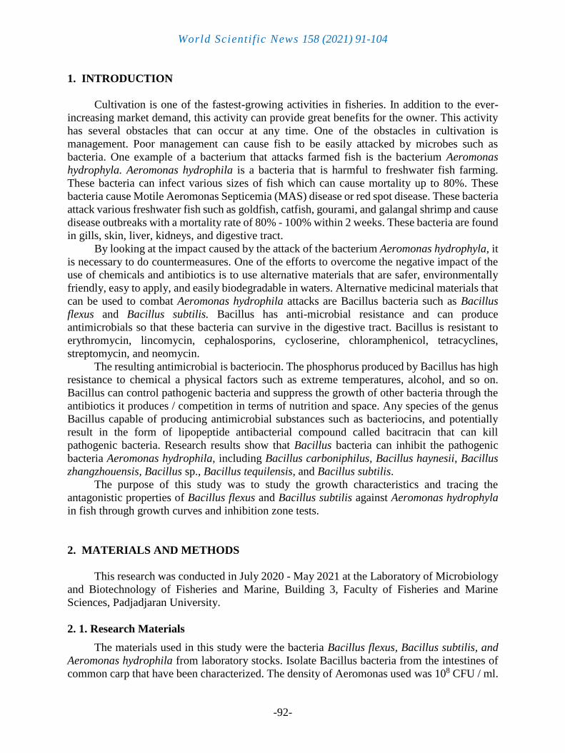
Table 2. Inhibition zone diameter of B. subtilis against A. hydrophila bacteria.
Treatment Deuteronomy 24th hour 48th hour 72nd hour
A (4000 ppm)
1 9.26 mm 8.67 mm 8.46 mm
2 - - -
3 - - -
Average 9.26 mm 8.67 mm 8.46 mm
B (Aquades)
1 - - -
2 - - -
3 - - -
Average - - -
C (Supernatant 100%)
1 - - -
2 - - -
3 - - -
Average - - -
World Scientific News 158 (2021) 91-104
-100-
Treatment Deuteronomy 24th hour 48th hour 72nd hour
D (Supernatant 50%)
1 - - -
2 - - -
3 - - -
Average - - -
E (Culture)
1 - - -
2 - - -
3 - - -
Average - - -
Based on Table 2, it can be seen that the average diameter of the inhibition zone formed
by chloramphenicol antibiotics as a positive control was 9.26 mm at 24 hours after incubation
and decreased the diameter of the inhibition zone to 8.67 mm after 48 hours of incubation
decreased the size of the inhibition zone diameter after 72 hours of incubation to 8.46 mm.
Antimicrobial activity is categorized as having a very strong sensitivity level if the
inhibition zone diameter reaches > 20 mm, the strong sensitivity level category is given if it has
an inhibition zone diameter of about 10-20 mm, the moderate sensitivity level category has an
inhibition zone diameter of about 5-10 mm, and the sensitivity level category is weak when the
diameter ranges from 6 mm. Based on the statement is known inhibition zone test results
Bacillus subtilis to the bacteria Aeromonas hydrophila belonging to the weaker categories.
No inhibition zone formation on paper discs was etched with the bacteria Bacillus flexus
and Bacillus subtilis can be caused by several factors. One of these factors is the condition of
the Aeromonas bacteria which is already resistant to antibiotics. One of the factors that affect
the inhibition zone diameter that is the turbidity suspension of bacteria. If the bacterial
suspension is less cloudy, the diameter of the inhibition zone will be bigger and the opposite is
true. If the suspense is more cloudy, the diameter will be smaller.
In addition, the thickness of the agar medium can also be a factor affecting the diameter
of the inhibition zone for bacterial growth. The thickness of the effective agar is about 4 mm.
If it is less than 4 mm the extract diffusion will be faster. Factors that influence antibacterial
activity are concentration, the content of antibacterial compounds, diffusion power, and the type
of bacteria that is inhibited.
In addition, the factors that affect the work of antimicrobial substances or substances
include microbial age, temperature, and antimicrobial ingredients. The formation of the
inhibition zone is highly dependent on the amount of antibacterial material that is dripped into
the disc, the solubility of the antibacterial agent on the media, the diffusion coefficient, and the
antibacterial effectiveness.

World Scientific News 158 (2021) 91-104
-101-
4. CONCLUSIONS
Based on the results, it is known that B. flexus bacteria experience an exponential phase
at the 6th to the 12th hour, after that it enters the stationary phase from the 14th hour to the
72nd hour. B. subtili bacteria experience an exponential phase at the 20th hour. Until the 40th
hour, then experiencing a stationary phase at the 42nd hour to the 72nd hour, and the growth of
A. hydrophila bacteria experiences an exponential phase at the 6th to the 28th hour, the
stationary phase at 30 to the hour 72nd. Meanwhile, the inhibition zone test results showed the
formation of the inhibition zone only in the positive control treatment in both Bacillus flexus
and Bacillus subtilis, the bacterial isolates supernatant, and the bacterial cultures of Bacillus
flexus and Bacillus subtilis were not formed.
References
[1] Otsuki N, Dang NH, Kumagai E, Kondo A, Iwata S, Morimoto C. Aqueous extract of
Carica papaya leaves exhibits anti-tumor activity and immunomodulatory effects. J
Ethnopharmacol. 2010 Feb 17; 127(3): 760-7. doi: 10.1016/j.jep.2009.11.024
[2] F Feliatra, Nursyirwani, A Tanjung, DS Adithiya, M Susanna and I Lukystyowati. The
Effectiveness of Heterotrophic Bacteria Isolated from Dumai Marine Waters of Riau,
Used as Antibacterial against Pathogens in Fish Culture. IOP Conference Series: Earth
and Environmental Science, Volume 116, 3rd International Conference on Tropical and
Coastal Region Eco Development 2017 2–4 October 2017, Yogyakarta, Indonesia.
2018, 116, 012034. https://doi.org/10.1088/1755-1315/116/1/012034
[3] Lee NK, Yeo IC, Park JW, Kang BS, Hahm YT. Isolation and characterization of a
novel analyte from Bacillus subtilis SC-8 antagonistic to Bacillus cereus. Journal of
Bioscience and Bioengineering. 2010 Sep; 110(3): 298-303. DOI:
10.1016/j.jbiosc.2010.03.002.
[4] Mulyani, Y., INP Aryantha, S. Suhandono and A. Pancoro. Intestinal Bacteria of
Common Carp (Cyprinus carpio L.) as a Biological Control Agent for Aeromonas.
Journal of Pure and Applied Microbiology 12 (2) (2018) 601-610.
https://dx.doi.org/10.22207/JPAM.12.2.18
[5] Xiong YW, Li XW, Wang TT, Gong Y, Zhang CM, Xing K, Qin S. Root exudates-
driven rhizosphere recruitment of the plant growth-promoting rhizobacterium Bacillus
flexus KLBMP 4941 and its growth-promoting effect on the coastal halophyte
Limonium sinense under salt stress. Ecotoxicol Environ Saf. 2020 May; 194: 110374.
doi: 10.1016/j.ecoenv.2020.110374
[6] Baranyi, J. and CP Single-cell and Population Lag Times as A Function of Cell Age.
Appl Environ Microbiol. 2008 Apr; 74(8): 2534–2536. doi: 10.1128/AEM.02402-07
[7] Setyati, WA, E. Martani, T. Subagiyo, and M. Zainuddin. Growth Kinetics and Protease
Activity of Isolate 36k from Mangrove Ecosystem Sediments, Karimunjawa, Jepara.
Journal of Marine Science 20 (3) (2015) 163–169
World Scientific News 158 (2021) 91-104
-102-
[8] W. A. Setyati, E. Martani, T. Triyanto, and M. Zainuddin, "Growth kinetics and
Protease Activity 36K Isolates Derived from Mangrove Ecosystem Sediment,
Karimunjawa, Jepara. Indonesian Journal of Marine Sciences, vol. 20, no. 3, pp. 163-
169, Sep. 2015. https://doi.org/10.14710/ik.ijms.20.3.163-169
[9] Walim Lili, Resvi Gumilar, Atikah Nurhayati, Rosidah, Effectivity of Solution
Mangosteen Rind (Garciana mangostana) as Medicine for Black Tilapia Juvenile
(Oreochromis niloticus Bleeker) when Infected by Aeromonas hydrophila. World
Scientific News 133 (2019) 56-70
[10] Rosidah, Maria Dewi Yunita, Isni Nurruhwati, Achmad Rizal, Histopathological
changes in gold fish (Carassius auratus (Linnaeus, 1758)) infected by Aeromonas
hydrophila bacteria with various densities. World Scientific News 142 (2020) 150-168
[11] G. Vivekanandhan, K. Savithamani, A.A.M. Hatha, P. Lakshmanaperumalsamy,
Antibiotic resistance of Aeromonas hydrophila isolated from marketed fish and prawn
of South India. International Journal of Food Microbiology, Volume 76, Issues 1–2,
2002, 165-168, https://doi.org/10.1016/S0168-1605(02)00009-0
[12] R. Harikrishnan & C. Balasundaram (2005) Modern Trends in Aeromonas hydrophila
Disease Management with Fish. Reviews in Fisheries Science, 13:4, 281-320, DOI:
10.1080/10641260500320845
[13] Afifah Shabirah, Rosidah, Yuniar Mulyani, Walim Lili, Effect of Types Isolated Lactic
Acid Bacteria on Hematocrit and Differential Leukocytes Fingerling Common Carp
(Cyprinus carpio L.) Infected with Aeromonas hydrophila bacteria. World News of
Natural Sciences 24 (2019) 22-35
[14] Atefeh Sheikhlar, Goh Yong Meng, Razak Alimon, Nicholas Romano, Mahdi Ebrahimi.
(2017) Dietary Euphorbia hirta Extract Improved the Resistance of Sharptooth Catfish
Clarias gariepinus to Aeromonas hydrophila. Journal of Aquatic Animal Health 29:4,
pages 225-235
[15] Yachan Ji, Jinquan Li, Zhendong Qin, Aihua Li, Zemao Gu, Xiaoling Liu, Li Lin, Yang
Zhou. (2015) Contribution of nuclease to the pathogenesis of Aeromonas hydrophila.
Virulence 6:5, pages 515-522
[16] Inácio Mateus Assane, Elielma Lima de Sousa, Gustavo Moraes Ramos Valladão,
Geovana Dotta Tamashiro, Eduardo Criscoulo-Urbinati, Diogo Teruo Hashimoto,
Fabiana Pilarski. (2021) Phenotypic and genotypic characterization of Aeromonas
jandaei involved in mass mortalities of cultured Nile tilapia, Oreochromis niloticus (L.)
in Brazil. Aquaculture 541, pages 736848.
[17] Sunita Kumari Yadav, Pujarini Dash, Pramoda Kumar Sahoo, Lalit C. Garg, Aparna
Dixit. (2021) Recombinant outer membrane protein OmpC induces protective immunity
against Aeromonas hydrophila infection in Labeo rohita. Microbial Pathogenesis 150,
pages 104727
[18] Ramasamy Harikrishnan, Subramanian Thamizharasan, Gunapathy Devi, Hien Van
Doan, Thipramalai Thankappan Ajith Kumar, Seyed Hossein Hoseinifar, Chellam
Balasundaram. (2020) Dried lemon peel enriched diet improves antioxidant activity,
World Scientific News 158 (2021) 91-104
-103-
immune response and modulates immuno-antioxidant genes in Labeo rohita against
Aeromonas sorbia. Fish & Shellfish Immunology 106, pages 675-684.
[19] Ayan Srivastava, Arup Mistri, Swati Mittal, Ajay Kumar Mittal. (2020) Alterations in
the epidermis of the carp, Labeo rohita (Cyprinidae: Cypriniformes), infected by the
bacteria, Aeromonas hydrophila: A scanning electron microscopic, histopathological
and immunohistochemical investigation. Journal of Fish Diseases 43: 8, pages 941-953
[20] Radhakrishnan Palanikani, Kanagaraj Muthu-Pandian Chanthini, Ramaiah Soranam,
Annamalai Thanigaivel, Sengodan Karthi, Sengottayan Senthil-Nathan, Arunachalam
Ganesan Murugesan. (2020) Efficacy of Andrographis paniculata supplements induce a
non-specific immune system against the pathogenicity of Aeromonas hydrophila
infection in Indian major carp (Labeo rohita). Environmental Science and Pollution
Research 27: 19, pages 23420-23436
[21] Murendeni Manavhela, Alfred Sichilima, Amidou Samie. (2020) Distribution and
Potential Effects of 17β-Estradiol (E2) on Aeromonas Diversity in Wastewater and Fish
Samples. Pakistan Journal of Biological Sciences 23: 3, pages 278-286.
[22] Junianto, Iskandar, Achmad Rizal, Windi Damayanti, Influence of Concentration of
Acetic Acid and Pepsin Enzyme in Nilem Fish Skin Collagen Extraction to the Amount
of Rendement Produced. World News of Natural Sciences 21 (2018) 164-170
[23] Vanessa M. da Rosa, Karine Ariotti, Caroline A. Bressan, Elisia G. da Silva, Magale
Dallaporta, Guerino B. Júnior, Silvio T. da Costa, Agueda C. de Vargas, Bernardo
Baldisserotto, Isabela A. Finamor, Maria A. Pavanato. (2019) Dietary addition of rutin
impairs inflammatory response and protects muscle of silver catfish (Rhamdia quelen)
from apoptosis and oxidative stress in Aeromonas hydrophila-induced infection.
Comparative Biochemistry and Physiology Part C: Toxicology & Pharmacology 226,
pages 108611
[24] Vito A. Mastrochirico-Filho, Raquel B. Ariede, Milena V. Freitas, Lieschen V.G. Lira,
John F.G. Agudelo, Fabiana Pilarski, Rafael V. Reis Neto, José M. Yáñez, Diogo T.
Hashimoto. (2019) Genetic parameters for resistance to Aeromonas hydrophila in the
Neotropical fish pacu (Piaractus mesopotamicus). Aquaculture 513, pages 734442
[25] Tayebeh Nemati, Seyed Ali Johari, Mehrdad Sarkheil. (2019) Will the antimicrobial
properties of ZnONPs turn it into a more suitable option than AgNPs for water
filtration? Comparative study in the removal of fish pathogen, Aeromonas hydrophila
from the culture of juvenile common carp (Cyprinus carpio). Environmental Science
and Pollution Research 26:30, pages 30907-30920.
[26] Alexanda Mzula, Philemon N. Wambura, Robinson H. Mdegela, Gabriel M. Shirima.
(2019) Phenotypic and molecular detection of Aeromonads infection in farmed Nile
tilapia in Southern highland and Northern Tanzania. Heliyon 5:8, pages e02220
[27] Yuying Fu, Qilan Cai, Yuqian Wang, Wanxin Li, Jing Yu, Guidi Yang, Wenxiong Lin,
Xiangmin Lin. (2019) Four LysR-type transcriptional regulator family proteins
(LTTRs) involved in antibiotic resistance in Aeromonas hydrophila. World Journal of
Microbiology and Biotechnology 35: 8

World Scientific News 158 (2021) 91-104
-104-
[28] Xianliang Zhao, He Chen, Zhaohui Jin, Li Li, Jie Zhang, Xianghui Kong. (2018) GC-
MS-based metabolomics analysis reveals L-aspartate enhances the antibiotic sensitivity
of neomycin sulfate-resistant Aeromonas hydrophila. Journal of Fish Diseases 41:12,
pages 1831-1841
[29] Shiqi Cao, Yi Geng, Zehui Yu, Longjun Deng, Weixiong Gan, Kaiyu Wang, Yangping
Ou, Defang Chen, Xiaoli Huang, Zhicai Zuo, Min He, Weiming Lai. (2018)
Acinetobacter lwoffii, an emerging pathogen for fish in Schizothorax genus in China.
Transboundary and Emerging Diseases 65:6, pages 1816-1822
[30] Jessyka Arruda da Cunha, Cecília de Ávila Scheeren, Viviane Pedroso Fausto, Larissa
Daiane Willrich de Melo, Bruno Henneman, Clarissa Piccinin Frizzo, Rodrigo de
Almeida Vaucher, Agueda Castagna de Vargas, Bernardo Baldisserotto. (2018) The
antibacterial and physiological effects of pure and nanoencapsulated Origanum
majorana essential oil on fish infected with Aeromonas hydrophila. Microbial
Pathogenesis 124, pages 116-121
[31] Soner Bilen, Oğuz Özkan, Kerem Alagöz, Keriman Yürüten Özdemir. (2018) Effect Of
dill (Anethum graveolens) and garden cress (Lepidium sativum) dietary
supplementation on growth performance, digestive enzyme activities and immune
responses of juvenile common carp (Cyprinus carpio). Aquaculture 495, pages 611-616
[32] J.A. Cunha, B.M. Heinzmann, B. Baldisserotto. (2018) The effects of essential oils and
their major compounds on fish bacterial pathogens – a review. Journal of Applied
Microbiology 125:2, pages 328-344
[33] V Jung-Schroers, M Adamek, S Harris, H Syakuri, A Jung, I Irnazarow, D Steinhagen.
(2018) Response of the intestinal mucosal barrier of carp (Cyprinus carpio) to a
bacterial challenge by Aeromonas hydrophila intubation after feeding with β-1,3/1,6-
glucan. Journal of Fish Diseases 41:7, pages 1077-1092
[34] Quinn, PJ, BK Markey, FC Leonard, ES Fitzpatrick, S. Fanning, and P. Hartigan. 2011.
Veterinary Microbiology and Microbial Disease. Willey-Blackwell Ltd, Oxford.