gross of esophagus
TRANSCRIPT
Dr. Monika Nema
GROSS OF ESOPHAGUS

Dr. Monika Nema
ANATOMY The esophagus is a muscular tubular
structure that measures approximately 25 cm in adult individuals.
It begins in the neck at the lower border of cricoid cartilage, opposite the sixth cervical vertebrae, descends along the front of vertebral column,through superior and posterior mediastinum,passes through the diaphragm and entering the abdomen,ends at the cardiac orifice of stomach,opposite the 11th thoracic vertebrae.

Dr. Monika Nema
GROSS Measure the dimensions of specimen. If proximal stomach included indicate
length along lesser and greater curvature
Ink outer surface of specimen.
Dr. Monika Nema
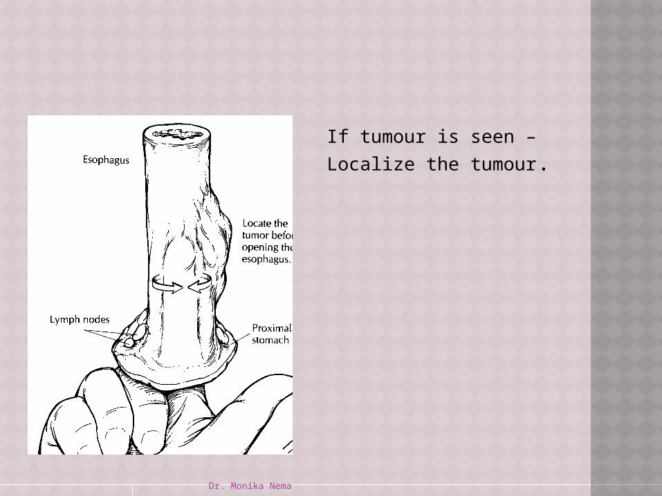
If tumour is seen –
Localize the tumour.

Dr. Monika Nema
Cut open the esophagus on the side opposite the tumour.
If a portion of the stomach is included, open along the greater curvature in continuity with the esophageal cut

Dr. Monika Nema
Measure the distance of tumour to resected margin in the fresh state. As fixation causes mucosa to retract, giving the appearance that the mucosal margins are closer to tumour than they actually are.

Dr. Monika Nema
IN THE OPEN SPECIMEN Observe the appearance and thickness
of esophageal wall. If stricture present , measure luminal
circumference of esophagus at point of narrowing and point of maximal dilation.
Inspect mucosa for hemorrhage,ulceration,scarring,puckering.
Wall: thickened? varices? Identify gastro-oesophageal junction.

Dr. Monika Nema
Normal gastric mucosa is velvety red and it contrasts sharply with smooth gray appearance of squamous epithelial lining the esophagus.
Dr. Monika Nema
TUMOUR GROSSLY APPARENT ??? If no- Try to identify the site of previous
biopsy. Biopsy site changes can be subtle.
Dr. Monika Nema
TUMOUR GROSSLY APPARENT ??? If yes- Note its dimensions. Its location with gastro-oesophageal
junction. Configuration of tumour-
Exophytic/fungating Endophytic/ulcerating Diffuse infilteration
Dr. Monika Nema
Also note- Proportion of tumour in esophagus v/s
stomach. Greatest dimension of each indiviual
component. Distance of tumour edge from margins
of resection.
Dr. Monika Nema
SECTIONS FOR HISTOLOGY
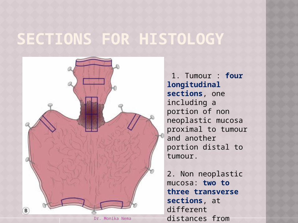
1. Tumour : four longitudinal sections, one including a portion of non neoplastic mucosa proximal to tumour and another portion distal to tumour.
2. Non neoplastic mucosa: two to three transverse sections, at different distances from tumour edge, proximally and/or distally,depending on location of tumour.

Dr. Monika Nema
Stomach, if present: two sections, one including gastroesophageal junction.
Proximal line of resection.
Distal line of resection. Lymph nodes:- Adjacent to tumour Proximal to tumour Distal to tumour Mention number of
lymph nodes found, size of largest; do they appear grossly involved by tumor?
Dr. Monika Nema
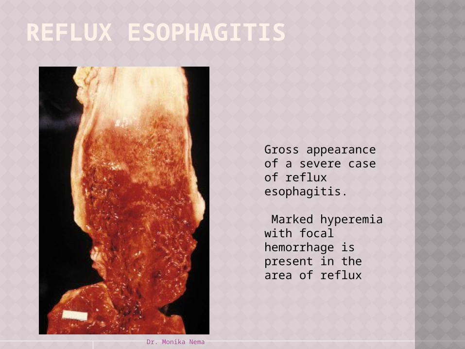
Gross appearance of a severe case of reflux esophagitis.
Marked hyperemia with focal hemorrhage is present in the area of reflux
REFLUX ESOPHAGITIS
Dr. Monika Nema
ESOPHAGEAL SQUAMOUS CELL CARCINOMA
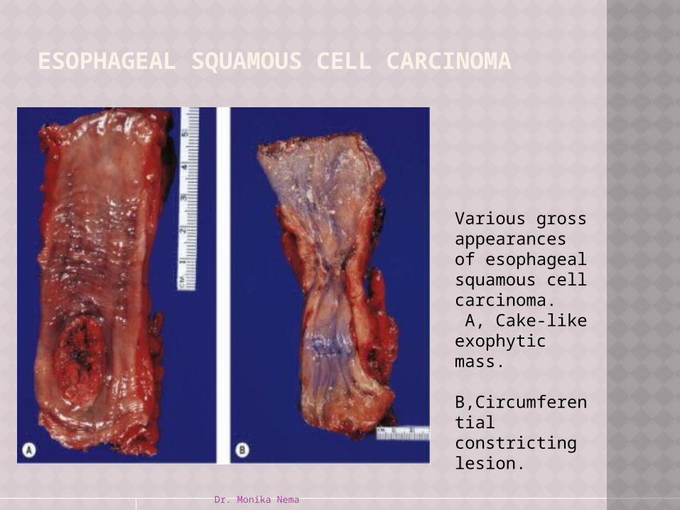
Various gross appearances of esophageal squamous cell carcinoma. A, Cake-like exophytic mass. B,Circumferential constricting lesion.

Dr. Monika Nema
ESOPHAGEAL SQUAMOUS CELL CARCINOMA
C, Elevated round nodule with central ulceration.
D, Widely invasive lesion with deep ulceration

Dr. Monika Nema
SARCOMATOID CARCINOMA OF ESOPHAGUS.
Gross appearance of sarcomatoid carcinoma of esophagus.
The tumor has a characteristic large size and polypoid shape.
The cardioesophageal junction is in the middle, and the stomach is at the bottom of the specimen.
Dr. Monika Nema
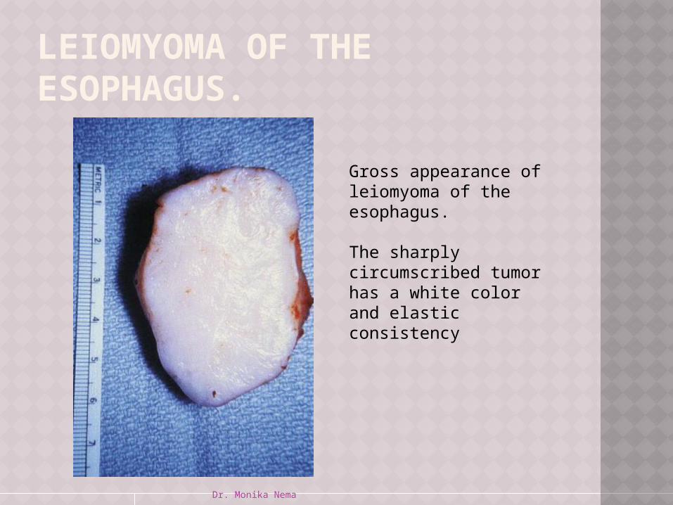
LEIOMYOMA OF THE ESOPHAGUS.
Gross appearance of leiomyoma of the esophagus.
The sharply circumscribed tumor has a white color and elastic consistency

Dr. Monika Nema
THANK YOU
SPEAKER:- Dr. MONIKA NEMA