groshong central venous catheters - c. r. bard...the groshong® central venous catheter incorporates...
TRANSCRIPT
Bard Access Systems
Groshong®
Central Venous CathetersLong Term
Instructions For Use

Table of Contents
Contents Page
Introduction ........................................................................ 1 Description Placement Schematics Groshong® Valve Function
Indications For Use............................................................ 5
Contraindications, Warnings, Cautions and Precautions .. 6
Possible Complications ..................................................... 11
Groshong® Central Venous Catheter Placement Procedures ...................................................... 12 Section A: Cutdown Section B: Percutaneous Section C: Tunneling Section D: Connector Attachment Section E: Catheter Securement
Groshong® Catheter Removal .......................................... 24
References ........................................................................ 25
Patient Information - Care and Maintenance ..................... 26 Catheter Damage Site Care Clamping the Catheter Flushing the Catheter
Changing the Injection Cap

1
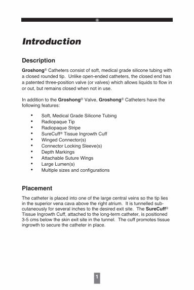
Introduction
Description
Groshong® Catheters consist of soft, medical grade silicone tubing with a closed rounded tip. Unlike open-ended catheters, the closed end has a patented three-position valve (or valves) which allows liquids to flow in or out, but remains closed when not in use.
In addition to the Groshong® Valve, Groshong® Catheters have the following features:
• Soft, Medical Grade Silicone Tubing • Radiopaque Tip • Radiopaque Stripe • SureCuff® Tissue Ingrowth Cuff • Winged Connector(s) • Connector Locking Sleeve(s) • Depth Markings • Attachable Suture Wings • Large Lumen(s) • Multiple sizes and configurations
Placement
The catheter is placed into one of the large central veins so the tip lies in the superior vena cava above the right atrium. It is tunnelled sub-cutaneously for several inches to the desired exit site. The SureCuff® Tissue Ingrowth Cuff, attached to the long-term catheter, is positioned 3-5 cms below the skin exit site in the tunnel. The cuff promotes tissue ingrowth to secure the catheter in place.
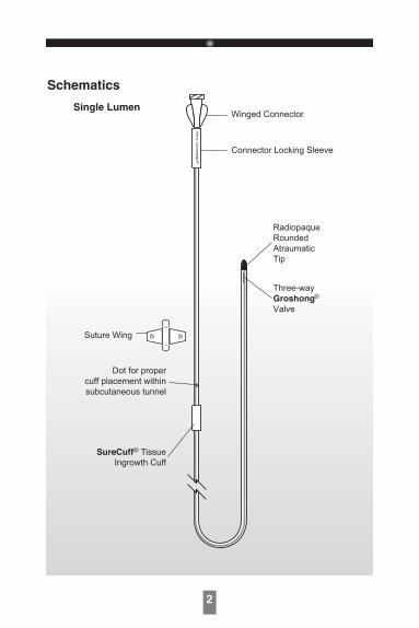
Winged Connector
Connector Locking Sleeve
Three-wayGroshongValve
RadiopaqueRoundedAtraumaticTip
Dot for propercuff placement withinsubcutaneous tunnel
Suture Wing
SureCuff® TissueIngrowth Cuff
PR
OX
GR
OS
HO
NG
®
®
Schematics
Single Lumen
2

Dual Lumen
3
Winged Connectors
Connector Locking Sleeve
Three-wayGroshongValve
RadiopaqueRoundedAtraumaticTip
Dot for propercuff placement withinsubcutaneous tunnel
SureCuff® TissueIngrowth Cuff
DIST G
RO
SHO
NG
®
PRO
X GR
OSH
ON
G®
Suture Wing
®
4
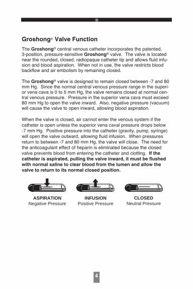
Groshong® Valve Function
The Groshong® central venous catheter incorporates the patented, 3-position, pressure-sensitive Groshong® valve. The valve is located near the rounded, closed, radiopaque catheter tip and allows fluid infu-sion and blood aspiration. When not in use, the valve restricts blood backflow and air embolism by remaining closed.
The Groshong® valve is designed to remain closed between -7 and 80 mm Hg. Since the normal central venous pressure range in the superi-or vena cava is 0 to 5 mm Hg, the valve remains closed at normal cen-tral venous pressure. Pressure in the superior vena cava must exceed 80 mm Hg to open the valve inward. Also, negative pressure (vacuum) will cause the valve to open inward, allowing blood aspiration.
When the valve is closed, air cannot enter the venous system if the catheter is open unless the superior vena caval pressure drops below -7 mm Hg. Positive pressure into the catheter (gravity, pump, syringe) will open the valve outward, allowing fluid infusion. When pressures return to between -7 and 80 mm Hg, the valve will close. The need for the anticoagulant effect of heparin is eliminated because the closed valve prevents blood from entering the catheter and clotting. If the
catheter is aspirated, pulling the valve inward, it must be flushed
with normal saline to clear blood from the lumen and allow the
valve to return to its normal closed position.
CLOSEDNeutral Pressure
ASPIRATIONNegative Pressure
INFUSIONPositive Pressure

5
The benefits provided by the Groshong® valve are:
1. Increased patient safety due to reduced risk of air embolism or bleedback.
2. Virtual elimination of heparin flushing to maintain catheter patency.
3. Reduced need for catheter clamping.
4 Reduced need for flushing when the catheter is not in use (only flushed every seven days with sterile normal saline when not in use).
Groshong® multi-lumen catheters have Groshong® valves which are rotated and staggered, allowing the concurrent infusion of incompatible drugs. Each lumen of a multi-lumen catheter is treated separately for maintenance and irrigation purposes.
Indications For Use
Groshong® Long-Term Catheters are designed for long-term vascular access and for use in patients that lack adequate peripheral venous access. They are available in single lumen and multi-lumen catheters.
All Groshong® central venous catheters are designed for the admin-istration of I.V. fluids, blood products, drugs, and parenteral nutrition solutions, as well as blood withdrawal.

6
Contraindications, Warnings, Cautions and Precautions
Contraindications
The device is contraindicated whenever:
• The presence of device related infection, bacteremia, or septicemia is known or suspected.
• The patient s body size is insufficient to accommodate the size of the implanted device.
• The patient is known or is suspected to be allergic to materials con-tained in the device.
• Severe chronic obstructive lung disease exists (percutaneous sub-clavian placement only.)
• Past irradiation of prospective insertion site.
• Previous episodes of venous thrombosis or vascular surgical proce-dures at the prospective placement site.
• Local tissue factors will prevent proper device stabilization and/or access.
Warnings:
• Intended for Single Patient Use. DO NOT REUSE. Bard Access Systems products are single use devices and should never be reimplanted. Reuse carries with it the attendant concern of cross-infection regardless of the cleaning or sterilization method. Resterilization of incompletely cleaned devices may not be effective. Any device that has been contaminated by blood should not be reused or resterilized.
7
• After use, this product may be a potential biohazard. Handle and discard in accordance with accepted medical practice and applica-ble local, state and federal laws and regulations.
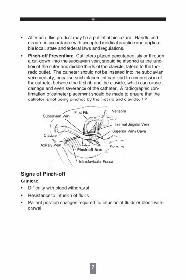
• Pinch-off Prevention: Catheters placed percutaneously or through a cut-down, into the subclavian vein, should be inserted at the junc-tion of the outer and middle thirds of the clavicle, lateral to the tho-racic outlet. The catheter should not be inserted into the subclavian vein medially, because such placement can lead to compression of the catheter between the first rib and the clavicle, which can cause damage and even severance of the catheter. A radiographic con-firmation of catheter placement should be made to ensure that the catheter is not being pinched by the first rib and clavicle. 1,2
First RibSubclavian Vein
Clavicle
Vertebra
Internal Jugular Vein
Superior Vena Cava
SternumPinch-off Area
Infraclavicular Fossa
Axillary Vein
Signs of Pinch-off
Clinical:
• Difficulty with blood withdrawal
• Resistance to infusion of fluids
• Patient position changes required for infusion of fluids or blood with-drawal

8
Radiologic:
• Grade 1 or 2 distortion on chest X-ray.
Pinch-off should be evaluated for degree of severity prior to explan-tation. Patients indicating any degree of catheter distortion at the clavicle/first rib area should be followed diligently. There are grades of pinch-off that should be recognized with appropriate chest x-ray as follows: 3,4
Grade
Grade 0
Grade 1
Grade 2
Grade 3
Severity
No distortion
Distortion presentwithout luminalnarrowing
Distortion presentwith luminal nar-rowing
Catheter transec-tion or fracture
Recommended Action
No action.
Chest x-ray should be taken every oneto three months to monitor progressionof pinch off to grade 2 distortion.Shoulder positioning during chest x-rays should be noted as it can con-tribute to changes in distortion grades.
Removal of the catheter should beconsidered.
Prompt removal of the catheter.
Cautions:
• Carefully read and follow all instructions prior to use.
• Federal (U.S.A.) law restricts this device to sale by or on the order of a physician.
• Only qualified healthcare practitioners should insert, manipulate and remove these devices.
Precautions:
Follow Universal Precautions when inserting and maintaining
the catheter.
Follow all contraindications, warnings, cautions, precautions and
instructions for all infusates as specified by its manufacturer.
9
Use aseptic techniques whenever the catheter lumen is opened or connected to other devices. Povidone-iodine is the suggested anti-septic to use with this device and components. Acetone and tincture of iodine should not be used because they could adversely affect the performance of the catheter and connectors. 10% acetone/70% isopropyl alcohol swabsticks used for dressing changes should not adversely affect the catheter.
I. Prior to beginning placement procedure, do the following: • Examine package carefully before opening to confirm its integrity
and that the expiration date has not passed. The device is supplied in a double sterile package and is non-pyrogenic. Do not use if
package is damaged, opened or the expiration date has passed. Sterilized by ethylene oxide. Do not Resterilize.
• Inspect kit for inclusion of all components.
• Fill (prime) the device with normal saline solution to help avoid air embolism.
• When using an introducer kit, verify that the catheter fits easily through the introducer sheath.
II. To avert device damage and/or patient injury during
placement: • Avoid accidental device contact with sharp instruments and
mechanical damage to the catheter material. Use only smooth-edged atraumatic clamps or forceps.
• Avoid perforating, tearing or fracturing the catheter when using a guidewire.
• Do not use the catheter if there is any evidence of mechanical
damage or leaking.
• Avoid sharp or acute angles during implantation which could com-promise the patency of the catheter lumen(s).
• Use suture wings to secure catheters.
• Do not place sutures directly around the catheter.
10
• When using percutaneous introducers:
- Carefully insert the introducer and catheter to avoid inadvertent penetration to vital structures in the thorax.
- To avoid blood vessel damage, do not allow the percutaneous
introducer sheath to remain indwelling in the blood vessel
without the internal support of a catheter or dilator.
- Simultaneously advance the sheath and dilator with rotational motion to help prevent sheath damage.
III. After insertion, observe the following precautions to
avoid device damage and/or patient injury: • Do not use the catheter if there is any evidence of mechanical
damage or leaking. Damage to the catheter may lead to rup-
ture, fragmentation and possible embolism and surgical remov-
al.
• Accessories and components used in conjunction with this device should incorporate Luer lock connections.
• If signs of extravasation exist, discontinue injections. Begin appropriate medical intervention immediately.
• Infusion pressure greater than 25 psi (172 kPa) may damage blood vessels and viscus and is not recommended. DO NOT USE A
SYRINGE SMALLER THAN 10 ml!

11
Possible Complications
The use of an indwelling central venous catheter provides an important means of venous access for critically ill patients; however, the potential exists for serious complications including the following:
• Air Embolism• Bleeding• Brachial Plexus Injury• Cardiac Arrhythmia• Cardiac Tamponade• Catheter or Cuff Erosion
Through Skin• Catheter Embolism• Catheter or Cuff Occlusion• Catheter Occlusion, Damage or Breakage due to Compression Between the Clavicle and First Rib• Catheter-related Sepsis • Endocarditis• Exit Site Infection• Exit Site Necrosis• Extravasation• Fibrin Sheath Formation• Hematoma• Hemothorax
• Hydrothorax• Intolerance Reaction to Implanted Device• Laceration of Vessels or Viscus• Perforation of Vessels or Viscus• Pneumothorax• Spontaneous Catheter Tip Malposition or Retraction• Thoracic Duct Injury• Thromboembolism• Venous Thrombosis• Ventricular Thrombosis• Vessel Erosion• Risks Normally Associated with Local and General Anesthesia, Surgery, and Post-Operative Recovery
These and other complications are well documented in medical litera-ture and should be carefully considered before placing the catheter.
12
Groshong® Central Venous Catheter Placement Procedures
Before beginning procedure, read the “Contraindications,
Warnings, Cautions and Precautions” and “Possible
Complications” sections of this manual.
Section A: Cutdown
1. Create sterile field and open tray.
2. Prep cutdown area, tunnel and tunnel exit areas.
3. Perform local anesthetic infiltration in cutdown area and along pathway chosen for the subcutaneous catheter tunnel.
4. Irrigate the catheter with sterile normal saline via the flushing hub:
Single-Lumen Catheters have catheter-stylet attached to the flushing hub.
Multi-Lumen Catheters have a flushing hub and no stylet.
5. Place patient in the Trendelenburg position with head turned away from the intended venipuncture site.
6. Surgically isolate the desired vessel through a small skin incision.
7. Insert the catheter assembly and advance to desired position in vessel by using the graduations on the catheter which are in 2.5 cm (3.5 Fr catheter) or 5 cm (all other catheters) intervals from the distal tip.
Refer to the “Warnings” section concerning Catheter Pinch-off.
13
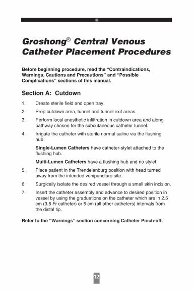
SuperiorVena Cava
Ventricle
Atrium
Catheter Tip Placement
8. Verify catheter tip location radio-graphically. The preferred loca-tion of the catheter tip is at the junction of the superior vena cava and the right atrium. Caution:
Avoid positioning the catheter
tip in the right atrium.
9. Measure catheter against chest wall of patient to determine desired location of SureCuff® tissue ingrowth cuff and exit site. Mark locations.
For Tunneling Instructions refer to Section C.
Section B: Percutaneous
1. Create sterile field and open tray.
2. Prep venipuncture, tunnel and exit site areas. Position drape.
3. Perform local anesthetic infiltration in venipuncture, tunnel and tunnel exit areas.
4. Irrigate the catheter with sterile normal saline via the flushing hub:
Single-Lumen Catheters have catheter-stylet integrated in the flushing hub.
Multi-Lumen Catheters have a flushing hub and no stylet.
5. Place patient in the Trendelenburg position with head turned away from the intended venipuncture site.
14
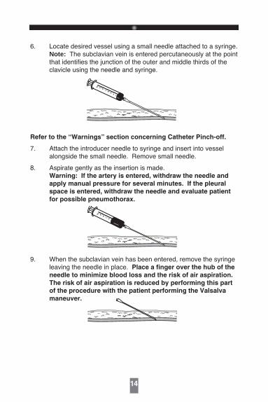
6. Locate desired vessel using a small needle attached to a syringe. Note: The subclavian vein is entered percutaneously at the point
that identifies the junction of the outer and middle thirds of the clavicle using the needle and syringe.
34
56
78
910
Refer to the “Warnings” section concerning Catheter Pinch-off.
7. Attach the introducer needle to syringe and insert into vessel alongside the small needle. Remove small needle.
8. Aspirate gently as the insertion is made. Warning: If the artery is entered, withdraw the needle and
apply manual pressure for several minutes. If the pleural
space is entered, withdraw the needle and evaluate patient
for possible pneumothorax.
34
56
78
910
9. When the subclavian vein has been entered, remove the syringe leaving the needle in place. Place a finger over the hub of the
needle to minimize blood loss and the risk of air aspiration.
The risk of air aspiration is reduced by performing this part
of the procedure with the patient performing the Valsalva
maneuver.

15
10. Straighten “J ” tip of guidewire with tip straightener and insert tapered end of tip straightener into the needle. Tip straightener should not be advanced over the guidewire beyond the guidewire tip. Caution: Do not insert guidewire beyond the bevel of
the needle while removing straightener from the needle, in
order to prevent guidewire shearing. Remove the tip straight-ener and advance the guidewire into the superior vena cava. Advance the guidewire as far as appropriate for the procedure. Verify correct positioning radiographically.
11. Gently withdraw and remove needle. Caution: If the guidewire must be withdrawn while the nee-
dle is inserted, remove both the needle and guidewire as a
unit to help prevent the needle from damaging or shearing
the guidewire.
12. Make a small (approx. 1 cm wide) incision parallel to the clavicle, positioning the guidewire at the center of the incision to permit proper entry of vessel dilator and sheath introducer.
(For Peel-Apart Introducer instructions proceed to step 20)

16
Intro-Eze® Introducer Instruction:
13. Advance the vessel dilator and sheath introducer as a unit over the exposed guidewire using a rotational motion. Advance it into the subclavian vein as a unit, leaving at least 2 cms of sheath exposed. Warning: Avoid vessel perforation.
14. Withdraw the vessel dilator and “J ” guidewire, leaving the sheath in place. Warning: Hold thumb over exposed orifice of
sheath to prevent air aspiration. The risk of air aspiration is
reduced by performing this part of the procedure with the
patient performing the Valsalva maneuver.
15. Advance the catheter through the sheath and into the vein.
17
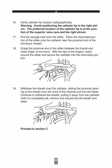
16. Verify catheter tip location radiographically. Warning: Avoid positioning the catheter tip in the right atri-
um. The preferred location of the catheter tip is at the junc-
tion of the superior vena cava and the right atrium.
17. Pull the storage tube from the slitter. Place the channeled por-tion of the slitter onto the catheter near the proximal end of the introducer sheath.
18. Grasp the proximal end of the slitter between the thumb and index finger of one hand. With the tips of the fingers, reach around the slitter and secure the catheter into the channeled por-tion.
19. Withdraw the sheath over the catheter, sliding the proximal open-ing of the sheath over the nose of the channel and into the blade. Continue to withdraw the sheath, pulling it away from the catheter until it is completely slit, remove and discard the slit sheath and slitter.
Proceed to section C.

18
Peel-Apart Sheath Introducer Instructions:
20. Advance the vessel dilator and sheath introducer as a unit over the exposed guidewire using a rotational motion. Advance it into the subclavian vein as a unit, leaving at least 2 cms of sheath exposed. Warning: Avoid vessel perforation.
21. Gently withdraw the vessel dilator and “J” guidewire, leaving the sheath in place.
22. Warning: Hold thumb over exposed orifice of sheath to
prevent air aspiration. The risk of air aspiration is reduced
by performing this part of the procedure with the patient per-
forming the Valsalva maneuver.

19
23. Insert catheter-stylet assembly into lumen of sheath and advance to desired position in vessel by using the graduations on the catheter which are in 2.5 cm (3.5 Fr catheter) or 5 cm (all other catheters) intervals from the distal tip.
24. Verify catheter tip location radiographically. Warning: Avoid positioning the catheter tip in the right atri-
um. The preferred location of the catheter tip is at the junc-
tion of the superior vena cava and the right atrium.
25. Grasp the two handles of the peel-apart sheath and pull outward and upward at the same time.
26. Peel the sheath away from the catheter completely. Make sure the catheter is not dislodged from vessel as sheath is removed.
27. Use the depth markings on the catheter to ensure proper inser-tion depth.
20
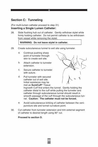
Section C: Tunneling
(For multi-lumen catheter proceed to step 31)Inserting a Single Lumen Catheter:
28. Slide flushing hub out of catheter. Gently withdraw stylet while firmly holding catheter. Do not permit catheter to be withdrawn from vessel while removing the stylet.
WARNING: Do not leave stylet in catheter.
29. Create subcutaneous tunnel to exit site using tunneler.
a. Continue pushing sharp point of tunneler through skin to create exit site.
b. Attach catheter to tunneler extension.
c. Secure catheter to tunneler with suture.
d. Pull tunneler with secured catheter out of exit site. Initial resistance may be met as SureCuff® Tissue Ingrowth Cuff first enters the tunnel. Gently holding the catheter distal to the cuff while pulling the tunneler and catheter through subcutaneous tunnel should result in smooth passage of the cuff through the subcutaneous tun-nel. Caution: The catheter must not be forced.
e. Avoid subcutaneous kinking of catheter between the veni-puncture site and tunnel curvature point.
30. Cut catheter from tunneler extension and trim external segment of catheter to desired length using 90° cut.
Proceed to section D.

21
Inserting a Multi-Lumen Catheter:
31. The multi-lumen catheter has no stylet that needs to be removed.
32. Create subcutaneous tunnel to exit site using tunneler.
a. Continue pushing sharp point of tunneler through skin to create exit site.
b. Cut flushing hub assembly from proximal end of catheter.
c. Attach catheter to tun-neler extension.
d. Secure catheter to tun-neler with suture.
e. Pull tunneler with secured catheter out of exit site. Initial resistance may be met as SureCuff® Tissue Ingrowth Cuff first enters the tunnel. Gently holding the catheter distal to the cuff while pulling the tunneler and catheter through subcutaneous tunnel should result in smooth passage of the cuff through the subcutaneous tunnel. Caution: The catheter must not be forced.
f. Avoid subcutaneous kinking of catheter between the veni-puncture site and tunnel curvature point.
33. Cut the extension tubes at the “Y” connection adjacent to the tun-neler to allow separation of the catheter lumens.
Section D: Connector Attachment
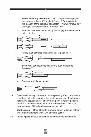
34. Secure connector(s) to catheter per the following instructions:
a. During Catheter Placement: Cut catheter at a 90° angle and trim external segment of catheter to desired length. For multi-lumen catheters, match the connector hub color to the color of the respective tubing stripes. Proceed to b.
22
When replacing connector. Using aseptic technique, cut the catheter off at a 90° angle, 0.5 in. (12.7 mm) distal to the location of the previous connector. This will remove any damaged catheter material. Proceed to b.
b Transfer clear connector locking sleeve (X) from connector onto catheter.
X
c. Firmly push catheter onto connector to position (Y).Y
d. Slide clear connector locking sleeve over catheter to Position (Y).
Y
e. Remove and discard stylet.
35. Draw blood through catheter to insure patency after placement is
complete, but before suturing the venipuncture site. If catheter is not patent, adjust catheter at curvature point to relieve possible restriction. Flush catheter with 10ml sterile saline solution to clear catheter of blood and create a saline lock.
Multi-Lumen — Draw blood through red lumen to insure patency and irrigate all lumens with 10ml of sterile saline.
36. Attach injection cap(s) or connect to intravenous fluid source.
23
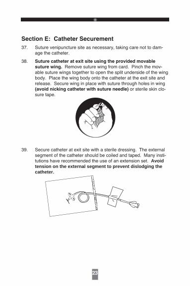
Section E: Catheter Securement
37. Suture venipuncture site as necessary, taking care not to dam-age the catheter.
38. Suture catheter at exit site using the provided movable
suture wing. Remove suture wing from card. Pinch the mov-able suture wings together to open the split underside of the wing body. Place the wing body onto the catheter at the exit site and release. Secure wing in place with suture through holes in wing (avoid nicking catheter with suture needle) or sterile skin clo-sure tape.
39. Secure catheter at exit site with a sterile dressing. The external segment of the catheter should be coiled and taped. Many insti-tutions have recommended the use of an extension set. Avoid tension on the external segment to prevent dislodging the catheter.
24
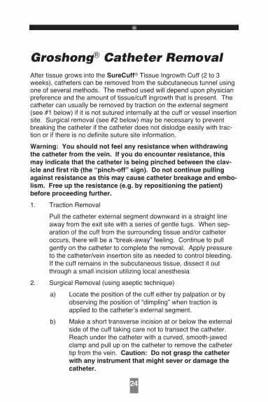
Groshong® Catheter RemovalAfter tissue grows into the SureCuff® Tissue Ingrowth Cuff (2 to 3 weeks), catheters can be removed from the subcutaneous tunnel using one of several methods. The method used will depend upon physician preference and the amount of tissue/cuff ingrowth that is present. The catheter can usually be removed by traction on the external segment (see #1 below) if it is not sutured internally at the cuff or vessel insertion site. Surgical removal (see #2 below) may be necessary to prevent breaking the catheter if the catheter does not dislodge easily with trac-tion or if there is no definite suture site information.
Warning: You should not feel any resistance when withdrawing
the catheter from the vein. If you do encounter resistance, this
may indicate that the catheter is being pinched between the clav-
icle and first rib (the “pinch-off” sign). Do not continue pulling
against resistance as this may cause catheter breakage and embo-
lism. Free up the resistance (e.g. by repositioning the patient)
before proceeding further.
1. Traction Removal
Pull the catheter external segment downward in a straight line away from the exit site with a series of gentle tugs. When sep-aration of the cuff from the surrounding tissue and/or catheter occurs, there will be a “break-away” feeling. Continue to pull gently on the catheter to complete the removal. Apply pressure to the catheter/vein insertion site as needed to control bleeding. If the cuff remains in the subcutaneous tissue, dissect it out through a small incision utilizing local anesthesia.
2. Surgical Removal (using aseptic technique)
a) Locate the position of the cuff either by palpation or by observing the position of “dimpling” when traction is applied to the catheter s external segment.
b) Make a short transverse incision at or below the external side of the cuff taking care not to transect the catheter. Reach under the catheter with a curved, smooth-jawed clamp and pull up on the catheter to remove the catheter tip from the vein. Caution: Do not grasp the catheter
with any instrument that might sever or damage the
catheter.
25
c) Dissect out the cuff. Transect the catheter on the exterior side of the cuff and remove the interior portion of the cath-eter and cuff through the incision.
Caution: Do not cut the catheter before removal from
vein to avoid catheter embolism.
d) Remove the exterior segment of the catheter by pulling it from the skin exit site.
e) Apply pressure to the catheter/vein insertion site as need-ed to control bleeding.
f) Close the incision with a suture as needed. Apply antibiot-ic ointment to incision and skin exit sites and an occlusive dressing to prevent air embolism through the tract.
References
1. Aitken, D.R. and Minton, J.P. “The Pinch-Off Sign: A Subclavian Catheters”, American Journal of Surgery, Vol. 148,
Nov. 1984, pp. 633-636.
2. Rubenstein, R.B., Alberty, R.E., et al. “Hickman® Catheter Separation”, JPEN, Vol. 9, No. 6, Nov./Dec. 1985, pp. 754-757.
3. Hinke, D.H.; Zandt-Stastny, D.A.; Goodman, L.R.; et al. Pinch-off syndrome: A complication of implantable subclavian venous access devices. Radiology 177: 353-356, 1990.
4. Ingle, Rebecca,; Nace, Corinne, Venous Access Devices: Catheter Pinch-off and Fracture, 1993, Bard Access Systems

Patient Information - Catheter Care and Maintenance
Catheter Damage
If the catheter or connection is damaged or dislodged during orafter surgery, immediately clamp the catheter with an atraumaticcatheter clamp or kink and tape it. The catheter should be repaired as soon as possible using the designated Groshong® repair kit for that particular catheter size. Damage close to the catheter hub can be repaired using the appropriate Groshong® replacement connector.
Site Care
Supplies you will need:
• Sterile gloves (if required)• 3 Alcohol swabsticks• Hydrogen peroxide• 3 Povidone iodine swabsticks• Povidone iodine ointment packet• 1 Alcohol wipe• Sterile 2 in. x 2 in. (5 cm x 5 cm) gauze dressing• 1 Sterile pre-cut 2 in. x 2 in. (5 cm x 5 cm) gauze dressing• Sterile cotton tipped applicators• 1 Sterile cover dressing (transparent or tape)• Tape
1. Clean the work surface by wiping with a paper towel that has been moistened with alcohol. Wipe dry or allow to air dry. Then place supplies on the cleaned surface.
2. Wash your hands thoroughly using warm soapy water. Rinse completely and dry using a clean towel or fresh paper tow-els.
3. Carefully open the dressing kit, or unwrap supplies, without touching the inside surfaces of the kits or wrappers.
26
4. Carefully remove the old dressing, starting from the top of the dressing and working downward. Remove the tape or dressing carefully to avoid irritating your skin or pulling on the catheter.
Caution: Do not use scissors or any sharp-edged instruments as they could damage the catheter.
5. Wash your hands again.
6. Do a careful observation of the exit site and the skin around it. If you notice anything unusual, finish the dressing procedure and then call your doctor.
7. If you are instructed to use gloves, put on the pair of sterile gloves following the procedure you were taught.
Be careful to not touch anything except the supplies being
used for site care.
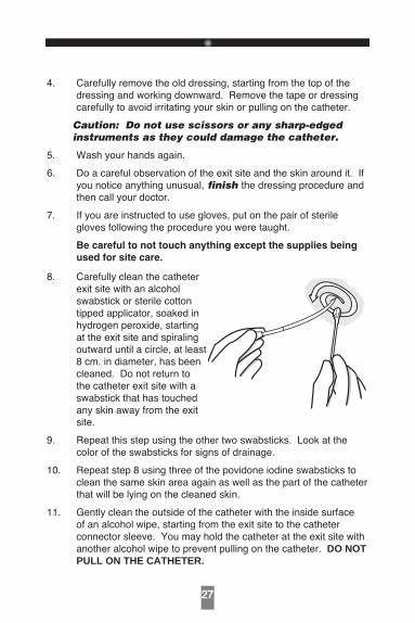
8. Carefully clean the catheter exit site with an alcohol swabstick or sterile cotton tipped applicator, soaked in hydrogen peroxide, starting at the exit site and spiraling outward until a circle, at least 8 cm. in diameter, has been cleaned. Do not return to the catheter exit site with a swabstick that has touched any skin away from the exit site.
9. Repeat this step using the other two swabsticks. Look at the color of the swabsticks for signs of drainage.
10. Repeat step 8 using three of the povidone iodine swabsticks to clean the same skin area again as well as the part of the catheter that will be lying on the cleaned skin.
11. Gently clean the outside of the catheter with the inside surface of an alcohol wipe, starting from the exit site to the catheter connector sleeve. You may hold the catheter at the exit site with another alcohol wipe to prevent pulling on the catheter. DO NOT PULL ON THE CATHETER.
27

Clamp
Here
12. Allow the povidone iodine on the skin and catheter to air dry at least two minutes.
13. Apply a small amount
of povidone iodine ointment to the exit site (optional).
14. Place the pre-cut gauze dressing over the ointment at the exit site fitting it snugly around the cathe-ter. Place the 2 in. x 2 in. (5 cm x 5 cm)gauze over the pre-cut gauze and catheter.
15. Apply the cover dressing (tape or transparent dressing) following the directions in the package as well as instructions from your doctor or nurse.
16. Coil the catheter, check to see that it is not kinked or pinched, and secure it to the chest or dressing with tape. This will prevent pulling of the catheter at the exit site and decrease irritation.
17. Always secure the catheter in such a way that you can easily see
the cap end. Your doctor or nurse will help you select the best method to secure the catheter. The type of clothing and normal activity will need to be considered in this selection. You should periodically look at the capped end to be sure it is intact.
28
Clamping the Catheter
Under normal circumstances, your catheter will not need to be clamped. If damage to the catheter occurs, the catheter should be clamped immediately.
a. Use only smooth-edged clamps.
b. Follow the directions or your doctor or nurse regarding when to clamp.
When should you clamp?
You should clamp if there is any damage to the catheter or the catheter connector or if there is any separation of the catheter and the catheter connector: Always have a clamp available for emergencies.
Flushing the Catheter
Supplies you will need:
• Alcohol or povidone iodine wipe.• 10ml syringe or larger with attached one inch needle filled with
5ml of normal saline, prepared for use.• Tape.
The steps in the procedure are:
1. Collect your supplies in a convenient place.
2. Wash your hands thoroughly.
3. Remove the tape that is around the cap.
4. Clean the cap with an alcohol or povidone iodine wipe. If you use the iodine wipe, allow the cap to air dry for two minutes—be sure not to touch the cap during this time. Do not blow on the area or allow the clean cap to dangle since this increases the chance of contamination of the area with germs.
5. Remove the needle cover and carefully insert the needle into the center of the catheter injection cap.
6. Inject the normal saline into the catheter. As you inject the last 0.5 ml, withdraw the needle from the injection cap. If you are
29
flushing the catheter of a child, do not flush too rapidly
because the child’s circulatory system is small and sensitive
to rapid changes in volume and pressure.
1� � 2� � 3� � 4� � 5� � �
7. Discard the syringe and needle into a biohazard container.
8. Re-tape the cap as outlined in the cap change procedure.
If you have a multi-lumen catheter, use a separate syringe to flush each lumen with sterile normal saline. Your doctor or nurse will give you
additional information for the care of multi-lumen catheters.
Changing the Injection Cap
Supplies you will need:
• Sterile injection cap.• Alcohol or povidone iodine wipe.• Tape.
The procedure to change the cap:
1. Wash your hands thoroughly.
2. Open the package of the new injection cap and prepare accord-ing to your instructions. Be sure the cap does not touch the outer surface of the package.
NOTE: You may need to prefill the injection cap with sterile nor-mal saline if it is a long cap with significant air space. Your doctor or nurse will teach you this additional procedure.
3. Remove the old tape from around the cap by unpeeling the tape. NEVER attempt to cut the tape with scissors as you may damage the catheter.
4. Using an alcohol or povidone iodine wipe, clean around the place where the cap is connected to the catheter. Allow to air dry.
30

5. While holding the catheter connector below the level of your
heart, unscrew the old cap and discard. (The fluid level in the catheter will drop part-way into the catheter if the connector is held above the level of your heart.)
6. Pick up the new cap only by the top and remove the sterile tip protector. Attach the new cap by firmly screwing it onto the cath-eter connector.
7. Cut a 5 cm piece of tape and make tabs on each end by folding back 1 cm. Apply the sticky part of the tape around the connec-tion of the cap and catheter and fasten securely.
8. Press ends of the tape together. The tabs on the end of the tape will enable you to remove it very easily.
31
An issued or revision date for these instructions is included for the user s information. In the event two years have elapsed between this date and product use, the user should contact Bard Access Systems to see if additional product information is available.
Revised Date: March 2015
BARD, Groshong, Intro-Eze and SureCuff are trademarks and/or registered trademarks of C.R. Bard, Inc.
0738860 / 1503R© 2015 C. R. Bard, Inc. All rights reserved.
Bard Access Systems, Inc.605 North 5600 WestSalt Lake City, Utah 84116 USA 801-522-5000
For Customer Service: 800-545-0890For Clinical Information: 800-443-3385www.bardaccess.com
Manufacturer