green synthesized silver nanoparticles destroy multidrug
TRANSCRIPT

Arabian Journal of Chemistry (2015) xxx, xxx–xxx
King Saud University
Arabian Journal of Chemistry
www.ksu.edu.sawww.sciencedirect.com
ORIGINAL ARTICLE
Green synthesized silver nanoparticles destroy
multidrug resistant bacteria via reactive oxygen
species mediated membrane damage
Abbreviations: Ag NPs, Silver nanoparticles; DAD, Disk agar diffusion; DLS, Dynamic light scattering; EDX, Energy-dispersive X-ray
Fourier transform infrared spectroscopy; MBC, Minimum bactericidal concentration; MIC, Minimum inhibitory concentration; MTT
Dimethylthiazol-2-yl)-2,5-Diphenyltetrazolium Bromide; PBS, Phosphate buffer saline; SEM, Scanning electron microscopy; TEM, Trans
electron microscopy; XRD, X-ray diffraction; Rh-B, Rhodamine B* Corresponding author.
E-mail address: [email protected] (S. Roy).
Peer review under responsibility of King Saud University.
Production and hosting by Elsevier
http://dx.doi.org/10.1016/j.arabjc.2015.08.0081878-5352 � 2015 The Authors. Production and hosting by Elsevier B.V. on behalf of King Saud University.This is an open access article under the CC BY-NC-ND license (http://creativecommons.org/licenses/by-nc-nd/4.0/).
Please cite this article in press as: Das, B. et al., Green synthesized silver nanoparticles destroy multidrug resistant bacteria via reactive oxygen species mediatbrane damage. Arabian Journal of Chemistry (2015), http://dx.doi.org/10.1016/j.arabjc.2015.08.008
Balaram Dasa, Sandeep Kumar Dash
a, Debasish Mandal
a, Totan Ghosh
b,
Sourav Chattopadhyay a, Satyajit Tripathy a, Sabyasachi Das a,
Sankar Kumar Dey c, Debasis Das b, Somenath Roy a,*
a Immunology and Microbiology Laboratory, Department of Human Physiology with Community Health, VidyasagarUniversity, Midnapore, West Bengal 721 102, IndiabDepartment of Chemistry, University of Calcutta, 92, A. P. C. Road, Kolkata 700 009, IndiacDepartment of Physiology, Santal Bidroha Sardha Satabarsiki Mahavidyalaya, Goaltore, Paschim Midnapore, West Bengal711 221, India
Received 1 May 2015; accepted 6 August 2015
KEYWORDS
Silver nanoparticles (Ag
NPs);
Green synthesis;
Antibacterial activity;
Staphylococcus aureus;
Escherichia coli
Abstract The growing need of antimicrobial agent for novel therapies against multi-drug resistant
bacteria has drawn researchers to green nanotechnology. Especially, eco-friendly biosynthesis of sil-
ver nanoparticles (Ag NPs) has shown its interesting impact against bacterial infection in laboratory
research. In this study, a simple method was developed to form Ag NPs at room temperature, bio-
reduction of silver ions from silver nitrate salt by leaf extract from Ocimum gratissimum. The Ag
NPs appear to be capped with plant proteins, but are otherwise highly crystalline and pure. The
Ag NPs have a zeta potential of �15 mV, a hydrodynamic diameter of 31 nm with polydispersity
index of 0.65, and dry sizes of 18 ± 3 nm and 16 ± 2 nm, based on scanning and transmission elec-
tron microscopy respectively. The minimum inhibitory concentration (MIC) of the Ag NPs against
a multi-drug resistant Escherichia coli was 4 lg/mL and the minimum bactericidal concentration
; FTIR,
, 3-(4,5-
mission
ed mem-

2 B. Das et al.
Please cite this article in press as: Das, B. et abrane damage. Arabian Journal of Chemistr
(MBC) was 8 lg/mL, while the MIC and MBC against a resistant strain of Staphylococcus aureus
were slightly higher at 8 lg/mL and 16 lg/mL respectively. Further, the Ag NPs inhibited biofilm
formation by both Escherichia coli and S. aureus at concentrations similar to the MIC for each
strain. Treatment of E. coli and S. aureus with Ag NPs resulted in damage to the surface of the cells
and the production of reactive oxygen species. Both mechanisms likely contribute to bacterial cell
death. In summary, this new method appears promising for green biosynthesis of pure Ag NPs with
potent antimicrobial activity.
� 2015 The Authors. Production and hosting by Elsevier B.V. on behalf of King Saud University. This is
an open access article under the CCBY-NC-ND license (http://creativecommons.org/licenses/by-nc-nd/4.0/).
1. Introduction
Different pathogenic bacteria and the antibiotic resistance by
bacteria, have become a great challenge in current times. Sev-
eral efforts are involved till now by the researchers in search of
new antibacterial agents. In the present scenario, nano-scale
materials have emerged up as novel antimicrobial agents owing
to their high surface area to volume ratio and the unique chem-
ical complexities. Nanotechnology is a rapidly growing field
for the purpose of manufacturing new materials at the nano-
scale level. This area is expected to open a new platform to
fight and prevent disease using nanomaterials. Among the
most promising nanomaterials with antibacterial properties,
metallic nanoparticles became the most interesting tool
(Morones et al., 2005; Albrecht et al., 2006).
Silver nanoparticles have become the focus of extensive
research due to its good antimicrobial efficacy against mul-
tidrug resistant bacteria, viruses and other eukaryotic micro-
organisms (Gong et al., 2007). Previous studies showed that
Ag NPs are capable of performing effective antibacterial prop-
erty against Staphylococcus aureus, Escherichia coli, Vibrio
cholera, Pseudomonas aeruginosa and Salmonella typhi
(Morones et al., 2005; Moyer et al., 1965; Li et al., 2010). In
medical treatment, silver nitrate is combined with sulfonamide
to form silver sulfadiazine cream, which was used as a broad-
spectrum antibacterial agent and was used for the treatment of
burns. It is also highly effective against bacteria such as E. coli,
S. aureus, Klebsiella sp., and Pseudomonas sp. and showed con-
siderable antifungal and antiviral activities (Fox and Modak,
1974). These applications strongly depend on the physico-
chemical properties of the produced Ag NPs such as particle
size and shape, size distribution and the surface charge
(Soukupov et al., 2008). Nanoparticles can be synthesized
using various approaches including chemical, physical, and
biological methods. Silver nanoparticles synthesized by chem-
ical methods are toxic to different normal cells and lead to
non-eco-friendly by-products which may disturb the normal
cells. Ag NPs were found to exert strong acute toxic effects
to various cultured cells. Only Ag NPs exposure, exhibited
significant cytotoxicity at higher doses and induced abnormal
cellular morphology, displaying cellular shrinkage and acquisi-
tion of an irregular shape (Kawata et al., 2009). So, an ever
increasing need for environmentally friendly, non-toxic proto-
cols for nanoparticle synthesis leads to the developing interest
in biological approaches which are free from the use of toxic
chemicals as by-products. Different biological aspects for
l., Green synthesized silver nanoparticley (2015), http://dx.doi.org/10.1016/j.ara
nanoparticles fabrications have been reported up to date whichinclude bacteria (Dickson, 1999; Pum and Sleytr, 1999; Joerger
et al., 2001; Nair and Pradeep, 2002), fungi (Mukherjeeet al., 2001; Ahmad et al., 2003; Duran et al., 2005) andplants (Singhal et al., 2011; Huang et al., 2007; Leela and
Vivekanandan, 2008; Singh et al., 2015). Due to the growingneeds of eco-friendly nanoparticles, green methods are usedfor the synthesis of various metal nanoparticles. But recently,
plant extract mediated nanoparticles fabrication proved asan advantageous way over other methods. Plant extracts medi-ated synthesis of nanoparticles is gaining importance due to itssimplicity and eco-friendliness (Awwad and Salem, 2012). The
mechanisms of NPs inhibiting bacterial growth remainunclear. It has been reported that the size and shape of NPscould affect their antibacterial activity (Zhou et al., 2012).
Studies suggested four mechanisms are hypothesized forantibacterial activity and these are firstly, accumulation anddissolution of nanoparticles in the bacterial membrane chang-
ing its permeability, with subsequent release of different intra-cellular biomolecules and dissipation of the proton motiveforce across the plasma membrane (Amro et al., 2000). Secondis generation of reactive oxygen species (ROSs) in the cell by
NPs, with subsequent oxidative damage to cellular structures(Applerot et al., 2012). Third is uptake of nanoparticles and/or metallic ions into cells, followed by depletion of intracellu-
lar ATP production, disruption of DNA replication and DNAdamage and fourth is nanoparticles and its active ions whichbind with different enzymes and inactivate them, resulting in
arrest of cellular respiration (Morones et al., 2005; Raffiet al., 2008; Rai et al., 2009). The nanoparticles get attachedto the cell membrane and also penetrate inside the bacteria
and form reactive oxygen species (ROS). The bacterial mem-brane contains sulfur-containing proteins and the silvernanoparticles interact with these proteins in the cell as wellas with the phosphorus containing compounds such as
DNA. Ag NPs destabilize plasma membrane potential anddepletion of levels of intracellular ATP by targeting bacterialmembrane resulting in bacterial cell death (Raffi et al., 2008;
Rai et al., 2009). So, recent studies suggested that generatingreactive oxygen species, damaging cellular enzymes (cellularrespiratory chain), disrupting cellular membrane, and DNA
damage ultimately lead to cell lysis and death.The antibacterial activity of green synthesized Ag NPs
against some drug-resistant bacteria has been established,
but further investigation is needed to determine whether these
particles could be an option for the treatment and prevention
of drug-resistant microbial infections.
s destroy multidrug resistant bacteria via reactive oxygen species mediated mem-bjc.2015.08.008

Antibacterial activity of silver nanoparticles 3
In this article, we designed rapid biosynthesis of novel AgNPs with a simple, non-toxic, cost-effective and eco-friendlymethod at ambient conditions using Ocimum gratissimum leaf
extract utilizing the reduced property of O. gratissimum leavesextract, which shows greater efficacy against different grampositive and gram negative bacteria even at low doses.
2. Materials and methods
2.1. Culture media and chemicals
All the microbiological media and chemicals were obtained
from HiMedia Laboratories, India, Merck Ltd., SRL Pvt.,Ltd., Mumbai. Ultrapure Milli Q water was used throughoutthe study.
2.2. Bacterial strains used in this study
Multidrug resistant E. coli (MC-2) and S. aureus (MMC-20)bacteria were previously isolated in our laboratory (Dash
et al., 2012; Chakraborty et al., 2011). The strains were subcul-tured and used throughout the study. These strains are alsoresistant to several traditional antibiotics.
2.3. Preparation of the leaf extract
Leaves of O. gratissimum were collected from Vidyasagar
University campus area, West Bengal., India. 20 g fresh leavesof O. gratissimum were collected and washed gently with dou-ble distilled water to remove dust particles. The leaves were
finely chopped and exposed to the sun until they were com-pletely dry. These materials were dissolved in distilled water(10 g dust/100 mL double distilled water) and filtered withWhatman filter paper No. 1. The filtrate was collected,
freeze-dried and stored at 4 �C until use.
2.4. Synthesis and purification of Ag NPs
Synthesis of Ag NPs using dried powder of O. gratissimum leafextract was done according to the method of Sintubin et al.,(2011) with some modifications. Silver nanoparticles (Ag
NPs) were synthesized by dissolving 10�3 M of silver nitratesalt (AgNO3) in 100 mL of deionized water and this solutionwas placed in a 250-mL reaction vessel. A total of 100 mg
freeze-dried plant leaf extract was added to the AgNO3 solu-tion at room temperature for the bio-reduction process. Afterthe addition of leaf extract, the pH value of the solution wasimmediately adjusted to 10.0 pH using a 7.7 M solution of
NaOH. The reaction vessel was thereafter shaken at a rotationrate of 150 rpm in the dark condition at 30 �C for 48 h. Thesolution containing Ag NPs was then collected and centrifuged
at 3000 rpm for 10 min for the removal of excess extractcomponents.
2.5. Characterization of Ag NPs
2.5.1. UV–vis spectroscopy
To observe the optical property of biosynthesized Ag NPs,samples were analyzed for UV–vis spectroscopic studies (Shi-madzu UV/vis 1800 spectrophotometer) at room temperatureoperated at a resolution of 1 nm between 190 and 1100 nm
ranges (Dash et al., 2014).
Please cite this article in press as: Das, B. et al., Green synthesized silver nanoparticlebrane damage. Arabian Journal of Chemistry (2015), http://dx.doi.org/10.1016/j.ara
2.5.2. Fourier transform infrared spectroscopy
Ag NPs were investigated by Fourier transform IR
spectroscopy with a PerkinElmer Spectrum RX I Fouriertransform IR system with a frequency ranging from 500 to4000 cm�1 and a resolution of 4 cm�1. The KBr pellet method
was used to prepare the samples (Chattopadhyay et al., 2013a).
2.5.3. Dynamic light scattering (DLS) and zeta potential
DLS analysis was done with a Zetasizer Nano ZS (Malvern
Instruments) according to standard method with some modifi-cations (Chattopadhyay et al., 2013a). The concentration ofthe Ag NPs was 100 lg/mL sonicated for 2 min, and dynamic
particle sizes were measured by suspending two drops of anaqueous suspension of NPs in 10 mL of Millipore water. Whenthe NPs had completely dispersed in water, they were analyzed
with a DLS analyzer. The experiments were repeated severaltimes to obtain the average size of the NPs.
The zeta potential of the Ag NPs was measured by using aZetasizer-Nano ZS (Malvern, Malvern Hills, U.K.). 1 mg/mL
Ag NPs suspension was prepared in Milli-Q water. Then thissuspension was used for the experiment (Dash et al., 2014).
2.5.4. Scanning electron microscopy
The particle size and microstructure were studied by high res-olution scanning electron microscopy (SEM; instrument from
Nikon, Japan) (Chattopadhyay et al., 2013a). In brief, AgNPs were suspended in deionized water at a concentration of1 mg/mL and then sonicated using a sonicator bath until thesample forms a homogenous suspension. For size measure-
ment, the sonicated stock solution of Ag NPs (1 mg/mL) wasdiluted 20 times. Then one drop of sonicated aqueous solutionwas taken on a glass plate and dried it. Then the sample was
gold coated and images were taken. SEM was used to charac-terize the size and shape of Ag NPs.
2.5.5. Transmission electron micrograph
The particle size and microstructure were studied by high res-olution transmission electron microscopy in a JEOL 3010,
Japan, operating at 200 kV according to the method ofChattopadhyay et al., (2012) with some modifications. In brief,Ag NPs were suspended in deionized water at a concentrationof 1 mg/mL then the sample was sonicated using a sonicator
bath until sample forms a homogenous suspension. For sizemeasurement, sonicated stock solution of all Ag NPs(0.5 mg/mL) was diluted 20 times. TEM was used to character-
ize the size and shape of the Ag NPs. A drop of the aqueousAg NPs suspension was placed on to carbon-coated coppergrid and this was dried in the air to get TEM image.
2.5.6. X-ray diffraction study
The solid state dispersions of Ag NPs were evaluated withX-ray powder diffraction. Diffraction patterns were obtained
using an XPERT-PRO diffractometer (PANalytical Ltd., theNetherlands) with a radius of 240 mm. The Cu Ka radiation(Ka 1.54060 A) was Ni filtered. A system of diverging and
receiving slits of 1� and 0.1 mm, respectively, was used.The pattern was collected with 40 kV of tube voltage and30 mA of tube current and scanned over the 2h range of
10–90�.
s destroy multidrug resistant bacteria via reactive oxygen species mediated mem-bjc.2015.08.008

4 B. Das et al.
2.5.7. EDX study
This technique determines the elemental composition of a sam-
ple. In this study it was used to confirm the presence of silver inthe particles as well as to detect the other elemental composi-tions of the particles. Beside identification of the elements pre-
sent in the sample by the use of EDX it is also possible toestimate their concentration. The particle solution was diluted100-fold in water and a drop of 10 lL diluted solution was
placed on a carbon stub and air-dried. The EDX spectrumwas obtained at an acceleration voltage of 20 kV and collectedfor 19 s. Mapping was completed using pseudo-colors to repre-sent the two-dimensional spatial distribution of energy emis-
sions of the chemical elements present in the sample.Analysis was done using JEOL JSM 6360 equipped with anEDX (energy dispersive X-ray) analyzer (Majumdar et al.,
2013).
2.6. Plasma protein binding assay
Plasma protein binding assay was performed by using humanplasma according to Chattopadhyay et al., (2013b). Humanplasma samples were collected from ten healthy individuals
according to institutional bioethical approval. Two milligramsper milliliter of nanosilver was mixed with 5 mL of 50 mMPBS and 0.5 mL of human plasma (8 mg/mL) was addedtogether and stirred vigorously within a shaking incubator
for 24 h at 37 �C. The nanoparticles were centrifuged at10,000 rpm for 10 min and supernatant was used to determinethe protein concentration by following the method of Lowry
et al., (1951). Plasma without NPs was used as a control toensure that there was no protein precipitation (Lowry et al.,1951).
2.7. Antibacterial activity determination
2.7.1. MIC and MBC determination
MIC and MBC were determined by a microdilution method,using Luria broth (Hi-media, India) and inoculums of2.5 � 105 CFU/mL. In brief, 10 lL (2.5 � 105 CFU/mL) of
each bacterial strain was added individually to 1 mL of nutri-ent broth (NB). Different concentrations of test particles (puresuspension of particles was formed by sonication and it acts as
a dissolved solution which accurately reflects the amount of sil-ver available in solution to act on the microorganisms) wereadded to the test tubes containing the test strains. After 24 h
of incubation, the MIC values were obtained by checking theturbidity of the bacterial growth. The MIC value correspondedto the concentration that inhibited 99% of bacterial growth
(Dash et al., 2012).The minimum bactericidal concentration (MBC) values of
the particles were determined according to the standardmethod (Dash et al., 2012). The MBC values were determined
by subculturing the MIC dilutions onto the sterile Muller Hin-ton agar plates incubated at 37 �C for 24 h. The lowest concen-tration of the nanoparticles which completely killed the tested
bacteria was observed and tabulated as MBC level. The MBCvalue corresponded to the concentration where 100% of thebacterial growth was arrested, compared to the positive con-
trol (no treatment). All assays were performed in the Biosafetycabinet.
Please cite this article in press as: Das, B. et al., Green synthesized silver nanoparticlebrane damage. Arabian Journal of Chemistry (2015), http://dx.doi.org/10.1016/j.ara
2.7.2. Tolerance level
The tolerance levels of each bacterial strain against Ag NPs
were determined according to the method of May et al.,(2006) using the following formula:
Tolerance ¼ MBC=MIC:
2.7.3. Disk agar diffusion (DAD)
Susceptibility of different multidrug resistant bacteria to greensynthesized Ag NPs was determined by the disk agar diffusion(DAD) technique according to Bauer et al., (1966). The test
bacterium taken from an overnight culture (inoculated froma single colony) was freshly grown for 4 h having 106 CFU/mL standardized against McFarland’s standard. With this cul-
ture, a bacterial lawn was prepared on Mueller–Hinton agar.Filter paper disks of 6-mm size were used to observe antibioticsusceptibility patterns against green synthesized Ag NPs. Silver
nanoparticles and AgNO3 filter paper disks were prepared byabsorbing 10 lL of 2 mg/mL Ag NPs and AgNO3 solutionrespectively. The diameter of zone of bacterial growth inhibi-tion surrounding the disk (including the disk) was measured
(Dash et al., 2012; Bauer et al., 1966).
2.7.4. Killing kinetic assay
Killing kinetic assay of E. coli and S. aureus strains was studiedagainst Ag NPs by the method of Guggenbichler et al., (1985).Bacterial growth after treatment (at their respective MBCvalues) was measured by quantifying cell viability at 0, 2, 4,
8, 12, 18 and 24 h after incubation with Ag NPs. The growthinhibition percentage was obtained with respect to the positivecontrol. Bacterial cell viability was measured spectrophotomet-
rically using Shimadzu UV/vis 1800 spectrophotometer.
2.7.5. Inhibition of biofilm formation
Virulence factor in terms of biofilm formation was measured
according to the method of Stepanovic et al. with some mod-ifications (Stepanovic et al., 2004). 20 lL (2.5 � 105 CFU/mL)of each bacterial strain was added individually to 2 mL of
nutrient broth (NB). Different concentrations of pure suspen-sion test particles (by sonication) were added to the test tubescontaining the test strains. Ag NPs treated bacterial cells were
grown overnight in LB broth and then normalized to identicaldensities based on OD600, and 5 lL was inoculated into 500 lLof LB broth in 10 mL borosilicate glass tubes. The tubes werethen incubated statically at 30 �C for 22 h. The tubes were
rinsed with distilled water, incubated with 600 lL of 0.1%crystal violet for 30 min, and rinsed again with distilled water.1.0 mL of dimethyl sulphoxide was then added, the tube was
vortexed and allowed to stand for 10 min, and the optical den-sity was measured in Shimadzu UV 1800 UV/vis spectro-photometer at a wavelength of 570 nm.
2.7.6. Bacterial cell viability assay
Bacterial cell viability was performed after 12 h of treatmentwith Ag NPs by 3-(4,5-dimethylthiazol-2-yl)-2,5-diphenyltetra
zolium bromide (MTT) method according to standard method(Mosmann, 1983). Drug treated bacterial cultures were cen-trifuged at 1000g for 10 min at 4 �C followed by repeated
washing for two times with sterile PBS (pH 7.4). Thereafter,the medium was replaced with fresh RPMI (without phenol
s destroy multidrug resistant bacteria via reactive oxygen species mediated mem-bjc.2015.08.008
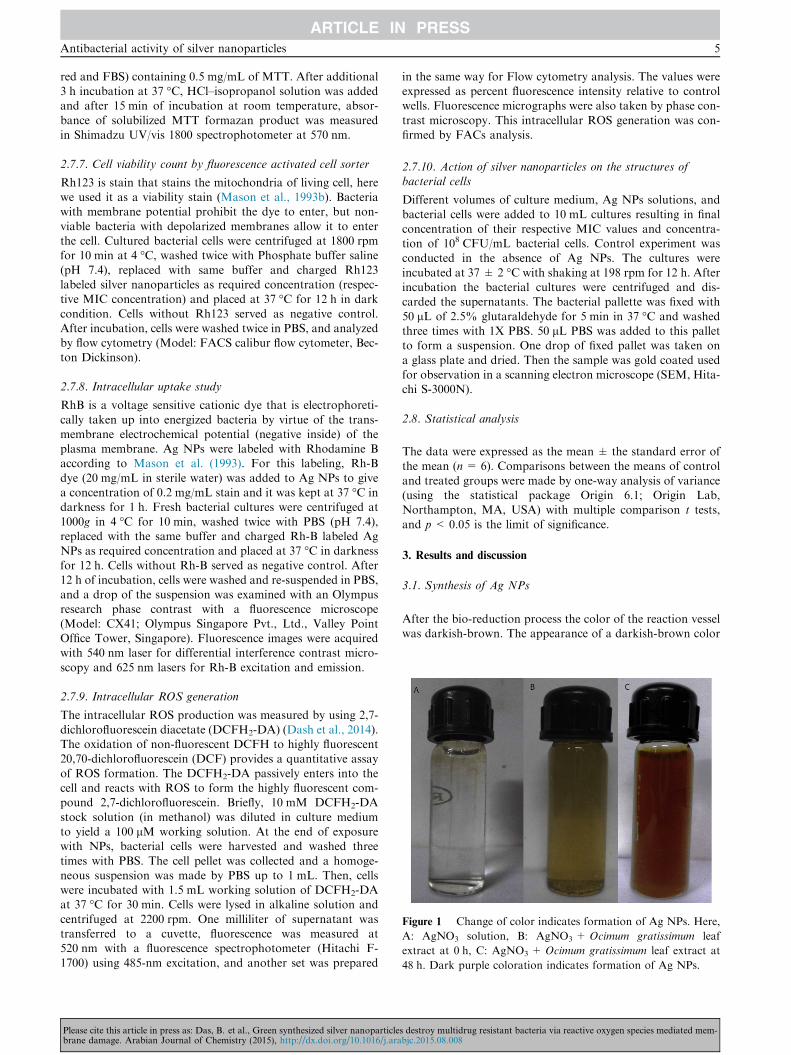
Figure 1 Change of color indicates formation of Ag NPs. Here,
A: AgNO3 solution, B: AgNO3 + Ocimum gratissimum leaf
extract at 0 h, C: AgNO3 + Ocimum gratissimum leaf extract at
48 h. Dark purple coloration indicates formation of Ag NPs.
Antibacterial activity of silver nanoparticles 5
red and FBS) containing 0.5 mg/mL of MTT. After additional3 h incubation at 37 �C, HCl–isopropanol solution was addedand after 15 min of incubation at room temperature, absor-
bance of solubilized MTT formazan product was measuredin Shimadzu UV/vis 1800 spectrophotometer at 570 nm.
2.7.7. Cell viability count by fluorescence activated cell sorter
Rh123 is stain that stains the mitochondria of living cell, herewe used it as a viability stain (Mason et al., 1993b). Bacteriawith membrane potential prohibit the dye to enter, but non-
viable bacteria with depolarized membranes allow it to enterthe cell. Cultured bacterial cells were centrifuged at 1800 rpmfor 10 min at 4 �C, washed twice with Phosphate buffer saline
(pH 7.4), replaced with same buffer and charged Rh123labeled silver nanoparticles as required concentration (respec-tive MIC concentration) and placed at 37 �C for 12 h in dark
condition. Cells without Rh123 served as negative control.After incubation, cells were washed twice in PBS, and analyzedby flow cytometry (Model: FACS calibur flow cytometer, Bec-ton Dickinson).
2.7.8. Intracellular uptake study
RhB is a voltage sensitive cationic dye that is electrophoreti-
cally taken up into energized bacteria by virtue of the trans-membrane electrochemical potential (negative inside) of theplasma membrane. Ag NPs were labeled with Rhodamine Baccording to Mason et al. (1993). For this labeling, Rh-B
dye (20 mg/mL in sterile water) was added to Ag NPs to givea concentration of 0.2 mg/mL stain and it was kept at 37 �C indarkness for 1 h. Fresh bacterial cultures were centrifuged at
1000g in 4 �C for 10 min, washed twice with PBS (pH 7.4),replaced with the same buffer and charged Rh-B labeled AgNPs as required concentration and placed at 37 �C in darkness
for 12 h. Cells without Rh-B served as negative control. After12 h of incubation, cells were washed and re-suspended in PBS,and a drop of the suspension was examined with an Olympusresearch phase contrast with a fluorescence microscope
(Model: CX41; Olympus Singapore Pvt., Ltd., Valley PointOffice Tower, Singapore). Fluorescence images were acquiredwith 540 nm laser for differential interference contrast micro-
scopy and 625 nm lasers for Rh-B excitation and emission.
2.7.9. Intracellular ROS generation
The intracellular ROS production was measured by using 2,7-
dichlorofluorescein diacetate (DCFH2-DA) (Dash et al., 2014).The oxidation of non-fluorescent DCFH to highly fluorescent20,70-dichlorofluorescein (DCF) provides a quantitative assay
of ROS formation. The DCFH2-DA passively enters into thecell and reacts with ROS to form the highly fluorescent com-pound 2,7-dichlorofluorescein. Briefly, 10 mM DCFH2-DA
stock solution (in methanol) was diluted in culture mediumto yield a 100 lM working solution. At the end of exposurewith NPs, bacterial cells were harvested and washed three
times with PBS. The cell pellet was collected and a homoge-neous suspension was made by PBS up to 1 mL. Then, cellswere incubated with 1.5 mL working solution of DCFH2-DAat 37 �C for 30 min. Cells were lysed in alkaline solution and
centrifuged at 2200 rpm. One milliliter of supernatant wastransferred to a cuvette, fluorescence was measured at520 nm with a fluorescence spectrophotometer (Hitachi F-
1700) using 485-nm excitation, and another set was prepared
Please cite this article in press as: Das, B. et al., Green synthesized silver nanoparticlebrane damage. Arabian Journal of Chemistry (2015), http://dx.doi.org/10.1016/j.ara
in the same way for Flow cytometry analysis. The values wereexpressed as percent fluorescence intensity relative to controlwells. Fluorescence micrographs were also taken by phase con-
trast microscopy. This intracellular ROS generation was con-firmed by FACs analysis.
2.7.10. Action of silver nanoparticles on the structures ofbacterial cells
Different volumes of culture medium, Ag NPs solutions, and
bacterial cells were added to 10 mL cultures resulting in finalconcentration of their respective MIC values and concentra-tion of 108 CFU/mL bacterial cells. Control experiment wasconducted in the absence of Ag NPs. The cultures were
incubated at 37 ± 2 �C with shaking at 198 rpm for 12 h. Afterincubation the bacterial cultures were centrifuged and dis-carded the supernatants. The bacterial pallette was fixed with
50 lL of 2.5% glutaraldehyde for 5 min in 37 �C and washedthree times with 1X PBS. 50 lL PBS was added to this palletto form a suspension. One drop of fixed pallet was taken on
a glass plate and dried. Then the sample was gold coated usedfor observation in a scanning electron microscope (SEM, Hita-chi S-3000N).
2.8. Statistical analysis
The data were expressed as the mean ± the standard error ofthe mean (n= 6). Comparisons between the means of control
and treated groups were made by one-way analysis of variance(using the statistical package Origin 6.1; Origin Lab,Northampton, MA, USA) with multiple comparison t tests,
and p < 0.05 is the limit of significance.
3. Results and discussion
3.1. Synthesis of Ag NPs
After the bio-reduction process the color of the reaction vesselwas darkish-brown. The appearance of a darkish-brown color
s destroy multidrug resistant bacteria via reactive oxygen species mediated mem-bjc.2015.08.008

200 400 600 800 1000 1200
0
1
2
3
4O
D
Wave length (nm)
Only extract Ag NPs
Figure 2 UV–vis spectroscopy analysis of Ocimum gratissimum
leaf extract and synthesized Ag NPs solution.
6 B. Das et al.
in reaction flasks was the indication of Ag NPs formation. Thecolor intensity increased as a function of time due to the reduc-tion of Ag+. The reduction of silver ions was visibly evident
from the color changes associated with it. Fig. 1 shows the onlyAgNO3 solution (A), the color changes before (B) and after (C)the process of reduction of the precursors as they transformed
into the Ag NPs and after different aging times during itspreservation under ambient conditions. After addition of thebiomass to the solution of silver nitrate, the solution changed
from colorless to darkish-brown. The dried biomass was foundto play a crucial role in the synthesis of Ag NPs. It is wellknown that color change of solutions is due to excitation ofsurface Plasmon vibrations with the Ag NPs (Mulvaney,
1996).
3.2. Characterization of nanoparticles
3.2.1. UV–vis spectroscopy
The absorption spectra of the Ag NPs are shown in Fig. 2. The
sample showed the characteristic surface-plasmon of Ag NPs.Ag NPs had a narrow band with a maximum at 415 nm. Theabsorption spectrum of triangular Ag NPs showed a maximumpeak between 420 and 450 nm with a blue or red shift when
particle size diminished or increased, respectively (Pal et al.,2007; Jana et al., 1999).
Figure 3 Fourier transform infrared (FTIR) spectroscopy analysis
Please cite this article in press as: Das, B. et al., Green synthesized silver nanoparticlebrane damage. Arabian Journal of Chemistry (2015), http://dx.doi.org/10.1016/j.ara
3.2.2. Fourier transform infrared spectroscopy
The FTIR measurements of green synthesized Ag NPs were
carried out to identify the possible interaction between proteinand Ag NPs. Results of FTIR study showed sharp absorptionpeaks at about 1631 and 3435 cm�1 (Fig. 3). Absorption peak
at 1631 cm�1 was assigned to the amide bond of proteins aris-ing due to the carbonyl stretch in proteins, and peaks at3435 cm�1 were assigned to OH stretching in alcohols and phe-
nolic compounds. The absorption peak at 1631 cm�1 was closeto that reported for native proteins (Macdonald and Smith,1996) which suggested that proteins were interacted withbiosynthesized nanoparticles and also their secondary struc-
ture was not affected during reaction with Ag+ ions or afterbinding with Ag nanoparticles (Fayaz et al., 2010). The bandwas observed at 1588 cm�1 due to the presence of aromatic
ring and band arises at 1452 cm�1, 1383 cm�1, and1349 cm�1 due to skeletal vibration of the organic substances.These IR spectroscopic studies confirmed that a carbonyl
group of amino acid residue and showed a strong binding abil-ity with metal suggested the formation of layer covering metalnanoparticles and acting as capping agent to prevent agglom-
eration and providing stability in the medium (Sathyavathiet al., 2010). These results confirmed the presence of possibleproteins acting as reducing and stabilizing agents.
3.2.3. Dynamic light scattering (DLS) and zeta potential
Average particle size, distribution and polydispersity index(PDI) of synthesized Ag NPs in solutions were evaluated by
DLS technique, which are shown in Fig. 4. The DLS patternrevealed that Ag NPs synthesized by green method had a Zaverage diameter of 31.45 nm according to the size distribu-tions in number in percentage and 20.53 nm according to the
number density distribution (q0) by relating the number %with PDI of 0.646 suggesting that the nanoparticles werehighly dispersive in aqueous medium. Dynamic light scattering
(DLS) measures the hydrodynamic diameter of nanoparticleswhich is greater than actual diameter obtained from SEMand TEM images. The zeta potential value of synthesized silver
nanoparticles was �15.2 mV and we assumed that Ag NPsshowed good stability in water due to the electrostatic repul-sion. The observed stability in combination with the measuredvalue for the zeta potential hints for an electrosteric mecha-
nism due to adsorption of components of the leaf extract tothe particles. These organic compounds act as spacers and pre-
of Ag NPs. KBr pallet method was applied to prepare samples.
s destroy multidrug resistant bacteria via reactive oxygen species mediated mem-bjc.2015.08.008

Figure 4 The hydro-dynamic size determination of Ag NPs by
dynamic light scattering (DLS) (Size Distribution Report by
Number).
Figure 6 Transmission electron microscopy (TEM) of synthe-
sized Ag NPs.
Antibacterial activity of silver nanoparticles 7
vent close contact between silver nanoparticles. The TEMimages support the steric stabilization mechanism.
3.2.4. Scanning electron microscopy
The size, shape and morphology of the green synthesized AgNPs (Ag NPs) were further characterized by SEM analysis.SEM images showed individual Ag NPs. The SEM morphol-ogy of Ag NPs showed that they had nearly triangular geom-
etry with a mean size of 18 ± 3 nm. This finding is representedin Fig. 5. The morphology of the synthesized Ag NPs by themethods was predominately triangular structure with well-
defined morphology.
3.2.5. Transmission electron micrography
The TEM images of the prepared Ag NPs are presented in
Fig. 6. It was revealed that Ag nanoparticles were predomi-nantly triangular in shape with maximum particles in sizerange with mean diameter of 16 ± 2 nm and are not in phys-
ical contact with each other. It was also observed that AgNPs were evenly distributed in the sample. The observed NPsize (SEM and TEM) was slightly larger than the hydrody-
namic diameter obtained from the DLS experiment. SEM orTEM describes the size in the dried state of the sample,whereas DLS measures the size in the hydrated state of thesample, so the size measured by DLS was a hydrodynamic
diameter and was larger scale range (Huang et al., 2010).However, one has to bear in mind that by SEM or TEM
Figure 5 Scanning electron microscopy (SEM) of synthesized Ag
NPs.
Please cite this article in press as: Das, B. et al., Green synthesized silver nanoparticlebrane damage. Arabian Journal of Chemistry (2015), http://dx.doi.org/10.1016/j.ara
analysis, we measured the image of dried particles. So, thistype of differential size range was obtained. The TEM imagerevealed that the nanoparticles were embedded in a dense
matrix which may be the organic stabilizing components ofthose leaf extracts.
3.2.6. X-ray diffraction study
The X-ray diffraction pattern of the Ag NPs synthesized bygreen method is shown in Fig. 7. Indexing process of powderdiffraction pattern was done and Miller Indices (hkl) to each
peak are assigned in first step. No spurious diffractions werenoticed due to absence of crystallographic impurities(Varshney et al., 2010). All the reflections correspond to pure
silver metal with face centered cubic symmetry. The intensityof peaks reflected the high degree of crystallinity of the AgNPs. The XRD showed that Ag NPs formed were highly crys-
talline in nature (Fig. 7). The data showed diffraction peaks at2h= 31.74, 45.74, 54.35, 57.04, 76.40 and 85.26 which can beindexed to (100), (103), (006), (105), (008) and (203) planesof pure silver nanoparticle (JCPDS 41-1402). It confirmed that
the main composition of the nanoparticles was silver. From thefull width at half maximum of the X-ray diffraction peak, thecrystalline size of the Ag NPs was calculated by using Scherrer
equation as follows:
Figure 7 X-ray diffraction (XRD) analysis of Ag NPs.
s destroy multidrug resistant bacteria via reactive oxygen species mediated mem-bjc.2015.08.008

Figure 8 Energy dispersive X-ray (EDX) spectrum of Ag NPs
showing presence of different phyto-elements as capping agents.0 hr 2 hr 4 hr 8 hr 12 hr 24 hr 30 hr
0
5
10
15
20
25
###
##
***
*
**
Prot
ein
cont
ent (
mg/
ml)
Time points in hour
% of Protein encapsulation with Ag NPs % of Protein content in supernatant
Figure 9 Plasma protein binding assay of Ag NPs. Graph
showing percentage of plasma protein binging and reduction of
protein content after binding. Values are expressed as the mean
± SEM of three experiments; superscripts indicate a significant
difference (p< 0.05) compared with the control group.
8 B. Das et al.
B ¼ kk=b cos h
where: B is the mean size of the ordered (crystalline) domains,which may be smaller or equal to the grain size; k is a dimen-sionless shape factor, with a value close to unity. The shape
factor has a typical value of about 0.9, but varies with theactual shape of the crystallite; k is the X-ray wavelength; b isthe line broadening at half the maximum intensity (FWHM),
after subtracting the instrumental line broadening, in radians.This quantity is also sometimes denoted as D(2h); h is theBragg angle.
The calculated sizes of Ag NPs were 20.29 nm at2h= 31.74� and 28.93 nm at 2h= 45.74�. The particle sizevalues derived from powder XRD plot by Scherrer equationare well matched with the particle size obtained from SEM
and TEM micrograph.The size range obtained from XRD was 20.29. As the size is
too much low, the NPs must be polycrystalline, and the single
crystal shows higher size range.
3.2.7. EDX study
Fig. 8 shows the EDX spectrum of the prepared Ag nanopar-
ticles. Silver (Ag) signal comes from the Ag nanoparticles andthe atomic percentage of silver is 31.39%. Except for Ag, therewere also some other peaks. The atomic percentages of Carbon
(C), Oxygen (O), Chlorine (Cl) and Sodium (Na) are 64.66%,1.24%, 1.08% and 1.63%, respectively. The carbon (C) signalcame from the adsorbed components of the leaf extract as well
as coating material of the instrument. The signals of O and Clmay be due to adsorption of plant element over Ag NPs. Thesignal of O may partly be coming from the atmosphere or –OHfrom the NaOH used for pH adjustment. Sodium signal may
be produced from the sodium hydroxide which was used forpH adjustment during synthesis of Ag NPs. Except carbon,other elements have a very low atomic percentage compared
to silver, and suggest the formation of pure Ag NPs.
Table 1 MIC, MBC and DAD values of Ag NPs against multidru
Bacterial strains MIC (lg/mL) MBC (lg/mL)
Multidrug resistant 4 8
Escherichia coli
Multidrug resistant 8 16
Staphylococcus aureus
Please cite this article in press as: Das, B. et al., Green synthesized silver nanoparticlebrane damage. Arabian Journal of Chemistry (2015), http://dx.doi.org/10.1016/j.ara
3.2.8. Plasma protein binding assay
The surface chemistry of nanomaterials has great effects on the
protein adsorption process. Some factors such as hydrophobicinteraction, electrostatic interaction, and specific chemicalinteractions between the protein and the adsorbent play impor-
tant roles. The binding of proteins to Ag NPs is shown inFig. 9. Our results supported that NPs can bind to differentplasma proteins. The knowledge of adsorption of albumin to
the NPs is very important because once in the body, blood pro-teins will adsorb to the particles and cells will then react withthe adsorbed proteins on the particles which will ultimately
affect cellular uptake and can alter biochemical activity.
3.3. Antibacterial activity of Ag NPs
3.3.1. MIC and MBC determination
Antimicrobial activity of biosynthesized Ag NPs against bothgram-negative E. coli and gram-positive S. aureus microorgan-
isms at different concentrations showed that they revealed astrong dose-dependent antimicrobial activity against both ofthe test microorganisms (Table 1). The antimicrobial activity
(MIC and MBC) of plant extract was absent until 1024 lg/mL against both of the bacterial strains. It was found that,as the concentration of biosynthesized nanoparticles was
increased, microbial growth decreases in both the cases. Bio-synthesized Ag NPs were observed to exhibit more antimicro-bial activity on gram-negative microorganism than gram-positive ones. Particular drug concentration was noted where
g resistant E. coli and S. aureus strains.
Tolerance (MBC/MIC ratio) DAD (mm)
2 4 lg/mL = 8 ± 0.5
16 lg/mL = 12 ± 0.6
2 8 lg/mL = 10 ± 0.5
32 lg/mL = 16 ± 1.0
s destroy multidrug resistant bacteria via reactive oxygen species mediated mem-bjc.2015.08.008
0
20
40
60
80
100 ####
#
#
****
**
Cel
l via
bilit
y (%
of c
ontro
l)
Time points in hour
S. aureus E. coli
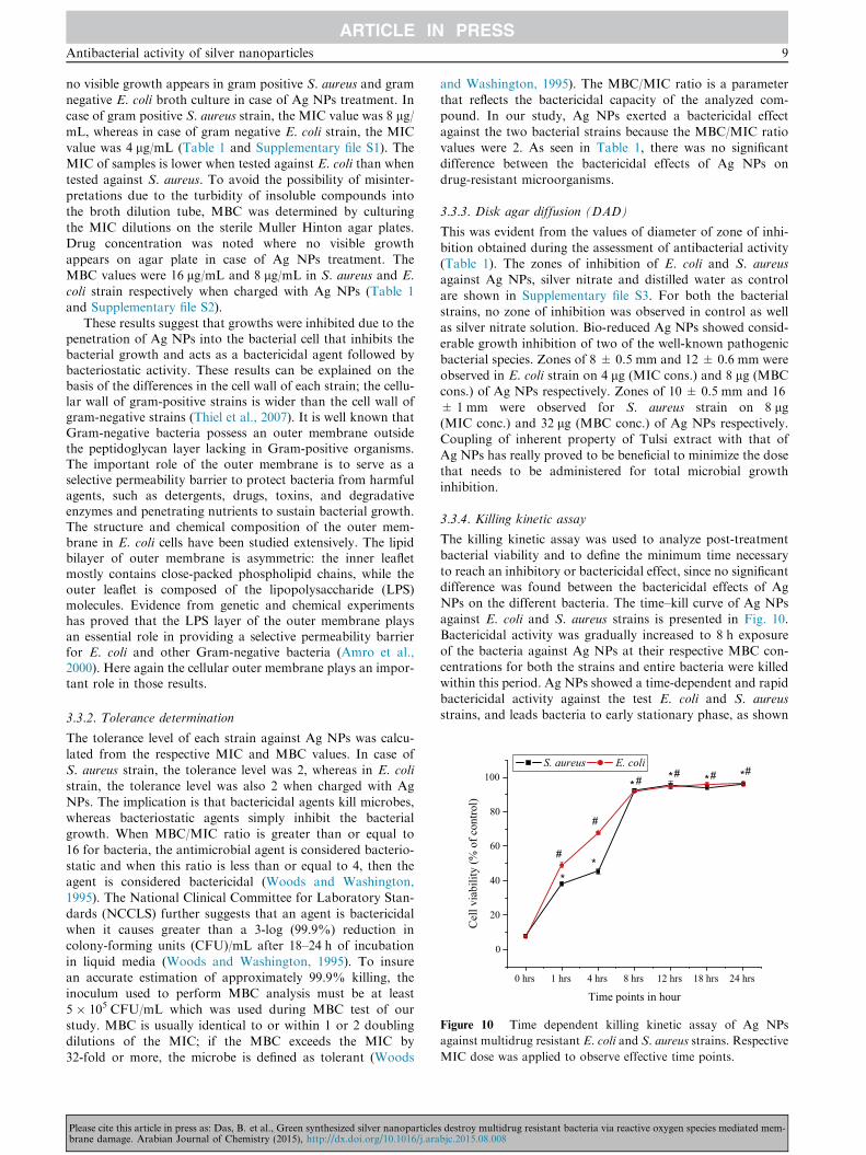
0 hrs 1 hrs 4 hrs 8 hrs 12 hrs 18 hrs 24 hrs
Figure 10 Time dependent killing kinetic assay of Ag NPs
against multidrug resistant E. coli and S. aureus strains. Respective
MIC dose was applied to observe effective time points.
Antibacterial activity of silver nanoparticles 9
no visible growth appears in gram positive S. aureus and gramnegative E. coli broth culture in case of Ag NPs treatment. Incase of gram positive S. aureus strain, the MIC value was 8 lg/mL, whereas in case of gram negative E. coli strain, the MICvalue was 4 lg/mL (Table 1 and Supplementary file S1). TheMIC of samples is lower when tested against E. coli than when
tested against S. aureus. To avoid the possibility of misinter-pretations due to the turbidity of insoluble compounds intothe broth dilution tube, MBC was determined by culturing
the MIC dilutions on the sterile Muller Hinton agar plates.Drug concentration was noted where no visible growthappears on agar plate in case of Ag NPs treatment. TheMBC values were 16 lg/mL and 8 lg/mL in S. aureus and E.
coli strain respectively when charged with Ag NPs (Table 1and Supplementary file S2).
These results suggest that growths were inhibited due to thepenetration of Ag NPs into the bacterial cell that inhibits thebacterial growth and acts as a bactericidal agent followed bybacteriostatic activity. These results can be explained on thebasis of the differences in the cell wall of each strain; the cellu-lar wall of gram-positive strains is wider than the cell wall ofgram-negative strains (Thiel et al., 2007). It is well known thatGram-negative bacteria possess an outer membrane outsidethe peptidoglycan layer lacking in Gram-positive organisms.The important role of the outer membrane is to serve as aselective permeability barrier to protect bacteria from harmfulagents, such as detergents, drugs, toxins, and degradativeenzymes and penetrating nutrients to sustain bacterial growth.The structure and chemical composition of the outer mem-brane in E. coli cells have been studied extensively. The lipidbilayer of outer membrane is asymmetric: the inner leafletmostly contains close-packed phospholipid chains, while theouter leaflet is composed of the lipopolysaccharide (LPS)molecules. Evidence from genetic and chemical experimentshas proved that the LPS layer of the outer membrane playsan essential role in providing a selective permeability barrierfor E. coli and other Gram-negative bacteria (Amro et al.,2000). Here again the cellular outer membrane plays an impor-tant role in those results.
3.3.2. Tolerance determination
The tolerance level of each strain against Ag NPs was calcu-
lated from the respective MIC and MBC values. In case ofS. aureus strain, the tolerance level was 2, whereas in E. colistrain, the tolerance level was also 2 when charged with Ag
NPs. The implication is that bactericidal agents kill microbes,whereas bacteriostatic agents simply inhibit the bacterialgrowth. When MBC/MIC ratio is greater than or equal to
16 for bacteria, the antimicrobial agent is considered bacterio-static and when this ratio is less than or equal to 4, then theagent is considered bactericidal (Woods and Washington,1995). The National Clinical Committee for Laboratory Stan-
dards (NCCLS) further suggests that an agent is bactericidalwhen it causes greater than a 3-log (99.9%) reduction incolony-forming units (CFU)/mL after 18–24 h of incubation
in liquid media (Woods and Washington, 1995). To insurean accurate estimation of approximately 99.9% killing, theinoculum used to perform MBC analysis must be at least
5 � 105 CFU/mL which was used during MBC test of ourstudy. MBC is usually identical to or within 1 or 2 doublingdilutions of the MIC; if the MBC exceeds the MIC by
32-fold or more, the microbe is defined as tolerant (Woods
Please cite this article in press as: Das, B. et al., Green synthesized silver nanoparticlebrane damage. Arabian Journal of Chemistry (2015), http://dx.doi.org/10.1016/j.ara
and Washington, 1995). The MBC/MIC ratio is a parameterthat reflects the bactericidal capacity of the analyzed com-pound. In our study, Ag NPs exerted a bactericidal effect
against the two bacterial strains because the MBC/MIC ratiovalues were 2. As seen in Table 1, there was no significantdifference between the bactericidal effects of Ag NPs on
drug-resistant microorganisms.
3.3.3. Disk agar diffusion (DAD)
This was evident from the values of diameter of zone of inhi-
bition obtained during the assessment of antibacterial activity(Table 1). The zones of inhibition of E. coli and S. aureusagainst Ag NPs, silver nitrate and distilled water as control
are shown in Supplementary file S3. For both the bacterialstrains, no zone of inhibition was observed in control as wellas silver nitrate solution. Bio-reduced Ag NPs showed consid-
erable growth inhibition of two of the well-known pathogenicbacterial species. Zones of 8 ± 0.5 mm and 12 ± 0.6 mm wereobserved in E. coli strain on 4 lg (MIC cons.) and 8 lg (MBCcons.) of Ag NPs respectively. Zones of 10 ± 0.5 mm and 16
± 1 mm were observed for S. aureus strain on 8 lg(MIC conc.) and 32 lg (MBC conc.) of Ag NPs respectively.Coupling of inherent property of Tulsi extract with that of
Ag NPs has really proved to be beneficial to minimize the dosethat needs to be administered for total microbial growthinhibition.
3.3.4. Killing kinetic assay
The killing kinetic assay was used to analyze post-treatmentbacterial viability and to define the minimum time necessary
to reach an inhibitory or bactericidal effect, since no significantdifference was found between the bactericidal effects of AgNPs on the different bacteria. The time–kill curve of Ag NPs
against E. coli and S. aureus strains is presented in Fig. 10.Bactericidal activity was gradually increased to 8 h exposureof the bacteria against Ag NPs at their respective MBC con-centrations for both the strains and entire bacteria were killed
within this period. Ag NPs showed a time-dependent and rapidbactericidal activity against the test E. coli and S. aureusstrains, and leads bacteria to early stationary phase, as shown
s destroy multidrug resistant bacteria via reactive oxygen species mediated mem-bjc.2015.08.008

Contro
l
0.5µg/m
l
1 µg/ml
2 µg/ml
4 µg/ml
8 µg/ml
16 µg/m
l
0
20
40
60
80
100
#
# # #*
*
**
#
*
Red
uctio
n of
cel
l via
bilit
y(%
of C
ontro
l)
Concentration of Ag NPs
E. coliS. aureus
Figure 12 Cell viability assay of Ag NPs treated multidrug
resistant E. coli and S. aureus strains. Cell viability was measured
by the MTT method as described in the Materials and methods.
Values are expressed as the mean ± SEM of three experiments;
superscripts indicate a significant difference (p< 0.05) compared
with the control group.
Contro
l
0.5 µg/m
l
1 µg/m
l
2 µg/m
l
4 µg/m
l
8 µg/m
l
16 µg/m
l
0
20
40
60
80
100
*
#
##
# #
*
** *
#
*Re
duct
ion
of b
io fi
lm fo
mm
atio
n(%
of C
ontro
l)
Concentration of Ag NPs
E. coliS. aureus
Figure 11 Reduction of bio-film formation of multidrug resis-
tant E. coli and S. aureus strains due to exposure of Ag NPs.
Values are expressed as the mean ± SEM of three experiments;
superscripts indicate a significant difference (p< 0.05) compared
with the control group.
10 B. Das et al.
in time–kill curves. In our study, silver nanoparticles wereeffective in inhibiting bacterial growth in a dose and timedependent manner. The growth curves of bacteria exposed to
Ag NPs indicated that it could inhibit the growth and repro-duction of both the bacteria. Yamanaka et al. (2005) foundthat silver ions require about 14 h to reduce an E. coli popula-tion from 107 to 101 CFU/mL. As mentioned by Pal et al.
(2007) the activity of nanoparticles might be similar to thatof silver ions. But in the present study, after 8 h of nanoparti-cles treatment, the bacterial cells were killed successfully.
3.3.5. Inhibition of biofilm formation
Formation of biofilm begins with irreversible binding of plank-tonic bacteria to any surfaces. The bacteria then form a com-
munity which adheres and synthesizes extracellular matrix,and matures and disperses around the site. The results showthat activity of Ag NPs is maximized or decreases biofilm for-
mation significantly (p < 0.05) at the concentration of4 lg/mL, with an inhibition rate of 65.2% against E. coliand also in case of S. aureus the significant inhibition concen-
tration was 8 lg/mL with an inhibition rate of 82.599%. It hasbeen reported that different antimicrobial activities againstplanktonic bacteria could lead to different extents of biofilminactivation by the Ag NPs. This is evident in Fig. 11. The pen-
etration rate of the biofilm may also differ between the grampositive and the gram negative strains. The inhibition effectof Ag NPs also reduced with the increase in the bacterial cell
number. The previous study has also documented that nega-tively charged Ag NPs can be electro-statically repulsed fromthe negatively charged surfaces of bacterial cells (Hong et al.,
2008). This suggests that the uptake of the Ag NPs could beremarkably reduced at the rate of increase in biofilm forma-tion. The ability of Ag NPs to agglomerate may also hinder
the activity of Ag NPs. They may be less efficient in penetrat-ing into the different extent of biofilm.
3.3.6. Cell viability by MTT assay and by FACS
Ag NPs significantly decreased (p < 0.05) the cell viability ofE. coli by 86.2% at 4 lg/mL; and 95.16% of S. aureus at
Please cite this article in press as: Das, B. et al., Green synthesized silver nanoparticlebrane damage. Arabian Journal of Chemistry (2015), http://dx.doi.org/10.1016/j.ara
8 lg/mL, which was also significant (Fig. 12). Flow cytometryand the MTT results were quite similar. Flow cytometric sus-ceptibility test showed that silver nanoparticles slaughtered E.
coli and S. aureus cells by 89.24% and 93.62%, respectively(Fig. 13). It may be due to the penetration of Ag NPs intothe bacterial cell that inhibits the bacterial growth and actsas a bactericidal agent followed by bacteriostatic activity.
3.3.7. Intracellular uptake study
Nanoparticles binding to the plasma membrane and cellular
uptake are probably a necessary condition for its exertion ofactivity. For assessing the Ag NPs uptake by E. coli and S. aur-eus, fluorescence images showed that Ag NPs treated E. coli andS. aureus cell successfully uptake the nanoparticles (Fig. 14). The
uptake of silver nanoparticles was higher in E. coli than in the S.aureus strains. The Rh-B successfully labeled with Ag NPs wasanalyzed by FTIR spectra (Supplementary Fig. S5) which con-
firms Rh-B labeled Ag NPs were taken up successfully andRh-B was not taken up independently.
Physical properties such as particle size, shape and surface
charge, play a crucial role in the uptake of nanoparticles. The cel-lular uptake of nanoparticles was done by a two step process:first, the binding step on the cell membrane and second, the inter-nalization step. The attachment of nanoparticles to cell mem-
brane seems to be most affected by the surface charge of theparticles. Variation of the particle surface charge could poten-tially control binding to the cell and direct NPs to cellular com-
partments. The fluorescence response to the Rh-B changes withthe cell membrane potential. Bacterial cell membrane is highlyelectronegative and Ag NPs are comparatively less negative
(nearly neutral charges). As both the bacterial cells had highlynegative surface charge the Ag NPs showed maximum uptakeowing to smaller size with nearly neutral surface charge, and it
was also noted that the stability of the drug played anotherimportant role in cellular uptake. In the second step, the internal-ization of nanoparticles occurs by endocytosis pathway (Zhanget al., 2008). During endocytosis process, cells readily uptake
nanomaterials by invaginating a small portion of the surface
s destroy multidrug resistant bacteria via reactive oxygen species mediated mem-bjc.2015.08.008

Figure 13 Flow cytometric cell viability test of E. coli and S. aureus strains against silver nanoparticles. A: Control – E. coli without Ag
NPs; B: Treated – E. coli with Ag NPs; C: Control – S. aureus without Ag NPs; D: Treated – S. aureus with Ag NPs.
Figure 14 Intracellular uptake of Ag NPs in multidrug resistant E. coli and S. aureus strains. A required amount of cells was treated with
Rhodamine B labeled Ag NPs (2 lg/mL) for 4 h. Intracellular uptake was examined using a fluorescence microscope. Here, A: E. coli, B:
S. aureus, (i) untreated cells and (ii): Ag NPs-Rh B treated cells.
Antibacterial activity of silver nanoparticles 11
plasma membrane and form a new intracellular vesicle aroundthe substance to transport inside the cells (Cooper, 2000).
3.3.8. Intracellular ROS generation
The generation of Reactive Oxygen Species has been shown tocontribute to Ag NP-triggered cytotoxicity in bacteria. In this
Please cite this article in press as: Das, B. et al., Green synthesized silver nanoparticlebrane damage. Arabian Journal of Chemistry (2015), http://dx.doi.org/10.1016/j.ara
study, we measure the intracellular reactive oxygen species(ROS), and 2,7-dichlorofluorescin-diacetate (DCFH2-DA)
was used as an intracellular ROS-indicator for the Ag NPstreated cells. After exposed to the nanoparticles, bacteria werestained with DCFH2DA for 30 min. We found that the AgNPs treated E. coli and S. aureus bacteria became DCF+,
s destroy multidrug resistant bacteria via reactive oxygen species mediated mem-bjc.2015.08.008
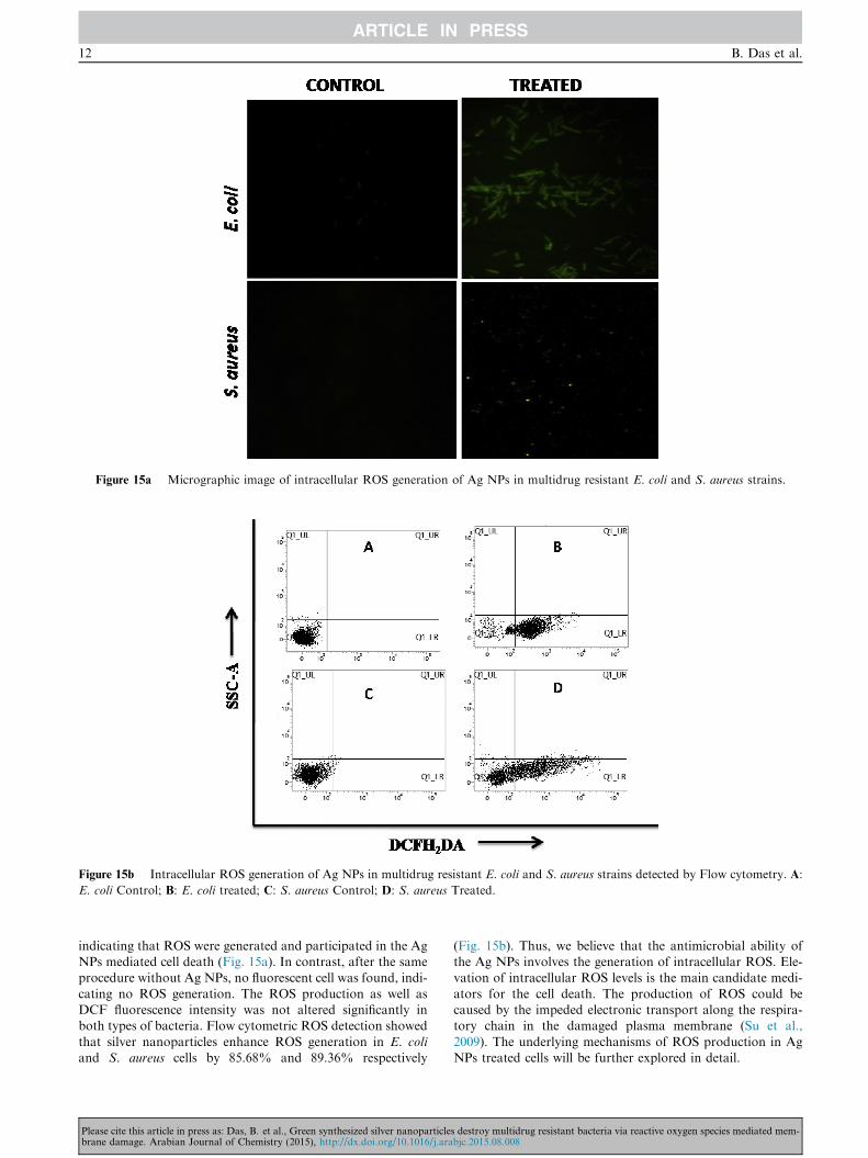
Figure 15a Micrographic image of intracellular ROS generation of Ag NPs in multidrug resistant E. coli and S. aureus strains.
Figure 15b Intracellular ROS generation of Ag NPs in multidrug resistant E. coli and S. aureus strains detected by Flow cytometry. A:
E. coli Control; B: E. coli treated; C: S. aureus Control; D: S. aureus Treated.
12 B. Das et al.
indicating that ROS were generated and participated in the AgNPs mediated cell death (Fig. 15a). In contrast, after the sameprocedure without Ag NPs, no fluorescent cell was found, indi-
cating no ROS generation. The ROS production as well asDCF fluorescence intensity was not altered significantly inboth types of bacteria. Flow cytometric ROS detection showed
that silver nanoparticles enhance ROS generation in E. coliand S. aureus cells by 85.68% and 89.36% respectively
Please cite this article in press as: Das, B. et al., Green synthesized silver nanoparticlebrane damage. Arabian Journal of Chemistry (2015), http://dx.doi.org/10.1016/j.ara
(Fig. 15b). Thus, we believe that the antimicrobial ability ofthe Ag NPs involves the generation of intracellular ROS. Ele-vation of intracellular ROS levels is the main candidate medi-
ators for the cell death. The production of ROS could becaused by the impeded electronic transport along the respira-tory chain in the damaged plasma membrane (Su et al.,
2009). The underlying mechanisms of ROS production in AgNPs treated cells will be further explored in detail.
s destroy multidrug resistant bacteria via reactive oxygen species mediated mem-bjc.2015.08.008

Figure 16 Action of Ag NPs on E. coli and S. aureus cells observed by SEM. A: E. coli Control; B: E. coli treated; C: S. aureus Control;
D: S. aureus Treated.
Antibacterial activity of silver nanoparticles 13
3.3.9. Action of silver nanoparticles on the structures of bacterial
cells
The electron micrographs by SEM of E. coli and S. aureus cellstreated and untreated with Ag NPs are shown in Fig. 16. Silver
nanoparticles in the membrane of the bacteria as well as in itsinterior were observed by electron microscopy. Electron micro-scopy determined the distribution and location of the silvernanoparticles, as well as the morphology of the bacteria after
treatment with silver nanoparticles. Micrograph by SEMshowed the surface of bacterial cells of control group (untreated)was smooth and showed typical characters of surface of native
cells, such as smooth and intact, and some filaments around cellswere obvious, while cells treated with Ag NPs damaged severely.Some cells showed large leakage, others misshapen and fragmen-
tary. Many pits and gaps appeared in the micrograph, and theirmembrane was fragmentary. The SEM micrograph of bacterialcells treated with Ag NPs showed that big gaps appeared in thecell membrane, and the bacteria were almost disorganized to sev-
eral parts and show many fragmentary bacteria. The mechanismby which the nanoparticles are able to penetrate the bacteria isnot understood completely, but studies suggest that when bacte-
rial cells were treated with silver, changes took place in its mem-brane morphology that produced a significant increase in itspermeability affecting proper transport through the plasma
membrane, leaving the bacterial cells incapable of properly reg-ulating transport through the plasma membrane, and resultinginto cell death. It is observed that silver nanoparticles have pen-
etrated inside the bacteria and have caused damage by interact-ing with phosphorus and sulfur containing compounds such asDNA, regulating enzymes (Morones et al., 2005). Silver tendsto have a high affinity to react with such compounds. These phe-
nomena suggest possible antibacterial mechanisms by which AgNPs inhibit bacterial growth, as well as cellular responses to theAg NPs treatment. Based on the present research, the action
model of Ag NPs may be described as Ag NPs making a break
Please cite this article in press as: Das, B. et al., Green synthesized silver nanoparticlebrane damage. Arabian Journal of Chemistry (2015), http://dx.doi.org/10.1016/j.ara
through the permeability of outer membrane firstly, resulting inthe leakage of cellular materials. Ag NPs enter the inner mem-
brane and produced ROS, thus inhibiting growth of cells. Simul-taneously, Ag NPs may affect some cellular components toinduce collapse of membrane, resulting in cell decomposition
and death eventually (Li et al., 2010).
4. Conclusion
This study represents a successful synthesis method of AgNPs through green route using O. gratissimum leaf extractas bio-reductant. Physical measurements suggested that thesynthesized nanoparticles were very small in size and highly
pure in nature, where plant products such as plant proteinsand phytochemicals served as capping agents. The antibac-terial activity of the particles was tested against multi-drug
resistant E. coli and S. aureus strains. It was manifestedthat Ag NPs showed the anti-bacterial activity and highereffectiveness were found against E. coli. Ag NPs also found
as an effective inhibitor of bio-film formations in both typesof strains. The bacterial killing mechanism is damaging thecell membrane followed by ROS generation. This nanoma-
terial is another option for treatment of multidrug resistantbacterial infection. So, this biogenic Ag NPs can be appliedfor the treatment of superbugs in a wide aspect of medicalfield.
Acknowledgments
The authors express gratefulness to the Vidyasagar University,Midnapore and CRNN, University of Calcutta for providingthe facilities to execute these studies. We are heartily thankful
to Mr. Joydeep Adhikari, Department of Chemistry, Univer-sity of Calcutta, 92, A. P. C. Road, Kolkata - 700 009, India,for EDX measurement.
s destroy multidrug resistant bacteria via reactive oxygen species mediated mem-bjc.2015.08.008
14 B. Das et al.
Appendix A. Supplementary material
Supplementary data associated with this article can be found,in the online version, at http://dx.doi.org/10.1016/j.arabjc.
2015.08.008.
References
Ahmad, A., Senapati, S., Khan, M.I., Kumar, R., Ramani, R.,
Srinivas, V., Sastry, M., 2003. Intracellular synthesis of gold
nanoparticles by a novel alkalotolerant actinomycete, Rhodococcus
species. Nanotechnology 14, 824–828.
Albrecht, M.A., Evan, C.W., Raston, C.L., 2006. Green chemistry and
the health implications of nanoparticles. Green Chem. 8, 417–432.
Amro, N.A., Kotra, L.P., Wadu-Mesthrige, K., Bulychev, A.,
Mobashery, S., Liu, G., 2000. High-resolution atomic force
microscopy studies of the Escherichia coli outer membrane:
structural basis for permeability. Langmuir 16, 2789–2796.
Applerot, G., Lellouche, J., Lipovsky, A., Nitzan, Y., Lubar, T.R.,
Gedanken, A., Banin, E., 2012. Understanding the antibacterial
mechanism of CuO nanoparticles: revealing the route of induced
oxidative stress. Small 8, 3326–3337.
Awwad, A.M., Salem, N.M., 2012. Green synthesis of silver nanopar-
ticles by mulberry leaves extract. Nanosci. Nanotechnol. 2, 125–
128.
Bauer, A.W., Kirby, W.M.M., Sherris, J.C., Turch, M., 1966.
Antibiotic susceptibility testing by a standardized single disk
method. Am. J. Clin. Pathol. 45, 493–496.
Chakraborty, S.P., KarMahapatra, S., Bal, M., Roy, S., 2011.
Isolation and identification of vancomycin resistant Staphylococcus
aureus from post operative pus sample. Al Ameen J. Med. Sci. 4,
152–168.
Chattopadhyay, S., Chakraborty, S.P., Laha, D., Baral, R., Pramanik,
P., Roy, S., 2012. Surface modified cobalt oxide nanoparticles: new
opportunities for anti-cancer drug development. Cancer Nano. 3,
13–23.
Chattopadhyay, S., Dash, S.K., Ghosh, T., Das, D., Pramanik, P.,
Roy, S., 2013a. Surface modification of cobalt oxide nanoparticles
using phosphonomethyl iminodiacetic acid followed by folic acid: a
biocompatible vehicle for targeted anticancer drug delivery. Cancer
Nano. 4, 103–116.
Chattopadhyay, S., Dash, S.K., Ghosh, T., Das, S., Tripathy, S.,
Mandal, D., Das, D., Pramanik, P., Roy, S., 2013b. Anticancer and
immunostimulatory role of encapsulated tumor antigen containing
cobalt oxide nanoparticles. J. Biol. Inorg. Chem. http://dx.doi.org/
10.1007/s00775-013-1044-y.
Cooper, G.M., 2000. The Cell: A Molecular Approach, second ed.
ASM Press, Washington, DC.
Dash, S.K., Chakraborty, S.P., Mandal, D., Roy, S., 2012. Isolation
and characterization of multi drug resistant uropathogenic
Escherichia coli from urine sample of urinary tract infected
patients. Int. J. Life Sci. Pharm. Res. 2, ISSN: 2250-0480.
Dash, S.K., Ghosh, T., Roy, S., Chattopadhyay, S., Das, D., 2014.
Zinc sulfide nanoparticles selectively induce cytotoxic and geno-
toxic effects on leukemic cells: involvement of reactive oxygen
species and tumor necrosis factor alpha. J. Appl. Toxicol. 34, 1130–
1144.
Dickson, D.P.E., 1999. Nanostructured magnetism in living systems. J.
Magn. Magn. Mater. 203, 46–49.
Duran, N., Marcato, P.D., Alves, O.L., De-Souza, G.I.H., Esposito,
E., 2005. Mechanistic aspects of biosynthesis of silver nanoparticles
by several Fusarium oxysporum strains. J. Nanobiotechnol. 3, 8.
Fayaz, A.M., Balaji, K., Girilal, M., Yadav, R., Kalaichelvan, P.T.,
Venketesan, R., 2010. Biogenic synthesis of silver nanoparticles and
their synergistic effect with antibiotics: a study against gram-
positive and gram-negative bacteria. Nanomed. Nanotechnol. Biol.
Med. 6, 103–109.
Please cite this article in press as: Das, B. et al., Green synthesized silver nanoparticlebrane damage. Arabian Journal of Chemistry (2015), http://dx.doi.org/10.1016/j.ara
Fox, C.L., Modak, S.M., 1974. Mechanism of silver sulfadiazine
action on burn wound infections. Antimicrob. Agents Chemother.
5, 582–588.
Gong, P., Li, H., He, X., Wang, K., Hu, J., Tan, W., Zhang, S., 2007.
Preparation and antibacterial activity of Fe3O4@Ag nanoparticles.
Nanotechnology 18, 604–611.
Guggenbichler, J.P., Semenitz, E., Konig, P., 1985. Kill kinetics and
regrowth pattern of bacteria exposed to antibiotic concentrations
simulating those observed in vivo. J. Antimicrob. Chemother. 15,
139–146.
Hong, S.H., Jeong, J., Shim, S., Kang, H., Kwon, S., Ahn, K.H.,
Yoon, J., 2008. Effect of electric currents on bacterial detachment
and inactivation. Biotechnol. Bioeng. 100, 379–386.
Huang, J., Li, Q., Sun, D., Lu, Y., Su, Y., Yang, X., Wang, H., Wang,
Y., Shao, W., He, N., Hong, J., Chen, C., 2007. Biosynthesis of
silver and gold nanoparticles by novel sundried Cinnamomum
camphora leaf. Nanotechnology 18. http://dx.doi.org/10.1088/0957-
4484/18/10/105104.
Huang, N.M., Lim, H.N., Radiman, S., Khiew, P.S., Chiu, W.S.,
Hashin, R., Chia, C.H., 2010. Sucrose ester micellar-mediated
synthesis of Ag nanoparticles and the antibacterial properties.
Colloids Surf. 353, 69–76.
Jana, N.R., Sau, T.K., Pal, T., 1999. Growing small silver particle as
redox catalyst. J. Phys. Chem. B 103, 115–121.
Joerger, R., Klaus, T., Granqvist, C.G., 2001. Biologically produced
silver–carbon composite materials for optically functional thin-film
coating. Adv. Mater. 12, 407–409.
Kawata, K., Osawa, M., Okabe, S., 2009. In vitro toxicity of silver
nanoparticles at noncytotoxic doses to HepG2 human hepatoma
cells. Environ. Sci. Technol. 43, 6046–6051.
Leela, A., Vivekanandan, M., 2008. Tapping the unexploited plant
resources for the synthesis of silver nanoparticles. Afr. J. Biotech-
nol. 7, 3162–3165.
Li, W.R., Xie, X.B., Shi, Q.S., Zeng, H.Y., OU-Yang, Y.S., Chen, Y.
B., 2010. Antibacterial activity and mechanism of silver nanopar-
ticles on Escherichia coli. Appl. Microbiol. Biotechnol. 85, 1115–
1122.
Lowry, O.H., Rosebrough, N.J., Farr, A.L., Randall, R.J., 1951.
Protein measurement with the Folin phenol reagent. J. Biol. Chem.
193, 265–275.
Macdonald, I.D.G., Smith, W.E., 1996. Orientation of cytochrome c
adsorbed on a citrate-reduced silver colloid surface. Langmuir 12,
706–713.
Majumdar, R., Bag, B.G., Maity, N., 2013. Acacia nilotica (Babool)
leaf extract mediated size-controlled rapid synthesis of gold
nanoparticles and study of its catalytic activity. Int. Nano Lett.
3, 53.
Mason, D., Allman, R., Lloyd, D., 1993. Uses of membrane potential
sensitive dyes with bacteria. In: Lloyd, D. (Ed.), Flow Cytometry in
Microbiology. Springer, London, pp. 67–82.
May, J., Shannon, K., King, A., 2006. Glycopeptide tolerance in
Staphylococcus aureus. J. Antimicrob. Chemother. 42, 189–197.
Morones, J.R., Elechiguerra, J.L., Camacho, A., Ramirez, J.T., 2005.
The bactericidal effect of silver nanoparticles. Nanotechnology 16,
2346–2353.
Mosmann, T., 1983. Rapid colorimetric assay for cellular growth and
survival: application to proliferation and cytotoxicity assays. J.
Immunol. Methods 65, 55–63.
Moyer, C.A., Brentano, L., Gravens, D.L., Margraf, H.W., Monafo
Jr., W.W., 1965. Treatment of large human burns with 0.5 per cent
silver nitrate solution. Arch. Surg. 90, 812–867.
Mukherjee, P., Ahmad, A., Mandal, D., Senapati, S., Sainkar, S.R.,
Khan, M.I., Ramani, R., Parischa, R., Ajayakumar, P.V., Alam,
M., Sastry, M., Kumar, R., 2001. Bioreduction of AuCl(4)(–) ions
by the fungus, Verticillium sp. and surface trapping of the gold
nanoparticles formed. Angew. Chem. Int. Ed. Engl. 40, 3585–3588.
Mulvaney, P., 1996. Surface plasmon spectroscopy of nanosized metal
particles. Langmuir 12, 788–800.
s destroy multidrug resistant bacteria via reactive oxygen species mediated mem-bjc.2015.08.008

Antibacterial activity of silver nanoparticles 15
Nair, B., Pradeep, T., 2002. Coalescence of nanoclusters and the
formation of sub-micron crystallites assisted by Lactobacillus
strains. Cryst. Growth Des. 2, 293–298.
Pal, S., Tak, Y.K., Song, J.M., 2007. Does the antibacterial activity
of silver nanoparticles depend on the shape of the nanoparticle?
A study of the Gram-negative bacterium Escherichia coli. Appl.
Environ. Microbiol. 73, 1712–1720.
Pum, D., Sleytr, U.B., 1999. The application of bacterial S-layers in
molecular nanotechnology. Trends Biotechnol. 17, 8–12.
Raffi, M., Hussain, F., Bhatti, T.M., Akhter, J.I., Hameed, A., Hasan,
M.M., 2008. Antibacterial characterization of silver nanoparticles
against E. coli ATCC-15224. J. Mater. Sci. Technol. 24, 192–196.
Rai, M., Yadav, A., Gade, A., 2009. Silver nanoparticles as a new
generation of antimicrobials. Biotechnol. Adv. 27, 76–83.
Sathyavathi, R., Krishna, M.B., Rao, S.V., Saritha, R., Rao, D.N.,
2010. Biosynthesis of silver nanoparticles using Coriandrum sativum
leaf extract and their application in nonlinear optics. Adv. Sci. Lett.
3, 1–6.
Singh, R., Shushni, M.A.M., Belkhei, A., 2015. Antibacterial and
antioxidant activities ofMentha piperita L. Arab. J. Chem. 8, 322–328.
Singhal, G., Bhavesh, R., Kasariya, K., Sharma, A.R., Singh, R.P.,
2011. Biosynthesis of silver nanoparticles using Ocimum sanctum
(Tulsi) leaf extract and screening its antimicrobial activity. J.
Nanopart. Res. 13, 2981–2988.
Sintubin, L., De Gusseme, B., Van der Meeren, P., Pycke, B.F.,
Verstraete, W., Boon, N., 2011. The antibacterial activity of
biogenic silver and its mode of action. Appl. Microbiol. Biotechnol.
91, 153–162.
Soukupov, J., Kvytek, L., Panacek, A., Nevecna, T., Zboril, R., 2008.
Comprehensive study on surfactant role on silver nanoparticles
Please cite this article in press as: Das, B. et al., Green synthesized silver nanoparticlebrane damage. Arabian Journal of Chemistry (2015), http://dx.doi.org/10.1016/j.ara
(NPs) prepared via modified Tollens process. Mater. Chem. Phys.
11, 77–81.
Stepanovic, C.S., Cirkoric, M.L., Ranin, L., Svabicviahocic, A.L.,
2004. Appl. Microbiol. 28, 326–432.
Su, H., Chou, C., Hung, D., Lin, S., Pao, I., Lin, J., Huang, F.L.,
Dong, R.X., Lin, J.J., 2009. The disruption of bacterial membrane
integrity through ROS generation induced by nanohybrids of silver
and clay. Biomaterials 30, 5979–5987.
Thiel, J., Pakstis, L., Buzby, S., Raffi, M., Ni, C., Pochan, D.J., Shah,
S.I., 2007. Antibacterial properties of silver-doped titania. Small 3,
799–803.
Varshney, R., Bhadauria, S., Gaur, M.S., 2010. Biogenic synthesis of
silver nanocubes and nanorods using sundried Stevia rebaudiana
leaves. Adv. Mater. Lett. 1, 232–237.
Woods, G.L., Washington, J.A., 1995. The clinician and the micro-
biology laboratory. In: Mandell, G., Bennett, J., Dolin, R. (Eds.),
Mandell, Douglas and Bennett’s Principles and Practice of Infec-
tious Diseases. Churchill Livingstone, Philadelphia, PA, pp. 169–
199.
Yamanaka, M., Hara, K., Kudo, J., 2005. Bactericidal actions of a
silver ion solution on Escherichia coli, studied by energy-filtering
transmission electron microscopy and proteomic analysis. Appl.
Environ. Microbiol. 71, 7589–7593.
Zhang, Y., Chen, Y., Wang, T., Zhou, J., Zhao, Y., 2008. Synthesis
and magnetic properties of nanoporous Co3O4 nanoflowers.
Microporous Mesoporous Mater. 114, 257–265.
Zhou, Y., Kong, Y., Kundu, S., Cirillo, J.D., Liang, H., 2012.
Antibacterial activities of gold and silver nanoparticles against
Escherichia coli and bacillus Calmette-Guerin. J. Nanobiotechnol.
10, 19.
s destroy multidrug resistant bacteria via reactive oxygen species mediated mem-bjc.2015.08.008