gougeon - ecdo documents/gougeon.pdf · analysis of apoptosis by flow-cytometry • antiviral...
TRANSCRIPT

Analysis of Apoptosis by FlowAnalysis of Apoptosis by Flow--cytometrycytometry
• Antiviral Immunity, Biotherapy and Vaccine Unit
Marie-Lise GOUGEON
2nd training course onConcepts and Methods in Programmed Cell Death
« Genetic Pathways and Techniques for Detection of Cell Death »Budapest, October 1, 2005

Cell shrinkingChromatin condensation
Cell fragmentation
Apoptotic bodies and phagocytosis
Apoptosis
Changes in morphology
Alteration of membraneintegrity
PS exposure
Drop in ∆ψm
caspase activation
Membrane alteration
Chromatin condensation
DNA fragmentation

Alteration of Morphology
Size
Granularity
Living cells
Early apoptoticLate apoptotic
Dead cells
Thymocytes
FACS acquisition
DXM24h
Petit et al. J Cell Biology, 130:157-167

Membrane integrity
Staining of apoptotic cells with :
YO-PRO-17-AAD
Staining of end staged dead cells with PI

Membrane integrity- Costaining with YO-PRO-1 and PI
YO-PRO-1
PI
Jurkat cellsCamptothecin 10mM
Co-staining with YO-PRO-1 and PI
UntreatedTreated
4hrs

QuickTime™ et undécompresseur TIFF (non compressé)
sont requis pour visionner cette image.
7-AAD stainingof ApoptoticCells
Lecoeur et al. J Imm Meth, 209:111-123

QuickTime™ et undécompresseur TIFF (non compressé)
sont requis pour visionner cette image.
Lecoeur et al. J Imm Meth, 209:111-123

Phosphatidylserine exposurePhosphatidylserine exposure
PS, normally located on the cytoplasmic surface of the cell membrane,becomes exposed to the extracellular environment. Annexin V (human vascular anticoagulant with high affinity for PS), allows the detection of PS.
Annexin-V
PIJurkat + camptothecin

Two Examples (Left and Right) of Triple Stained(HO-AnnexFITC and PI) DHD Cells
Blue light
UV light
Intact cells are blue, apoptotic cells are green, late apoptotic necroticcells are multilabelled.
Broken nucleus
QuickTime™ et undécompresseur TIFF (non compressé)
sont requis pour visionner cette image.
Multiparametric Analysis of Apoptosis:FSC/7-AAD/Annexin-V/ISNT
Lecoeur et al. J Imm Meth, 209:111-123
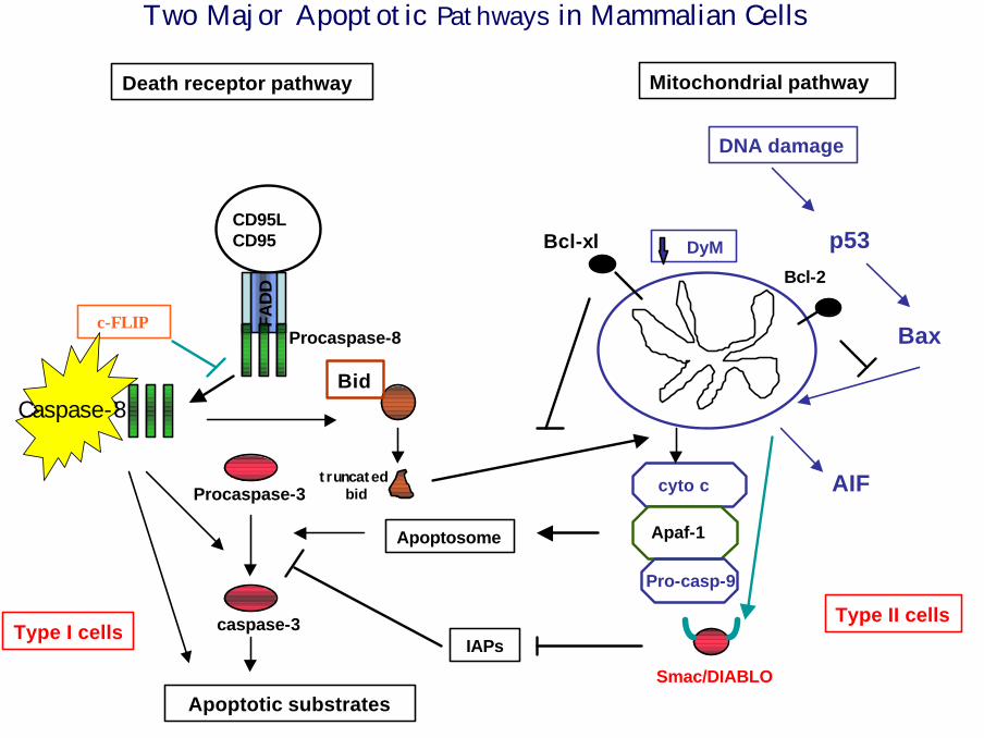
Death receptor pathway
Apoptosome
IAPs
Bid
truncatedbid
CD95LCD95
FAD
D
Procaspase-8
caspase-8
Procaspase-3
caspase-3
Apoptotic substrates
c-FLIP
Two Major Apoptotic Pathways in Mammalian Cells
Type I cells
AIFcyto c
Apaf-1
Pro-casp-9
DyM
DNA damage
Mitochondrial pathway
p53
Bax
Bcl-xl
Bcl-2
Smac/DIABLO
Type II cells
Caspase-8

Caspase activation
FLICA: caspase inhibitor, binds to the reactive cysteinson active caspases and inhibits further enzymatic activity.Unbound FLICA diffuses out of the cell and is washed away;The green fluorescent signal measures the amount of active caspase that was present at the time FLICA was added.
Jurkat cellsCamptothecin 10mM
Co-staining with FLICA and PI4hrs

Untreated Jurkat cells
Camptothecin-treated cells
Caspase-8 reagent
PI
PI+, caspase- (Dead)
PI-, caspase+ (Apoptotic)
Detection of active caspase-8
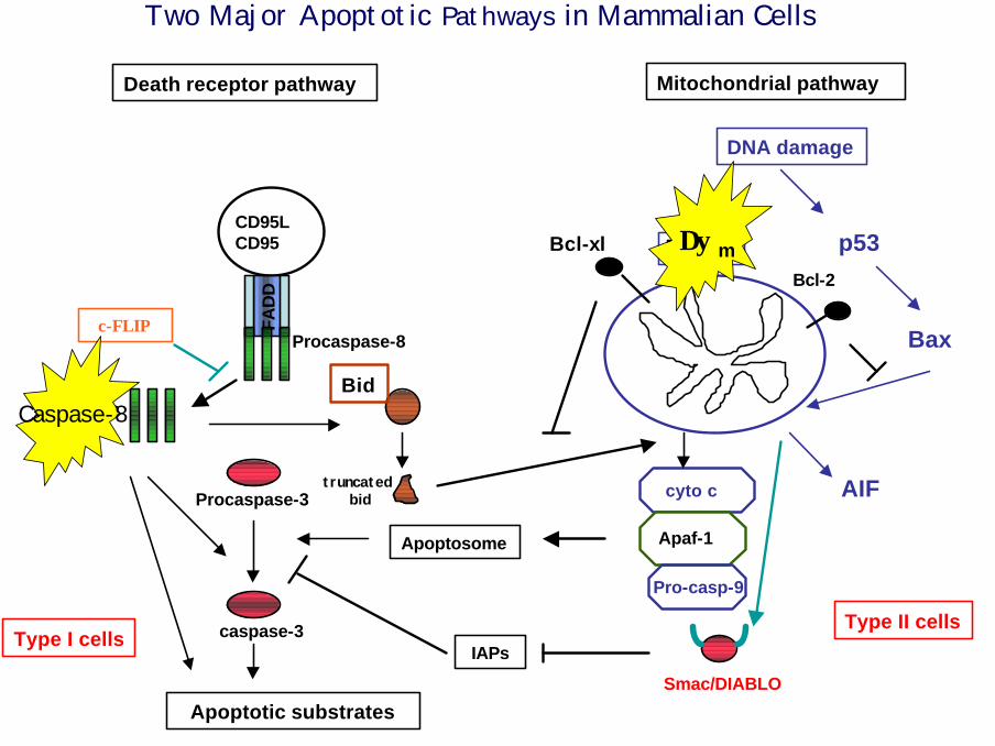
Death receptor pathway
Apoptosome
IAPs
Bid
truncatedbid
CD95LCD95
FAD
D
Procaspase-8
caspase-8
Procaspase-3
caspase-3
Apoptotic substrates
c-FLIP
Two Major Apoptotic Pathways in Mammalian Cells
Type I cells
AIFcyto c
Apaf-1
Pro-casp-9
DyM
DNA damage
Mitochondrial pathway
p53
Bax
Bcl-xl
Bcl-2
Smac/DIABLO
Type II cells
Caspase-8
∆ψm

Mitochondrial Structure and Function
∆ψm assessment using DIOC6(3) and JC-1 dyes:
DIOC6(3) accumulates in the mitochondrial matrix underthe influence of the ∆ψm
JC-1 forms aggregates under high mitoch ∆ψm, which fluoresce in red, whereas the monomeric form fluoresces in green.
Mitochondrial structure analysis:Nonyl Acridine Orangeincorporation. Reveals alteration of cardiolipids in mitochmembrane. 1 cardiolipid molecule binds 2 NAO molecules=>decreased fluorescence means decreased or altered cardiolipids.

Thymocytes stained with JC1 following incubation :
4 C
37 C
DXM 1µM
Living
Apoptotic
Petit et al. J Cell Biology, 130:157-167

Kinetics of mitochondrial alterations in thymocytes treated with DXM
DIOC6(3)
NAO
DNA fragmentation
Petit et al. J Cell Biology, 130:157-167

Confocal microscopy analysis of apoptotic thymocytes
DIOC6(3)JC-1
J-aggregatesMonomeres
Living thymocytes Apoptotic thymocytes
PI/NAO Extinctionof mito NAOFluo/PI+ nuclei
Petit et al. J Cell Biology, 130:157-167

Time course analysis of early events in DXM-apoptosis
∆ψm
Fragmented DNA
Trypan blue staining
Petit et al. J Cell Biology, 130:157-167

Strategies for Phenotyping Apoptotic Lymphocytesin a complex population
Method for the simultaneous Detection of Both Surface
and Intracellular Molecules on apoptotic cells

Objectives:
1- To determine the frequency of apoptotic cellswithin a complex population (peripheral lymphocytes)
2- To identify the phenotype of apoptotic cells

QuickTime™ et undécompresseur TIFF (non compressé)
sont requis pour visionner cette image.
Lecoeur et al. J Imm Meth, 209:111-123

QuickTime™ et undécompresseur TIFF (non compressé)
sont requis pour visionner cette image.
Lecoeur et al. J Imm Meth, 209:111-123

QuickTime™ et undécompresseur TIFF (non compressé)
sont requis pour visionner cette image.
ISNT does not allow the proper detection of surface molecules on apop cells
Lecoeur et al. J Imm Meth, 209:111-123

QuickTime™ et undécompresseur TIFF (non compressé)
sont requis pour visionner cette image.
Impact of detergents on cell morphology
Lecoeur et al. J Imm Meth, 217:11-26

QuickTime™ et undécompresseur TIFF (non compressé)
sont requis pour visionner cette image.
EtOH is inappropriate for the phenotyping of apoptotic cells
Lecoeur et al. J Imm Meth, 217:11-26

QuickTime™ et undécompresseur TIFF (non compressé)
sont requis pour visionner cette image.
Strategy for identification of both surface and intracellular molecules on apop cells
Lecoeur et al. J Imm Meth, 217:11-26

Intracellular cytokine detection in apoptotic cells
log
7-A
AD
log
7-A
AD
log caspase-3contrôle
PBMC
PPIBrefeldin A
Multiparametric FACS analysis(staining with CD3-,CD4-,CD8-,Cytokine-mAbs and 7-AAD)
%
23 %
log 7-AAD
log
IFN
-γ
CD3 gated

QuickTime™ et undécompresseur TIFF (non compressé)
sont requis pour visionner cette image.
Intracellular cytokine detection in apoptotic cells
Lecoeur et al. J Imm Meth, 217:11-26

The example of apoptosis measurement in the context of HIV infection

Regulatory Pathway of Mature T Lymphocyte Apoptosis
RestingT Cell
ActivatedT Cell IL-2
Activation
ProliferatingT cell
Low or no Ag Secondary Ag stimulation
Memory T cell
Cytokine deprivation death Antigen-dependent death- Antigen-independent- Induced by withdrawal of IL-2- Prevented by Bcl-2, Bcl-xl- Eliminates excessive T cells
after antigen clearance.
- Antigen-dependent- Mediated by FasL or TNF- Blocked by FLIPs- Limits response to repeated Ag stimulation
Ag/HIV
Cycling
Ag/HIV
FasL or TNFBcl-2
Fas
TNFR

Apoptotic pathways triggered by HIV
ML Gougeon, Nature Rev Immunology 2003

HIV-infection primes T lymphocytes for spontaneous and activation-induced apoptosis
Gougeon et al. J Immunology, 156:3509

Quantification of apoptosis in CD4 and CD8 T cells with 7-AAD staining
Gougeon et al. J Immunology, 156:3509
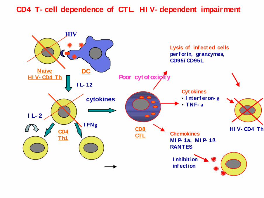
CD4 T-cell dependence of CTL. HIV-dependent impairment
NaiveNaiveHIVHIV--CD4 ThCD4 Th
DCDC
HIV
IL-12
CD4 CD4 Th1Th1
IL-2IFNγ
cytokines
Lysis of infected cellsperforin, granzymes, CD95/CD95L
CD8 CD8 CTLCTL
HIV-CD4 Th
Cytokines• Interferon-γ• TNF-α
ChemokinesMIP-1a, MIP-1ßRANTES
Inhibitioninfection
Poor cytotoxicityPoor cytotoxicity

Impact of premature apoptosis at the levelof CD8 cytotoxic T cells

Low Bcl-2 CD8 High Bcl-2 CD8
HIV disease evolution is associated with progressive accumulation of low Bcl-2 CD8 T cells
Bcl-2
Boudet et al.J. Immunol.156:2580

Down-regulation of Bcl-2 expression primes patients’ CD8 T cells for apoptosis
Boudet et al.J. Immunol.156:2580

In vivo Characteristics of low Bcl-2 CD8 T cells in patients’blood and Lymph nodes
T CD8
HLA-DR
TiA1
Perforin
Characteristics of activated CTL: HLA-DR+, CD38+, CD28-, express cytotoxic granules
CD38
Bcl-2IL-2R
IL-2, IL-15
5-15% in blood, > 60% in LN
Boudet et al.J. Immunol.156:2580

Impact of premature apoptosis at theCD4 T cell level

PBMCs
Triple staining with mAbs: surface CD3/CD8intracellular cytokines nuclear 7-AAD (apoptosis)
PMA + PHA +IonomycinBrefeldin A
33 % %
23 %
33 % 30 %
23 %
log 7-AAD
log
TNF-
α
Ex-vivo quantification of cytokine-producing precursors
Gated on T CD3
Lecoeur H, Ledru E, Gougeon M-LJ Imm Methods 217:11
FACS analysis

Decrease of the frequency of IL-2 and TNF-α -producing T cell precursors
Homme
log CD3+
log
IL-2
control HIV+39 % 14 %
CD3+
60
50
40
30
20
10
0
IL-2
**
60
50
40
30
20
10
IFN-γ
**
60
50
40
30
20
10
0
TNF-α IL-4 IL-13
*
% p
ositi
ve c
ells
0
controls HIV+ CD4 > 29 % 13 % < CD4 < 28 % CD4 < 13 % *p < 0.05 vs contrôles
60
50
40
30
20
10
0
60
50
40
30
20
10
0
Ledru et al. J. Immunol. 160:3194

Decreased frequency of IL-2 and TNF-α T cell producers is related to their priming for activation-induced apoptosis
CD8+CD4+
0
20
40
60
80
100
CD8+CD4+
0
20
40
60
80
100
Controls HIV+ Patients
%A
popt
ose
% A
popt
ose
IL-2
IFN
-γTN
F-α
IL-2
IFN
-γTN
F-α
IL-2
IFN
-γTN
F-α
IL-2
IFN
-γTN
F-α
* * *
6040200
60
40
20
0
p < 0.0001 r = -0.58
6040200
60
40
20
0
NS r = O.10
6040200
60
40
20
0
p < 0.02 r = -0.33
% A
popt
osis
with
in t
he s
ubse
t
% cytokine-producing CD3+ T cells
IL-2
IL-2 IFN-γ
TNF-α
Ledru et al. J. Immunol. 160:3194

1- Quantitative and qualitative defect in CD4 Thelper cells
2- Defect in CTL CD8 maturation
3- Altered type-1 cytokine expression
TT--cell priming for apoptosis contributes to:cell priming for apoptosis contributes to:
Defective HIVDefective HIV--specific immunityspecific immunityUncontrolled Uncontrolled viral replicationviral replicationCollapse of the Immune system =>AIDSCollapse of the Immune system =>AIDS

References
P.X.PETIT,LECOEUR H, ZORN E.DAUGUET C.MIGNOTTE B.,GOUGEON ML Alterations in mitochondrial structure and function are early events of dexamethasone-induced apoptosis. J. Cell Biology. (1995) 130: 157-167
F. BOUDET, LECOEUR H, GOUGEON M-LApoptosis associated with ex-vivo down-regulation of Bcl-2 and up-regulation of Fas in potential cytotoxic CD8+ T lymphocytes during HIV infection. J. Immunol. (1996),156:2282
M-L. GOUGEON, LECOEUR H, DULIOUST A, ENOUF M. G., CROUVOISIER M., GOUJARD C. DEBORD T., MONTAGNIER L. Programmed cell death in peripheral lymphocytes from HIV-infected persons : the increased susceptibility to apoptosis of CD4 and CD8 T cellscorrelates with lymphocyte activation and with disease progression;J. Immunol. (1996), 156:3509
H. LECOEUR and M-L. GOUGEON.Comparative analysis of flow cytometric methods for apoptosis quantitation in thymocytes and human peripheral bloodlymphocytes of controls and HIV+ persons. Evidence forinterferences of granulocytes and erythrocytes. J. Immunol. Methods (1996)198:87-99
M-L GOUGEON, LECOEUR H., BOUDET F., LEDRU E., MARZABAL S., BOULLIE, S., ROUE R., NAGATA S., HEENEY J. Lack of chronic immune activation in HIV-infected chimpanzees correlates with the resistance of T cells to Fas/Apo-1 (CD95)-induced apoptosis and preservation of a Th1 phenotype. J. Immunol. (1997) 158:2964
H. LECOEUR, LEDRU E., PREVOST M-C., GOUGEON M-L. Strategies for phenotyping apoptotic peripheral human lymphocytes comparing ISNT,Annexin-V and 7-AAD cytofluorometric methods. J. Immunol. Methods (1997) 209:11-20
E. LEDRU, LECOEUR H, GARCIA S, DEBORD T, GOUGEON M-L.Differential susceptibility to activation-induced apoptosis among peripheral Th1subsets.Correlation with Bcl-2 expression and consequences for AIDS pathogenesis. J. Immunology (1998), 160: 3194-3206
H. LECOEUR, LEDRU E, GOUGEON M-L.A cytofluorometric method for the simultaneous detection of both intracellular and surfaceantigens on apoptotic peripheral lymphocytes. J. Immunol. Methods (1998), 217:11-26
E LEDRU, N CHRISTEFF, O PATEY, J-C MELCHIOR, M-L GOUGEON. Alteration of TNFa T cell homeostasis following HAART. Implication in thedevelopment of HIV-associated lipodystrophy syndrome.BLOOD (2000) 95:3191-98
H LECOEUR, MC PRÉVOST AND M-L GOUGEON. Oncosis is associated to exposure of phosphatidylserine residues on the outside layer of plasma membrane. A reconsideration of the specificity of the AnnexinV-Propidium Iodide assay. Cytometry 2001, 44:65-72
M-L GOUGEON. Apoptosis as an HIV strategy to escape immune attack. Nature Review Immunology, 2003
M-L GOUGEON. To kill or be killed: how HIV exhausts the immune system. Cell Death Diff. 2005, i n press