glycoproteogenomics: setting the course for next
TRANSCRIPT

REVIEW
Glycoproteogenomics: Setting the Course for
Next-generation Cancer Neoantigen Discovery for
Cancer Vaccines
Jose Alexandre Ferreira1,2,3,*,#, Marta Relvas-Santos
1,2,4,#, Andreia Peixoto
1,2,
Andre M.N. Silva 4, Lucio Lara Santos 1,2,3
1Experimental Pathology and Therapeutics Group, Portuguese Institute of Oncology, Porto 4200-072, Portugal2 Institute of Biomedical Sciences Abel Salazar, University of Porto, Porto 4050-313, Portugal3Porto Comprehensive Cancer Center (P.ccc), Porto 4200-072, Portugal4REQUIMTE-LAQV, Department of Chemistry and Biochemistry, Faculty of Sciences of the University of Porto, Porto4169-007, Portugal
Received 11 August 2020; revised 25 January 2021; accepted 1 March 2021
Available online 9 June 2021
Handled by Song Liu
KEYWORDS
Glycoproteogenomics;
Oncoproteogenomics;
Cancer neoantigens;
Glycosylation;
Precision oncology
Abstract Molecular-assisted precision oncology gained tremendous ground with high-throughput
next-generation sequencing (NGS), supported by robust bioinformatics. The quest for genomics-
based cancer medicine set the foundations for improved patient stratification, while unveiling a wide
array of neoantigens for immunotherapy. Upfront pre-clinical and clinical studies have successfully
used tumor-specific peptides in vaccines with minimal off-target effects. However, the low muta-
tional burden presented by many lesions challenges the generalization of these solutions, requiring
the diversification of neoantigen sources. Oncoproteogenomics utilizing customized databases for
protein annotation by mass spectrometry (MS) is a powerful tool toward this end. Expanding
the concept toward exploring proteoforms originated from post-translational modifications (PTMs)
will be decisive to improve molecular subtyping and provide potentially targetable functional nodes
with increased cancer specificity. Walking through the path of systems biology, we highlight that
alterations in protein glycosylation at the cell surface not only have functional impact on cancer pro-
gression and dissemination but also originate unique molecular fingerprints for targeted therapeu-
tics. Moreover, we discuss the outstanding challenges required to accommodate glycoproteomics in
oncoproteogenomics platforms. We envisage that such rationale may flag a rather neglected
research field, generating novel paradigms for precision oncology and immunotherapy.
* Corresponding author.
E-mail: [email protected] (Ferreira JA).# Equal contribution.
Peer review under responsibility of Beijing Institute of Genomics, Chinese Academy of Sciences / China National Center for Bioinformation and
Genetics Society of China.
Genomics Proteomics Bioinformatics 19 (2021) 25–43
Genomics Proteomics Bioinformatics
www.elsevier.com/locate/gpbwww.sciencedirect.com
https://doi.org/10.1016/j.gpb.2021.03.0051672-0229 � 2021 The Authors. Published by Elsevier B.V. and Science Press on behalf of Beijing Institute of Genomics, Chinese Academy of Sciences /China National Center for Bioinformation and Genetics Society of China.This is an open access article under the CC BY-NC-ND license (http://creativecommons.org/licenses/by-nc-nd/4.0/).

Neoantigen-based cancer vaccines: status and mile-
stones for clinical translation
Targeted therapeutics, alone or in combination with chemo
and radiotherapy, have constituted a crucial milestone in themanagement of cancer patients, being particularly importantfor those at advanced stage facing limited therapeutic options
[1,2]. Over the last ten years, a plethora of antibodies has beendeveloped targeting proteins overexpressed by cancer cells,which play key roles in relevant oncogenic pathways and
tumor vasculature development [3,4]. A few antibody-basedtreatments have already been introduced in clinical practiceand many more are undergoing the late phase of clinical trials
[5,6]. Cancer antibodies have also been shown to inhibit cancergrowth and spread by blocking key cellular processes andinducing antibody-dependent cellular cytotoxicity (ADCC),promoting cancer cell elimination [7]. The introduction of anti-
bodies capable of inhibiting checkpoint molecules responsibleby immune tolerance to cancer cells such as PD-1, PD-L1,and CTLA-4 has decisively boosted cancer immunotherapy,
showing effective results, especially in combination with chemoand radiotherapy [8,9].
Currently, many worldwide reference oncological centers
provide comprehensive treatment options, which are electedaccording to the molecular features of the targeted lesionsand adjusted to the spatio-temporal evolution over the courseof disease management. However, the enormous potential of
antibody-based targeted therapeutics has been, to some extent,limited by off-target toxicity and high tumor molecular hetero-geneity [10]. On the other hand, a pre-existing immunogenic
fingerprint translated by high mutational burden appears tobe a pre-requisite for effective immunotherapies, limiting thegeneralization of immune checkpoint inhibitors [11].
The limitations inherent to antibody-based immunotherapyhave prompted a quest for cancer neoantigens, i.e., peptides(segments of proteins) specifically found on the surface of can-
cer cells. Although classically associated to alterations in pro-tein primary sequences due to non-synonymous mutations, itbecame widely accepted that these neoantigens may also arisefrom altered splicing mechanisms, gene fusions, endogenous
retroelements, and other processes occurring at the genomeand transcriptome levels [12–14]. Post-translational modifica-tions (PTMs) may also decisively contribute to unique cancer
molecular signatures, which can be explored to unleashimmune responses against cancer cells [15,16] (Figure 1).However, at early stages of neoantigen discovery, the
immunotherapy field has neglected the potential of proteinneoantigens due to tremendous molecular heterogeneity andlimited potential for ‘‘one fits all” pharmacological solutions
[17]. In recent years, the technological readiness of high-throughput molecular characterization platforms has chal-lenged this concept. Next-generation DNA sequencing hasallowed rapid tackle of the cancer genome for mutations that
have been subsequently explored in vaccine formulations [18].Moreover, mass spectrometry (MS) has been used to quantita-tively characterize cancer neoantigens [19–21], and increasingly
sophisticated bioinformatics tools are aiding in the real-timeidentification of most suited protein species to include in vac-cine formulations [22]. Finally, lab-scale peptide synthesizers
allow real-time production of small quantities of diverse mole-cules for vaccines under good manufacturing practice (GMP)
conditions, providing an opportunity for vaccine productionin loco [23]. Increasing numbers of improved vaccine deliveryvehicles are also emerging, which are designed to boost immune
responses against otherwise less immunogenic cancer neoanti-gens [24]. So far, pre-clinical studies in mice support the excel-lent therapeutic potential of whole genome-based multivalent
cancer vaccines for melanomas and neuroblastomas [25–28].These solutions have remarkably reduced tumor burden and,in some cases, completely eradicated tumors in animal models,
while generating an immunological memory capable of pre-venting metastasis [25–28]. This has provided blueprints onhow such approaches can be translated into clinical applica-tions in humans. Namely, a phase I clinical trial is ongoing to
evaluate the safety and effect of personalized neoantigen vacci-nes for pancreatic cancer based on next-generation sequencing(NGS) and major histocompatibility complex (MHC) affinity
prediction algorithm (NCT03558945). The study hypothesizesthat personalized neoantigen vaccines will be safe and capableof generating measurable neoantigen-specific CD4+ and
CD8+ T cell responses. Moreover, combination therapies withimmune checkpoint inhibitors have allowed to unleash previ-ously compromised immune responses in pre-clinical trials
[29,30]. Such findings fostered phase I/II clinical trials focusedon personalized cancer vaccines derived from mutated peptidesin combination with nivolumab and ipilimumab, in patientswith metastatic non-small cell lung cancer, microsatellite stable
colorectal cancer, gastroesophageal adenocarcinoma, andmetastatic urothelial cancer (NCT03953235 andNCT03639714). These initiatives illustrate the materialization
of precision oncology and will, most likely, constitute the nextcornerstone in cancer management.
While no longer considering blue-sky approaches, the effi-
ciency of patient-tailored cancer neoantigen vaccines is stillchallenged by the low mutational frequency presented by manylesions [31]. More strikingly, comparative genomics has shown
that, in some cases, metastases present a rather limited array ofmutations in comparison to primary lesions [32]. As such,extending neoantigen discovery beyond peptide identificationbased on genomic sequencing stands as the next logical step.
The emerging field of proteogenomics, exploring customizedprotein sequence databases by integrating genomics and tran-scriptomics data, provides a unique tool to interrogate the can-
cer proteome [33]. In fact, the integration of genomics andtranscriptomics is crucial for protein identification, bringingproteomics one step closer to the exome. Accordingly, paired
genomics, transcriptomics, and proteomics data of samplesfrom the same tumors have demonstrated that the proteomecontains novel information that cannot be discerned throughgenomic analysis alone [34].
An additional level of molecular complexity arises from thearray of PTMs that decisively define protein biophysical andbiochemical properties as well as functional roles. PTMs pro-
vide microenvironmental context and exponentially increasethe number of protein species defined by the exome, leveraginga more complete view of tumor molecular heterogeneity and
cancer biology [35]. Particularly, glycosylation is amongst themain PTMs of membrane proteins and it is well establishedthat cancer cells present altered glycosylation patterns in com-
parison to corresponding healthy tissues [36,37]. A significantnumber of studies have also consensually postulated an arrayof glycan modifications that appear of pancarcinoma nature[38,39]. Moreover, many reports advocate that such molecular
26 Genomics Proteomics Bioinformatics 19 (2021) 25–43

features are responsible for generating unique glycopeptide sig-
natures at the cell surface [40]. Identifying these distinctivecancer-specific glycoproteoforms will provide bispecific (atthe glycan and protein levels) molecular targets, with the
potential to limit off-target effects.The proof of concept regarding the potential of glycan-
based proteogenomics has been highlighted in two recent stud-ies, supporting the pursuit of more comprehensive research
efforts [41,42]. As such, this review focuses on illustrating therole of proteogenomics as a decisive tool for systems biologyand, ultimately, precision oncology. Focus is set on the impor-
tance of addressing protein glycosylation and integrating gly-comics and glycoproteomics into neoantigen discoveryplatforms, envisaging the generalization of cancer vaccines to
tumors of distinct molecular natures.
Oncoproteogenomics toward neoantigen discovery
Cancer biomarker discovery has been mostly centered on thegenome and transcriptome, with less, but growing emphasis,
on the proteome. While the complexity of the tumor genomic
background is being rapidly uncovered by large dimensionsequencing studies [32,43,44], translation of its findings intoresulting proteome remodeling remains poorly understood
and explored. In fact, many reports disclosed a landscape ofgenetically relevant alterations and dysfunctional transcrip-tomes that reflect large numbers of non-synonymous single-nucleotide variants (SNVs), insertions and deletions (indels),
alternative splicing variants, copy-number aberrations, andabnormal fusion genes [45]. However, the transcriptome fre-quently fails to mirror the proteome’s abundance, diversity
[46,47], and consequent functional impact on tumor initiation,progression, dissemination, and response to treatment. Thishas limited the easy translation of elegant transcriptome-
derived patient stratification models into the everyday practiceof most pathology laboratories. It has also delayed the identi-fication of clinically relevant proteins for rational design of tar-
geted therapeutics. The number of protein surrogates and/ortargetable biomarkers for molecular-based patient stratifica-tion reaching the clinics remains scarce. The few successfulexamples include prostate-specific antigen (PSA) for detection
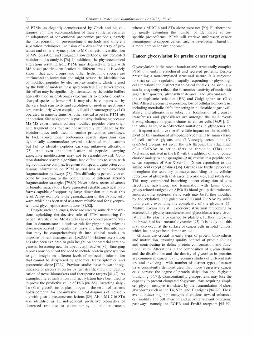
Figure 1 Neoantigens generated by modifications at DNA, RNA, and protein levels as well as by PTMs
Cancer cells frequently express unique protein species that are not present in healthy tissues (neoantigens), holding tremendous potential
for targeted therapies and immunotherapy. Neoantigens may derived primarily from alterations in genome but also in RNA processing
and other events underlying protein synthesis. Protein maturation with PTMs, such as glycosylation and phosphorylation, adds a second
layer of specificity, which is valuable toward more effective targeted therapeutics. Beyond genetic alterations, epigenetic regulation plays a
key role in governing gene expression as well as processing, significantly contributing to the formation of a wide array of proteoforms
either directly or indirectly modulating the expression of enzymes involved in PTMs of proteins. Me, methyl; Ac, acetyl; SLeA, sialyl-Lewis
A; SLeX, sialyl-Lewis X; S3T, sialyl-3-T; STn, sialyl-Tn; GPI, glycosylphosphatidylinositol; PTM, post-translational modification.
Ferreira JA et al / Glycoproteogenomics in Cancer 27

[48], carcinoembryonic antigen (CEA) for patient managementand prognosis [49], and CA125/mucin 16 (MUC16), which is adiagnostic marker and holds potential for targeted therapies
[50], in prostate, colon, and ovarian cancer, respectively.On the other side of the equation, the initial enthusiasm
with oncoproteomics in the post-genomic era, which started
20 years ago, has been gradually losing ground. This is mostlydue to protein identifications in conventional proteomicsworkflows relying on general protein databases that fail to
reflect the uniqueness of the cancer transcriptome [51], limitingthe breadth of identified biomarkers. Oncoproteogenomics hasarisen from the pressing need to correct this discrepancy,enabling the accurate interpretation of large datasets generated
by modern high-resolution mass spectrometers. Proteoge-nomics exploits sample-matched genomics and transcriptomicsdatasets to customize protein annotation, providing definitive
proofs of protein translation [52]. The most used approachesfor generating protein databases include whole-genome orexome sequencing, with emphasis on six-frame translation of
whole genome sequences, which enables the identification ofpreviously undiscovered exons and open reading frames [53].Available exon annotations on the human genome may also
be explored to generate junction sequences for all possibleexons in a gene. DNA/RNA sequencing is also available toobtain SNV data and corresponding tumor-specific proteinsequences [54].
When applied to cancer, proteogenomics is an exciting newconcept, which attempts to detect tumor-specific changes inproteoforms that result from mutations or an altered tran-
scription process, rather than solely focusing on improvingproteome characterization (Figure 2). Over the past five years,these methodologies have become sufficiently mature to sup-
port the transition from proof of concept studies in cell linesand animal models to large-scale translational studies usinghuman samples. Several examples show how oncoproteoge-
nomics may complement and improve on the molecular sub-typing and prognostication of breast [55], gastric [41],colorectal [46], and ovarian [56] tumors beyond previousgenomics- and transcriptomics-based patterns. Most of these
reports provide proteome-based networks highlighting func-tional protein nodules for targeted intervention. Furthermore,the depth of tumor proteogenomics profiling has led to the
identification of breast cancer protein neoantigens that resultfrom previously undescribed gene variants and non-codinggene regions, defying old paradigms and expanding our under-
standing of cancer molecular biology [20]. Such molecular sig-natures are possibly the consequence of significant cancergenome instability and arise as strong candidates forimmunotherapy.
Despite several examples of considerable technologyreadiness, when facing clinical translation, some outstandingchallenges persist for oncoproteogenomics. Perhaps the most
pressing difficulty relates to the fact that databases of putativeprotein sequences derived from genomics experiments are sig-nificantly larger than those explored in conventional pro-
teomics, which dangerously increases the probability of falsepositives during protein annotation. Moreover, the incorpora-tion of large genomics datasets into proteomics poses a signif-
icant computational challenge, translated by long processingtimes and frequently high false discovery rates [57]. This is fur-ther aggravated by the low relative abundance of peptidesequences derived from genetic abnormalities over protein
species arising from genomics predictions. Alternatively,libraries inferred directly from RNA-seq, expressed sequencetag (EST), and cDNA bring us one step closer to the exome,
while significantly reducing the amount of generated data incomparison to whole-genome sequencing [58]. Furthermore,transcriptome-generated databases allow the proteomic identi-
fication of RNA editing products, splice junctions, and fusionproteins. Nevertheless, oncoproteogenomics continues to be afast-evolving field, and more detailed information on the
opportunities and limitations of analytical and bioinformaticstools to assist database customization and accommodatinggenomics information can be found in recent reviews [59,60].Another key aspect is the capacity to translate proteogenomics
data into relevant targets for immunotherapy. This aspect hasbeen elegantly tackled by several computational pipelines toelect peptides with higher binding affinity to MHC-I molecules
[61–63]. Moreover, neural networks exploiting annotated dataon the binding kinetics of known peptides are being used torefine these models to estimate cancer neoantigen affinity to
a wide array of different MHC classes and haplotypes, provid-ing means for a more educated election of suitable immuno-gens [64–66]. However, even if a peptide has strong MHC
binding prediction, this may be ineffective if upstream process-ing such as proteolysis prevents the actual loading of that pep-tide. Accordingly, several software are now available to aidprediction of proteasome specificity and protease cleavage
sites, including NetChop20S, NetChopCterm, and Pro-teaSMM for MHC class I antigens, and PepCleaveCD4 andMHC-II-NP for MHC class II antigens, offering a second level
of predicted peptide quality control [67–70]. The notion thatmany predicted neoantigens may never constitute proteolysisproducts capable of being presented by MHC has again
prompted proteomics. MHC molecules and associated pep-tides are currently being isolated from different cancers fortandem mass spectrometry (MS/MS) identification, generating
key data for training machine learning algorithms to improveneoantigen prediction [71]. The thorough accomplishment ofsuch goals will be decisive for translating oncoproteogenomicsinto clinically useful targets. Finally, critical challenges persist
concerning the capacity of oncoproteogenomics to infer on rel-evant functional mechanisms adopted by cancer systems atmultiple biological scales, ultimately pinpointing key biologi-
cal network nodes for intervention [72]. A plethora of reportsshows that this goal requires a comprehensive understandingof PTMs [39,73]. PTMs are key for defining and regulating
protein functions, degradation pathways, and even cellularlocations, and many times play an essential role in the signal-ing pathways that define cell fate [72]. PTMs, such as phospho-rylation, methylation, and glycosylation, are pivotal in the
rapid modulation of protein functions in response to microen-vironmental cues, providing potential links between metabolicalterations and protein activity [74]. Therefore, incorporating
PTMs into oncoproteogenomics setups will bring us one stepcloser to systems biology settings, even though at the expensesof a new set of interdisciplinary analytical challenges.
PTMs are often of a transient nature, but exponentiallyincrease the number of proteoforms, raising problems foraccurate protein identification. Such dynamics are often
accompanied by a complex and non-templated molecularorganization, as is the case of glycosylation. As a result, morethan half of the spectral information generated by MS/MSexperiments usually remains unassigned due to the presence
28 Genomics Proteomics Bioinformatics 19 (2021) 25–43

Figure 2 Integrated multiomics data toward discovery of potential clinically relevant biomarkers and targeted therapeutics
Oncoproteogenomics concerns genomics and transcriptomics data from tumor samples, which is used for generating customized
databases, to support protein annotation. The inclusion of glycomics information in the workflow allows more effective protein
identification by MS/MS, including glycosylation site mapping, illustrating its value to gain insight on molecular information that cannot
be achieved by the other omics toward precision oncology. Overall oncoproteogenomics is supported by several bioinformatics tools,
which also contributes for identification of more suitable antigens toward cancer vaccines. WGS, whole-genome sequencing; SNV, single-
nucleotide variant; indel, insertion and deletion; MS/MS, tandem mass spectrometry; HexNAc, N-acetylhexosamine; Hex, hexose.
Ferreira JA et al / Glycoproteogenomics in Cancer 29
of PTMs, as elegantly demonstrated by Chick and his col-leagues [75]. The accommodation of these subtleties requiresan adaptation of conventional proteomics protocols, namely
the incorporation of pre-enrichment methods and differentseparation techniques, inclusion of a diversified array of pro-teases and other enzymes prior to MS analysis, diversification
of MS ionization and fragmentation methods, and dedicatedbioinformatics analysis [76]. In addition, the physicochemicalalterations resulting from PTMs may decisively interfere with
MS-based protein identification at different levels. It is widelyknown that acid groups and other hydrophilic species aredetrimental to ionization and might reduce the identificationof modified peptides by electrospray analysis, which is used
by the bulk of modern mass spectrometers [77]. Nevertheless,this effect may be significantly attenuated by the acidic buffersgenerally used in proteomics experiments to produce positive
charged species at lower pH. It may also be compensated bythe very high sensitivity and resolution of modern spectrome-ters, particularly when coupled to liquid chromatography (LC)
operated in nano-settings. Another critical aspect is PTM siteannotation. Site assignment is particularly challenging becauseMS/MS experiments involving modified peptides often origi-
nate fragment ions that are not accurately identifiable by thebioinformatics tools used in routine proteomics workflows.In fact, conventional proteomics search algorithms candynamically accommodate several anticipated modifications
but fail to identify peptides carrying unknown alterations[75]. And even the identification of peptides carryingexpectable modifications may not be straightforward. Com-
mon database search algorithms face difficulties to score withhigh-confidence complex fragment ion spectra quite often con-taining information on PTM neutral loss or non-canonical
fragmentation pathways [78]. This difficulty is generally over-come by resorting to the combination of different MS/MSfragmentation strategies [79,80]. Nevertheless, recent advances
in bioinformatics tools have generated reliable analytical plat-forms capable of supporting large dimension studies at thislevel. A key example is the generalization of the Byonic soft-ware, which has been used as a more reliable tool for glycopro-
tein and glycopeptide annotations [81,82].Despite such challenges, there are already some demonstra-
tions upholding the decisive role of PTM monitoring for
patient stratification. Most studies have explored phosphoryla-tion to demonstrate its decisive role for pinpointing relevantdisease-associated molecular pathways and how this informa-
tion may be comprehensively fit into clinical models toimprove patient management [56,83,84]. Histone acetylationhas also been explored to gain insight on endometrial carcino-genesis, foreseeing new therapeutic approaches [85]. Emerging
reports now point out the need to include protein glycosylationto gain insight on different levels of molecular informationthat cannot be deciphered by genomics, transcriptomics, and
proteomics alone [37,39]. Previous studies have shown the sig-nificance of glycosylation for patient stratification and identifi-cation of novel biomarkers and therapeutic targets [41,42]. As
example, altered sialylation and fucosylation have been used toimprove the predictive value of PSA [86–88]. Targeting sialyl-Tn (STn) glycoforms of plasminogen in the serum of patients
holds potential for non-invasive clinical diagnosis of individu-als with gastric precancerous lesions [89]. Also, MUC16-STnwas identified as an independent predictive biomarker ofdecreased response to chemotherapy in bladder cancer,
whereas MUC16 and STn alone were not [90]. Furthermore,by greatly extending the number of identifiable cancer-specific proteoforms, PTMs will retrieve unforeseen cancer
neoantigens to support cancer vaccine development based ona more comprehensive approach.
Cancer glycosylation for precise cancer targeting
Glycosylation is the most abundant and structurally complex
PTM of membrane-anchored and secreted proteins. Despitepresenting a non-templated structural nature, it is subjectedto strict cellular regulation, rapidly responding to physiologi-cal alterations and distinct pathological contexts. As such, gly-
can heterogeneity reflects the harmonized activity of nucleotidesugar transporters, glycosyltransferases, and glycosidases inthe endoplasmic reticulum (ER) and Golgi apparatus (GA)
[36]. Altered glycogene expression, loss of cellular homeostasis,including metabolic shifts impacting in nucleotide sugar avail-ability, and alterations in subcellular localization of glycosyl-
transferases and glycosidases are amongst the main eventsdriving changes in glycan chains in cancer cells [36,91]. Onthe other hand, loss-of-function mutations in glycogenes arenot frequent and have therefore little impact on the establish-
ment of this malignant glycophenotype [92]. The main classesof cell surface glycans are O-N-acetylgalactosamine (O-GalNAc) glycans, set up in the GA through the attachment
of a GalNAc to serine (Ser) or threonine (Thr), andN-glycans, initiated in the ER with the addition of an oligosac-charide moiety to an asparagine (Asn) residue in a peptide con-
sensus sequence of Asn-X-Ser/Thr (X corresponding to anyamino acid except proline) [36]. Glycans are further processedthroughout the secretory pathways according to the cellular
repertoire of glycosyltransferases, glycosidases, and substrates.This may comprehend branching and/or elongation of corestructures, sialylation, and termination with Lewis bloodgroup-related antigens or ABO(H) blood group determinants,
amongst other epitopes. Sialic acids may be further modifiedby O-acetylation, and galactose (Gal) and GlcNAc by sulfa-tion, greatly expanding the complexity of the glycome [36].
Mature glycans may still experience structural remodeling byextracellular glycosyltransferases and glycosidases freely circu-lating in the plasma or carried by platelets, further increasing
the breadth of its structural dynamics [93]. It is likely that thismay also occur at the surface of cancer cells in solid tumors,which has not yet been demonstrated.
Glycans are crucial in early steps of protein biosynthesisand maturation, ensuring quality control of protein foldingand contributing to define protein conformation and func-tional roles. Alterations in the composition of glycan chains
and the distribution and the density of glycosites in proteinsare common in cancer [39]. Glycomics studies of different nat-ure and involving a wide number of distinct types of cancer
have consistently demonstrated that more aggressive cancercells increase the degree of protein sialylation and N-glycanbranching [36,91]. Concomitantly, glycoproteins may lose the
capacity to present elongated O-glycans, thus acquiring simplecell glycophenotypes translated by the accumulation of shortglycoforms such as the Tn, STn, and T antigens [94–96]. Theseevents induce major phenotypic alterations toward enhanced
cell motility and cell invasion and activate relevant oncogenicpathways, namely the EGFR and ErbB2 receptors [95–99].
30 Genomics Proteomics Bioinformatics 19 (2021) 25–43
The expression of truncated glycans further impacts the inter-actions of cancer cells with the immune system. Examplesinclude STn antigen recognition by Siglec-15 on macrophages
and dendritic cells, and sialyl-T (ST) antigen recognition bySiglec-9 on neutrophils and Siglec-7 on natural killer cells[100]. These interactions trigger an array of inhibitory signals
on antigen-presenting cells, ultimately precluding T cell-mediated responses against tumor cells, as demonstrated byrecent studies [101,102]. It is likely that a deeper understanding
of functional interactions between glycans and glycoconjugateswith immune system intermediates may constitute the nextcornerstone for immune checkpoint inhibition and, poten-
tially, a more rational design of cancer vaccines. Cancer cellsalso often overexpress sialylated Lewis antigens such assialyl-Lewis A (SLeA) and X (SLeX). These are terminal epi-
topes of glycan chains in proteins and glycolipids that play adecisive role in cancer cell hematogenous dissemination byinteracting with E-selectin on activated endothelial cells
[103]. Moreover, they promote cancer cell adhesion to plateletsthrough P-selectin and lymphocytes through L-selectin, whichare critical aspects to support survival in circulation and evade
immune responses [104]. There are many other examples ofhow glycans impact key cancer hallmarks, as elegantly high-lighted by recent reviews [39,105].
There are numerous studies associating STn, SLeA, and
other glycoforms with worst prognosis and metastatic spread[106,107]. Notably, STn and SLeA glycans are rarely expressedby healthy tissues, where they are confined to cells specialized
in secretion across the lumen of the gastrointestinal and respi-ratory tracts as well as in secreted mucins, acting as a protec-tive barrier against pathogens and contributing to
immunological homeostasis [37,108]. These glycans are alsonot expressed in blood cells in circulation, standing as poten-tial biomarkers [109,110]. This has prompted their exploitation
with relative success for targeted therapeutics andimmunotherapy in pre-clinical and clinical settings, withemphasis on chimeric antigen receptor T (CAR-T) cells andcancer vaccines, as reviewed recently [111,112]. Namely,
CAR-Ts designed against cancer cells expressing the Tn andSTn antigens hold potential toward this objective, given theirrestricted expression pattern in healthy human tissues
[111,113]. On the other hand, glycan-based vaccines, while cap-able of inducing high antibody titters, have not been able toinduce effective and long-lasting cellular immunity against
tumors [114,115].To this date, the establishment of immunization strategies
capable of overcoming the low immunogenicity and immuno-
suppressive nature of cancer-associated glycans remains anoutstanding challenge. However, the concept of neoantigensdoes not apply to glycans alone, [116] with the only knownexception coming from the incorporation of N-
glycolylneuraminic acid (Neu5Gc; sialic acid) from dietarysources such as red meat, which induces the formation ofxenoantigens in human glycan chains [117]. Neoantigen signa-
3
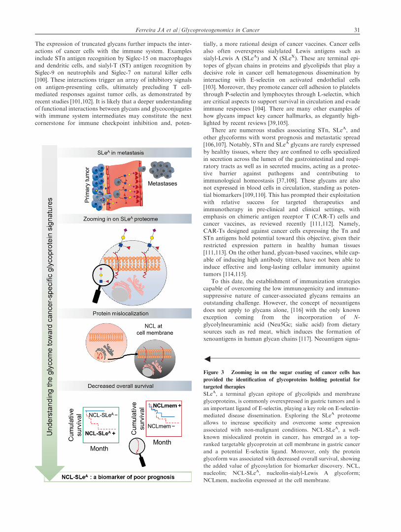
Figure 3 Zooming in on the sugar coating of cancer cells has
provided the identification of glycoproteins holding potential for
targeted therapies
SLeA, a terminal glycan epitope of glycolipids and membrane
glycoproteins, is commonly overexpressed in gastric tumors and is
an important ligand of E-selectin, playing a key role on E-selectin-
mediated disease dissemination. Exploring the SLeA proteome
allows to increase specificity and overcome some expression
associated with non-malignant conditions. NCL-SLeA, a well-
known mislocalized protein in cancer, has emerged as a top-
ranked targetable glycoprotein at cell membrane in gastric cancer
and a potential E-selectin ligand. Moreover, only the protein
glycoform was associated with decreased overall survival, showing
the added value of glycosylation for biomarker discovery. NCL,
nucleolin; NCL-SLeA, nucleolin-sialyl-Lewis A glycoform;
NCLmem, nucleolin expressed at the cell membrane.
Ferreira JA et al / Glycoproteogenomics in Cancer 31

tures may be obtained from glycopeptides, granting bispeci-ficity both via the glycan and the peptide chain [118,119]. Sup-porting this hypothesis, glycoarray-based studies have
identified autoantibodies against distinct mucin variable tan-dem repeat glycoproteoforms in the serum of cancer patients[120,121]. Autoantibodies showed cancer-specific recognition
of targeted glycans, regardless of disease-associated variationsin glycan density and distribution in peptide chains, supportingthe existence of molecular fingerprints of disease that should be
explored for cancer targeting. Accordingly, understanding therepertoire of glycans stands out as a decisive step for zoomingin on the cancer glycoproteome. We have recently explored
32 Genomics Proteomics Bioinformatics 19 (2021) 25–43

this approach to demonstrate the presence of unusual glyco-proteins at the cell surface, such as the case of nucleolin(NCL) [122]. NCL is mostly confined to the nucleus of healthy
cells, but it may migrate to the cell surface where it appearsglycosylated in different types of solid tumors [122–126](Figure 3). These are striking observations since NCL presents
neither hydrophobic transmembrane domains nor a targetingsignal to the cell membrane that could justify glycosylation[127]. The mechanisms governing this protein mislocalization
remain to be fully elucidated; nevertheless, it becomes clearthat this cancer-specific alteration presents enormous potentialfor precise targeting. Many similar examples have beenreported in the literature, which set an important novel para-
digm for biomarker research and can be comprehensivelyexplored in the future [128–130].
When taking into account the relevance of integrating gly-
can information into protein surrogates, there are severalexamples on how considering cancer-associated glycoformsof classical biomarkers has improved their clinical value
(PSA, MUC16, CEA, and CD133) [86,90,131–133]. Moreover,focusing on glycans on HER2, PD-1, and PD-L1 has allowedto provide important footprints for patient stratification, bet-
ter predict treatment response, and improve the efficacy oftherapeutic antibodies [102,134–139]. In summary, glycosyla-tion provides functional and microenvironmental contexts tocell membrane proteins, and targeting glycosylated moieties
may lead to previously unforeseen glycoproteomics signaturesat the cell surface, paving the way toward disease-specificglycoproteoforms.
Glycomics and glycoproteomics: concepts, strategies,
and challenges facing biomarker discovery
The comprehensive combination of glycomics and glycopro-teomics will be crucial toward the identification of novel cancer
biomarkers of clinical value, with emphasis on unforeseencancer-specific signatures reflecting molecular alterations at
the genome, transcriptome, proteome, and metabolome levels.Glycomics addresses the repertoire of glycans in a biologicalsystem and is a critical milestone for identification of glyco-
neoantigens. MS is by far the most widely used analytical toolin a field that has been progressing fast, backed by the increas-ing sensitivity and resolution of mass spectrometers [140,141].
Moreover, important steps have been made toward protocolstandardization [142], automatization [143], and bioinformat-ics [144], setting the foundations for carbohydrate metrology.
However, despite intense research and robust proofs of con-cept, the implementation of MS-based glycome signatures inclinical practice has yet to be established and poses significantanalytical, clinical, and regulatory difficulties, namely the lack
of well-defined glycan standards as well as biased reproducibil-ity resulting from a wide array of MS architectures. Notably,lectin microarrays of variable architectures have been pro-
posed as high-throughput alternative technologies, whichmay be particularly important in the context of liquid biopsies,but of limited use for glycoantigen discovery [145]. Facing
mature technology, the field should now push toward a com-prehensive, large-scale interrogation of the human glycome.This will decisively prompt our understanding of glycan diver-
sity in health and disease, including the foundations to asser-tively tackle the glycoproteome.
Glycomics
Glycan analysis poses a significant analytical challenge due toits non-templated and structural heterogeneity and frequentco-existence of isomeric/isobaric structures in the same sample
[146,147]. MS remains the gold standard technique, but sup-port from nuclear magnetic resonance (NMR), complementaryenzymatic methods, and immunoassays may be required for
more detailed structural elucidation. Typical protocols initiatewith the selective release of glycans from glycoproteins byeither enzymatic or chemical methods. N-glycans are isolated
by peptide-N-glycosidase F (PNGase F) or PNGase A diges-
3
Figure 4 Glycomics and glycoproteomics conventional workflows for the identification of potential targetable glycobiomarkers
A. Immobilization of extracted glycoproteins from biological material on PVDF membrane allows consecutive release of N-glycans and
O-glycans by PNGase F digestion and reductive b-elimination, respectively. This protocol comprises analysis of the released and reduced
glycans by PGC–nanoLC–ESI–MS/MS. B. An achievement for the field of (glyco)proteomics in the last years was the introduction of
MALDI Imaging, which adds spatial information to molecular specificity provided by MS. The main steps of a MALDI-MSI protocol for
protein and N-glycan analyses are very similar. After removal of paraffin in FFPE tissues, PNGase F or trypsin is applied on the tissue
section, to release N-glycans from glycoproteins or obtain digested peptides, followed by application of MALDI matrix. Besides the
average mass spectrum obtained for each tissue section, the distribution of the most abundant glycans or peptides can be visualized
individually across the tissue. Moreover, identification of N-glycans and/or peptides can be confirmed by MS/MS on tissue extracts. For
the sequential analysis of peptides on the same tissue section used previously for N-glycan analysis, an intermediate washing step to
remove MALDI matrix, N-glycans, and PNGase F is necessary in addition to the antigen retrieval step. C. The information generated by
glycomics experiments is pivotal for guiding glycoproteomics by employing a glycan-targeted proteomics approach. Therefore, this
includes an initial enrichment of extracted cell membrane proteins for the glycan of interest, followed by separation of glycoproteins
according to the molecular weight and analysis by nanoLC–MS/MS using a bottom-up approach. Combined bioinformatics tools are
employed toward curated data, through comparison with transcriptomics data, protein expression data from healthy and tumor tissues,
and evaluation of subcellular location. PVDF, polyvinylidene fluoride; PNGase F, peptide-N-glycosidase F; EIC, extracted ion
chromatogram; PGC, porous graphitized carbon; LC, liquid chromatography; ESI, electrospray ionization; FFPE, formalin-fixed
paraffin-embedded; a-CHCA, a-cyano-4-hydroxycinnamic acid; MALDI-MSI, matrix-assisted laser desorption/ionization mass
spectrometry imaging; PNA, peanut agglutinin; VVA, Vicia villosa agglutinin; NeuAse, neuraminidase.
Ferreira JA et al / Glycoproteogenomics in Cancer 33

tion, whereas O-GalNAc glycans are obtained by reductiveb-elimination through chemical methods [148].
Focusing on N-glycans, PNGase F digestion has the advan-
tage of tagging substituted Asn residues in protein chains,facilitating downstream glycosite identification by glycopro-teomics [149]. This can be achieved in proteins in solutions,
immobilized in polyacrylamide gels, and even in tumorsections [150–152]. However, the immobilization of purifiedglycoproteins or protein mixtures on polyvinylidene fluoride
(PVDF) membranes prior to enzymatic and chemical treat-ments significantly facilitates glycan release from minuteamounts of biological material (nano-femtomole) [153](Figure 4A). Notably, PNGase F works well for glycoproteins,
whereas PNGase A is more recommended for deglycosylationof glycopeptides [154,155]. Moreover, PNGase A differs fromPNGase F in the fact that it is able to cleave N-linked glycans
with or without a(1,3)-linked core fucose residues, whereasPNGase F is incapable of cleaving glycans showing thisparticular structural feature [156]. Glycans may then be
directly analyzed by MS in their native form, providing anoverview on the main classes of available structures. Alterna-tively, they may be resolved by LC into isomeric structures,
as advocated by glycomics consensus guidelines [157]. Theupfront identification of regioisomers may be accomplishedby LC–MS/MS using different types of columns [hydrophilicinteraction (HILIC) [158], zwitterionic interaction, porous
graphitized carbon (PGC) [159], or C4-C18 reverse phase[160]]. However, complementary sample treatment with siali-dases and other specific exoglycosidases may be necessary for
more detailed structural characterization of complex stereoiso-mers. A promising approach to overcome this limitation is toinclude ion mobility spectrometry (IMS) as an additional
gas-phase separation dimension, which will probably consti-tute routine in a near future [161]. Glycans may be derivatizedprior to analysis to facilitate chromatography and improve
ionization properties. The most popular, well-established,and widely applied derivatization method is permethylation,allowing to address both N- and O-glycans at an omics scale.Permethylation stabilizes labile sugars such as fucose and sialic
acids, preventing fucose migration and enabling detection bysoft ionization methods, such as matrix-assisted laser desorp-tion/ionization (MALDI) and electrospray ionization (ESI)
[162,163]. Moreover, it renders glycans more hydrophobic,thus facilitating separation by conventional C18 reverse phaseLC columns and positive ionization by both MALDI and ESI,
and improving sensitivity. Another popular derivatizationapproach for N-glycans comprehends reductive amination,which resorts to labels such as 2-aminobenzamide or2-aminobenzoic acid, allowing fluorescence detection and
quantification [164]. The introduction of fluorescent tags hasalso permitted the construction of MS-independent platformsfor rapid N-glycomics, crucial for the generalization of
glycomics analysis at a wider scale. Other less exploredapproaches for quantitative analysis of N-glycans include iso-baric tags, such as aminoxyTMT [165] and QUANTITY [166].
These tags can be conjugated with the reducing end ofN-glycans, allowing their quantification byLC–MSusing reportions. Currently, underivatized glycan analysis has been gaining
ground with the introduction of PGC–LC columns that allow agood separation of isomeric structures. Moreover, importantsteps have been made toward standardization, with the recentestablishment of PGC–LC–MS N-glycan retention libraries
and elution mapping resources [167]. While the field evolvestoward less time-consuming PGC platforms, permethylationstill remains by far the most used derivatization method for
glycan analysis at the micromolar scale. In addition, production spectra of permethylated glycans are greatly informativeby providing ions derived from both glycosidic linkages and
cross-ring fragmentations [168,169], and several libraries existfor the interpretation of LC chromatograms [160,170].
Analysis of O-glycans has been particularly challenging due
to the lack of enzymatic approaches. In fact, until recentlyO-glycosidase was the only available enzyme to address thisobjective. However, this enzyme catalyzes the removal of core1 and core 3 O-linked disaccharides from glycoproteins but
does not act on more elongated O-glycans that are commonlyfound in human cell glycome [171]. Notably, O-proteases, suchas StcE (acting on mucins) and OpeRATOR, have been
recently introduced for studying O-glycopeptides [172,173],holding true potential to improve sequence coverage, glycositemapping, and glycoform analysis. Accordingly, O-glycomics
analysis still relies on chemical methods that often degradethe protein backbone and thus significantly reduce the sensitiv-ity of analysis. However, addition of a reducing agent in the
most widely employed O-glycan release strategy, reductiveb-elimination, prevents the glycan degradation resulting from‘‘peeling reactions”. The limitation of lacking enzymaticapproaches has been recently addressed by the introduction
of a semi-quantitative method that exploits a mimetic of theTn antigen (benzyl-GalNAc) as a scaffold to determine thestructure of more extended glycan chains [174,175]. Moreover,
it increased sensitivity of analysis by 100–1000 folds whencompared to chemical methods and identified a more complexrepertoire of O-glycans [174]. However, while extremely ele-
gant, this technique is limited to cell culture-based approaches,being unsuited to support studies in vivo or ex vivo. In addi-tion, many new tools have been introduced in the recent years
to overcome several limitations associated with the analysisand quantification of both N- and O-glycans. For example,solid-phase chemoenzymatic approaches have been proposedto improve N- and O-glycomics [176–178] and several chemical
methods have been introduced for easy identification of differ-ent types of sialic acid linkages in glycan chains (a2,3, a2,6,a2,8, or a2,9) [179], which are decisive for defining functional
traits. Overall, MS has paved the way for functional glycomicsand glycobiomarker discovery. In this context, another majorachievement was the introduction of MS imaging, which
enables analysis in situ using fresh but also formalin-fixedembedded tissues with high sensitivity [180,181]. The general-ization of this approach confers spatial resolution to glycomicsand glycoproteomics studies and paves the way for more
robust and context-customized glycoproteomics (Figure 4B).Collectively, the field has reached the maturity to supportlarge-scale multiomics studies.
Glycoproteomics
The bulk of genetic and epigenetic alterations in protein glyco-
sylation pathways occurring in cancer cells are reflected on thecell surface [182,183]. These include changes not only in thestructure, length, and charge of glycan chains but also in the
abundance and occupancy of glycosites in a protein, whichdecisively shape protein functions and concomitantly provide
34 Genomics Proteomics Bioinformatics 19 (2021) 25–43

molecular signatures for targeted therapeutics andimmunotherapy [39,91,184]. Providing information on the nat-ure and abundance of glycosylated proteins, as well as on the
distribution and composition of glycosites for a given biologi-cal milieu, has been the subject of glycoproteomics. Neverthe-less, cell membrane glycoproteins constitute a small portion of
the proteome [185]. A sample pre-enrichment for the glycans ofinterest, guided by a prior knowledge of the glycome, is oftenelected as the starting point to overcome this limitation
[90,122]. Samples may be pre-enriched for species of interestby physical methods, immunoprecipitation, and affinity chro-matography targeting specific glycans with antibodies or lec-tins, prior to analysis by MS [80,122,186]. Digestions with
glycosidases may also be introduced to generate desired gly-costructures for lectin affinity chromatography prior to MSanalysis. As a typical example, many studies addressing glyco-
proteins carrying short-chain STn antigens in cancer employsialidases to generate the Tn antigen, enabling enrichment withthe Vicia villosa agglutinin (VVA) lectin [90]. A similar ratio-
nale has been used to enrich samples for proteins carryingST antigens using the peanut agglutinin (PNA) lectin [187](Figure 4C). Building on these protocols, in the last decade,
Vakhrushev et al. [80] developed an elegant strategy forhigh-throughput identification of O-GalNAc glycosites byexploiting genome editing technologies. This strategy is basedon knockout of COSMC (C1GALT1-specific chaperone),
which restricts O-glycosylation to the simplest Tn and STnantigens suitable for VVA enrichment [80,186]. Thissimplification of cell glycosylation has allowed to overcome
O-glycosylation heterogeneity, facilitating MS/MS-basedglycopeptide identification, which significantly contributes toexpanding current knowledge of human O-glycoproteome
[80,186].MS coupled to LC using nano-flow stands as the gold stan-
dard technique for glycoprotein annotation from minute
amounts of starting material [188]. The development of highly
sensitive and accurate mass spectrometer analyzers enableddeep proteome mining, providing the identification and quan-tification of a vast array of proteins and peptides while simul-
taneously informing on their PTMs [141]. Currently, standardionization methods for protein and PTM analyses involveMALDI and nano-ESI. Additionally, the employment of
hybrid mass analyzers, such as quadrupole time-of-flight(QTOF), time-of-flight/time-of-flight (TOF/TOF), ion trap/or-bitrap (IT/Orbitrap), and quadrupole/orbitrap (Q/Orbitrap),
which allow MS/MS experiments, has contributed to increas-ing confidence in protein identification and accurate mappingof PTM sites [76]. Moreover, the new generation of mass spec-trometers with a tribrid architecture, namely combining quad-
rupole, linear ion trap, and orbitrap mass analyzers, willenhance protein coverage, improve fragmentation, and pro-vide more comprehensive identification and characterization
of proteomes [189].Protein and PTM identification by MS can be achieved by
different strategies. The most widely used approach consists in
a bottom-up analysis of peptides derived from the digestion ofproteins with different proteases by nanoLC–MS/MS. Con-ventionally, C18 reverse phase columns have been shown to
be sufficiently versatile for chromatography prior to MS[190]. However, HILIC chromatography enables good separa-tion of glycopeptides from protein mixtures, as well as efficientseparation between neutral and sialoglycopeptides, while
allowing more efficient structural characterization, mainly inthe presence of a neutral or zwitterionic stationary phase[191,192]. Noteworthily, glycosylation often renders proteins
less prone to proteases, limiting the success of theseapproaches [193]. As such, many studies combine differentbroad-spectrum proteolytic enzymes to increase protein cover-
age and the chances of glycopeptide identification. Alterna-tively, the middle-down approach enables the analysis oflarge peptides resulting from mild proteolysis [194]. Finally,
top-down analysis contemplates the identification of intact
Figure 5 Onco-glycoproteogenomics toward safer and more effective immunotherapy and precision oncology
The comprehensive combination of the different omics is crucial for discovery of unforeseen tumor unique molecular signatures.
Moreover, it will allow a thorough understanding of microenvironmental and functional contexts of glycoproteoforms, toward the
rational design of targeted therapeutics.
Ferreira JA et al / Glycoproteogenomics in Cancer 35

protein mass by MS followed by direct ion dissociation in thegas phase [195]. Relative and absolute quantification of pro-teins and PTMs has been classically achieved by 2-DE, before
MS-based approaches emerged [196]. MS-based methodsinvolve stable isotopic metabolic labeling [stable isotope label-ing by amino acids in cell culture (SILAC) and stable isotope
labeling with amino acids in mammal (SILAM)] and post-metabolic labeling [isobaric tags for relative and absolutequantification (iTRAQ), tandem mass tag (TMT), and
isotope-coded affinity tag (ICAT)] [197]. However, with theincreased resolution and sensitivity of modern mass spectrom-eters, label-free quantification (LFQ) methods have been gain-ing ground. These methods generally include the
computational analysis of MS ion intensity, spectral counting,chromatogram peak area determination, or targetedapproaches, such as selected reaction monitoring (SRM)
[197]. MS/MS, with different fragmentation methods, providesmore structural information, such as glycosites. Collision-induced dissociation (CID) is the most used and easily
available ion fragmentation methodology. It consists in thecollision of selected molecular ions with an inert gas (argon,nitrogen, or helium), leading to the fragmentation of proto-
nated amide linkages, while frequently favoring neutral lossof glycan moieties [198]. Although capable of providing signif-icant information on peptide sequence, CID does not provideideal diagnostic ion information for the identification of
glycopeptides. Contrastingly, electron capture dissociation(ECD) induces preferential cleavage of peptide backbones atthe N-Ca bond, preventing the glycan-associated neutral loss,
and thus being a better approach for glycopeptide identifica-tion [199]. More recently, electron transfer dissociation(ETD) was developed, showing many similarities with ECD;
however, it can be performed in ion trap mass spectrometersor even in QTOF type instruments [200]. Moreover, high-energy collision dissociation (HCD) was implemented essen-
tially in orbitrap platforms. Although similar to CID, inHCD, fragmentation is carried at higher collision energies,ensuring accurate glycopeptide diagnosis through the genera-tion of typical glycan oxonium ions [201]. Lastly, electron-
transfer/higher-energy collision dissociation (EThcD) is ahybrid dissociation method, resulting from combination ofETD and HCD. It provides higher peptide sequence coverage
by simultaneously providing HCD diagnostic glycan ions andETD-derived peptide fragments with preserved information onmodification sites, facilitating PTM site assignment [202].
Namely, it has shown promising results for glycoproteomicsanalysis by providing both glycan and peptide fragment spec-tral information through cleavage of amide and glycosidiclinkages [203]. Regardless of the methodological approach
selected for protein identification, there is still a gap betweenprotein identification and biomarker discovery. In silicoapproaches have revealed more accurate assignments and iden-
tified relevant glycobiomarkers for clinical translation [204].This strategy encompasses several bioinformatics tools to val-idate protein identification (SequestHT, Proteome Discoverer,
and SwissProt database), glycosylation sites (NetNGlyc andNetOGlyc), biological functions (Panther, STRING, Cytos-cape, and UniProtKB), and associations to histological data
(Oncomine and The Human Protein Atlas) [204] (Figure 4C).Recently, a novel in silico prediction approach for mucin-type O-GalNAcylation termed ISOGlyP (https://isoglyp.utep.edu/) has also been introduced to assist the identification of
potential glycoproteins [205]. Moreover, Byonic software hasemerged as a powerful tool toward automated identificationof glycopeptide mass spectra, at level of glycosite modification.
It allows the use of a generic glycan list, composed by a widearray of N- and O-glycan structures, or a customized list forsearch and identification of modified peptides. Nevertheless,
it still requires manual validation [81,82]. In summary, giventhe large panoply of methodological approaches, it is now pos-sible to personalize workflows toward more accurate access to
cellular and tissue glycome and glycoproteome, ultimatelyfacilitating clinical translation.
Glycoproteogenomics: a new concept in the cross-
road between nucleic acids, proteins, and carbo-
hydrates
The full characterization of the glycoproteome remains adaunting analytical enterprise whose success is directly linked
to the level of understanding about the nature of proteoformsand glycoforms. The array of proteoforms available for glyco-sylation is dependent on the genome and the subsequent eventsthat culminate in protein synthesis and maturation [35]. On the
other hand, glycosylation is not a direct gene product, butrather the result of a highly regulated process mediated by awide array of glycosyltransferases, glycosidases, chaperones,
and sugar donors along protein secretory pathways [39]. Assuch, not a single omics can accurately predict glycoproteome,making it necessary to adapt conventional strategies to accom-
modate molecular information of distinct origins. This hasbeen, to some extent, partially minimized by shaping conven-tional proteomics workflows to accommodate the presence of
glycans. However, significant amounts of glycoproteomicsdata generated by nanoLC–MS/MS runs remain undeciphereddue to the lack of genome-customized protein databases usedfor protein annotation.
Glycoproteogenomics is a rather novel concept thatattempts to bridge genes, proteins, and PTMs to bring newawareness to protein functions and ultimately provide
targetable protein nodes (Figure 5). To our knowledge, theconcept was first explored by Rolland et al. [42] in 2017, byintegrating N-glycoproteomics and transcriptome sequencing
to identify relevant signaling networks and therapeutic targetsin lymphomas. In the following year, Mun et al. [41] combinedmRNA, protein, phosphorylation, and N-glycosylation data toidentify different subtypes of diffuse gastric cancer in young
populations. PTMs are particularly important for definingsubtypes associated with immune- and invasion-related path-ways. However, the term glycoproteogenomics was only for-
mally introduced by Lin et al. [206] in a study concerninghuman serum a-2-HS-glycoprotein. The authors have demon-strated correlations between gene polymorphisms and changes
in glycosylation for this protein [206]. A wide variability ofproteoforms was also observed, suggesting need for quantita-tive profiling to foresee improved biomarkers for different dis-
ease states. Taken together, these studies elegantly highlightthe pivotal role played by glycans in human biology and theimportance of associating different omics for improved cancerbiomarkers. However, it remains crucial to progress beyond
incrementality toward more integrated and interconnectedapproaches.
36 Genomics Proteomics Bioinformatics 19 (2021) 25–43
In a broader sense, we conceptualize that glycoproteoge-nomics addresses the protein species resulting from a widearray of events, including gene polymorphisms and mutations,
differential transcription, and glycosylation occurring in bio-logical milieus. These events generate a wide number of glyco-proteoforms for the same protein, which require integrated
analytical workflows for comprehensive characterization. Pro-teogenomics has led the way by providing the conceptualframework to accomplish this goal. Adapting current
approaches to accommodate the structural diversity of glycansand the physicochemical subtleties introduced by glycosylationin proteins will be the next logical step toward glycoproteoge-nomics. We anticipate that genomics and glycomics-
customized protein identification methods will be crucial forunderstanding the role of glycoproteins in cancer. The identi-fication of unforeseen molecular signatures is plausible to pave
the way for neoantigen discovery and novel therapeutics.
Concluding remarks
Novel sources of cancer neoantigens are urgently needed toaddress cold tumors, showing low mutational frequencies, lackof T cell infiltration, and poor response to current immune
checkpoint inhibitors. Glycans are essential components ofbiological systems that hold enormous potential toward thisend, since they are present at the cell surface and can be easily
targeted by ligands of different natures, including lectins of theimmune system. Nevertheless, they are often neglected in thecontext of biomarker research facing the enormous amount
of information on the cancer genome, transcriptome, and pro-teome. The technological readiness of high-throughput geno-mics and proteomics platforms has not only prompted
cancer neoantigen discovery but also led to the generation ofeffective cancer vaccines. Moreover, genomics, transcrip-tomics, and more recently, proteogenomics have improvedthe molecular subtyping of human tumors and currently con-
stitute a paramount framework toward precision oncology.Nevertheless, zooming in on the glycoproteome of cancer cellsbacked by a profound understanding about the cancer gly-
come may provide yet unforeseen molecular signatures, consti-tuting another crucial milestone toward systems biology.However, mapping of the glycoproteome cannot be achieved
without bridging different sources of information. It becomesimperative to systematize procedures to bring together geno-mics and transcriptomics information, enabling the accurate
customization of proteomics databases with the integrationof cancer-specific gene polymorphisms and alternative splicingtranscripts. It is also mandatory to integrate PTMs that deci-sively define molecular functions. In this sense, glycosylation
holds tremendous potential, as it provides well-known glycancancer signatures and confers functional and metabolic con-texts to the membrane proteome. Furthermore, setting the
aim in cancer glycoproteogenomics-derived peptide identifica-tion will provide bispecific neoantigens. This wish list comeswith outstanding computational challenges, aiming at integrat-
ing and extracting information from multiple layers of data.Nevertheless, it will undoubtedly unveil novel and even unex-pected molecular signatures, providing a more thoroughunderstanding of biological systems, improved tumor classifi-
cation capacity, and a more rational design of personalizedcancer therapies.
CRediT author statement
Jose Alexandre Ferreira: Conceptualization, Methodology,Software, Data curation, Writing - original draft, Supervision,
Project administration, Funding acquisition. Marta Relvas-
Santos:Methodology, Software, Data curation, Writing - orig-inal draft, Visualization. Andreia Peixoto: Writing - review &
editing. Andre M.N. Silva: Software, Resources, Writing -review & editing, Supervision. Lucio Lara Santos: Resources,Writing - review & editing, Supervision, Project administra-tion, Funding acquisition. All authors read and approved the
final manuscript.
Competing interests
The authors have declared no competing interests.
Acknowledgments
The authors wish to acknowledge the Portuguese Foundationfor Science and Technology (FCT) for the PhD grants (Grant
Nos. SFRH/BD/111242/2015 awarded to AP andSFRH/BD/146500/2019 awarded to MRS) and the FCT assis-tant researcher grant (Grant No. CEECIND/03186/2017awarded to JAF). FCT is co-financed by European Social Fund
(ESF) under Human Potential Operation Programme (POPH)from National Strategic Reference Framework (NSRF). Theauthors also acknowledge the FCT funding for CI-IPOP
research unit (Grant No. PEst-OE/SAU/UI0776/201) andLAQV-REQUIMTE research unit (Grant No.UIDB/50006/2020), the Portuguese Oncology Institute of
Porto Research Centre (Grant Nos. CI-IPOP-29-2020,CI-IPOP-58-2020, and CI-IPOP-Proj.70-bolsa2019-GPTE),and the PhD Program in Biomedical Sciences of
ICBAS-University of Porto in Portugal. The authors alsothank the ‘‘Early stage cancer treatment, driven by context ofmolecular imaging (ESTIMA)” framework (Grant No.NORTE-01-0145-FEDER-000027) and the IPO-Score (Grant
No. DSAIPA/DS/0042/2018) for financial support. This workwas financed by European Regional Development Fund(ERDF) through the COMPETE 2020 – Operational Pro-
gramme for Competitiveness and Internationalisation (POCI),Portugal 2020, and by Portuguese funds through FCT / Min-istry for Science, Technology and Higher Education (MCTES).
ORCID
0000-0002-0097-6148 (Jose Alexandre Ferreira)
0000-0001-5764-5414 (Marta Relvas-Santos)0000-0001-9514-0775 (Andreia Peixoto)0000-0001-5554-7714 (Andre M.N. Silva)0000-0002-0521-5655 (Lucio Lara Santos)
References
[1] Bayat Mokhtari R, Homayouni TS, Baluch N, Morgatskaya E,
Kumar S, Das B, et al. Combination therapy in combating
cancer. Oncotarget 2017;8:38022–43.
Ferreira JA et al / Glycoproteogenomics in Cancer 37
[2] Sharma RA, Plummer R, Stock JK, Greenhalgh TA, Ataman O,
Kelly S, et al. Clinical development of new drug-radiotherapy
combinations. Nat Rev Clin Oncol 2016;13:627–42.
[3] Yang L, Shi P, Zhao G, Xu J, Peng W, Zhang J, et al. Targeting
cancer stem cell pathways for cancer therapy. Signal Transduct
Target Ther 2020;5:8.
[4] Azevedo R, Ferreira JA, Peixoto A, Neves M, Sousa N, Lima A,
et al. Emerging antibody-based therapeutic strategies for bladder
cancer: a systematic review. J Control Release 2015;214:40–61.
[5] Seebacher NA, Stacy AE, Porter GM, Merlot AM. Clinical
development of targeted and immune based anti-cancer thera-
pies. J Exp Clin Cancer Res 2019;38:156.
[6] Kaplon H, Reichert JM. Antibodies to watch in 2019. MAbs
2019;11:219–38.
[7] Zahavi D, AlDeghaither D, O’Connell A, Weiner LM. Enhanc-
ing antibody-dependent cell-mediated cytotoxicity: a strategy for
improving antibody-based immunotherapy. Antibody Therapeu-
tics 2018;1:7–12.
[8] Seidel JA, Otsuka A, Kabashima K. Anti-PD-1 and anti-CTLA-
4 therapies in cancer: mechanisms of action, efficacy, and
limitations. Front Oncol 2018;8:86.
[9] Afonso J, Santos LL, Longatto-Filho A, Baltazar F. Compet-
itive glucose metabolism as a target to boost bladder cancer
immunotherapy. Nat Rev Urol 2020;17:77–106.
[10] Rosenblum D, Joshi N, Tao W, Karp JM, Peer D. Progress and
challenges towards targeted delivery of cancer therapeutics. Nat
Commun 2018;9:1410.
[11] Kim JY, Kronbichler A, Eisenhut M, Hong SH, van der Vliet
HJ, Kang J, et al. Tumor mutational burden and efficacy of
immune checkpoint inhibitors: a systematic review and meta-
analysis. Cancers (Basel) 2019;11:1798.
[12] Kahles A, Lehmann KV, Toussaint NC, Huser M, Stark SG,
Sachsenberg T, et al. Comprehensive analysis of alternative
splicing across tumors from 8,705 patients. Cancer Cell
2018;34:211–24.
[13] Yang W, Lee KW, Srivastava RM, Kuo F, Krishna C, Chowell
D, et al. Immunogenic neoantigens derived from gene fusions
stimulate T cell responses. Nat Med 2019;25:767–75.
[14] Smith CC, Selitsky SR, Chai S, Armistead PM, Vincent BG,
Serody JS. Alternative tumour-specific antigens. Nat Rev Cancer
2019;19:465–78.
[15] Mnatsakanyan R, Shema G, Basik M, Batist G, Borchers CH,
Sickmann A, et al. Detecting post-translational modification
signatures as potential biomarkers in clinical mass spectrometry.
Expert Rev Proteomics 2018;15:515–35.
[16] RodrIguez E, Schetters STT, van Kooyk Y. The tumour glyco-
code as a novel immune checkpoint for immunotherapy. Nat
Rev Immunol 2018;18:204–11.
[17] Wirth TC, Kuhnel F. Neoantigen targeting-dawn of a new era in
cancer immunotherapy? Front Immunol 2017;8:1848.
[18] Lancaster EM, Jablons D, Kratz JR. Applications of next-
generation sequencing in neoantigen prediction and cancer
vaccine development. Genet Test Mol Biomarkers
2020;24:59–66.
[19] Wang Q, Douglass J, Hwang MS, Hsiue EH, Mog BJ, Zhang M,
et al. Direct detection and quantification of neoantigens. Cancer
Immunol Res 2019;7:1748–54.
[20] Johansson HJ, Socciarelli F, Vacanti NM, Haugen MH, Zhu Y,
Siavelis I, et al. Breast cancer quantitative proteome and
proteogenomic landscape. Nat Commun 2019;10:1600.
[21] Gubin MM, Zhang X, Schuster H, Caron E, Ward JP, Noguchi
T, et al. Checkpoint blockade cancer immunotherapy targets
tumour-specific mutant antigens. Nature 2014;515:577–81.
[22] Dhanda SK, Mahajan S, Paul S, Yan Z, Kim H, Jespersen MC,
et al. IEDB-AR: immune epitope database-analysis resource in
2019. Nucleic Acids Res 2019;47:W502–6.
[23] Truex NL, Holden RL, Wang BY, Chen PG, Hanna S, Hu Z,
et al. Automated flow synthesis of tumor neoantigen peptides for
personalized immunotherapy. Sci Rep 2020;10:723.
[24] Scheetz L, Park KS, Li Q, Lowenstein PR, Castro MG,
Schwendeman A, et al. Engineering patient-specific cancer
immunotherapies. Nat Biomed Eng 2019;3:768–82.
[25] Oberli MA, Reichmuth AM, Dorkin JR, Mitchell MJ, Fenton
OS, Jaklenec A, et al. Lipid nanoparticle assisted mRNA
delivery for potent cancer immunotherapy. Nano Lett
2017;17:1326–35.
[26] Ali OA, Emerich D, Dranoff G, Mooney DJ. In situ regulation
of DC subsets and T cells mediates tumor regression in mice. Sci
Transl Med 2009;1:8ra19.
[27] Barker SE, Grosse SM, Siapati EK, Kritz A, Kinnon C,
Thrasher AJ, et al. Immunotherapy for neuroblastoma using
syngeneic fibroblasts transfected with IL-2 and IL-12. Br J
Cancer 2007;97:210–7.
[28] Morandi F, Frassoni F, Ponzoni M, Brignole C. Novel
immunotherapeutic approaches for neuroblastoma and malig-
nant melanoma. J Immunol Res 2018;2018:8097398.
[29] Liu L, Wang Y, Miao L, Liu Q, Musetti S, Li J, et al.
Combination immunotherapy of MUC1 mRNA nano-vaccine
and CTLA-4 blockade effectively inhibits growth of triple
negative breast cancer. Mol Ther 2018;26:45–55.
[30] Tahtinen S, Feola S, Capasso C, Laustio N, Groeneveldt C,
Ylosmaki EO, et al. Exploiting preexisting immunity to enhance
oncolytic cancer immunotherapy. Cancer Res 2020;80:2575–85.
[31] Chu Y, Liu Q, Wei J, Liu B. Personalized cancer neoantigen
vaccines come of age. Theranostics 2018;8:4238–46.
[32] Priestley P, Baber J, Lolkema MP, Steeghs N, de Bruijn E, Shale
C, et al. Pan-cancer whole-genome analyses of metastatic solid
tumours. Nature 2019;575:210–6.
[33] Park H, Bae J, Kim H, Kim S, Kim H, Mun DG, et al. Compact
variant-rich customized sequence database and a fast and
sensitive database search for efficient proteogenomic analyses.
Proteomics 2014;14:2742–9.
[34] Zhang B, Whiteaker JR, Hoofnagle AN, Baird GS, Rodland
KD, Paulovich AG. Clinical potential of mass spectrometry-
based proteogenomics. Nat Rev Clin Oncol 2019;16:
256–68.
[35] Aebersold R, Agar JN, Amster IJ, Baker MS, Bertozzi CR, Boja
ES, et al. How many human proteoforms are there? Nat Chem
Biol 2018;14:206–14.
[36] Azevedo R, Peixoto A, Gaiteiro C, Fernandes E, Neves M, Lima
L, et al. Over forty years of bladder cancer glycobiology: where
do glycans stand facing precision oncology? Oncotarget
2017;8:91734–64.
[37] Fernandes E, Sores J, Cotton S, Peixoto A, Ferreira D, Freitas
R, et al. Esophageal, gastric and colorectal cancers: looking
beyond classical serological biomarkers towards glycopro-
teomics-assisted precision oncology. Theranostics
2020;10:4903–28.
[38] Kudelka MR, Ju T, Heimburg-Molinaro J, Cummings RD.
Simple sugars to complex disease—mucin-type O-glycans in
cancer. Adv Cancer Res 2015;126:53–135.
[39] Peixoto A, Relvas-Santos M, Azevedo R, Santos LL, Ferreira
JA. Protein glycosylation and tumor microenvironment alter-
ations driving cancer hallmarks. Front Oncol 2019;9:380.
[40] Bassani-Sternberg M, Coukos G. Mass spectrometry-based
antigen discovery for cancer immunotherapy. Curr Opin
Immunol 2016;41:9–17.
[41] Mun DG, Bhin J, Kim S, Kim H, Jung JH, Jung Y, et al.
Proteogenomic characterization of human early-onset gastric
cancer. Cancer Cell 2019;35:111–24.
[42] Rolland DCM, Basrur V, Jeon YK, McNeil-Schwalm C, Fermin
D, Conlon KP, et al. Functional proteogenomics reveals
38 Genomics Proteomics Bioinformatics 19 (2021) 25–43

biomarkers and therapeutic targets in lymphomas. Proc Natl
Acad Sci U S A 2017;114:6581–6.
[43] ICGC/TCGA Pan-Cancer Analysis of Whole Genomes Consor-
tium. Pan-cancer analysis of whole genomes. Nature
2020;578:82–93.
[44] Adalsteinsson VA, Ha G, Freeman SS, Choudhury AD, Stover
DG, Parsons HA, et al. Scalable whole-exome sequencing of cell-
free DNA reveals high concordance with metastatic tumors. Nat
Commun 2017;8:1324.
[45] PCAWG Transcriptome Core Group, Calabrese C, Davidson
NR, Demircioglu D, Fonseca NA, He Y, et al. Genomic basis
for RNA alterations in cancer. Nature 2020;578:129–36.
[46] Zhang B, Wang J, Wang X, Zhu J, Liu Q, Shi Z, et al.
Proteogenomic characterization of human colon and rectal
cancer. Nature 2014;513:382–7.
[47] Vogel C, Marcotte EM. Insights into the regulation of protein
abundance from proteomic and transcriptomic analyses. Nat
Rev Genet 2012;13:227–32.
[48] Barry MJ. Clinical practice. Prostate-specific-antigen testing for
early diagnosis of prostate cancer. N Engl J Med
2001;344:1373–7.
[49] Becerra AZ, Probst CP, Tejani MA, Aquina CT, Gonzalez MG,
Hensley BJ, et al. Evaluating the prognostic role of elevated
preoperative carcinoembryonic antigen levels in colon cancer
patients: results from the National Cancer Database. Ann Surg
Oncol 2016;23:1554–61.
[50] Felder M, Kapur A, Gonzalez-Bosquet J, Horibata S, Heintz J,
Albrecht R, et al. MUC16 (CA125): tumor biomarker to cancer
therapy, a work in progress. Mol Cancer 2014;13:129.
[51] Sheynkman GM, Shortreed MR, Cesnik AJ, Smith LM.
Proteogenomics: integrating next-generation sequencing and
mass spectrometry to characterize human proteomic variation.
Annu Rev Anal Chem (Palo Alto Calif) 2016;9:521–45.
[52] Alfaro JA, Sinha A, Kislinger T, Boutros PC. Onco-proteoge-
nomics: cancer proteomics joins forces with genomics. Nat
Methods 2014;11:1107–13.
[53] Ang MY, Low TY, Lee PY, Wan Mohamad Nazarie WF,
Guryev V, Jamal R. Proteogenomics: from next-generation
sequencing (NGS) and mass spectrometry-based proteomics to
precision medicine. Clin Chim Acta 2019;498:38–46.
[54] Reuter JA, Spacek DV, Pai RK, Snyder MP. Simul-seq:
combined DNA and RNA sequencing for whole-genome and
transcriptome profiling. Nat Methods 2016;13:953–8.
[55] Mertins P, Mani DR, Ruggles KV, Gillette MA, Clauser KR,
Wang P, et al. Proteogenomics connects somatic mutations to
signalling in breast cancer. Nature 2016;534:55–62.
[56] Zhang H, Liu T, Zhang Z, Payne SH, Zhang B, McDermott JE,
et al. Integrated proteogenomic characterization of human high-
grade serous ovarian cancer. Cell 2016;166:755–65.
[57] Nesvizhskii AI. Proteogenomics: concepts, applications and
computational strategies. Nat Methods 2014;11:1114–25.
[58] Lowe R, Shirley N, Bleackley M, Dolan S, Shafee T. Transcrip-
tomics technologies. PLoS Comput Biol 2017;13:e1005457.
[59] Pereira R, Oliveira J, Sousa M. Bioinformatics and computa-
tional tools for next-generation sequencing analysis in clinical
genetics. J Clin Med 2020;9:132.
[60] D’Souza M, Sulakhe D, Wang S, Xie B, Hashemifar S, Taylor A,
et al. Strategic integration of multiple bioinformatics resources
for system level analysis of biological networks. Methods Mol
Biol 2017;1613:85–99.
[61] Paul S, Sidney J, Sette A, Peters B. TepiTool: a pipeline for
computational prediction of T cell epitope candidates. Curr
Protoc Immunol 2016;114:18.19.1–24.
[62] Abella JR, Antunes DA, Clementi C, Kavraki LE. APE-Gen: a
fast method for generating ensembles of bound peptide-MHC
conformations. Molecules 2019;24:881.
[63] Nielsen M, Lundegaard C, Blicher T, Lamberth K, Harndahl M,
Justesen S, et al. NetMHCpan, a method for quantitative
predictions of peptide binding to any HLA-A and -B locus
protein of known sequence. PLoS One 2007;2:e796.
[64] Wu J, Wang W, Zhang J, Zhou B, Zhao W, Su Z, et al.
DeepHLApan: a deep learning approach for neoantigen predic-
tion considering both HLA-peptide binding and immunogenic-
ity. Front Immunol 2019;10:2559.
[65] Zhao T, Cheng L, Zang T, Hu Y. Peptide-major histocompat-
ibility complex class I binding prediction based on deep learning
with novel feature. Front Genet 2019;10:1191.
[66] Baldauf MC, Gerke JS, Kirschner A, Blaeschke F, Effenberger
M, Schober K, et al. Systematic identification of cancer-specific
MHC-binding peptides with RAVEN. Oncoimmunology 2018;7:
e1481558.
[67] Nielsen M, Lundegaard C, Lund O, Kesmir C. The role of the
proteasome in generating cytotoxic T-cell epitopes: insights
obtained from improved predictions of proteasomal cleavage.
Immunogenetics 2005;57:33–41.
[68] Tenzer S, Peters B, Bulik S, Schoor O, Lemmel C, Schatz MM,
et al. Modeling the MHC class I pathway by combining
predictions of proteasomal cleavage, TAP transport and MHC
class I binding. Cell Mol Life Sci 2005;62:1025–37.
[69] Hoze E, Tsaban L, Maman Y, Louzoun Y. Predictor for the
effect of amino acid composition on CD4+ T cell epitopes
preprocessing. J Immunol Methods 2013;391:163–73.
[70] Paul S, Karosiene E, Dhanda SK, Jurtz V, Edwards L, Nielsen
M, et al. Determination of a predictive cleavage motif for eluted
major histocompatibility complex class II ligands. Front
Immunol 2018;9:1795.
[71] Purcell AW, Ramarathinam SH, Ternette N. Mass spectrometry-
based identification of MHC-bound peptides for immunopep-
tidomics. Nat Protoc 2019;14:1687–707.
[72] Boja ES, Rodriguez H. Proteogenomic convergence for under-
standing cancer pathways and networks. Clin Proteomics
2014;11:22.
[73] Vasaikar S, Huang C, Wang X, Petyuk VA, Savage SR, Wen B,
et al. Proteogenomic analysis of human colon cancer reveals new
therapeutic opportunities. Cell 2019;177:1035–49.
[74] Metallo CM, Vander Heiden MG. Understanding metabolic
regulation and its influence on cell physiology. Mol Cell
2013;49:388–98.
[75] Chick JM, Kolippakkam D, Nusinow DP, Zhai B, Rad R,
Huttlin EL, et al. A mass-tolerant database search identifies a
large proportion of unassigned spectra in shotgun proteomics as
modified peptides. Nat Biotechnol 2015;33:743–9.
[76] Silva AMN, Vitorino R, Domingues MRM, Spickett CM,
Domingues P. Post-translational modifications and mass spec-
trometry detection. Free Radic Biol Med 2013;65:925–41.
[77] Greer SM, Cannon JR, Brodbelt JS. Improvement of shotgun
proteomics in the negative mode by carbamylation of peptides
and ultraviolet photodissociation mass spectrometry. Anal Chem
2014;86:12285–90.
[78] Eng JK, McCormack AL, Yates JR. An approach to correlate
tandem mass spectral data of peptides with amino acid sequences
in a protein database. J Am Soc Mass Spectrom 1994;5:976–89.
[79] Nardiello D, Palermo C, Natale A, Quinto M, Centonze D.
Strategies in protein sequencing and characterization: multi-
enzyme digestion coupled with alternate CID/ETD tandem mass
spectrometry. Anal Chim Acta 2015;854:106–17.
[80] Vakhrushev SY, Steentoft C, Vester-Christensen MB, Bennett
EP, Clausen H, Levery SB. Enhanced mass spectrometric
mapping of the human GalNAc-type O-glycoproteome with
SimpleCells. Mol Cell Proteomics 2013;12:932–44.
[81] Bern M, Kil YJ, Becker C. Byonic: advanced peptide and protein
identification software. Curr Protoc Bioinformatics 2012;Chap-
ter 13:Unit13.20.
[82] Lee LY, Moh ES, Parker BL, Bern M, Packer NH, Thaysen-
Andersen M. Toward automated N-glycopeptide identification
in glycoproteomics. J Proteome Res 2016;15:3904–15.
Ferreira JA et al / Glycoproteogenomics in Cancer 39

[83] Chen YJ, Roumeliotis TI, Chang YH, Chen CT, Han CL, Lin
MH, et al. Proteogenomics of non-smoking lung cancer in East
Asia delineates molecular signatures of pathogenesis and pro-
gression. Cell 2020;182:226–44.
[84] Clark DJ, Dhanasekaran SM, Petralia F, Pan J, Song X, Hu Y,
et al. Integrated proteogenomic characterization of clear cell
renal cell carcinoma. Cell 2020;180:207.
[85] Dou Y, Kawaler EA, Cui Zhou D, Gritsenko MA, Huang C,
Blumenberg L, et al. Proteogenomic characterization of endome-
trial carcinoma. Cell 2020;180:729–48.
[86] Llop E, Ferrer-Batalle M, Barrabes S, Guerrero PE, Ramirez M,
Saldova R, et al. Improvement of prostate cancer diagnosis by
detecting PSA glycosylation-specific changes. Theranostics
2016;6:1190–204.
[87] Gratacos-Mulleras A, Duran A, Asadi Shehni A, Ferrer-Batalle
M, Ramirez M, Comet J, et al. Characterisation of the main PSA
glycoforms in aggressive prostate cancer. Sci Rep 2020;10:18974.
[88] Haga Y, Uemura M, Baba S, Inamura K, Takeuchi K,
Nonomura N, et al. Identification of multisialylated LacdiNAc
structures as highly prostate cancer specific glycan signatures on
PSA. Anal Chem 2019;91:2247–54.
[89] Gomes C, Almeida A, Ferreira JA, Silva L, Santos-Sousa H,
Pinto-de-Sousa J, et al. Glycoproteomic analysis of serum from
patients with gastric precancerous lesions. J Proteome Res
2013;12:1454–66.
[90] Cotton S, Azevedo R, Gaiteiro C, Ferreira D, Lima L, Peixoto
A, et al. Targeted O-glycoproteomics explored increased sialy-
lation and identified MUC16 as a poor prognosis biomarker in
advanced-stage bladder tumours. Mol Oncol 2017;11:895–912.
[91] Pinho SS, Reis CA. Glycosylation in cancer: mechanisms and
clinical implications. Nat Rev Cancer 2015;15:540–55.
[92] Hansen L, Lind-Thomsen A, Joshi HJ, Pedersen NB, Have CT,
Kong Y, et al. A glycogene mutation map for discovery of
diseases of glycosylation. Glycobiology 2015;25:211–24.
[93] Lee-Sundlov MM, Ashline DJ, Hanneman AJ, Grozovsky R,
Reinhold VN, Hoffmeister KM, et al. Circulating blood and
platelets supply glycosyltransferases that enable extrinsic extra-
cellular glycosylation. Glycobiology 2017;27:188–98.
[94] Radhakrishnan P, Dabelsteen S, Madsen FB, Francavilla C,
Kopp KL, Steentoft C, et al. Immature truncated O-glycophe-
notype of cancer directly induces oncogenic features. Proc Natl
Acad Sci U S A 2014;111:E4066–75.
[95] Freitas D, Campos D, Gomes J, Pinto F, Macedo JA, Matos R,
et al. O-glycans truncation modulates gastric cancer cell signaling
and transcription leading to a more aggressive phenotype.
EBioMedicine 2019;40:349–62.
[96] Peixoto A, Fernandes E, Gaiteiro C, Lima L, Azevedo R, Soares
J, et al. Hypoxia enhances the malignant nature of bladder
cancer cells and concomitantly antagonizes protein O-glycosyla-
tion extension. Oncotarget 2016;7:63138–57.
[97] Lin M-C, Chien P-H, Wu H-Y, Chen S-T, Juan H-F, Lou P-J,
et al. C1GALT1 predicts poor prognosis and is a potential
therapeutic target in head and neck cancer. Oncogene
2018;37:5780–93.
[98] Tajadura-Ortega V, Gambardella G, Skinner A, Halim A, Van
Coillie J, Schjoldager KTG, et al. O-linked mucin-type glycosy-
lation regulates the transcriptional programme downstream of
EGFR. Glycobiology 2020;31:200–10.
[99] Ferreira IG, Pucci M, Venturi G, Malagolini N, Chiricolo M,
Dall’Olio F. Glycosylation as a main regulator of growth and
death factor receptors signaling. Int J Mol Sci 2018;19:580.
[100] Rodrigues E, Macauley MS. Hypersialylation in cancer: modu-
lation of inflammation and therapeutic opportunities. Cancers
(Basel) 2018;10:207.
[101] Perdicchio M, Cornelissen LA, Streng-Ouwehand I, Engels S,
Verstege MI, Boon L, et al. Tumor sialylation impedes T cell
mediated anti-tumor responses while promoting tumor associ-
ated-regulatory T cells. Oncotarget 2016;7:8771–82.
[102] Perdicchio M, Ilarregui JM, Verstege MI, Cornelissen LA,
Schetters ST, Engels S, et al. Sialic acid-modified antigens impose
tolerance via inhibition of T-cell proliferation and de novo
induction of regulatory T cells. Proc Natl Acad Sci U S A
2016;113:3329–34.
[103] Trinchera M, Aronica A, Dall’Olio F. Selectin ligands sialyl-
Lewis a and sialyl-Lewis x in gastrointestinal cancers. Biology
(Basel) 2017;6:16.
[104] Borsig L. Selectins in cancer immunity. Glycobiology
2018;28:648–55.
[105] Vajaria BN, Patel PS. Glycosylation: a hallmark of cancer?
Glycoconj J 2017;34:147–56.
[106] Hauselmann I, Borsig L. Altered tumor-cell glycosylation
promotes metastasis. Front Oncol 2014;4:28.
[107] Oliveira-Ferrer L, Legler K, Milde-Langosch K. Role of protein
glycosylation in cancer metastasis. Semin Cancer Biol
2017;44:141–52.
[108] Ferreira JA, Magalhaes A, Gomes J, Peixoto A, Gaiteiro C,
Fernandes E, et al. Protein glycosylation in gastric and colorectal
cancers: toward cancer detection and targeted therapeutics.
Cancer Lett 2017;387:32–45.
[109] Neves M, Azevedo R, Lima L, Oliveira MI, Peixoto A, Ferreira
D, et al. Exploring sialyl-Tn expression in microfluidic-isolated
circulating tumour cells: a novel biomarker and an analytical
tool for precision oncology applications. Nat Biotechnol
2019;49:77–87.
[110] Furukawa Y, Tara M, Ohmori K, Kannagi R. Variant type of
sialyl Lewis X antigen expressed on adult T cell leukemia cells is
associated with skin involvement. Cancer Res 1994;54:6533–8.
[111] Posey Jr AD, Schwab RD, Boesteanu AC, Steentoft C, Mandel
U, Engels B, et al. Engineered CAR T cells targeting the cancer-
associated Tn-glycoform of the membrane mucin MUC1 control
adenocarcinoma. Immunity 2016;44:1444–54.
[112] Steentoft C, Migliorini D, King TR, Mandel U, June CH, Posey
Jr AD. Glycan-directed CAR-T cells. Glycobiology
2018;28:656–69.
[113] Loureiro LR, Feldmann A, Bergmann R, Koristka S, Berndt N,
Arndt C, et al. Development of a novel target module redirecting
UniCAR T cells to sialyl Tn-expressing tumor cells. Blood
Cancer J 2018;8:81.
[114] Mettu R, Chen C-Y, Wu C-Y. Synthetic carbohydrate-based
vaccines: challenges and opportunities. J Biomed Sci 2020;27:9.
[115] Nishat S, Andreana PR. Entirely carbohydrate-based vaccines:
an emerging field for specific and selective immune responses.
Vaccines (Basel) 2016;4:19.
[116] Reily C, Stewart TJ, Renfrow MB, Novak J. Glycosylation in
health and disease. Nat Rev Nephrol 2019;15:346–66.
[117] Yehuda S, Padler-Karavani V. Glycosylated biotherapeutics:
immunological effects of N-glycolylneuraminic acid. Front
Immunol 2020;11:21.
[118] Malaker SA, Penny SA, Steadman LG, Myers PT, Loke JC,
Raghavan M, et al. Identification of glycopeptides as posttrans-
lationally modified neoantigens in leukemia. Cancer Immunol
Res 2017;5:376–84.
[119] Sharma P, Marada V, Cai Q, Kizerwetter M, He Y, Wolf SP,
et al. Structure-guided engineering of the affinity and specificity
of CARs against Tn-glycopeptides. Proc Natl Acad Sci U S A
2020;117:15148–59.
[120] Blixt O, Bueti D, Burford B, Allen D, Julien S, Hollingsworth M,
et al. Autoantibodies to aberrantly glycosylated MUC1 in early
stage breast cancer are associated with a better prognosis. Breast
Cancer Res 2011;13:R25.
[121] Wandall HH, Blixt O, Tarp MA, Pedersen JW, Bennett EP,
Mandel U, et al. Cancer biomarkers defined by autoantibody
signatures to aberrant O-glycopeptide epitopes. Cancer Res
2010;70:1306–13.
[122] Fernandes E, Freitas R, Ferreira D, Soares J, Azevedo R,
Gaiteiro C, et al. Nucleolin-Sle A glycoforms as E-selectin
40 Genomics Proteomics Bioinformatics 19 (2021) 25–43
ligands and potentially targetable biomarkers at the cell surface
of gastric cancer cells. Cancers (Basel) 2020;12:861.
[123] Fujiki H, Watanabe T, Suganuma M. Cell-surface nucleolin acts
as a central mediator for carcinogenic, anti-carcinogenic, and
disease-related ligands. J Cancer Res Clin Oncol
2014;140:689–99.
[124] Losfeld ME, Khoury DE, Mariot P, Carpentier M, Krust B,
Briand JP, et al. The cell surface expressed nucleolin is a
glycoprotein that triggers calcium entry into mammalian cells.
Exp Cell Res 2009;315:357–69.
[125] Losfeld ME, Leroy A, Coddeville B, Carpentier M, Mazurier J,
Legrand D. N-glycosylation influences the structure and self-
association abilities of recombinant nucleolin. FEBS J
2011;278:2552–64.
[126] Dorota HŁ, Sylwia KK, Weronika D, Anna L. The lectin-
binding pattern of nucleolin and its interaction with endogenous
galectin-3. Cell Mol Biol Lett 2014;19:461–82.
[127] Hovanessian AG, Puvion-Dutilleul F, Nisole S, Svab J, Perret E,
Deng JS, et al. The cell-surface-expressed nucleolin is associated
with the actin cytoskeleton. Exp Cell Res 2000;261:312–28.
[128] Monferran S, Paupert J, Dauvillier S, Salles B, Muller C. The
membrane form of the DNA repair protein Ku interacts at the
cell surface with metalloproteinase 9. EMBO J 2004;23:3758–68.
[129] Sajic T, Ciuffa R, Lemos V, Xu P, Leone V, Li C, et al. A new
class of protein biomarkers based on subcellular distribution:
application to a mouse liver cancer model. Sci Rep 2019;9:6913.
[130] Lee K, Byun K, Hong W, Chuang HY, Pack CG, Bayarsaikhan
E, et al. Proteome-wide discovery of mislocated proteins in
cancer. Genome Res 2013;23:1283–94.
[131] Hsiao CJ, Tzai TS, Chen CH, Yang WH, Chen CH. Analysis of
urinary prostate-specific antigen glycoforms in samples of
prostate cancer and benign prostate hyperplasia. Dis Markers
2016;2016:8915809.
[132] Zhao Q, Zhan T, Deng Z, Li Q, Liu Y, Yang S, et al. Glycan
analysis of colorectal cancer samples reveals stage-dependent
changes in CEA glycosylation patterns. Clin Proteomics
2018;15:9.
[133] Mak AB, Blakely KM, Williams RA, Penttila PA, Shukalyuk
AI, Osman KT, et al. CD133 protein N-glycosylation processing
contributes to cell surface recognition of the primitive cell
marker AC133 epitope. J Biol Chem 2011;286:41046–56.
[134] Peiris D, Spector AF, Lomax-Browne H, Azimi T, Ramesh B,
Loizidou M, et al. Cellular glycosylation affects Herceptin
binding and sensitivity of breast cancer cells to doxorubicin and
growth factors. Sci Rep 2017;7:43006.
[135] Dong Y, Li W, Gu Z, Xing R, Ma Y, Zhang Q, et al. Inhibition
of HER2-positive breast cancer growth by blocking the HER2
signaling pathway with HER2-glycan-imprinted nanoparticles.
Angew Chem Int Ed Engl 2019;58:10621–5.
[136] Hsu JM, Li CW, Lai YJ, Hung MC. Posttranslational modifi-
cations of PD-L1 and their applications in cancer therapy.
Cancer Res 2018;78:6349–53.
[137] Wang M, Wang J, Wang R, Jiao S, Wang S, Zhang J, et al.
Identification of a monoclonal antibody that targets PD-1 in a
manner requiring PD-1 Asn58 glycosylation. Commun Biol
2019;2:392.
[138] Lee HH, Wang YN, Xia W, Chen CH, Rau KM, Ye L, et al.
Removal of N-linked glycosylation enhances PD-L1 detection
and predicts anti-PD-1/PD-L1 therapeutic efficacy. Cancer Cell
2019;36:168–78.
[139] Chen D, Tan S, Zhang H, Wang H, He W, Shi R, et al. The FG
loop of PD-1 serves as a ‘‘hotspot” for therapeutic monoclonal
antibodies in tumor immune checkpoint therapy. iScience
2019;14:113–24.
[140] Everest-Dass AV, Moh ESX, Ashwood C, Shathili AMM,
Packer NH. Human disease glycomics: technology advances
enabling protein glycosylation analysis - part 1. Expert Rev
Proteomics 2018;15:165–82.
[141] Eliuk S, Makarov A. Evolution of orbitrap mass spectrometry
instrumentation. Annu Rev Anal Chem (Palo Alto Calif)
2015;8:61–80.
[142] Kolarich D, Rapp E, Struwe WB, Haslam SM, Zaia J, McBride
R, et al. The minimum information required for a glycomics
experiment (MIRAGE) project: improving the standards for
reporting mass-spectrometry-based glycoanalytic data. Mol Cell
Proteomics 2013;12:991–5.
[143] Walsh I, Nguyen-Khuong T, Wongtrakul-Kish K, Tay SJ, Chew
D, Jose T, et al. GlycanAnalyzer: software for automated
interpretation of N-glycan profiles after exoglycosidase diges-
tions. Bioinformatics 2019;35:688–90.
[144] Rojas-Macias MA, Mariethoz J, Andersson P, Jin C, Venkatakr-
ishnan V, Aoki NP, et al. Towards a standardized bioinformatics
infrastructure for N- and O-glycomics. Nat Commun
2019;10:3275.
[145] Shang Y, Zeng Y, Zeng Y. Integrated microfluidic lectin barcode
platform for high-performance focused glycomic profiling. Sci
Rep 2016;6:20297.
[146] Mulloy B, Dell A, Stanley P, J HP. Structural analysis of glycans.
In: Varki A, Cummings RD, Esko JD, Stanley P, Hart GW, Aebi
M, et al. editors. Essentials of glycobiology. New York: Cold
Spring Harbor; 2015, p. 639–52.
[147] Narimatsu Y, Joshi HJ, Nason R, Van Coillie J, Karlsson R, Sun
L, et al. An atlas of human glycosylation pathways enables
display of the human glycome by gene engineered cells. Mol Cell
2019;75:394–407.
[148] Morelle W, Michalski JC. Analysis of protein glycosylation by
mass spectrometry. Nat Protoc 2007;2:1585–602.
[149] Rudd P, Karlsson NG, Khoo KH, Packer NH. Glycomics and
glycoproteomics. In: Varki A, Cummings RD, Esko JD, Stanley
P, Hart GW, Aebi M, editors. Essentials of glycobiology. New
York: Cold Spring Harbor; 2015. p. 653–66.
[150] Plummer Jr TH, Elder JH, Alexander S, Phelan AW, Tarentino
AL. Demonstration of peptide: N-glycosidase F activity in endo-
beta-N-acetylglucosaminidase F preparations. J Biol Chem
1984;259:10700–4.
[151] Kuster B, Wheeler SF, Hunter AP, Dwek RA, Harvey DJ.
Sequencing of N-linked oligosaccharides directly from protein
gels: in-gel deglycosylation followed by matrix-assisted laser
desorption/ionization mass spectrometry and normal-phase
high-performance liquid chromatography. Anal Biochem
1997;250:82–101.
[152] Powers TW, Jones EE, Betesh LR, Romano PR, Gao P,
Copland JA, et al. Matrix assisted laser desorption ionization
imaging mass spectrometry workflow for spatial profiling anal-
ysis of N-linked glycan expression in tissues. Anal Chem
2013;85:9799–806.
[153] Jensen PH, Karlsson NG, Kolarich D, Packer NH. Structural
analysis of N- and O-glycans released from glycoproteins. Nat
Protoc 2012;7:1299–310.
[154] Tarentino AL, Plummer Jr TH. Oligosaccharide accessibility to
peptide: N-glycosidase as promoted by protein-unfolding
reagents. J Biol Chem 1982;257:10776–80.
[155] Tarentino AL, Trimble RB, Plummer Jr TH. Enzymatic
approaches for studying the structure, synthesis, and processing
of glycoproteins. Methods Cell Biol 1989;32:111–39.
[156] Tretter V, Altmann F, Marz L. Peptide-N4-(N-acetyl-beta-
glucosaminyl)asparagine amidase F cannot release glycans with
fucose attached alpha 1?3 to the asparagine-linked N-acetyl-
glucosamine residue. Eur J Biochem 1991;199:647–52.
[157] Campbell MP, Abrahams JL, Rapp E, Struwe WB, Costello CE,
Novotny M, et al. The minimum information required for a
glycomics experiment (MIRAGE) project: LC guidelines. Gly-
cobiology 2019;29:349–54.
[158] Wuhrer M, de Boer AR, Deelder AM. Structural glycomics
using hydrophilic interaction chromatography (HILIC) with
mass spectrometry. Mass Spectrom Rev 2009;28:192–206.
Ferreira JA et al / Glycoproteogenomics in Cancer 41
[159] Kolarich D, Windwarder M, Alagesan K, Altmann F. Isomer-
specific analysis of released N-glycans by LC-ESI MS/MS with
porous graphitized carbon. Methods Mol Biol 2015;1321:427–35.
[160] Hu Y, Shihab T, Zhou S, Wooding K, Mechref Y. LC-MS/MS
of permethylated N-glycans derived from model and human
blood serum glycoproteins. Electrophoresis 2016;37:1498–505.
[161] Fernandez-Lima F, Kaplan DA, Suetering J, Park MA. Gas-
phase separation using a trapped ion mobility spectrometer. Int J
Ion Mobil Spectrom 2011;14:93–8.
[162] Zhou S, Dong X, Veillon L, Huang Y, Mechref Y. LC-MS/MS
analysis of permethylated N-glycans facilitating isomeric char-
acterization. Anal Bioanal Chem 2017;409:453–66.
[163] Wuhrer M, Koeleman CA, Hokke CH, Deelder AM. Mass
spectrometry of proton adducts of fucosylated N-glycans: fucose
transfer between antennae gives rise to misleading fragments.
Rapid Commun Mass Spectrom 2006;20:1747–54.
[164] Ruhaak LR, Zauner G, Huhn C, Bruggink C, Deelder AM,
Wuhrer M. Glycan labeling strategies and their use in identifi-
cation and quantification. Anal Bioanal Chem
2010;397:3457–81.
[165] Hahne H, Neubert P, Kuhn K, Etienne C, Bomgarden R, Rogers
JC, et al. Carbonyl-reactive tandem mass tags for the proteome-
wide quantification of N-linked glycans. Anal Chem
2012;84:3716–24.
[166] Yang S, Wang M, Chen L, Yin B, Song G, Turko IV, et al.
QUANTITY: an isobaric tag for quantitative glycomics. Sci Rep
2015;5:17585.
[167] Abrahams JL, Campbell MP, Packer NH. Building a PGC-LC-
MS N-glycan retention library and elution mapping resource.
Glycoconj J 2018;35:15–29.
[168] Zhao C, Xie B, Chan SY, Costello CE, O’Connor PB.
Collisionally activated dissociation and electron capture dissoci-
ation provide complementary structural information for
branched permethylated oligosaccharides. J Am Soc Mass
Spectrom 2008;19:138–50.
[169] Lemoine J, Fournet B, Despeyroux D, Jennings KR, Rosenberg
R, de Hoffmann E. Collision-induced dissociation of alkali metal
cationized and permethylated oligosaccharides: influence of the
collision energy and of the collision gas for the assignment of
linkage position. J Am Soc Mass Spectrom 1993;4:197–203.
[170] Zhou S, Hu Y, Mechref Y. High-temperature LC-MS/MS of
permethylated glycans derived from glycoproteins. Electrophore-
sis 2016;37:1506–13.
[171] Koutsioulis D, Landry D, Guthrie EP. Novel endo-alpha-N-
acetylgalactosaminidases with broader substrate specificity. Gly-
cobiology 2008;18:799–805.
[172] Malaker SA, Pedram K, Ferracane MJ, Bensing BA, Krishnan
V, Pett C, et al. The mucin-selective protease StcE enables
molecular and functional analysis of human cancer-associated
mucins. Proc Natl Acad Sci U S A 2019;116:7278–87.
[173] Yang S, Onigman P, Wu WW, Sjogren J, Nyhlen H, Shen RF,
et al. Deciphering protein O-glycosylation: solid-phase chemoen-
zymatic cleavage and enrichment. Anal Chem 2018;90:8261–9.
[174] Kudelka MR, Antonopoulos A, Wang Y, Duong DM, Song X,
Seyfried NT, et al. Cellular O-Glycome Reporter/Amplification
to explore O-glycans of living cells. Nat Methods 2016;13:81–6.
[175] Kudelka MR, Nairn AV, Sardar MY, Sun X, Chaikof EL, Ju T,
et al. Isotopic labeling with cellular O-glycome reporter/ampli-
fication (ICORA) for comparative O-glycomics of cultured cells.
Glycobiology 2018;28:214–22.
[176] Yang S, Hu Y, Sokoll L, Zhang H. Simultaneous quantification
of N- and O-glycans using a solid-phase method. Nat Protoc
2017;12:1229–44.
[177] Adamczyk B, Stockmann H, O’Flaherty R, Karlsson NG, Rudd
PM. High-throughput analysis of the plasma N-glycome by
UHPLC. Methods Mol Biol 2017;1503:97–108.
[178] Zhang T, Madunic K, Holst S, Zhang J, Jin C, Ten Dijke P, et al.
Development of a 96-well plate sample preparation method for
integrated N- and O-glycomics using porous graphitized carbon
liquid chromatography-mass spectrometry. Mol Omics
2020;16:355–63.
[179] de Haan N, Yang S, Cipollo J, Wuhrer M. Glycomics studies
using sialic acid derivatization and mass spectrometry. Nat Rev
Chem 2020;4:229–42.
[180] Powers TW, Holst S, Wuhrer M, Mehta AS, Drake RR. Two-
dimensional N-glycan distribution mapping of hepatocellular
carcinoma tissues by MALDI-imaging mass spectrometry.
Biomolecules 2015;5:2554–72.
[181] Heijs B, Holst S, Briaire-de Bruijn IH, van Pelt GW, de Ru AH,
van Veelen PA, et al. Multimodal mass spectrometry imaging of
N-glycans and proteins from the same tissue section. Anal Chem
2016;88:7745–53.
[182] Ju T, Lanneau GS, Gautam T, Wang Y, Xia B, Stowell SR, et al.
Human tumor antigens Tn and sialyl Tn arise from mutations in
Cosmc. Cancer Res 2008;68:1636–46.
[183] Dall’Olio F, Trinchera M. Epigenetic bases of aberrant glyco-
sylation in cancer. Int J Mol Sci 2017;18:998.
[184] Shental-Bechor D, Levy Y. Effect of glycosylation on protein
folding: a close look at thermodynamic stabilization. Proc Natl
Acad Sci U S A 2008;105:8256–61.
[185] Apweiler R, Hermjakob H, Sharon N. On the frequency of
protein glycosylation, as deduced from analysis of the SWISS-
PROT database. Biochim Biophys Acta 1999;1473:4–8.
[186] Steentoft C, Vakhrushev SY, Vester-Christensen MB, Schjolda-
ger KT, Kong Y, Bennett EP, et al. Mining the O-glycoproteome
using zinc-finger nuclease-glycoengineered SimpleCell lines. Nat
Methods 2011;8:977–82.
[187] Campos D, Freitas D, Gomes J, Magalhaes A, Steentoft C,
Gomes C, et al. Probing the O-glycoproteome of gastric cancer
cell lines for biomarker discovery. Mol Cell Proteomics
2015;14:1616–29.
[188] Wuhrer M, Koeleman CA, Hokke CH, Deelder AM. Protein
glycosylation analyzed by normal-phase nano-liquid chromatog-
raphy–mass spectrometry of glycopeptides. Anal Chem
2005;77:886–94.
[189] Senko MW, Remes PM, Canterbury JD, Mathur R, Song Q,
Eliuk SM, et al. Novel parallelized quadrupole/linear ion trap/
Orbitrap tribrid mass spectrometer improving proteome cover-
age and peptide identification rates. Anal Chem
2013;85:11710–4.
[190] Thaysen-Andersen M, Packer NH. Advances in LC-MS/MS-
based glycoproteomics: getting closer to system-wide site-specific
mapping of the N- and O-glycoproteome. Biochim Biophys Acta
2014;1844:1437–52.
[191] Jensen PH, Mysling S, Hojrup P, Jensen ON. Glycopeptide
enrichment for MALDI-TOF mass spectrometry analysis by
hydrophilic interaction liquid chromatography solid phase
extraction (HILIC SPE). Methods Mol Biol 2013;951:131–44.
[192] Palmisano G, Lendal SE, Engholm-Keller K, Leth-Larsen R,
Parker BL, Larsen MR. Selective enrichment of sialic acid-
containing glycopeptides using titanium dioxide chromatogra-
phy with analysis by HILIC and mass spectrometry. Nat Protoc
2010;5:1974–82.
[193] Goettig P. Effects of glycosylation on the enzymatic activity and
mechanisms of proteases. Int J Mol Sci 2016;17:1969.
[194] Pandeswari PB, Sabareesh V. Middle-down approach: a choice
to sequence and characterize proteins/proteomes by mass spec-
trometry. RSC Adv 2019;9:313–44.
[195] Toby TK, Fornelli L, Kelleher NL. Progress in top-down
proteomics and the analysis of proteoforms. Annu Rev Anal
Chem (Palo Alto Calif) 2016;9:499–519.
[196] Ning F, Wu X, Wang W. Exploiting the potential of 2DE in
proteomics analyses. Expert Rev Proteomics 2016;13:901–3.
[197] Ankney JA, Muneer A, Chen X. Relative and absolute quan-
titation in mass spectrometry-based proteomics. Annu Rev Anal
Chem (Palo Alto Calif) 2018;11:49–77.
42 Genomics Proteomics Bioinformatics 19 (2021) 25–43

[198] Kolli V, Dodds ED. Energy-resolved collision-induced dissoci-
ation pathways of model N-linked glycopeptides: implications
for capturing glycan connectivity and peptide sequence in a
single experiment. Analyst 2014;139:2144–53.
[199] Mormann M, Paulsen H, Peter-Katalinic J. Electron capture
dissociation of O-glycosylated peptides: radical site-induced
fragmentation of glycosidic bonds. Eur J Mass Spectrom
(Chichester) 2005;11:497–511.
[200] Kim MS, Pandey A. Electron transfer dissociation mass spec-
trometry in proteomics. Proteomics 2012;12:530–42.
[201] Scott NE, Parker BL, Connolly AM, Paulech J, Edwards AV,
Crossett B, et al. Simultaneous glycan-peptide characterization
using hydrophilic interaction chromatography and parallel
fragmentation by CID, higher energy collisional dissociation,
and electron transfer dissociation MS applied to the N-linked
glycoproteome of Campylobacter jejuni. Mol Cell Proteomics
2011;10:M000031-MCP201.
[202] Frese CK, Altelaar AF, van den Toorn H, Nolting D, Griep-
Raming J, Heck AJ, et al. Toward full peptide sequence coverage
by dual fragmentation combining electron-transfer and higher-
energy collision dissociation tandem mass spectrometry. Anal
Chem 2012;84:9668–73.
[203] Yu Q, Wang B, Chen Z, Urabe G, Glover MS, Shi X, et al.
Electron-transfer/higher-energy collision dissociation (EThcD)-
enabled intact glycopeptide/glycoproteome characterization. J
Am Soc Mass Spectrom 2017;28:1751–64.
[204] Azevedo R, Silva AMN, Reis CA, Santos LL, Ferreira JA. In
silico approaches for unveiling novel glycobiomarkers in cancer.
J Proteomics 2018;171:95–106.
[205] Mohl JE, Gerken TA, Leung MY. ISOGlyP: de novo prediction
of isoform specific mucin-type O-glycosylation. Glycobiology
2021;31:168–72.
[206] Lin YH, Zhu J, Meijer S, Franc V, Heck AJR. Glycoproteoge-
nomics: a frequent gene polymorphism affects the glycosylation
pattern of the human serum fetuin/alpha-2-HS-glycoprotein.
Mol Cell Proteomics 2019;18:1479–90.
Ferreira JA et al / Glycoproteogenomics in Cancer 43