glutathione depletion is linked with th2 polarization in mice with a
TRANSCRIPT

Glutathione depletion is linked with Th2 polarization in mice with a retrovirus-induced 1
immunodeficiency syndrome, MAIDS: role of pro-glutathione molecules as immunotherapeutics 2
3
Serena Brundu,a Linda Palma,a Giusi Giada Picceri,a Daniela Ligi,a Chiara Orlandi,a Luca Galluzzi,a 4
Laura Chiarantini,a Anna Casabianca,a Giuditta Fiorella Schiavano,a Martina Santi,a Ferdinando 5
Mannello,a Kathy Green,b Michaël Smietana,c Mauro Magnani,a Alessandra Fraternalea# 6
7
Department of Biomolecular Sciences, University of Urbino Carlo Bo, Urbino (PU), Italya; Department 8
of Microbiology and Immunology, Geisel School of Medicine at Dartmouth, Lebanon, New Hampshire 9
USAb; Institut des Biomolécules Max Mousseron, UMR 5247 CNRS Université de Montpellier, 10
Montpellier, Francec 11
12
Running Head: Glutathione and Th2 cytokines in MAIDS 13
14
15
#Address correspondence to Alessandra Fraternale, [email protected] 16
17
Abstract: 250 words 18
Text: 6587 19
20
21
22
23
24
JVI Accepted Manuscript Posted Online 25 May 2016J. Virol. doi:10.1128/JVI.00603-16Copyright © 2016, American Society for Microbiology. All Rights Reserved.
on March 18, 2018 by guest
http://jvi.asm.org/
Dow
nloaded from

1
Abstract 25
LP-BM5 murine leukemia virus injection causes murine AIDS, a disease characterized by many 26
dysfunctions of immunocompetent cells. To establish whether the disease is characterized by 27
glutathione imbalance, reduced glutathione (GSH) and cysteine were quantified in different organs. A 28
marked redox imbalance, consisting in GSH and/or cysteine depletion, was found in the lymphoid 29
organs, such as the spleen and lymph nodes. Moreover, a significant decrease in cysteine and GSH 30
levels was measured in pancreas and brain respectively at 5 weeks post infection. Th2 immune 31
response was predominant at all the times investigated as revealed by the expression of Th1/Th2 32
cytokines. Furthermore, an investigation of the activation status of peritoneal macrophages showed that 33
the expression of genetic markers of alternative activation, namely, Fizz1, Ym1, and Arginase1, was 34
induced. Conversely, expression of iNOS, a marker of classical activation of macrophages, was 35
detected only when Th1 cytokines were more expressed. In vitro studies revealed that during the very 36
early phases of infection, GSH depletion and down-regulation of interleukin-12 p40 mRNA were 37
correlated with the level of the infecting dose of LP-BM5 used to infect macrophages. Treatment of 38
LP-BM5-infected mice with I-152, a N-acetyl-cysteine supplier, restored GSH/cysteine levels in the 39
organs, reduced the expression of alternatively activated macrophage markers, and increased IFN-γ 40
production while decreased Th2 cytokines, such as IL-4 and IL-5. Our findings thus establish a link 41
between GSH deficiency and Th1/Th2 disequilibrium in LP-BM5 infection, and I-152 can be used to 42
restore the GSH level and a balanced Th1/Th2 response in infected mice. 43
44
IMPORTANCE: This study represents the first report of association between Th2 polarization and 45
alteration of the redox state in LP-BM5 infection. Moreover, it is shown the evidence that LP-BM5 46
infection causes a decrease in thiol content of peritoneal macrophages which can influence IL-12 47
production. 48
on March 18, 2018 by guest
http://jvi.asm.org/
Dow
nloaded from

2
The restoration of GSH levels by GSH-replenishing molecules can represent a new therapeutic avenue 49
to fight this retroviral infection re-establishing the Th1/Th2 balance. The immunotherapy based on the 50
use of pro-GSH molecules would permit to more effectively combat LP-BM5 infection and probably 51
all those viral infections characterized by GSH deficiency and Th1/Th2 imbalance. 52
53
on March 18, 2018 by guest
http://jvi.asm.org/
Dow
nloaded from

3
Introduction 54
LP-BM5 murine leukemia virus infection causes a profound and broad immunodeficiency in 55
susceptible mouse strains such as C57BL/6 (B6). This disease is known as MAIDS and is characterized 56
by early polyclonal T- and B-cell activation, splenomegaly, lymphadenopathy, 57
hypergammaglobulinemia, decreased T and B cell responses, increased susceptibility to opportunistic 58
pathogens, and the development of terminal B cell lymphomas and neurological dysfunctions (1-4). 59
MAIDS is characterized by physiological abnormalities which are similar to those observed in the early 60
stages of human AIDS. Hence, this model may provide insights into the immunosuppressive changes 61
characterizing human retrovirus infections, which in turn could lead to new antiretroviral drugs and 62
new pharmacological approaches (5-10). 63
LP-BM5-infected mice develop functional and phenotypic changes in leukocyte populations leading to 64
a profound immunodeficiency, with a shift in cytokine profiles from a Th1 to Th2 phenotype (11-15). T 65
cell differentiation from naïve Th0 cells into Th1 or Th2 depends on the cytokine milieu induced during 66
the innate activation of macrophages (16); for example, interleukin (IL)-12 and TNF-α activate the Th1 67
T cell, whereas IL-10 cytokine favors Th2 T cell development (17). The glutathione-redox balance in 68
antigen presenting cells (APC) has a critical role in the development of innate immune responses. Low 69
levels of GSH decrease the secretion of IL-12 and lead to the polarization from the Th1 cytokine 70
profile towards Th2 response patterns; on the contrary, high levels of GSH favor Th1 response (17, 18). 71
Th1 cells, which produce interferon (IFN)-γ and IL-2, evoke cell-mediated immunity. Th2 cells, which 72
produce IL-4, IL-5, IL-6, IL-9, IL-10, and IL-13, evoke strong antibody responses (including those of 73
the IgE class) (19). 74
Th1 immune response is known to be suppressed during infections by different pathogens such as HIV, 75
Leishmania and Mycobacterium tuberculosis (20-22). Interestingly, studies also show that patients with 76
AIDS or tuberculosis have an altered glutathione balance (23-25), and HIV-associated disturbances in 77
on March 18, 2018 by guest
http://jvi.asm.org/
Dow
nloaded from
4
GSH have been associated with impaired survival (26). The shift towards a Th2-dominant state and a 78
deficiency in the induction of specific Th1 cells observed during infections may compromise host 79
protection against intracellular pathogens. Undoubtedly, there are several factors involved in the 80
functional alterations associated with cell-mediated immunity; however, GSH depletion appears to be 81
an important feature which can contribute to the imbalance in Th1/Th2 response (20-26). 82
In the present study, to evaluate whether GSH depletion could be involved in the progression of 83
MAIDS, GSH and cysteine contents were studied in spleen and lymph nodes, where CD4+T cells are 84
central to the induction and progression of both B and T cell abnormalities in MAIDS (27). Moreover, 85
it has been reported that LP-BM5 infection is accompanied by a pancreatitis-like injury (28) and by 86
central nervous system (CNS) damage (29-31). Hence, to investigate whether thiol content could play a 87
role in the alterations of these organs, GSH and cysteine levels were also measured in the pancreas and 88
brain. 89
The Th1/Th2 cytokine profile was also investigated when thiol measurements were made. Moreover, it 90
is known that the cytokine environment influences the development of macrophages into different 91
subsets (32). Classically activated macrophages (CAMФ), induced by IFN-γ and lipopolysaccharide 92
(LPS), are important players in the elimination of various pathogens. Th2 immune response mediators, 93
i.e. IL-4 and IL-13, antagonize CAMФ and induce the development of alternatively activated 94
macrophages (AAMФ) (32). 95
AAMФ are associated with Fizz1, Ym1 and Arginase1 (Arg1) expression suggesting that these genes 96
can constitute useful markers for the identification of AAMФ. Indeed, they have been found to be 97
abundantly expressed in peritoneal macrophages after infection with Th2-inducing pathogens (33-35). 98
Therefore, we focused on the identification of genes that are expressed differentially in AAMФ to 99
correlate the recruitment of AAMФ with a polarized Th2 immune response in this model. At all the 100
time points investigated, a redox imbalance, consisting in GSH and/or cysteine depletion, was found in 101
on March 18, 2018 by guest
http://jvi.asm.org/
Dow
nloaded from

5
several organs. Moreover, a prevalent Th2 immune response associated with an alternative pathway of 102
macrophage activation was found to characterize LP-BM5 infection. The treatment of LP-BM5-103
infected mice with I-152 [N-(N-acetyl-L-cysteinyl)-S-acetylcysteamine], a pro-drug of N-acetyl-104
cysteine (NAC) and beta-mercaptoethylamine (cysteamine), which was already successfully used both 105
as an antiviral and as an immunomodulator (36-39), could restore the intra-organ GSH content and a 106
balanced Th1/Th2 response. These data show that, similarly to what happens in other viral infections 107
(20, 26, 40-44), Th1/Th2 imbalance can be mediated by GSH-dependent mechanisms in MAIDS as 108
well. Consequently, the restoration of intracellular GSH levels can represent the basis for a new 109
therapeutic intervention for this pathology and for all those characterized by GSH deficiency and 110
down-regulation of Th1 immune response. 111
112
113
114
115
116
117
118
119
120
121
122
123
124
125
on March 18, 2018 by guest
http://jvi.asm.org/
Dow
nloaded from

6
Materials and methods 126
127
Ethics Statement 128
In the first series of experiments housing and treatment of the mice were in compliance with the 129
recommendations in the Guide for the Care and Use of Laboratory Animals by the Health Ministry, law 130
116, 1992. Experiments were done with the approval of the Committee on the Ethics of Animal 131
Experiments of the University of Urbino Carlo Bo. 132
In the second series of experiments housing and treatment of the mice were in compliance with the 133
recommendations in the Guide for the Care and Use of Laboratory Animals by the Health Ministry, law 134
26/2014 and the experiments were done upon authorization nr. 279/2015-PR by the Health Ministry. 135
136
Mice 137
Four-week-old female C57BL/6 (B6) mice were purchased from Charles River Laboratories Italy and 138
housed in the animal facility of the Department of Biomolecular Sciences (University of Urbino) 139
approved by the Health Ministry. 140
141
LP-BM5 virus inoculation 142
The LP-BM5 viral mixture was prepared co-culturing G6 cells with uninfected SC-1cells as previously 143
described (4). C57BL/6 mice were infected by two successive intraperitoneal (i.p.) injections at 24-hr 144
intervals of 100 µL of the LP-MB5 MuLV virus stock containing 0.25 units each of reverse 145
transcriptase (RT). At 2, 5, 9 weeks after virus inoculation, 5 uninfected mice and 5 LP-BM5-infected 146
mice were sacrificed. In a second experiment, uninfected and infected mice groups were replicated and 147
two infected groups receiving either I-152 or placebo were added. 148
149
on March 18, 2018 by guest
http://jvi.asm.org/
Dow
nloaded from

7
Treatment of LP-BM5-infected mice with I-152 150
Mice were infected as described above. One hour post each virus inoculation, 15 mice received i.p. 151
injections of I-152 (30 µmoles/mouse in 200 µL of 0.9% NaCl) or placebo (200 µL NaCl 0.9%). 152
Treatments were repeated three times a week for a total of 9 weeks. I-152 was synthesized as 153
previously described (36). 154
155
Quantification of thiol species in mouse organs and peritoneal macrophages 156
Thiol content in the spleen, lymph nodes, pancreas and brain of uninfected or infected mice was 157
determined at 2, 5, 9 weeks after virus inoculation following a HPLC method which has recently been 158
validated according to US and European standards (45). Briefly, a portion of organ (10-20 mg) was 159
immersed in 500 L of precipitating solution (100 mL containing 1.67 g of glacial metaphosphoric 160
acid, 0.2 g of disodium EDTA, and 30 g of NaCl); then it was homogenized and sonicated. The sample 161
was kept in ice for 10 min and then centrifuged at 12,000×g for 10 min at 4°C. Fifteen µL of 0.3 M 162
Na2HPO4 were added to 60 µL of the acid extract and immediately after 45 µL DTNB were added. 163
DTNB solution was prepared dissolving 20 mg of DTNB in 100 mL of sodium citrate solution (1% 164
w/v). The mixture was stirred for 1 min and then left at room temperature for another 5 min and finally 165
used for quantification of cysteine and GSH by HPLC. A Teknokroma tracer excel 120 ODSA 5 µm 166
15×0.46 protected by a guard column was used in these studies. The mobile phase consisted of 10 mM 167
KH2PO4 solution, pH 6.0 (buffer A) and buffer A containing 60% (v/v) of acetonitrile (buffer B). The 168
elution conditions were as follows: 10 min 100% buffer A, followed by an increase to 100% buffer B in 169
15 min; this condition was maintained for 5 min. The gradient was returned to 100% buffer A in 3 min. 170
The flow rate was 1 mL/min, the injection volume was 50 µL, and detection was at 330 nm. 171
Thiol determination in peritoneal macrophages was performed as previously described (46). The 172
removed macrophage cells were immediately lysed with 100 µL of lysis buffer (0.1% Triton X-100, 0.1 173
on March 18, 2018 by guest
http://jvi.asm.org/
Dow
nloaded from

8
M Na2HPO4, 5 mM EDTA, pH 7.5). Thereafter, 15 µL of 0.1 N HCl and 140 µL of precipitating 174
solution were added. After centrifugation, the supernatants were collected and 25% (v/v) Na2HPO4 0.3 175
M and immediately after 10% (v/v) DTNB were added. Then the samples were submitted to the same 176
procedure and analyzed by HPLC as described above for the organ samples. 177
178
Quantification of oxidized glutathione in organs 179
Glutathione oxidized form (GSSG) in the spleen, lymph nodes, pancreas and brain of uninfected or 180
infected mice was evaluated at 2, 5, and 9 weeks after virus inoculation. 181
A portion of organ (20-40 mg) was put into an Eppendorf tube containing 500 µL of 20% 182
metaphosphoric acid. The sample was homogenized and then sonicated as described above for the 183
quantification of thiol species in organs. After centrifugation, the acid extracts were neutralized through 184
1 M Tris HCl and used for GSSG measurements, which were performed spectrophotometrically as 185
previously described (47). 186
187
Cytokine assay 188
The production of Th1 cytokines (IL-2, IL-12, IFN-γ) and Th2 cytokines (IL-5, IL-4, IL-10) from 189
splenocytes obtained from uninfected or infected mice was determined. Briefly, spleen lymphocytes 190
were seeded in duplicate at a density of 5x10⁵ per well in 96-well tissue culture plates in 200 µL of 191
RPMI 1640 complete medium (10% heat-inactivated foetal bovine serum, 100 U/mL penicillin, 100 192
μg/mL streptomycin). They were incubated for 72 h at 37°C in a 5% CO2 atmosphere. Supernatants 193
were collected and stored at -80°C until analysis. The cytokines were determined by a multiplex 194
biometric ELISA-based immunoassay, containing fluorescently dyed magnetic beads covalently 195
conjugated with a monoclonal antibody specific for a target protein according to the manufacturer’s 196
instructions (Bioplex, Bio-Rad Lab., Inc., Hercules, CA, USA). Soluble molecules were measured 197
on March 18, 2018 by guest
http://jvi.asm.org/
Dow
nloaded from

9
using the commercially available kit (Pro™ Mouse Cytokine Th1/Th2 Panel 8-Plex, including IL-2, IL-198
4, IL-5, IL-10, IL-12(p70), GM-CSF, IFN-γ, TNF-α). 199
Each experiment was performed at least in duplicate according to the manufacturer’s instructions. 200
Levels of all cytokines were determined using a Bio-Plex 200 array reader, based on Luminex X-Map 201
Technology (BioRad Labs, Hercules, CA, USA), which detects and quantifies multiple targets in a 96-202
well plate with a single small fluid volume (50 µL). The interleukin concentrations (expressed as 203
pg/mL) were calculated through a standard curve using the software provided by the manufacturer 204
(Bio-Plex Manager Software v.6.1). 205
206
Spleen expression profile 207
The expression of 11 cytokines in spleen from uninfected and infected mice at 2, 5, 9 weeks after virus 208
inoculation was assessed using custom RT2 profiler PCR array (SABiosciences). The genes in the PCR 209
array, including two housekeeping genes and three internal controls, are listed in Table 1. 210
Total RNA was extracted from 8-10 mg of spleen tissue preserved in RNA later solution (Qiagen) using 211
the RNeasy mini kit (Qiagen). The RNA was quantified using the spectrophotometer UV-2401PC 212
(Shimadzu). The cDNA was synthesized from 0.8 μg of total RNA using the RT2 first strand cDNA 213
synthesis kit (Qiagen) according to the manufacturer’s instructions. Real-time PCR reactions (20 µL 214
total volume) were carried out in custom RT2 profiler PCR array (100-well disc format) with RT2 215
SYBR® Green ROX FAST Mastermix (Qiagen) using a Rotor-Gene 6000 (Corbett Life Science). The 216
PCR protocol consisted of one cycle of 10 min at 95°C, followed by 40 cycles of denaturation at 95°C 217
for 15 s, annealing and extension at 60°C for 1 min. The cDNA from uninfected mice was used as a 218
calibrator. Data analysis was based on the ΔΔCt method: the raw data were normalized using reference 219
housekeeping genes, and the analysis was performed using the web-based RT2 Profiler PCR Array data 220
analysis software (SABiosciences). 221
on March 18, 2018 by guest
http://jvi.asm.org/
Dow
nloaded from

10
Determination of IgE in mouse plasma 222
At 2, 5 and 9 weeks after virus inoculation, plasma IgE levels were determined by ELISA using a kit 223
from Bethyl Laboratories, Inc. (Texas) according to the manufacturer’s instructions. Standard curve for 224
IgE was obtained using the recombinant standard protein provided by the manufacturer. 225
226
Murine peritoneal macrophage isolation 227
The peritoneal exudate cells of C57BL/6 (B6) mice were obtained by peritoneal lavage as previously 228
described (46). Macrophages were incubated for 24 hours in 35 mm Ø dishes with RPMI1640 (Sigma) 229
supplemented with 10% heat-inactivated foetal bovine serum (Euroclone), 2 mM L-glutamine, 100 230
U/mL penicillin, 100 μg/mL streptomycin (Sigma) and then processed differently according to the 231
determinations that were to be made. 232
233
Peritoneal macrophage expression profile 234
To study the macrophage expression of Fizz1 (NM_020509), Ym1 (NM_009892), Arg1 (NM_007482) 235
and iNOS (NM_010927) in uninfected and infected mice, peritoneal macrophages, isolated and 236
cultured as described above, were lysed in phenol:chlorophorm:isoamyl alcohol (25:24:1) and collected 237
in microcentrifuge tubes. Total RNA from peritoneal macrophages was extracted according to 238
Sambrook et al. (48) and subsequently processed for contaminant DNA removal with Dnase Free Kit 239
(Thermo Fisher Scientific, Waltham, MA USA). The RNA concentration was evaluated with a 240
NanoDrop ND-1000 Spectrophotometer (NanoDrop Technologies, Wilmington, DE, USA) and 0.5 μg 241
of the total RNA was utilized for cDNA synthesis in a 10 μL reaction mix using the PrimeScript RT 242
Master Mix (Takara). Real-time PCR analysis was performed in a 7500 Real-Time PCR system 243
(Applied Biosystems) with the software package SDS 1.4.0. PCR reactions were assembled in a 25 μL 244
volume using the 2× master mix Hot-Rescue Real Time PCR Kit-SG (Diatheva, Fano, Italy) and 360 245
on March 18, 2018 by guest
http://jvi.asm.org/
Dow
nloaded from

11
nM of each primer. Primer oligonucleotides were purchased from Sigma Aldrich (St. Louis, MO, 246
USA). Primers sequences were as follows: Actin -Forward (5' TCC ACC CGC GAG CAC A 3') -247
Reverse (5' ACA TGC CGG AGC CGT TGT 3'); Arg1 -Forward (5' TCA ACA CTC CCC TGA CAA C 248
3') -Reverse (5' CAG ATA TGC AGG GAG TCA CC 3'); Fizz1 -Forward (5' TTG CCA ATC CAG CTA 249
ACT AT 3') -Reverse (5' CAG TGG TCC AGT CAA CGA 3'); iNOS -Forward (5'CCC TTC CGA AGT 250
TTC TGG CAG C3') -Reverse (5'CCC TTC CGA AGT TTC TGG CAG C3'); Ym1 -Forward (5' ACA 251
GGT CTG GCA ATT CTT CT 3') -Reverse (5' AAA GGC ATA GAT CAG GTG AGT A 3'). For each 252
sample, three replicates were run, corresponding to 10 nanograms of total RNA. Thermal cycling was 253
performed as follows: 10 min at 95°C; 45 cycles of denaturation at 95°C for 15 seconds, annealing at 254
either 61°C (Actin, Ym1, Arg1) or 55°C (Fizz1, iNOS) for 15 seconds and extension at 72°C for 35 255
seconds. At the end of the PCR cycles, a melting curve was generated to verify the specificity of the 256
PCR products. Relative amounts of mRNAs for selected target genes were calculated according to the 257
2^-ΔΔCt comparative method using Actin (NM_007393) as a reference gene; infected mice were 258
considered as calibrators (49). 259
260
In vitro infection of murine peritoneal macrophages with LP-BM5 261
Murine peritoneal macrophages, isolated and cultured as described above, were infected with LP-BM5 262
retroviral complex containing either 0.5 or 1.0 units of RT. After virus removal, the cells were washed 263
twice and at 3 and 6 days post infection, BM5d DNA content, IL-12 p40 mRNA expression and thiol 264
species were quantified. 265
266
Quantification of IL-12 p40 mRNA in peritoneal macrophages infected with LP-BM5 267
IL-12 p40 mRNA in murine peritoneal macrophages infected in vitro was quantified as previously 268
described (46). Briefly, total RNA was isolated from about 2.5x10⁵ macrophages using the RNeasy 269
on March 18, 2018 by guest
http://jvi.asm.org/
Dow
nloaded from

12
mini kit (Qiagen) and 0.8 µg was used for cDNA synthesis (RT2 first strand cDNA synthesis kit, 270
Qiagen) according to the manufacturer’s instructions. Real time PCR reactions (50 µL total volume) 271
were performed with a Hot-Rescue Real-Time PCR Kit-SG (Diatheva, Fano, Italy), using a 7500 Real-272
Time PCR instrument (Applied Biosystems) with the Sequence Detection System software package 273
(version 1.4.0). mRNA levels were normalized to 100 ng of total RNA. 274
275
Quantification of BM5d DNA in peritoneal macrophages infected with LP-BM5 and in organs of 276
LP-BM5-infected mice 277
BM5d DNA was quantified in peritoneal macrophages infected in vitro with LP-BM5 and in spleen, 278
lymph nodes, pancreas and brain of LP-BM5-infected mice. 279
At 3 and 6 days post LP-BM5 infection, cellular DNA was isolated from about 2.5x10⁵ macrophages 280
using the QIAamp DNA Mini Kit (Qiagen) according to the manufacturer’s instructions. 281
Total cellular DNA was isolated from frozen tissue (10-20 mg) and purified by lysis-phenol method 282
(50). The DNA concentration was evaluated with a NanoDrop ND-1000 Spectrophotometer (NanoDrop 283
Technologies, Wilmington, DE, USA) and the absorbance ratios of 260/280 and 260/230 were used to 284
control the quality; all samples had a ratio of about 1.8 and 2.0 respectively, and were accepted as 285
“pure” DNA. BM5d DNA quantification was performed by SYBR Green I real-time PCR, as 286
previously described (50). Reactions were carried out on the Applied Biosystems 7500 Real-Time PCR, 287
in a final volume of 50 µL, using 2× master mix Hot-Rescue Real Time PCR Kit-SG (Diatheva srl, 288
Fano, Italy) and 50-100 ng of DNA. The copy number of BM5d DNA was calculated by interpolation 289
of the experimentally determined standard curve and was normalized to 100 ng of DNA. 290
291
292
293
on March 18, 2018 by guest
http://jvi.asm.org/
Dow
nloaded from

13
Statistical analysis 294
Statistical analysis was performed using the Mann-Whitney test for all the parameters, with the 295
exception of determinations in murine macrophages infected in vitro with LP-BM5, which were 296
performed by the Student’s t test. A p value of less than 0.05 was considered significant. 297
298
on March 18, 2018 by guest
http://jvi.asm.org/
Dow
nloaded from

14
Results 299
GSH and cysteine levels in organs of LP-BM5-infected mice 300
To assess whether LP-BM5 infection caused GSH depletion, GSH and cysteine levels were quantified 301
in the lymphoid organs (spleen and lymph nodes) known to be the sites where viral loads are higher 302
(50). In mice with MAIDS, pancreatic lesions of the exocrine system with a predominance of 303
infiltrating Th2 cells have been described (28); moreover, CNS damage and cognitive deficits develop 304
concurrently with the immunodeficiency (29-31). Hence, thiol content was also analyzed in pancreas 305
and brain. A decrease in the thiol species was observed in all organs (Fig. 1), in particular in spleen and 306
lymph nodes as expected given the higher viral load found in these organs (data not shown). 307
Specifically, we can observe that in infected mice: 308
I) spleen content of GSH was significantly depleted at 5 and 9 weeks post infection and 309
cysteine was significantly lower at all the time points studied; 310
II) lymph node content of GSH showed a significant decrease at 5 and 9 weeks post infection, 311
while noteworthy differences were not recorded in cysteine levels; 312
III) pancreas levels of GSH were unchanged while cysteine content was significantly decreased 313
at 5 weeks post infection; 314
IV) brain content of GSH showed a significant decrease at 5 weeks post infection and cysteine 315
content was always unchanged. 316
GSSG content was evaluated in all organs, but significant differences were not found when these 317
values were compared with those of uninfected mice (data not shown). 318
319
320
321
322
on March 18, 2018 by guest
http://jvi.asm.org/
Dow
nloaded from

15
Expression of Th1 and Th2 cytokines in LP-BM5-infected mice 323
Expression of Th2 (IL-5, IL-4, IL-10) and Th1 (IL-2, IL-12, IFN-γ) cytokines as well as of TNF-α and 324
GM-CSF was investigated at 2, 5 and 9 weeks after virus inoculation. To this end, both spleen mRNA 325
and proteins secreted in the supernatants of unstimulated spleen cells obtained from uninfected mice or 326
infected mice were analyzed (Fig. 2). 327
Quantitative analysis revealed that all cytokines were detectable in all mice, including uninfected ones. 328
Regarding the comparison between uninfected and infected mice, at 2 weeks post infection (Fig. 2A, 329
white columns), cytokine analysis showed that the concentrations of all the cytokines involved in 330
Th1/Th2 response were statistically increased in infected mice compared to uninfected animals. 331
Specifically, IL-5 levels showed a 100-fold increase; IL-4 and IL-2 were about 10-fold higher; IL-10, 332
IL-12 and IFN-γ levels were almost 2-fold higher. 333
At 5 weeks post infection (Fig. 2A, black columns), levels of IL-5, IL-4 and IL-2 released in culture 334
media from spleen cells of infected mice were about 40-, 15- and 8-fold higher than those measured in 335
uninfected mice, respectively. On the other hand, levels of IL-10, IL-12 and IFN-γ were not statistically 336
different from those found in uninfected mice. 337
At both 2 and 5 weeks, concentrations of TNF-α and GM-CSF in infected mice were not significantly 338
different from those measured in uninfected animals. 339
Real-time PCR analysis of cDNA from the spleens of uninfected and infected mice confirmed this 340
trend of cytokine production, although to a lesser extent. It is noteworthy that the levels of IL-5 341
transcripts in the infected animals were significantly higher than those measured in uninfected animals 342
at all the times studied (Fig. 2B). 343
The results at 9 weeks post infection were comparable to those obtained at 5 weeks (data not shown). 344
345
346
on March 18, 2018 by guest
http://jvi.asm.org/
Dow
nloaded from

16
IgE levels in LP-BM5-infected mice 347
IgE quantification in plasma showed that IgE levels increased significantly in infected mice compared 348
to uninfected animals both at 2 and 5 weeks post infection (Fig. 3). At 9 weeks post infection 349
differences between the two groups were not found (data not shown). 350
351
Expression of genetic markers of macrophage activation in LP-BM5-infected mice 352
It is reported that Th2-polarizing cytokines also prompt macrophages to an alternative activation 353
profile. In particular, AAMФ express a peculiar set of genes in mice including Arg1, Fizz1 and Ym1 354
(33-35). Therefore, peritoneal macrophages from uninfected and infected mice were compared for 355
mRNA expression of these polarization markers (Fig. 4). Indeed, Ym1, which was expressed at very 356
low levels in controls, underwent a robust induction at all the time points tested. Arg1 was also 357
significantly up-regulated at 2 and 9 weeks post infection, although to a lesser extent than Ym1. On the 358
other hand, Fizz1 showed the same expression levels in both control and infected mice. Nonetheless, 359
the alternative polarization profile of macrophages from infected mice was highlighted by the down-360
modulation of iNOS expression. Indeed, at 2 weeks post infection, we observed iNOS expression in 3 361
out of 5 mice, while it remained undetectable at subsequent post infection time points and in most 362
uninfected controls at all the time points. 363
364
Thiol content and expression of IL-12 p40 mRNA in murine peritoneal macrophages infected 365
with LP-BM5 366
The relationship between intracellular thiol content, IL-12 p40 mRNA expression and virus load during 367
the early phases of infection was investigated. To this aim, murine peritoneal macrophages were 368
infected in vitro with 0.5 and 1 RT units of LP-BM5, and GSH and cysteine contents as well as IL-12 369
p40 mRNA expression were determined at 3 and 6 days post infection. The results reported in Fig. 5 370
on March 18, 2018 by guest
http://jvi.asm.org/
Dow
nloaded from

17
show that the LP-BM5 DNA copy numbers measured are proportional to the amount of RT units of LP-371
BM5 RT used to infect the macrophages (Fig. 5 lower left graph). In macrophages infected with the 372
low dose (0.5 RT units), GSH level was already significantly reduced at 6 days post infection. While, 373
using the high dose (1 RT units) both GSH and cysteine contents were significantly lower compared 374
with uninfected macrophages at 3 and 6 days post infection (Fig. 5 upper graphs). Moreover, IL-12 p40 375
mRNA levels were down-regulated compared with uninfected macrophages reaching a significant 376
inhibition at 6 days post infection in macrophages infected with the low dose (Fig. 5 lower right graph). 377
Whilst, in macrophages infected with the high dose, IL-12 p40 mRNA was undetectable at either time 378
point. 379
380
Effect of I-152 in LP-BM5-infected mice 381
A second in vivo experimental LP-BM5 infection was conducted to detect the immunomodulatory 382
activity of I-152 molecule. I-152 is a precursor of N-acetyl-cysteine (NAC) and beta-383
mercaptoethylamine (cysteamine), able to increase in vitro intracellular GSH content and with antiviral 384
properties (36). Herein, the capacity of I-152 to increase thiol levels in organs of LP-BM5-infected 385
mice and to enhance Th1 immune response was studied. 386
The antiviral effect of I-152 was previously demonstrated in MAIDS model (37), and herein confirmed 387
as revealed by reduced splenomegaly and lymphadenopathy, this effect particularly evident at 9 weeks 388
of treatment (Fig. 6). 389
The capacity of I-152 to restore the intracellular content of the thiol species in LP-BM5-infected mice 390
was assessed by measuring GSH and cysteine levels in all organs and the results obtained in the spleen 391
and lymph nodes, which are those undergoing the most significant GSH/cysteine depletion, have been 392
reported (Fig. 7). We can observe that I-152 treatment was able to re-establish levels of GSH and 393
cysteine to values of uninfected mice both in spleen and lymph nodes, while significant decreases in 394
on March 18, 2018 by guest
http://jvi.asm.org/
Dow
nloaded from
18
both thiol species were confirmed in infected mice (data not shown). In Fig. 7 only the results obtained 395
at 2 weeks post infection are shown since the results obtained at the following time points were 396
comparable to those obtained at 2 weeks. 397
To investigate whether the restoration of GSH/cysteine levels through I-152 treatment could influence 398
Th1/Th2 cytokine released in culture media from spleen cells of infected I-152-treated mice, expression 399
of Th2 (IL-5, IL-4, IL-10) and Th1 (IL-2, IL-12, IFN-γ) cytokines was investigated at 2, 5 and 9 weeks 400
after viral infection. Quantitative analysis revealed that at 2 weeks post infection, levels of IL-5 were 401
significantly decreased, meanwhile IFN-γ was significantly increased compared with infected and not 402
treated animals (Fig. 8 white columns). At 5 weeks post infection the levels of IL-5 and IL-4 secreted 403
by the spleens of infected and treated mice were significantly lower than those produced by infected 404
ones (Fig. 8 black columns). Real-time PCR analysis of cDNA displayed a significant decrease in the 405
levels of IL-5 transcripts of infected I-152-treated mice at 2 weeks post infection (data not shown). The 406
results at 5 weeks post infection were comparable to those obtained at 2 weeks (data not shown). 407
I-152 treatment also influenced the activation status of peritoneal macrophages, reducing the 408
expression of AAMΦ markers in an apparent time-dependent fashion. In particular, although Arg1 and 409
Fizz1 were significantly down-modulated already after 2 weeks post infection, at 5 weeks both infected 410
and infected I-152-treated mice expressed similar levels of the same genes. On the other hand, a more 411
pronounced effect of I-152 was seen at 9 weeks post infection, not only because Ym1, Arg1 and Fizz1 412
were decreased, but also because, in two out of five mice analyzed, the expression of iNOS was 413
induced two-fold compared with the highest level observed in infected mice 2 weeks post infection 414
(Fig. 9). 415
For all the parameters examined, the results obtained from infected mice treated with placebo were not 416
different from infected untreated mice. 417
418
on March 18, 2018 by guest
http://jvi.asm.org/
Dow
nloaded from

19
Discussion 419
420
An alteration in the intracellular redox balance characterizes several viral infections and the 421
progression of viral-induced diseases (51). It has been demonstrated that intracellular redox status 422
alterations are often associated with depletion of GSH (23-25). In this investigation we found that 423
important redox perturbations consisting in GSH and/or cysteine decrease, occur in organs of LP-BM5-424
infected mice. In particular, spleen and lymph nodes, which are the organs containing the highest 425
BM5d content and undergoing the most significant phenotypic as well as proliferative changes (11, 50), 426
showed the most significant drops of GSH and/or cysteine. It will be interesting to learn more about the 427
mechanism(s) by which this viral infection induces a decrease in intracellular GSH content. In fact, in 428
some cases GSH depletion occurs at early time points after viral infection (52); in other cases, 429
increased levels of inflammatory cytokines can induce GSH depletion, which in turn, may activate 430
redox-sensitive transcription factors, such as NF-κB, leading to downstream signal transduction events 431
favoring viral expression (53). In pancreas and brain, where viral loads were measured significantly 432
lower than in spleen and lymph nodes (data not shown), we found less dramatic decreases in the thiol 433
species analyzed. These data suggest that the extent of the decrease in GSH and cysteine levels could 434
be linked to the viral load of organ and this hypothesis was demonstrated in in vitro experiments. 435
Indeed, the results obtained from in vitro infection of macrophages showed that GSH depletion occurs 436
at the very early stages of infection and depends on the viral load suggesting that it may be a direct 437
effect of the viral infection. However, this aspect requires a deeper investigation. 438
Although not as important as those found in spleen and lymph nodes, the GSH perturbation observed in 439
pancreas and brain may have a role in the exocrine pancreatitis and encephalopathy characterizing LP-440
BM5 infection. In fact, the decrease in pancreatic cysteine may be due to the use of the aminoacid for 441
on March 18, 2018 by guest
http://jvi.asm.org/
Dow
nloaded from

20
GSH synthesis which would be necessary as a consequence of the up-regulation of glutathione-S-442
transferase previously described (54). 443
In brain, a significant decrease in GSH level was measured after 5 weeks of infection. In MAIDS 444
spatial learning and memory deficits were described although brain viral titer has been found very low 445
(29-31, 50). Hence, it is not possible to establish whether the presence of the virus is responsible for the 446
neuronal alterations observed (31). Actually, we cannot establish the cause of the GSH depletion found, 447
but we could suggest that it may contribute to the neuronal alterations described in MAIDS, as already 448
observed in other neurodegenerative disorders such as Alzheimer’s disease (55). This aspect is worth 449
further investigations; in fact, it would be interesting to evaluate the capacity of GSH precursors able to 450
cross the blood brain barrier to diminish the encephalopathy that develops in these mice by 451
replenishing GSH level (56). The finding that I-152 could increase GSH/cysteine levels in brain of 452
infected mice (not shown) is very interesting and it will be subject of future studies. 453
LP-BM5 infection can affect the balance of Th subset expression promoting Th2 response (11-15), 454
although C57BL/6 mice are predisposed towards the production of Th1 cytokines (57). In this 455
investigation a central role of Th2 cells in LP-BM5 infection was shown by using a multiplexed assay, 456
which allowed us to detect all the cytokines in supernatants of unstimulated spleen cells at every time 457
point examined, including those at lower concentrations (58, 59). The results, corroborated by cytokine 458
expression analysis through PCR array, showed that the infection was characterized by two main 459
phases distinguished by different cytokine profiles. During the first two weeks, LP-BM5 viral infection 460
induced a CD4+T cell response characterized by expression of Th1 and Th2 cytokines, with a 461
prevalence of Th2 cytokines, as revealed by high levels of IL-5 and IL-4. The progress of viral 462
infection (5 or 9 weeks post infection) was characterized by a general decrease in cytokine production, 463
although a prevalence of Th2 cytokines persisted; in fact, IL-4 and IL-5 were still present at very high 464
on March 18, 2018 by guest
http://jvi.asm.org/
Dow
nloaded from

21
concentrations (p<0.01), while IL-12 as well as IFN-γ returned to control values, and IL-2 was the only 465
Th1 cytokine still present at a significantly higher concentration (p<0.05). 466
The prevalent Th2 response was also supported by IgE production, which is the most distinctive effect 467
of IL-4 in vivo (60), although IgE switching in LP-BM5-infected mice could also be regulated by IL-4-468
independent pathways (12, 13). The preferential activation of Th2-type cells has been accounted for by 469
the important role of B cells in in vivo T cell activation in this infection where antigen presentation by 470
B cells favors the activation of Th2 clones (11, 61, 62). The results reported herein provide further 471
insight into the role that GSH and macrophages could have in disease progression and Th1/Th2 472
cytokine production during LP-BM5 infection. In fact, as already observed, low levels of GSH were 473
measured in all organs examined, and in particular, in spleen and lymph nodes. At 2 weeks post 474
infection, when both Th1 and Th2 responses were present with a prevalence of the latter, GSH 475
depletion was not observed, but reduced cysteine levels in spleen and lymph nodes (statistically 476
significant in spleen) were measured. Diminished cysteine levels may be a consequence of the use of 477
this aminoacid in GSH synthesis. At this time point, viral replication is low and cysteine stores may be 478
sufficient to replenish normal GSH levels. Indeed, a correlation between redox alteration and LP-BM5 479
infection in the very early phases of infection is supported by in vitro studies conducted on 480
macrophages where the extent of decreases in GSH and cysteine depended on the level of the infecting 481
dose, and reduced IL-12 p40 mRNA were expressed. These results are in agreement with previous 482
studies showing that decreased intracellular GSH content is linked to reduced IL-12 secretion and to 483
skewing towards Th2 response (17, 18). 484
It is interesting to note that at 5 or 9 weeks post infection, when significantly high levels of Th2 485
cytokines, i.e. IL-4 and IL-5, were still present, an altered redox state was found. 486
A strict correlation between GSH content and Th response in LP-BM5 infection was finally 487
demonstrated when LP-BM5-infected mice were treated with I-152, a precursor of NAC and 488
on March 18, 2018 by guest
http://jvi.asm.org/
Dow
nloaded from

22
cysteamine, which can be used for GSH synthesis. The antiviral effect of I-152 was assessed in vitro 489
against HIV-1 (36). Moreover, we have previously demonstrated that day treatment with I-152 at a 490
concentration, in molar equivalent, about ten times lower than GSH, moderated the development of 491
MAIDS symptoms and BM5d proviral DNA in lymphoid organs (37). In the present work I-152, 492
administered three times a week instead of daily, was able to restore GSH and cysteine levels in all the 493
organs examined, including spleen and lymph nodes. The treatment, despite the lower doses of I-152 494
previously used (37), was still able to reduce the signs of the disease, i.e. splenomegaly and 495
lymphadenopathy. Moreover, at 2 weeks post infection I-152 administration favored and inhibited the 496
production of IFN-γ and IL-5 respectively; while at 5 weeks post infection a significant inhibition of 497
Th2 immune response, as revealed by the low concentrations of IL-4 and IL-5, was observed. 498
Consistent with the well known reciprocal relationship between the production of IFN-γ and IL-4 (63), 499
the increase in IFN-γ levels by splenocytes from infected I-152-treated mice was accompanied by 500
decreased IL-4 levels in splenocytes from such mice. Hence, in agreement with previous in vitro and in 501
vivo works showing that GSH levels influence IL-12 production and Th response patterns (17, 18, 38, 502
39), we can conclude that also in MAIDS Th1/Th2 cytokines can be influenced by the redox state. 503
Moreover, the molecules able to increase the intracellular GSH content can favor and/or inhibit Th1 504
and Th2 cytokines respectively, thus re-establishing a Th1/Th2 balance. 505
In agreement with a Th2 prevalent immune response, we demonstrated that LP-BM5 infection was 506
characterized by a recruitment of AAMФ at every time point studied. The expression of genes coding 507
for Fizz1, Arg1, and Ym1, was always induced. In contrast, expression of iNOS, which is a marker of 508
classical activation of macrophages, was detected at the early stages of infection (2 weeks) when Th1 509
cytokines were more expressed and only in about 50% of animals. These results are in agreement with 510
other studies showing a correlation between a Th2 cytokine environment and high expression of Fizz1 511
and/or Ym1 in peritoneal macrophages (33, 34). Hence, we have clearly established the phenotype of 512
on March 18, 2018 by guest
http://jvi.asm.org/
Dow
nloaded from

23
AAMФ during LP-BM5 infection, but the exact function of these cells has yet to be characterized. It 513
has to be considered that this study analyzes in vivo the net outcome of even the small changes from a 514
wide variety of cell types. Also, M1 and M2 macrophages are not strictly separable clones as Th1 and 515
Th2. Rather, they can present with a full array of phenotypes between the two most extreme. In this 516
context, it is the propensity of the whole environment that determines the overall response of the 517
organism (64). It is known that AAMФ produce low IL-12 levels (32), contributing to the switch 518
between Th1 and Th2 responses in several diseases (21-23, 65). On the other hand, it is possible to 519
regulate the amount of IL-12 secreted by increasing the intracellular GSH levels and an IL-12low-520
macrophage phenotype could be changed into an IL-12high-macrophage phenotype (66). In agreement 521
with an inhibited Th2 immune response and in particular with reduced IL-4 levels through I-152 522
treatment, the expression of genes characterizing AAMФ was found reduced. In addition, at 9 weeks 523
post infection the expression of the M1 gene iNOS was found to be increased, suggesting that the 524
switch from M2 to M1-like macrophages could be a slower mechanism, sustained by the Th2Th1 525
switch. In fact, IL-4 can act as a stimulus to drive the expression of many M2 markers (34, 67), and the 526
data presented here show that I-152 could be used to change the M2 macrophage phenotype. It would 527
be interesting, given the results obtained on the effect of I-152 treatment on peritoneal macrophages, to 528
test the molecule versus other cells of the myeloid lineage, in the context of LP-BM5 retrovirus 529
induced disease. Specifically, recent reports have detailed augmented monocytic MDSC (M-MDSCs) 530
production during LP-BM5 induced disease including immunodeficiency (68-71). Because the 531
mechanism of suppression by these M-MDSCs for inhibition of T-cell responses was fully dependent 532
on iNOS/NO, whereas iNOS/NO was responsible for about 50% of the inhibition of B-cell responses, it 533
would be interesting to study I-152 treatment to determine whether M-MDSC numbers and suppressive 534
activity can be increased or decreased in LP-BM5-infected mice. The present study shows that in LP-535
BM5 infection, GSH depletion and AAMФ could have an important role in determining the Th2-536
on March 18, 2018 by guest
http://jvi.asm.org/
Dow
nloaded from

24
immune response and that I-152 treatment may represent a useful approach to fight this retroviral 537
infection (Fig 10). The main advantage of using I-152 and presumably other GSH-replenishing 538
molecules, derives from their dual mechanism of action: on one hand they can directly inhibit viral 539
replication, on the other hand they can re-establish a correct GSH content in the antigen presenting 540
cells favoring the production of cytokines which induce the Th1 immune response. In conclusion, 541
GSH-replenishing molecules could be considered as new tools by acting as both immunomodulators 542
and antivirals. 543
544
Acknowledgements: We wish to thank Professor Timothy C. Bloom for his linguistic revision of the 545
article and Dr. Davide Sisti for his advices on statistics. 546
on March 18, 2018 by guest
http://jvi.asm.org/
Dow
nloaded from

25
References 547
1. Mosier DE. 1986. Animal models for retrovirus-induced immunodeficiency disease. 548
Immunol Invest 15:233-261. 549
2. Mosier DE, Yetter RA, Morse HC 3rd. 1985. Retroviral induction of acute 550
lymphoproliferative disease and profound immunosuppression in adult C57BL/6 mice. J Exp 551
Med 161:766-784. 552
3. Hartley JW, Fredrickson TN, Hartley JW, Yetter RA, Morse HC 3rd. 1989. Retrovirus-553
induced murine acquired immunodeficiency syndrome: natural history of infection and 554
differing susceptibility of inbred mouse strains. J Virol 63:1223-1231. 555
4. Klinken SP, Fredrickson TN, Hartley JW, Yetter RA, Morse HC 3rd. 1988. Evolution of 556
B cell lineage lymphomas in mice with a retrovirus-induced immunodeficiency syndrome, 557
MAIDS. J Immunol 140:1123-1131. 558
5. Cao L, Butler MB, Tan L, Draleau KS, Koh WY. 2012. Murine immunodeficiency virus-559
induced peripheral neuropathy and associated cytokine responses. J Immunol 189:3724-3733. 560
6. Dias AS, Bester MJ, Britz RF, Apostolides Z. 2006. Animal models used for the evaluation 561
of antiretroviral therapies. Curr HIV Res 4:431-446. 562
7. Eiseman JL, Yetter RA, Fredrickson TN, Shapiro SG, MacAuley C, Bilello JA. 1991. 563
Effect of 3’azidothymidine administered in drinking water or by continuous infusion on the 564
development of MAIDS. Antiviral Res 16:307-326. 565
8. Palamara AT, Garaci E, Rotilio G, Ciriolo MR, Casabianca A, Fraternale A, Rossi L, 566
Schiavano GF, Chiarantini L, Magnani M. 1996. Inhibition of murine AIDS by reduced 567
glutathione. AIDS Res Hum Retroviruses 12:1373-1381. 568
on March 18, 2018 by guest
http://jvi.asm.org/
Dow
nloaded from

26
9. Fraternale A, Casabianca A, Tonelli A, Vallanti G, Chiarantini L, Brandi G, Celeste AG, 569
Magnani M. 2000. Inhibition of murine AIDS by alternate administration of azidothymidine 570
and fludarabine monophosphate. J Acquir Immune Defic Syndr 23:209-220. 571
10. Fraternale A, Casabianca A, Tonelli A, Chiarantini L, Brandi G, Magnani M. 2001. New 572
drug combinations for the treatment of murine AIDS and macrophage protection. Eur J Clin 573
Invest 31:248-252. 574
11. Gazzinelli RT, Makino M, Chattopadhyay SK, Snapper CM, Sher A, Hügin AW, Morse 575
HC 3rd. 1992. CD4+ subset regulation in viral infection. Preferential activation of Th2 cells 576
during progression of retrovirus-induced immunodeficiency in mice. J Immunol 148:182-577
188. 578
12. Morawetz RA, Gabriele L, Rizzo LV, Noben-Trauth N, Kühn R, Rajewsky K, Müller W, 579
Doherty TM, Finkelman F, Coffman RL, Morse HC 3rd. 1996. Interleukin (IL)-4-580
independent immunoglobulin class switch to immunoglobulin (Ig)E in the mouse. J Exp Med 581
184:1651-1661. 582
13. Morse HC 3rd, McCarty T, Giese NA, Taddesse-Heath L, Grusby MJ. 1999. STAT6-583
deficient mice exhibit normal induction of murine AIDS and expression of immunoglobulin E 584
following infection with LP-BM5 murine leukemia viruses. J Virol 73:7093-7095. 585
14. Morawetz RA, Doherty TM, Giese NA, Hartley JW, Müller W, Kühn R, Rajewsky K, 586
Coffman R, Morse HC 3rd. 1994. Resistance to murine acquired immunodeficiency 587
syndrome (MAIDS). Science 265:264-266. 588
15. Sher A, Gazzinelli RT, Oswald IP, Clerici M, Kullberg M, Pearce EJ, Berzofsky JA, 589
Mosmann TR, James SL, Morse HC 3rd. 1992. Role of T-cell derived cytokines in the 590
downregulation of immune responses in parasitic and retroviral infection. Immunol Rev 591
127:183-204. 592
on March 18, 2018 by guest
http://jvi.asm.org/
Dow
nloaded from

27
16. Desmedt M, Rotiers P, Dooms H, Fiers W, Grooten J. 1998. Macrophages induce cellular 593
immunity by activating Th1 cell responses and suppressing Th2 cell responses. J Immunol 594
160:5300-5308. 595
17. Dobashi K, Aihara M, Araki T, Shimizu Y, Utsugi M, Iizuka K, Murata Y, Hamuro J, 596
Nakazawa T, Mori M. 2001. Regulation of LPS induced IL-12 production by IFN-γ and IL-597
4 through intracellular glutathione status in human alveolar macrophages. Clin Exp Immunol 598
124:290-296. 599
18. Peterson JD, Herzenberg LA, Vasquez K, Waltenbaugh C. 1998. Glutathione levels in 600
antigen-presenting cells modulate Th1 versus Th2 response patterns. Proc Natl Acad Sci USA 601
95:3071-3076. 602
19. Romagnani S. 2000. T-cell subsets (Th1 versus Th2). Ann Allergy Asthma Immunol 85:9-18. 603
20. Becker Y. 2004. The changes in the T helper 1 (Th1) and T helper 2 (Th2) cytokine balance 604
during HIV-1 infection are indicative of an allergic response to viral proteins that may be 605
reversed by Th2 cytokine inhibitors and immune response modifiers--a review and 606
hypothesis. Virus Genes 28:5-18. 607
21. Dwivedi VP, Bhattacharya D, Chatterjee S, Prasad DV, Chattopadhyay D, Van Kaer L, 608
Bishai WR, Das G. 2012. Mycobacterium tuberculosis directs T helper 2 cell differentiation 609
by inducing interleukin-1β production in dendritic cells. J Biol Chem 287:33656-33663. 610
22. Piedrafita D, Proudfoot L, Nikolaev AV, Xu D, Sands W, Feng GJ, Thomas E, Brewer J, 611
Ferguson MA, Alexander J, Liew FY. 1999. Regulation of macrophage IL-12 synthesis by 612
Leishmania phosphoglycans. Eur J Immunol 29:235-244. 613
23. Venketeraman V, Millman A, Salman M, Swaminathan S, Goetz M, Lardizabal A, David 614
Hom, Connell ND. 2008. Glutathione levels and immune responses in tuberculosis patients. 615
Microb Pathog 44:255-261. 616
on March 18, 2018 by guest
http://jvi.asm.org/
Dow
nloaded from

28
24. Helbling B, von Overbeck J, Lauterburg BH. 1996. Decreased release of glutathione into 617
the systemic circulation of patients with HIV infection. Eur J Clin Invest 26:38-44. 618
25. Morris D, Ly J, Chi PT, Daliva J, Nguyen T, Soofer C, Chen YC, Lagman M, 619
Venketaraman V. 2014. Glutathione synthesis is compromised in erythrocytes from 620
individuals with HIV. Front Pharmacol 5:73. 621
26. Herzenberg LA, De Rosa SC, Dubs JG, Roederer M, Anderson MT, Ela SW, Deresinski 622
SC, Herzenberg LA. 1997. Glutathione deficiency is associated with impaired survival in 623
HIV disease. Proc Natl Acad Sci USA 94:1967-1972. 624
27. Yetter RA, Buller RM, Lee JS, Elkins KL, Mosier DE, Fredrickson TN, Morse HC 3rd. 625
1988. CD4+T cells are required for development of a murine retrovirus-induced 626
immunodeficiency syndrome (MAIDS). J Exp Med 168:623-635. 627
28. Kawauchi Y1, Suzuki K, Watanabe S, Yamagiwa S, Yoneyama H, Han GD, Palaniyandi 628
SS, Veeraveedu PT, Watanabe K, Kawachi H, Okada Y, Shimizu F, Asakura H, Aoyagi 629
Y, Narumi S. 2006. Role of IP-10/CXCL10 in the progression of pancreatitis-like injury in 630
mice after murine retroviral infection. Am J Physiol Gastrointest Liver Physiol 291:G345-631
G354. 632
29. Kustova Y, Espey MG, Sung EG, Morse D, Sei Y, Basile AS. 1998. Evidence of neuronal 633
degeneration in C57B1/6 mice infected with the LP-BM5 leukemia retrovirus mixture. Mol 634
Chem Neuropathol 35:39-59. 635
30. Sei Y, Arora PK, Skolnick P, Paul IA. 1992. Spatial learning impairment in a murine model 636
of AIDS. FASEB J 6:3008-3013. 637
31. Sei Y, Kustova Y, Li Y, Morse HC 3rd, Skolnick P, Basile AS. 1998. The encephalopathy 638
associated with murine acquired immunodeficiency syndrome. Ann NY Acad Sci 840:822-639
834. 640
on March 18, 2018 by guest
http://jvi.asm.org/
Dow
nloaded from

29
32. Fraternale A, Brundu S, Magnani M. 2015. Polarization and repolarization of 641
macrophages. J Clin Cell Immunol 6:319. 642
33. Donnelly S, O'Neill SM, Sekiya M, Mulcahy G, Dalton JP. 2005. Thioredoxin peroxidase 643
secreted by Fasciola hepatica induces the alternative activation of macrophages. Infect 644
Immun 73:166-173. 645
34. Raes G, De Baetselier P, Noël W, Beschin A, Brombacher F, Hassanzadeh Gh G. 2002. 646
Differential expression of FIZZ1 and Ym1 in alternatively versus classically activated 647
macrophages. J Leukoc Biol 71:597-602. 648
35. Chang NC, Hung SI, Hwa KY, Kato I, Chen JE, Liu CH, Chang AC. 2001. A 649
macrophage protein, Ym1, transiently expressed during inflammation is a novel mammalian 650
lectin. J Biol Chem 276:17497-17506. 651
36. Oiry J, Mialocq P, Puy JY, Fretier P, Dereuddre-Bosquet N, Dormont D, Imbach JL, 652
Clayette P. 2004. Synthesis and biological evaluation in human monocyte-derived 653
macrophages of N-(N-acetyl-L-cysteinyl)-S-acetylcysteamine analogues with potent 654
antioxidant and anti-HIV activities. J Med Chem 47:1789-1795. 655
37. Fraternale A, Paoletti MF, Casabianca A, Orlandi C, Schiavano GF, Chiarantini L, 656
Clayette P, Oiry J, Vogel J-U, Cinatl J Jr, Magnani M. 2008. Inhibition of murine AIDS by 657
pro-glutathione (GSH) molecules. Antiviral Res 77:120-127. 658
38. Fraternale A, Paoletti MF, Dominici S, Caputo A, Castaldello A, Millo E, Brocca-Cofano 659
E, Smietana M, Clayette P, Oiry J, Benatti U, Magnani M. 2010. The increase in intra-660
macrophage thiols induced by new pro-GSH molecules directs the Th1 skewing in ovalbumin 661
immunized mice. Vaccine 28:7676-7682. 662
on March 18, 2018 by guest
http://jvi.asm.org/
Dow
nloaded from

30
39. Fraternale A, Paoletti MF, Dominici S, Buondelmonte C, Caputo A, Castaldello A, 663
Tripiciano A, Cafaro A, Palamara AT, Sgarbanti R, Garaci E, Ensoli B, Magnani M. 664
2001. Modulation of Th1/Th2 immune responses to HIV-1 Tat by new pro-GSH molecules. 665
Vaccine 29:6823-6829. 666
40. Nucci C, Palamara AT, Ciriolo MR, Nencioni L, Savini P, D’Agostini C, Rotilio G, 667
Cerulli L, Garaci E. 2000. Imbalance in corneal redox state during herpes simplex virus 1-668
induced keratitis in rabbits. Effectiveness of exogenous glutathione supply. Exp Eye Res 669
70:215-220. 670
41. Buhl R, Jaffe HA, Holroyd KJ, Wells FB, Mastrangeli A, Saltini C, Cantin AM, Crystal 671
RG. 1989. Systemic glutathione deficiency in symptom-free HIV-seropositive individuals. 672
Lancet 2:1294-1298. 673
42. Ciriolo MR, Palamara AT, Incerpi S, Lafavia E, Buè MC, De Vito P, Garaci E and 674
Rotilio G. 1997. Loss of GSH, oxidative stress, and decrease of intracellular pH as sequential 675
steps in viral infection. J Biol Chem 272:2700-2708. 676
43. Price TO, Ercal N, Nakaoke R and Banks WA. 2005. HIV-1 viral proteins gp 120 and Tat 677
induce oxidative stress in brain endothelial cells. Brain Res 1045:57-63. 678
44. Okuda M, Li K, Beard MR, Showalter LA, Scholle F, Lemon SM and Weinman SA. 679
2002. Mitochondrial injury, oxidative stress, and antioxidant gene expression are induced by 680
hepatitis C virus core protein. Gastroenterol 122:366-375. 681
45. Brundu S, Nencioni L, Celestino I, Coluccio P, Palamara AT, Magnani M, Fraternale A. 682
2016. Validation of a reversed-phase high performance liquid chromatography method for the 683
simultaneous analysis of cysteine and reduced glutathione in mouse organs. Oxid Med Cell 684
Longev 2016:1746985. doi:10.1155/2016/1746985. 685
on March 18, 2018 by guest
http://jvi.asm.org/
Dow
nloaded from

31
46. Fraternale A, Crinelli R, Casabianca A, Paoletti MF, Orlandi C, Carloni E, Smietana M, 686
Palamara AT, Magnani M. 2013. Molecules altering the intracellular thiol content modulate 687
NF-kB and STAT-1/IRF-1 signalling pathways and IL-12 p40 and IL-27 p28 production in 688
murine macrophages. PLoS One 8:e57866. 689
47. Fiorani M, De Sanctis R, Menghinello P, Cucchiarini L, Cellini B, Dachà M. 2001. 690
Quercetin prevents glutathione depletion induced by dehydroascorbic acid in rabbit red blood 691
cells. Free Rad Res 34:639-648. 692
48. Sambrook J, Fritsch EF, Maniatis T. 1989. Molecular Cloning: A Laboratory Manual, p 693
E3-E4, 2nd ed, vol 3. Cold Sping Harbor Laboratory Press, New York, NY. 694
49. Pfaffl MW. 2001. A new mathematical model for relative quantification in real-time RT-PCR. 695
Nucleic Acids Res 29:e45. 696
50. Casabianca A, Orlandi C, Fraternale A, Magnani M. 2004. Development of a real-time 697
PCR assay using SYBR Green I for provirus load quantification in a murine model of AIDS. 698
J Clin Microbiol 42:4361-4364. 699
51. Beck MA, Handy J, Levander OA. 2000. The role of oxidative stress in viral infections. 700
Ann N Y Acad Sci 917:906-912. 701
52. Palamara AT, Perno CF, Ciriolo MR, Dini L, Balestra E, D'Agostini C, Di Francesco P, 702
Favalli C, Rotilio G, Garaci E. 1995. Evidence for antiviral activity of glutathione: in vitro 703
inhibition of herpes simplex virus type 1 replication. Antiviral Res 27:237-253. 704
53. Staal FJ1, Roederer M, Herzenberg LA, Herzenberg LA. 1990. Intracellular thiols 705
regulate activation of nuclear factor kappa B and transcription of human immunodeficiency 706
virus. Proc Natl Acad Sci U S A 87:9943-9947. 707
54. Löhr JM, Faissner R, Koczan D, Bewerunge P, Bassi C, Brors B, Eils R, Frulloni L, 708
Funk A, Halangk W, Jesenofsky R, Kaderali L, Kleeff J, Krüger B, Lerch MM, Lösel R, 709
on March 18, 2018 by guest
http://jvi.asm.org/
Dow
nloaded from

32
Magnani M, Neumaier M, Nittka S, Sahin-Tóth M, Sänger J, Serafini S, Schnölzer M, 710
Thierse HJ, Wandschneider S, Zamboni G, Klöppel G. 2010. Autoantibodies against the 711
exocrine pancreas in autoimmune pancreatitis: gene and protein expression profiling and 712
immunoassays identify pancreatic enzymes as a major target of the inflammatory process. Am 713
J Gastroenterol 105:2060-2071. 714
55. Saharan S, Mandal PK. 2014. The emerging role of glutathione in Alzheimer's disease. J 715
Alzheimers Dis 40:519-529. 716
56. Zeevalk GD, Manzino L, Sonsalla PK, Bernard LP. 2007. Characterization of intracellular 717
elevation of glutathione (GSH) with glutathione monoethyl ester and GSH in brain and 718
neuronal cultures: relevance to Parkinson's disease. Exp Neurol 203:512-520. 719
57. O'Garra A1, McEvoy LM, Zlotnik A. 1998. T-cell subsets: chemokine receptors guide the 720
way. Curr Biol 8:R646-R649. 721
58. Fu Q, Schoenhoff FS, Savage WJ, Zhang P, Van Eyk JE. 2010. Multiplex assays for 722
biomarker research and clinical application: translational science coming of age. Proteomics 723
Clin Appl 4:271-284. 724
59. Prabhakar U, Eirikis E, Reddy M, Silvestro E, Spitz S, Pendley C 2nd, Davis HM, 725
Miller BE. 2004. Validation and comparative analysis of a multiplexed assay for the 726
simultaneous quantitative measurement of Th1/Th2 cytokines in human serum and human 727
peripheral blood mononuclear cell culture supernatants. J Immunol Methods 291:27-38. 728
60. Chatila TA. 2004. Interleukin-4 receptor signaling pathways in asthma pathogenesis. Trends 729
Mol Med 10:493-499. 730
61. Cerny A, Hügin AW, Hardy RR, Hayakawa K, Zinkernagel RM, Makino M, Morse HC 731
3rd. 1990. B cells are required for induction of T cell abnormalities in a murine retrovirus-732
induced immunodeficiency syndrome. J Exp Med 171:315-320. 733
on March 18, 2018 by guest
http://jvi.asm.org/
Dow
nloaded from

33
62. Gajewski TF, Pinnas M, Wong T, Fitch F. W. 1991. Murine Th1 and Th2 clones proliferate 734
optimally in response to distinct antigen-presenting cell populations. J Immunol 46:1750-735
1758. 736
63. Farrar JD, Asnagli H, Murphy KM. 2002. T helper subset development: roles of 737
instruction, selection, and transcription. J Clin Invest 109:431-435. 738
64. Mills CD, Kincaid K, Alt JM, Heilman MJ, Hill AM. 2000. M-1/M-2 macrophages and the 739
Th1/Th2 paradigm. J Immunol 164:6166-6173. 740
65. Hamza T, Barnett JB, Li B. 2010. Interleukin 12 a key immunoregulatory cytokine in 741
infection applications. Int J Mol Sci 11:789-806. 742
66. Koike Y, Hisada T, Utsugi M, Ishizuka T, Shimizu Y, Ono A, Murata Y, Hamuro J, Mori 743
M, Dobashi K. 2007. Glutathione redox regulates airway hyperresponsiveness and airway 744
inflammation in mice. Am J Respir Cell Mol Biol 37:322-329. 745
67. Nelson SM, Lei X, Prabhu KS. 2011 Selenium levels affect the IL-4–induced expression of 746
alternative activation markers in murine macrophages. J Nutr 141:1754-1761. 747
68. Green KA, Cook WJ, Green WR. 2013. Myeloid-Derived suppressor cells in murine 748
retrovirus-induced AIDS inhibit T- and B-cell responses In Vitro that are used to define the 749
immunodeficiency. J Virol 87:2058-2071. 750
69. O’Connor MA, Vella JL, Green WR. 2015. Reciprocal relationship of T regulatory cells 751
and monocytic myeloid-derived suppressor cells in LP-BM5 murine retrovirus-induced 752
immunodeficiency. J Gen Virol 97:509-522. 753
70. Green KA, Wang L, Noelle RJ, Green WR. 2015. Selective involvement of the checkpoint 754
regulator VISTA in suppression of B-cell, but not T-cell, responsiveness by monocytic 755
myeloid-derived suppressor cells from mice infected with an immunodeficiency-causing 756
retrovirus. J Virol 89:9693-9698. 757
on March 18, 2018 by guest
http://jvi.asm.org/
Dow
nloaded from
34
71. Connor MA, Fu W, Green KA, Green WR. 2016. Subpopulations of M-MDSCs from mice 758
infected by an immunodeficiency-causing retrovirus and their differential suppression of T-vs 759
B-cell responses. Virol 485:263-273. 760
761
on March 18, 2018 by guest
http://jvi.asm.org/
Dow
nloaded from

35
Legends to the Figures 762
FIG 1 Decreased GSH and cysteine levels in organs of LP-BM5-infected mice. GSH (left panels) and 763
cysteine (right panels) levels were quantified in the spleen, lymph nodes, pancreas and brain of 764
uninfected (white squares) or infected mice (black circles) at 2, 5, 9 weeks after virus inoculation. 765
Mouse infection as well as GSH and cysteine determination are described in the Materials and Methods 766
section. Values are expressed as µmoles/g organ. *p<0.05, **p<0.01 vs Uninfected at the same time 767
point. 768
FIG 2 Predominant Th2 cytokine expression in LP-BM5-infected mice. Fig. 2A: production of Th2 769
cytokines (IL-5, IL-4, and IL-10) and Th1 cytokines (IL-2, IL-12, IFN-γ) from splenocytes obtained 770
from uninfected or LP-BM5-infected mice. Infected and uninfected mice were sacrificed at 2 (white 771
columns) or at 5 weeks (black columns) after infection, spleen cells were cultured and supernatants 772
harvested at 72 h for assays of all cytokines. Cytokine assays were performed as described in the 773
Materials and Methods section. Fold change was calculated for every infected mouse dividing the value 774
of the cytokine expressed as pg/ml by the mean value of the corresponding cytokine of uninfected 775
controls. Values represent the mean ± S.D. of 5 animals. *p<0.05, **p<0.01 vs Uninfected mice at the 776
same time point. 777
Fig. 2B: expression of IL-5 gene. The fold change respect to uninfected controls was calculated using 778
2-ΔΔCt method with normalization of the raw data to the housekeeping gene GAPDH. Data analysis 779
was performed using the web-based RT2 Profiler PCR Array data analysis software (SABiosciences). 780
Values represent the mean of 3 animals and were obtained at 2 and 5 weeks post infection. The error 781
bars represent 95% confidence intervals. *p<0.05 vs Uninfected mice at the same time point. 782
FIG 3 Increased IgE levels in LP-BM5-infected mice. Plasma IgE level was measured in uninfected 783
and infected mice at 2 and 5 weeks after viral infection. Mouse infection and IgE determination are 784
on March 18, 2018 by guest
http://jvi.asm.org/
Dow
nloaded from

36
described in the Materials and Methods section. Values represent the mean ± S.D. of 5 animals. 785
*p<0.05 vs Uninfected mice at the same time point. 786
FIG 4 Alternative activation of macrophages in LP-BM5-infected mice. mRNA levels of Ym1, Arg1, 787
Fizz1 and iNOS were determined in uninfected and infected mice as reported in the Materials and 788
Methods section. β−actin was used as internal reference gene. Relative quantification was performed 789
by Real Time RT-PCR according to the 2^-ΔΔCt comparative method and data are expressed as mean 790
fold changes compared to the calibrator sample (one infected mouse at each time after infection) ± S.D. 791
from 5 animals. *p<0.05, ** p<0.01 vs Uninfected mice at the same time point. 792
FIG 5 Decreased thiol content and inhibited expression of IL-12 p40 mRNA in macrophages infected 793
with LP-BM5. GSH, cysteine levels, LP-BM5d DNA and IL-12 p40 were analyzed in murine 794
peritoneal macrophages infected with 0.5 and 1 units of RT at 3 and 6 days post infection. Infection of 795
the macrophages and the different parameters were performed as described in the Materials and 796
Methods section. Values represent the mean ± S.D. of 3 samples. *p<0.05, **p<0.01; *** p<0.005 vs 797
Uninfected at the same time point. 798
FIG 6 Inhibition of splenomegaly and lymphadenopathy in LP-BM5-infected mice treated with I-152. 799
Mice were infected as described in the Materials and Methods section and treated i.p. with I-152 (30 800
µmoles/mouse) three days a week for a total of 9 weeks. Values represent the mean ± S.D. of 5 animals 801
and were obtained at 9 weeks post infection. *p<0.05. 802
FIG 7 Increased GSH and cysteine levels in spleen and lymph nodes of LP-BM5-infected mice treated 803
with I-152. Mice were infected as described in the Materials and methods section and treated i.p. with 804
I-152 (30 µmoles/mouse) three days a week for a total of 9 weeks. GSH and cysteine were determined 805
following the procedure described in the Materials and Methods section. Results refer to those obtained 806
at 2 weeks post infection. Values represent the mean ± S.D. of 5 animals and are expressed as µmoles/g 807
organ. 808
on March 18, 2018 by guest
http://jvi.asm.org/
Dow
nloaded from

37
FIG 8 Inhibition of Th2 cytokine expression in LP-BM5-infected mice treated with I-152. The 809
production of Th1 cytokines (IL-2, IL-12, IFN-γ) and Th2 cytokines (IL-5, IL-4, IL-10) from 810
splenocytes obtained from infected mice, untreated or treated with I-152 was determined. Mice were 811
infected as reported in the Materials and Methods section and at 2 (white columns) and 5 weeks (black 812
columns) after infection spleen cells were cultured and supernatants harvested at 72 h for assays of all 813
cytokines. Cytokine assays were performed as described in the Materials and Methods section. Fold 814
change was calculated for every infected I-152-treated mouse dividing the value of the cytokine 815
expressed as pg/ml by the mean value of the corresponding cytokine of infected mice. Values represent 816
the mean ± S.D. of 5 animals. *p<0.05, vs infected mice at the same time point. 817
FIG. 9 Inhibition of AAMΦ marker genes by I-152. mRNA levels of Ym1, Arg1, Fizz1 and iNOS were 818
determined in infected I-152-treated mice compared with those of infected mice. β-actin was used as 819
internal reference gene. Relative quantification was performed by Real Time RT-PCR according to the 820
2^-ΔΔCt comparative method and data are expressed as mean fold changes compared to the calibrator 821
sample (one infected mouse at each time after infection) ± S.D. from 5 animals. *p<0.05, ** p<0.01 vs 822
infected mice at the same time point. The expression of iNOS at 9 weeks was calibrated to the value of 823
infected mice at 2 weeks. Because iNOS was expressed only in two out of five mice, the statistical 824
analysis is missing. 825
FIG. 10 Inhibition of murine AIDS (MAIDS) by GSH-replenishing molecules. A: LP-BM5 infection is 826
characterized by several immune dysfunctions including splenomegaly and lymphadenopathy (1-4) and 827
reduced glutathione (GSH) deficiency which favors a Th2 immune response as well as an alternatively 828
activated macrophage phenotype distinguished by the expression of the genetic markers Fizz1, Ym1, 829
and Arg1. 830
on March 18, 2018 by guest
http://jvi.asm.org/
Dow
nloaded from

38
B: The inhibitory effect of I-152 on MAIDS can be considered the result of two mechanisms of action: 831
on one hand it can directly hamper viral replication, on the other hand it can inhibit and stimulate Th2 832
and Th1 cytokine production respectively re-establishing a balanced Th1/Th2 immune response. 833
834
on March 18, 2018 by guest
http://jvi.asm.org/
Dow
nloaded from


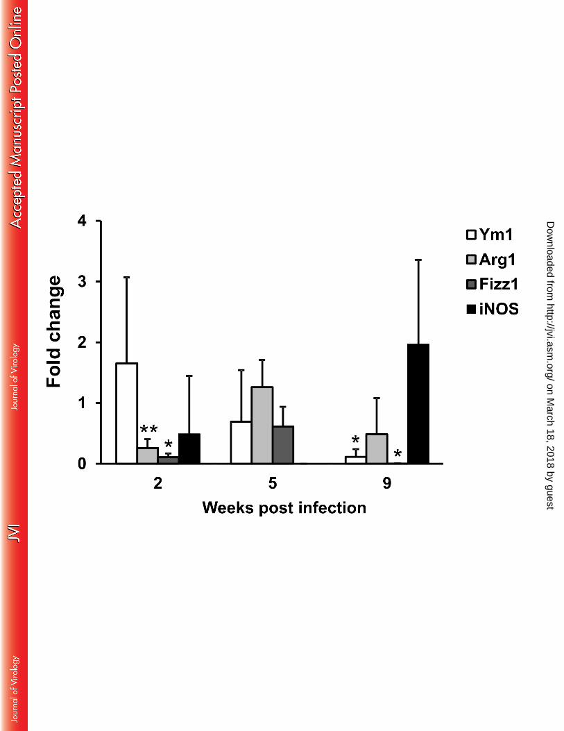


Table 1. PCR array format (16 genes; 6 samples/disc) and list of genes tested.
Position Gene Symbol Gene description Gene RefSeq #
1 IFNG Interferongamma NM_008337
2 IL10 Interleukin 10 NM_010548
3 IL2 Interleukin 2 NM_008366
4 IL27 Interleukin 27 NM_145636
5 IL4 Interleukin 4 NM_021283
6 IL5 Interleukin 5 NM_010558
7 Irf1 interferon regulatory factor 1 NM_008390
8 Nfkb1 Nuclear Factor Of Kappa Light Polypeptide
Gene Enhancer In B-Cells 1, p105
NM_008689
9 Stat1 signal transducer and activator of
transcription 1
NM_009283
10 tnf tumor necrosis factor NM_013693
11 IL12b Interleukin 12b NM_008352
12 B2M beta-2-microglobulin NM_009735
13 Gapdh glyceraldehyde-3-phosphate dehydrogenase NM_008084
14 MGDC Mouse Genomic DNA Contamination
control
-
15 RTC Reverse transcription control -
16 PPC Positive PCR control -
on March 18, 2018 by guest
http://jvi.asm.org/
Dow
nloaded from