glutamate mutase - vanderbilt university · glutamate mutase coenzyme b 12 ch h ch3 ......
TRANSCRIPT

121
241
Coenzyme B12-Co
adenosyl
methylaspartate
His-16
Glutamate mutase
pdb code: 1I9C
(2S,3S)-3-methyl-L-aspartate
HO2C CO2H
NH2glutamate
mutase
Coenzyme B12CO2H
NH2
CH3
HO2C
L-glutamate
242
O
OPO O
G
O O
O
T PO
O
O
OP OO
O
A
OO
O
CPO
OO
Nucleic Acids 1869: Miescher- gelatinous material from cell nuceli of white blood cells containing
organophosphorous compounds- nuclein (chromatin)- discovery of nucleic acids 1891: Kossel- identified the DNA bases A, T, G and C
identified D-ribose in nucleic acids 1889: Altman- purified DNA 1901: Ascoli- identified U in RNA 1910 : DNA and RNA realized to be separate entities. DNA (thymus) RNA (yeast) 1929: Levene & Jacobs- identified 2’-deoxyribose in DNA 1920’s - 1950’s: structures of nucleosides and nucleotides - Alexander Todd (Nobel Prize, 1959) 1928: Griffith- first to propose that DNA was genetic material; not widely accepted 1931: Levine & Bass- first proposed structure. Believed to be part
of chromosome physiology, but NOT genetic material 1944: Avery, MacLeod & McCarty- Strong evidence that DNA
is genetic material 1950: Chargaff- careful analysis of DNA from a wide variety of
organisms. Content of A,T, C & G varied widely according to the organism, however: A=T and C=G (Chargaff’s Rule)
1952: Hershey & Chase: followed the transfer of 32P-labelled DNA from T2 bacteriophage to infected bacterial cytoplasm (1969 Nobel Prize)
1953: Watson & Crick- structure of DNA (Nobel Prize with M. Wilkens, 1962)

122
243
Nucleoside, nucleotides and nucleic acids
nucleoside nucleotide nucleic acid
sugar
base
sugar
base
phosphate
Blackburn et al. Ch. 2
244
Sugar: D-ribose
CHOOHHOHHOHH
CH2OHOH
H
HHO OH
H HO
HO
H
OH
HHO OH
H HO
HO
β-D-ribofuranose α-D-ribofuranose
H
OH
HHO H
H HO
HO
β-D-2-ribofuranose
Stereochemistry: As drawn, the side above the plane of the ring is β the side below the plane of the ring is α
123
245
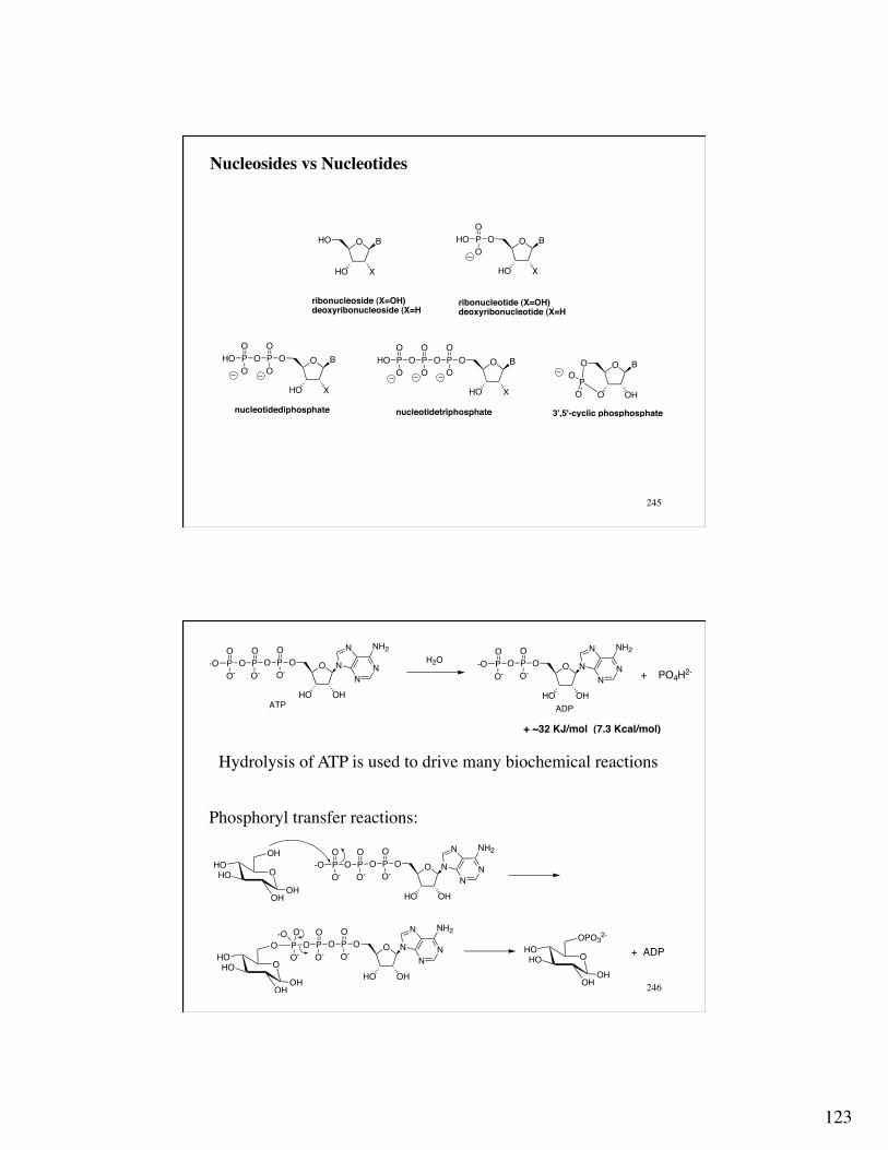
Nucleosides vs Nucleotides
O
HO X
HO B
ribonucleoside (X=OH)deoxyribonucleoside (X=H
ribonucleotide (X=OH)deoxyribonucleotide (X=H
O
HO X
O BPHOO
O
O
HO X
O BPOO
OPHOO
OO
HO X
O BPOO
OPOO
OPHOO
O
nucleotidediphosphate nucleotidetriphosphate
O
O OH
O B
PO
O
3',5'-cyclic phosphosphate
246
O N
N
NN
NH2
HO OH
OPOO-
OPO
OO-
P-OO-
O
ATP
H2OO N
N
NN
NH2
HO OH
OPOO-
OPO
-OO-
ADP
+ PO4H2-
+ ~32 KJ/mol (7.3 Kcal/mol)
Hydrolysis of ATP is used to drive many biochemical reactions
Phosphoryl transfer reactions:
OHOHO
OHOH
OH
OHOHO
OHOH
O OPO--O
O- OHOHO
OHOH
OPO32-
+ ADP
O N
N
NN
NH2
HO OH
OPOO-
OPO
OO-
P-OO-
O
O N
N
NN
NH2
HO OH
OPOO-
OPO
O-

124
247
N
N N
N
NH2
H2N
HN
N N
N
O
HN
N N
N
O
Adenine (R=H)Adenosine (R= furanose, A)
RGuanine (R=H)Guanosine (R= furanose, G)
Hypoxanthine (R=H)Inosine (R=furanose, I)
R R
N
N
NH2
O
R
NH
N
O
O
NH
N
O
O
N
N
NH2
O
RR RCytosine (R=H)Cytidine (R=furanose, C)
Thymine (R=H)Thymidine (R=furanose, T)
Uracil (R=H)Uridine (R=furnaose, U)
5-Methylcytidine (R=furnaose)
Bases A. Purines
B. Pyrimidines
DNA contains A, C, G, T all with 2'-deoxyribose!RNA contains A, C, G, U all with ribose!!The stereochemistry of the base is β
248
Numbering System
O
N
NN
NH
Purine
1
2
34
567
89
1'BaseHO
4'
5'
HO OH
Ribonucleoside
N
N
12
34
5
6
Pyrimidine
N
NH
O
OR
N
NHN
N1
345
62
Uridine
R
O
NH2
1
23
4
56
7
89
Guanosine
O6
2'3' O2
O4
N2

125
249
Nucleoside Conformation:
NH
N
NO
NH2N
O
OH
HOO
OH
HON
NH
O
O
O
OH
HO
HN
N
NO
H2N N
O
OH
HON
HN
O
O
Anti conformation Syn conformation
5-member ring conformation: envelope
C2' - endo conformation!found in B-form DNA!
O
OH
R
H
RO
H
N
N
NNH
NH2
O
7.0 Å
O
O HR
H
RO
H
N
N
NNH
NH2
O5.9 Å
C3' - endo conformation!found in A-form DNA!
250
Watson & Crick: double helix
Initial “like-with-like”, parallel helix: Does not fit with with Chargaff’s Rule: A = T G = C
Wrong tautomers !!
Watson, J. D. The Double Helix, 1968
N
NN
N
NH
dR
N
NN
N
NH
dR
HN
N
dR
O H
O
N
N
dR
OH
O
purine - purine pyrimidine - pyrimidine
N
N
dR
N H
O
N
N
dR
NH
O
H
N
NN
N
O
N
H
dR
N
NN
N
OH
dR
HH
H
NH
H
H

126
251
O
OR
RON
N
N
O
HH
O
OR
RO
NN N
NO
NH
H
H
NN
R
N
O
HH
RNN N
NO
NH
H
HN3 N1
N2
Hydrogen BondDonor
Hydrogen BondDonor
Hydrogen BondAcceptor
Hydrogen BondAcceptor
3'
5'
Hydrogen BondA cceptor
Hydrogen BondDonor
5'
O2
N4 O6
3'
Antiparallel C-G Pair
O
OR
RON
N
O
OO
OR
RO
NN N
NN
H
HH
NN
R
O
O
RNN N
NN
H
HH
N1N3
5'
3'
3'
5'
Hydrogen BondDonorN6
Hydrogen BondAcceptor
Hydrogen BondAccep tor O4
Hydrogen BondDonor
Antiparallel T-A Pair
Complimentary Base- Pairing in Nucleic Acids: antiparallel double helix
“Molecular Structure of Nucleic Acids” Watson J. D.; Crick, F. H. C. Nature 1953, 171, 737-8
252
B-DNA
Minor groove
Minor groove
Major groove
Major groove
pdb code: 1bna

127
253
DNA Grooves
cytidine guanosine
thymidine adenosine
minor groove
major groove
minor groove
major groove
254
DNA Sequences are written 5’ to 3’
5’-ATCGCAT-3’
5’-d(ATCGCAT)-3’
Writing DNA sequences
HO
A
P
T
P
C
P
G
P
C
P
A
P
T
OH
5' 3'

128
255
Melting Temperature (Tm): a measure a duplex stability
Double stranded
Single strand
Δ
Hybridzed: highly ordered
structure
Unhybridized: less ordered, random coil
Base stacking causes a deceases in the net UV absorbance when DNA is hybridized (double stranded) versus unhybridized.
Upon thermal denaturation (melting), the UV absorbance increases:
hyperchromicity
Blackburn et al. Ch. 2.5.1, 11.1.1
256
DNA melting curve:
0
0.05
0.1
0.15
0.2
0 20 40 60 80 100
Tm= 65 °C
annealingmelting
T (°C)
Abs
(260
nm
)
5’-d(GCT AGC GAG TCC)-3’!3’-d(CGA TCG CTC AGG)-5’
double stranded
single stranded
Tm is often taken as a measure of DNA duplex stability Tm is a measure of ΔG not ΔH The Tm is dependent upon the length and sequence of the
oligonucleotide (CG/AT ratio) and the ionic strength of the medium

129
257
DNA processing enzymes: DNA replication:
Helicase: Unwinds double stranded DNA DNA polymerase: replicates DNA using each strand as a template for the newly synthesized strand.
“It has not escaped our attention that the specific pairing we have postulated immediately suggests a possible copying mechanism for the genetic material.”
Watson & Crick
Blackburn et al. Ch. 3.6, 5.3, 6.6.4
258
DNA Polymerase: the new strand is replicated from 5’ to 3’ DNA polymerase are Mg2+ ion dependent Details regarding the mechanism of recognition of dNTP incorporation for the growing DNA strand are not fully understood

130
259
DNA Ligase: ATP-dependent enzyme that will join (ligate) two DNA segment by catalyzing the formation of the phosphodiester bond of a terminal 5’-phosphate of one oligonucleotide and the 3’-hydroxyl group of another oligonucleotide.
2-O3PO
OH 2-O3PO
OH
2-O3PO OH5'
5'
5'3' 3'
3'
DNA LigasePhosphodiesterbonds
OPO32-
OH
OH
2-O3PO5' 3'
3' 5'
OH
2-O3PO5'
3'OPO3
2-
OH3'
5'
DNA Ligase
260
Restriction Enzymes (endonucleases): enzymes that will cleave double stranded DNA at specific, known sequences.
5’-d(G-A-A-T-T-C)-3’!3’-d(C-T-T-A-A-G)-5’!!!5’-d(G-G-A-T-C-C)-3’!3’-d(C-C-T-A-G-G)-5’!
EcoR I BAM HI
5'
3'
3'
5'
5'
3'
3'
5'
OH 2-O3PO
2-O3PO OH
restrictionenzyme
“sticky ends”
Werner Arber, Daniel Nathans and Hamilton Smith 1978 Nobel Prize in Medicine & Physiology

131
261
Type I: cleaves at a site very distant from the recognition sequence; Mg2+, SAM and ATP dependent.
Type II: cleaves DNA at or near the recognition sequence; Mg2+ dependent. i.e., EcoR I, BamHI, Bbs I
BbsI: GAAGACNNNNNNN! !CTTCTGNNNNNNN
Recognition sequence Cleavage: blunt end
5'
3'
3'
5'
3'
5'
OH 2-O3PO
2-O3PO OH
Type 2restrictionenzyme
3'
5'
262
Polymerase Chain Reaction (PCR): method of amplifying DNA using DNA polymerase and cycling temperature
Heat stable DNA Polymerases:
Taq: thermophilic bacteria (hot springs)- no proof reading Pfu: geothermic vent bacteria- proof reading
Mg 2+ two Primer DNA strands (synthetic, large excess) one sense primer and one antisense primer one Template DNA strand dNTP’s O B
HO
OPOO-
OPO
OO-
P-OO-
O
KARY B. MULLIS, 1993 Nobel Prize in Chemistry for his invention of the polymerase chain reaction (PCR) method.
Blackburn et al. Ch. 5.2.3, 5.4.2

132
263
A typical PCR temperature cycle Denaturation: 94 °C 0.5 - 1 min Annealing: 55-68 °C 0.5 - 1 min 5 °C below the
Tm of the primer Extension: 72 °C 1 min + 1 min per Kb
of DNA # of cycles 25 - 35 Final extension 72 °C 10 min
264
Polymerase Chain Reaction
For a PCR animation go to: http://www.blc.arizona.edu/INTERACTIVE/recombinant3.dna/pcr.html
5'
3'
3'
5'
95 °C
denaturation
5' 3'
3' 5'
anneal (+) and (-) primers
55 - 68 °C5' 3'
3' 5'
3'3'
72 °CTaq, Mg 2+, dNTPs
extension
5'
3'
3'
5'
5'
3'
3'
5'
95 °C
denaturation
2nd cycle
5'
3'
3'
5'
5'
3'
3'
5'
amplification of DNA
2 copies of DNA

133
265
1 x 2 = 2 x 2 = 4 x 2 = 8 x 2 = 16 x 2 = 32 x 2 = 64 x 2 = 128 x 2 = 256 x 2 = 512 x 2 = 1,024 x 2 = 2,048 x 2 = 4,096 x 2 = 8,192 x 2 = 16,384 x 2 = 32,768 x 2 = 65,536 x 2 = 131,072 x 2 = 262,144 x 2 = 524,288 x 2 = 1,048,576
$$ The Power of Compounded Interest $$
In principle, over one million copies per original, can be obtained after just twenty cycles
266
Oligonucleotide-Based Site-directed mutagenesis
DNA mRNA protein
Blackburn et al. Ch. 5.6

134
267
3’-GAG ATG ACA CCC AAA-5’!5’-CTC TAC TGT GGG TTT-3’!! -Leu Tyr Cys Gly Phe-!!!!3’-GAG ATG CGA CCC AAA-5’!5’-CTC TAC GCT GGG TTT-3’!! -Leu Tyr Ala Gly Phe-!
268
23 BspD I23 Cla I29 HinD III
185 EcoR V229 Nhe I
375 BamH I
562 Sph I622 EcoN I651 Sal I
939 Eag I972 Nru I
1063 BspM I
1353 Bsm I1369 Sty I
1425 Ava I1444 Bal I1444 Msc I
1650 Bsg I1664 BspE I1664 BspM II1668 BsaB I
2066 Pvu II2124 Esp3 I
Tth111 I 2219Bsa I 2227
Bst1107 I 2246Xca I 2246
Nde I 2297Afl III 2475
AlwN I 2886
Eam1105 I 3363Bsa I 3435
Ase I 3539Pst I 3609
Pvu I 3735
Sca I 3846
Ssp I 4170Aat II 4286
EcoR I 4361
pBR322
4363 base pairsUnique Sites
Restriction Map

135
269
MICHAEL SMITH, 1993 Nobel Prize in chemistry for his fundamental contributions to the establishment of oligonucleotide-based, site-directed mutagenesis and its development for protein studies.
EcoR I
BamH ICys Ala
EcoR 1BamH1
Cys Ala
BamH I
EcoR IEcoR I
BamH I
3'-GAT CCC NNN CTC TAC ACA GGG TTT NNN--------------------------5'5'-CTA GGG NNN GAG ATG GCT CCC AAA NNN-3'
Cys
Ala
BAM H1 plasmid (template)
PCR primer with mutation
PCR amplifies a new insert with the mutation
Cut the PCR fragments with EcoR1 and BamH1
Ligate the new insert (with the desired mutation) back into the envelope
envelope
insert
(−) antisense (+) sense
270
Amplification of the mutant sequence by PCR TGTACA Codes for Cys
ACA
TGTGCT
Denature andanneal primers
(-) Primer contains the mutation for Ala
extend
ACATGTGCTACA
Denature andanneal primers
ACAGCT
TGT
ACAGCT
GCT
extend
ACAGCT
TGT
ACAGCT
GCTCGA ACA
(+) primer

136
271
ACAGCT
TGT
ACAGCT
GCTCGA ACA
Denature andanneal primers
ACAGCT GCT
GCT
GCTCGA
GCT
ACA
GCT
ACAGCT
TGT
DNA strands with the desired mutation are being amplified much faster than strands without the mutation; after ~25 rounds of PCR, the amount of DNA that does not have the desire mutation become negligible (<< 1 in a million)
GCT GCT
GCT
GCTCGA
GCT
ACA
GCT
ACAGCT
TGTCGA
CGA
ACA
Extend
CGA
CGA
272
Nucleoside Synthesis: important class of chemotherapeutic agents (anticancer and antivirals) important reagents for biotechnology
O
HO
HO NNH
O
O
F
O
HO
HO NN
NH2
OO
HO
HO NNH
O
O
F3C
O
N3
HO NNH
O
OOHO N
N
NH2
OOHO N
N
N
NH
O
S
O OHNN
H2N
OOHO N
N
NH2
O
ddIAZT ddC (-)-3TC
Anti-Cancer Nucleosides
Anti-Viral Nucleosides
d4C
F
F
Blackburn et al. Ch. 3.1

137
273
Chemical Synthesis of Ribonucleosides via Vorbrüggen Glycosylation
O
AcO OAc
AcOOAc
N
N OTMS
O
N
N OTMS
OTMS
O
AcO O
AcOO
AcO
AcO
O
O
SiMe3
O
AcO OAc
AcO NNH
O
OO
HO OH
HO NNH
O
O
SnCl4, CH3CN
Cl -
+
β-Ribonucleoside
NH3, MeOH
+
O
274
O
AcO OAc
AcOOAc
N
N OTMS
OTMS
O
AcO OAc
AcO NNH
O
OSnCl4, CH3CN
ribose-tetraacetate β-1'-stereochemistry
O
AcO OAc
AcOOAc
N
N OTMS
OTMS
O
AcO OAc
AcO NNH
O
OSnCl4, CH3CN
arabinose-tetraacetate α-1'-stereochemistry
The stereochemistry of the C2-ester group of the furanose-tetraester controls the stereochemistry of the glycosylation reaction

138
275
Exocyclic amino groups of C, A and G require protecting for the Vorbrüggen Glycosylation reaction
HN
NH
O
OR
N
NTMSO
OTMSR
N
NH
O
HN R
O
R= H, CH3
R= CH3, CH(CH3)2
N
NTMSO
N R
OTMS
N
N
HN Ph
O
N
NH N
N
N Ph
OTMS
N
N(H3C)3Si
N
NH
O
N
NH N
H
O
RR= CH3, Ph
N
N
OTMS
N
N N
OTMS
R(H3C)3Si
276
Chemical Conversion of Ribonucleoside to 2’-Deoxyribonucleoside: Free radical deoxygenation of the 2’-hydroxyl group
O
HO OH
HO NNH
O
O O
O OH
OSi
SiO
iPr
iPr
iPr iPr
O
HO
HO
NNH
O
O
PhOC(S)Cl,pyridine
NNH
O
O
β-Ribonucleoside
β−2'-Deoxyribonucleoside
2) nBu4N+F-, THF
1) nBu3SnH, AIBN PhCH3, reflux
(iPr2SiCl)2O,pyridine
O
O O
OSi
SiO
iPr
iPr
iPr iPrS
OPh
NNH
O
O
139
277
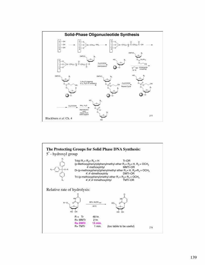
Solid-Phase Oligonucleotide Synthesis!
Blackburn et al. Ch. 4
OH O
O
O
Si (CH2)3 NH2
O
O
O
Si (CH2)3 NH
C (CH2)2
OCO
OH
O
O
O
Si (CH2)3 NH
C (CH2)2
OCO
OB1
O
DMTrO
OB
O
PO
O
O
B1
O
DMTrO
CN
OB1
HO
PO
O
O
B2
O
HO
O -
P N (iPr)2
O
O
B1
O
DMTrO
CN
OB1
O
PO
O
O
B2
O
HO
OCN
OH
OH
OB1
O
HO
SILICA
SILICA
SILICA
SILICA
Cl3CCOOH
Cl3CCOOH
NH3, H2O
1) Ac2O (capping)2) I2, H2O (P oxidation)
Repeat Cycle
1H-tetrazole(coupling)
deprotection-and-
cleave from solid support
OB1
O
PO
O
O
B2
O
DMTrO
OCN
(detritylation)NN N
NH
Cl3CCOOH
278
The Protecting Groups for Solid Phase DNA Synthesis: 5’- hydroxyl group
O
OH
N
HO
O
HN
O
OR
O
OH
N
HO
HO
HN
O
O20°C
80% AcOH (aq)
R = Tr ! 48 hr.!R= MMTr 2 hr.!R= DMTr 15 min.!R= TMTr 1 min. (too labile to be useful)!
C O RR2
R1
R3
Trityl R1= R2= R3 = H ! !Tr-OR!(p-Methoxyphenyl)diphenylmethyl ether R1= R2= H, R3 = OCH3 !!
!4'-methoxytrityl ! !MMTr-OR!Di-(p-methoxyphenyl)phenylmethyl ether R1= H, R2=R3 = OCH3! 4',4'-dimethoxytrity ! !DMTr-OR!Tri-(p-methoxyphenyl)methyl ether R1= R2= R3 = OCH3! 4',4',4'-trimethoxytrityl !TMTr-OR!
Relative rate of hydrolysis:

140
279
Protecting groups for the exocyclic amino groups
All are removed with NH3/H2O at 80° C
N
NO
HN
O
N
N
HN Ph
O
N
N N
NH
O
N
N NH
O
N
NH
O
N
N N N
N
NO
HN
O
N
N
HN
O
N
N N
NH
O
N
N NH
O
OPh
OPh
dR
dR
dR dR dR
dRdR
280
Phosphoramidite reagents: the monomeric building blocks for solid-phase DNA synthesis
OHO B
HO
Base is protected
DMTr-Cl
pyridine
ODMTrO B
HO
NP
O
ClCN
[(CH3)2CH]2NH
ODMTrO B
OP
O N[CH(CH3)2]NC
for solid-phase RNA synthesis OHO B
HO
Base is protected
ODMTrO B
HOOH
NP
O
ClCN
[(CH3)2CH]2NH
ODMTrO B
OP
O N[CH(CH3)2]NC
Si Cl
Ag+OH
DMTr-Cl
pyridine
ODMTrO B
HO O Si
ODMTrO B
OHSi O+
70 : 30
O Si
silyl group removed with fluoride ion (F-)

141
281
sequence selective chemical cleavage Detection: 5'-32P-labeling
A: Cleavage at the 3'-side of guanosines with a) dimethylsulfate b) Δ, c) piperidine.
O
O
O N
N
N
NH
O
NH2
32P
P-O OO
OS OCH3H3COO
OH
O
O
32P
P-O OO
OH
O
O
O N
N
N
NH
O
NH2
32P
P-O OO
H3C
OH
O
O
32P
P-O OO
NH
H H
HN
N
N
N
NH
O
NH2
H3C
O-
P-O OO
OHO
32P
NH
H
O
O
O
32P
P-O OO
OH
+
H2O, Δ
lose
+ +
DNA Sequencing: Maxam-Gilbert Sequencing Blackburn et al. Ch. 5.1
Maxam, A. M.; Gilbert, W. Methods Enzymol. 1980, 65, 499-558
282
Maxam-Gilbert Sequencing B. Cleavage at the 3'-side of adenosines and guanosines with a) dimethyl sulfate b) HCO2H, c) piperidine.
O
O
O N
N
N
N
NH232P
P-O OO
OS OCH3H3COO
OH
O
O
32P
P-O OO
OH
O
O
O N
N
N
N
NH232P
P-O OO
CH3
OH
O
O
32P
P-O OO
NH
H HHN
HCO2H, H2O
NH
N
N
N
NH2
CH3
O-
P-O OO
OHO
32P
NH
H
O
O
O
32P
P-O OO
OH+
+
+ +
lose

142
283
Maxam-Gilbert Sequencing
C. Cleavage at the 3'-side of thymidines and cytidines with a) hydrazine, b) Δ, c) piperidine.
O
O
O N
32P
P-O OO
HNO
O
CH3H2N NH2 O
O
O N
32P
P-O OO
HNO
O
CH3
HN NH2
H+
O
O
O N
32P
P-O OO
NH2O NH
HN
O
OH
O
O
32P
P-O OO
N
H H
OH
O-
O
32P
P-O OO
NNH2
HHN
NH2
O O
H2O
284
Maxam-Gilbert Sequencing D. Cleavage at the 3'-side of cytidines with a) hydrazine, 1.5 M NaCl, b) Δ, c) piperidine.
O
O
O N
32P
P-O OO
NO
NH2
H2N NH2 O
O
O N
32P
P-O OO
NO
NH2
HN NH2
H+
OH
O
O
32P
P-O OO
N
H H
OH
O-
O
32P
P-O OO
NNH2
HHN
NH2
O O
O
O
O N
32P
P-O OO
NH2
O NHHN
NH2
O
O
O N
32P
P-O OO
HNO
NH2
NH
NHH+
+
1.5 M NaCl

143
285
Separation and detection of 32P-labeled DNA fragments by polyacrylamide gel electrophosesis (PAGE). DNA fragments separated based upon charge, which is proportional to the length of the DNA fragment.
Maxam-Gilbert Sequencing
Larger fragments
Smaller fragments
3’
5’-32P
GTAACGTAATCACAG
G A & G C C & T
286
Sanger Sequencing Enzymatic replication of the DNA fragment to be sequnced with DNA polymerase, Mg+2, ddNTP's and 32P-labeled primers.
ddNTP’s
N
NN
N
NH2
OOPOO-
OPO
O-OP
O-O
O-
NH
NN
N
O
OOPOO-
OPO
O-OP
O-O
O-
ddATP
NH2
ddGTP
OOPOO-
OPO
O-OP
O-O
O-
OOPOO-
OPO
O-OP
O-O
O-
ddTTP
ddCTP
N
NH
O
O
N
N
NH2
O

144
287
Sanger Sequencing
Larger fragments
Smaller fragments
GTAACGTAATCACAG
ddA ddG ddC ddT
CATTGCATTAGTGTC
5'32P
3'
5'
3'
32P-5' 3'
primer template
288
Sanger sequencing with flourescent ddNTP's
N
NN
NH2
O-3 H3O9P3O
HN
ONCH3
O
OO O -
CH3H3C
NH
NN
O
O-3 H3O9P3O
HN
ONCH3
O
OO O -
NH2
OO O -
O
-3 H3O9P3O
N
N
NH2
O
HN O
NH3C
(CH2)2O
CH3 CH3
OO O -
CH3H3CO
-3 H3O9P3O
HN
N
O
O
HN O
NH3C
(CH2)2O
CH3 CH3
ddA ddG
ddT ddC
Excitation: ~ 490 nM, Emission: ddT= 526 nm. ddC= 519 nm, ddA= 512 nm, ddG= 505 nm

145
289
Sanger sequencing using flourescent ddNTP's terminated DNA strands are separated by capillary electrophoresis
290
Pyrosequencing
Ronaghi, M., Genome Res. 2001, 11, 3-11
Three enzyme system where the incorporation of a complimentary nucleotide opposite a template DNA strand results in the emission of a photon.
O
HO OH
AOPOS
OPOO
OPOO
O
is used in place of ATP - not a substrate for ATP-sulfurylase & luciferase
5'-ACCTTGATACCATCTAGGA----------------3' AGATCCT----------------5'
dNTPs are added sequentially one at a time
dTTP, Mg2+, DNA polymerase
5'-ACCTTGATACCATCTAGGA----------------3'- TAGATCCT----------------5'
O P OO
OP OO
O
+
O
HO OH
AOPOO
OO3S
ATP-Sulfurylase
ATP + SO42-
Luciferase, O2
oxyluciferase + P2O72- + AMP + photon
Adenosine- 5’-phosphosulfate (APS)

146
291
5'-ACCTTGATACCATCTAGGA----------------3' TAGATCCT----------------5'
dNTPs are added sequentially one at a time
dGTP, Mg2+, DNA polymerase
5'-ACCTTGATACCATCTAGGA----------------3'- GGTAGATCCT----------------5'
O P OO
OP OO
O
+
O
HO OH
AOPOO
OO3S
ATP-Sulfurylase
2 ATP + SO42-
Luciferase, O2
oxyluciferase + P2O72- + AMP + 2 photon
2
292
pyrogram
5’-----TCCCCACCGAAACCCCAACGTCAAC---------3’ AGGGGTGGCTTTGGGGTTGCAGTTG---------5’

147
293
Apyrase is a fourth enzyme that is added that will hydrolyze the dNTP and ATP to the corresponding dNMP and PPi between dNTP additions. The rate of hydrolysis of apyrase is slower that than the other three enzymes. Alternatively, a biotinated template is used which is bound to streptavidin coated beads. The immobilized template DNA is washed between dNTP addition streptavidin•biotin--–––-5’-ACCTTGATACCATCTAGGA-------3’ AGATCCT-------5'
NHHN
S
O
CO2H
H H
Biotin
NHHN
S
O
H H HN
O O
O-DMTr
PNiPr2
O CN
KD= 10-15
Sequencing by Reversible Chain Termination
O
O
BPPPO
N3
linker O
N3Dye
P
–O2C
CO2–
CO2– tris(2-carboxyethyl)phosphine (TCEP): Reduces azides to an amino groups 3’-O-blocked fluorescently
labeled dNTP
O N3
linker O
N3Dye
DNA polymerase
3'-O-blocked-fluorescent dNTPs
chain elongation is blocked
read fluorscence = a specific base
TCEP OHlinker OH
3' 3'DNA polymerase
3'-O-blocked-fluorescent dNTPs
linker OH
Olinker O
3'
N3
Dye
read fluorscence = a specific base
Etc.N3
chain elongation is blocked
C C C C C C
CTAG
C T G C A G

148
HN
NO
O NH
OO O
N3O N
H
O HN
O
N BN
F F
O
ON3
PPPO
N
NO
NH2 NH
OO O
N3O N
H
O HN
O
O
ON3
PPPO
O
CO2–
HN
HN EtEt +
N
N
N
NH2NH
OO O
N3O N
H
O HN
O
O
ON3
PPPO
O
CO2–
NN
N
HN
N
ONH
OO O
N3O N
H
O HN
O
O
ON3
PPPO
H2N
N
N
SO3–
SO3–
dTTP (blue)
dCTP (green)
dATP (yellow)
dGTP (red)
Reversible Chain-Terminating, Fluorescent dNTPs
296
OP
OP
OAOP
OP
OAOP
OP
OP
OP
OP
OP
OP
OP
OH
OAOP
OH OP
OAOP
OP
OBOP
OP
OBOP
OP
OP
OP
A-OP
OAOP
OAOP
OP
OP
B-OP
OAOP
OAOP
OBOP
OBOP
OCOP
OCOP
OA
OA
OBP
OBP
OCP
OCP
D-OPO
AO
DOP
OAO
DOP
OBP
OBP
OCP
OCP
OP
OP
hν
Coupling
hν
Coupling
hν
Coupling
Light-Directed, Spatially Addressable Parallel Synthesis

149
297
Photoremovable protecting group: o-nitrobenzyl ether derivatives
OO B
OP
O N[CH(CH3)2]NC
O
O
NO2Mechanism: hν (λ ~ 350 nm)
ROO
O N
H
O
O
hνRO
O
O N
H
O
O
ROO
O NO
OH
ROO
O NO
O+H
ROO
O NOH
O+
ROO
O NO
O
HH2O
H-OH2+
ROH +O
O NO
O
298
Spatially addressable: 8-mer chip: 65, 536 different sequences 12-mer chip: 1,677,216 different sequences
DNA fragment DNA–fluorophore
Place DNA on the chip, then wash away non-specific hybridization after 1-10 hrs. Raise temperature and “melt” away partially hybridized sequences.
3’-ACGGTGCG! CGGTGCGA !
!GGTGCGAG! GTGCGAGA! TGCGAGAA! GCGAGAAT etc!!3’-ACGGTGCGAGAAT---5’ (from the chip)!5’-TGCCACGCTCTTA---3’!

150
299 Blackburn et al. Ch. 3.4, 3.5
Nucleotide Biosynthesis
CHOOHHOHHOHH
CH3OP
OPO
HO OH
OHATP AMP
D-ribose-5-phosphate
OPO
HO OH
OPP
5-Phosphoribosyl-1- pyrophosphate (PRPP)
300
Purine Biosynthesis
PRPPO
HO OH
PO NH2 O
HO OH
PO
H2N
HNO
glutamine glutamate
glycine+ ATP
10-formyl-tetrahydrofolate
HCO3-, ATP
No biotin
O
HO OH
PO
NH
HNO
H
O
O
HO OH
PO
NH
HNNH2
H
O
ATPO
HO OH
PO N
N
NH2
glutamine + ATP
+
- H2O
glutamate + ADP+ Pi
amidophospho-ribosyl transferase
glycinamide ribonucleotide (GAR)
synthaseGAR
transformylase
O
HO OH
PO N
N
NH2
CO2-
O
HO OH
PO N
N
NH2
NH2
O
aspartate, ATP
10-formyl-tetrahydrofolate
H X
O" "
O
HO OH
PO N
N
NH
NH2
O
CHO
formylglycinamidine (FGAM) synthase
Aminoimidazoleribonucleotide (AIR) synthase
AIR carboxylase
aminoimidazole succinylocarboxamide
ribonucleotide (SAICAR) synthase
aminoimidazole carboxamide
ribonucleotide (AICAR) transformylase
O
HO OH
PO N
N
NH2
NH
OCO2
-CO2
-
-fumaric acid-ADP, -Pi
adenylosuccinatelyase
IMPcyclohydrolase
O
HO OH
PO N
N
N
NH
O
Inosine monophosphate(IMP)
- H2O
!ATP!

151
301
Folate derivatives – one-carbon donors
N
N N
N
H2N
NH2
N
NH
O CO2-
CO2-
RR= CH3, methotrexateR= H, aminopterin
HN
N N
N
H2N
O
NH
NH
O CO2-
Folic Acid
HN
N NH
HN
H2N
O
NH
NH
O CO2-
CO2-
CO2-
Tetrahydrofolate
HN
N NH
N
H2N
O
NH
NH
O CO2-
CO2-NADH
NADH
Dihydrofolate
dihydrofolatereductase
dihydrofolatereductase
Dihydrofolate reductase inhibitors
HN
N NH
HN
H2N
O
N
OH
NH
O CO2-
10-formyl-tetrahydrofolate
CO2-
NH
N
HNN
H2N O
N
NH
O CO2-
CO2-
5,10-methylene-tetrahydrofolate
O
H X X CH2OH" "
= " "=O
H H-or-
HN
N NH
N
H2N
O
NH
NH
O CO2-
CO2-
5-methyl-tetrahydrofolate
X CH3=" "
CH3
302
5,10-methene-THF to 5-formyl-THF
5,10-methene-THF to 5-methyl-THF
NH
N
HNN
H2N O
N
NH
O CO2-
CO2-
5,10-methylene-THFdehydrogenase
NADP
NH
N
HNN
H2N O
N
NH
O CO2-
CO2-
+
5,10-methenyl-THFcyclohydrolase
H2O
NH
N
HNN
H2N O
N
NH
O CO2-
CO2-
OH
HN
N NH
HN
H2N
O
N
OH
NH
O CO2-
10-formyl-tetrahydrofolate
CO2-
HN
N NH
N
H2N
O
NH
NH
O CO2-
CO2-
CH3
NH
N
HNN
H2N O
N
NH
O CO2-
CO2-
NH
N
HNN
H2N O
NH
CH2
NH
O CO2-
CO2-
5,10-methylene-THFreductase
NADPH, FAD
H+
5-methyl-tetrahydrofolate

152
303
Purine Biosynthesis (con’t)
O
HO OH
PO N
N
N
NH
O
O
HO OH
PO N
N
N
N
NH2
Inosine monophosphate(IMP)
aspartate, GTP
fumaric acid+ GDP + Pi
Adenosine monophosphate(AMP)
adenylosuccinate synthase,adenylosuccinate lyase
NAD+ + H2O
O
HO OH
PO N
N
NH
NH
O
O O
HO OH
PO N
N
N
NH
O
NH2
Xanthosine monophosphate(XMP)
glutamine + ATP
Guanosine monophosphate(GMP)
IMPdehydrogenase
GMP synthase
NADH + H+
glutamate+ ADP+Pi
304
IMP Dehydrogenase
OO3PO
HO OH
2-N
N
NNH
O
NR
CONH2
NAD+IMP
Cys-S
H2O OO3PO
HO OH
2-N
N
HNNH
O
O
XMP
+ + NADH + H+
Cys-S

153
305
Pyrimidine Biosynthesis
O
NH2-O3PO N
HCO2
-
CO2H
H2N
OHCO3
- + ATP+ glutamine
Aspartic acidZn2+,
- H2O
carbamoylphosphatesynthase
aspartatecarbamoyltransferasecarbamoyl
phosphate
dihydroorotase
HN
NH
CO2-
O
O
HN
NH
CO2-
O
O
PRPPO
HO OH
PO NCO2
-
O
OHN
dihydroorotate
NAD+ / FAD
Dihydroorotatedehydrogenase
Orotate monophosphate(OMP)
orotate
orotatephosphoribosyl
transferase
306
O
HO OH
PO NCO2
-
O
OHN
- CO 2
Orotate monophosphate(OMP)
OMPdecarboxylase
O
HO OH
PO N
HNO
O
O
HO OH
PPPO N
HNO
O
O
HO OH
PPPO N
NO
NH2
Uridine monophosphate(UMP)
1) 2 ATP
Cytidine triphosphate(CTP)
Uridine triphosphate(UTP)
ATP + GTPglutamine
CTPsynthase
O
HO
PO N
HNO
O
Ribonucleotidereductase
dUMP
O
HO
PO N
HNO
O
CH3
methylene-tetrahydrofolate
Thymidine monophosphate(TMP)
thymidylatesynthase

154
307
Ribonucleotide Reductase O
HO OH
PO B RibonucleotideReductase O
HO
PO B
FeO
OH2O
CAsp Fe
OO
H2OOO
O
Glu
Glu
O
µ-oxo-bridged diferric clusier
OTyr
Cys S
Class I
S
Fe S
FeFe
S Fe
S
Class III
O
HO OH
S AH3C
H3NCO2
ETO
HO OH
A
O
proteinHN
protein
O
proteinHN
protein
glycine radical
Coenzyme B12
Cys S
Class IIClass IV
OTyr
Mn MnO
308
Mechanism of Ribonucleoside Reductase
OH H
OHHO
PnO B
Radical
OH
OHO
PnO B
Radical-H
HB:
OH
OHO
PnO B
S CysH
O
HO
PnO B
-H2O
S Cys
O
HO
PnO BO
HO
PnO B
S Cys S Cys
S CysH
O
HO
PnO B
H
S CysS Cys
ET
H+
O
HHO
PnO B
H
Radical-H
O
HHO
PnO B
H
Radical
H
ribonucleoside-5P
2'-deoxyribonucleoside-5P

155
309
Mechanism and Inhibition of Thymidylate Synthase
O N
HN
2- O3PO
HO
O
O
-S Cys
N
HNN
HN
O
H2N
CH2
+
NHRX
dUMP (X= H)
O N
HN
2- O3PO
HO
O
O
CH3
dTMP
N
HNN
HN
O
H2N
+ NHR
Dihydrofolate-S CysGlu-CO2-
Glu-CO2-
310
Ternary complex of thymidylate synthase with 5’-fluoro-dUMP and 5,10-dihydrotetrahydrofolate
FdUMP
Cys-161
Tyr-108
9,10-CH2-THF
pdb code: 1B02

156
311
DNA Methylation: 5-methyl-2’-deoxycytidine
O N
N
O
O
O
NH2
S Cys
SO2CCH3
NH3O N
N
NN
NH2
HO OHO N
N
O
O
O
NH2
S Cys
CH3
SO2C
NH3O N
N
NN
NH2
HO OH+ +
312
5-Methyl-C (Epigenetic) sequencing
N
N
DNA
NH2
O
NaHSO3
sodiumbisulfite
N
N
DNA
NH2
OO3S
basic pH
H2ON
NH
DNA
O
OO3S N
NH
DNA
O
O
N
N
DNA
NH2
O
NaHSO3
sodiumbisulfite
N
N
DNA
NH2
OO3S
H3C H3C
Sodium bisulfite converst C’s to U’s, but has no effect on 5-methyl-C

157
313
Antiviral nucleosides: 2,3-dideoxynucleosides as reverse transcriptase inhibitors
Protein Biosynthesis
DNA mRNA Protein transcription translation
Retrovirus (hepatitis B, HIV, HPV) ssRNA + proteins (reverse transcriptase, integrase, protease)
RNA ds DNA poly-protein
Blackburn et al. Ch. 3.7 H. Temin & D. Baltimore
1975 Nobel Prize in Medicine or Physiology
reverse transcription
reverse transcriptase
intgrase
insertion into host cell genome
proteins protease
314
Mechanism of action of 2,3-dideoxynucleosides: recall Sanger sequencing- termination of a growing DNA chain by enzymatic incorporation of a 2,3-dideoxynucleosides
RT RT
Incorporation of a ddNTP Termination of elongation Truncated DNA also inhibits RT

158
315
O
N3
HO NNH
O
OOHO N
N
NH2
OOHO N
N
N
NH
O
S
O OHNN
H2N
OOHO N
N
NH2
O
ddIAZT ddC (-)-3TCd4C
Nucleoside-based reverse transcriptase inhibitors
OHO B O2-O3PO B O
2-O3PO3PO B
O2-O3PO3PO3PO B
ddNMP ddNDP
ddNTP
2,3-dideoxynucleoside is enzymatically phosphorylated at the 5-hydroxyl to the ddNTP, which is the active RT inhibitor
Pro-drug: administered in an inactive formed but is transformed in vivo into the active drug
316
DNA as a target for therapeutic agents: Intercalation: • planar aromatic molecules “slide” between stacked bases • intercalators span the minor and major grooves of DNA • driven by hydrophobic interactions • intercalators stabilize DNA structure • causes an “unwinding” and elongation of DNA in the area of
the intercalation
NH2N
NH2
+
Br -
OCH3 O
O OH
OH
O
R
OH
O
OH3C
OHNH2
Ethidium BromideR=H, daunomycinR=OH, adriamycin
N
N
O
O
OOH
Campthothecin
NH
NH3C
CH3
Ellipticine

159
317
Topological relationship of DNA: supercoiling, tangling, knotting for replication (transcription), the supercoiling of DNA must first be “relaxed”
Topoisomerase (mammalian) DNA gyrase (bacteria)
Corbett, A.H.; Osheroff, N. Chem. Res. Toxicol. 1993, 6, 585-597 Topo II movies: http://berger.berkeley.edu/Pages/Teaching.html
Topo II inhibitors stabilize the covalent DNA•Topo II complex (protein bound double strand cleaved DNA), which causes chromosomal abnormalities and leads to apoptosis
Mechanism of the DNA cleavage step OO B
O
P_ O O
OO B
O
Tyr
O _
H+
OO B
HO
P O _
OO B
O
O
Tyr O
colvalent enzyme-substrate
intermediate
ATP
318
Intercalation
pdb code: 110D!
major groove
minor groove
major groove
OCH3 O
O OH
OH
O
OH
O
OH3C
OHNH2
daunomycin

160
319
Ribosomal Protein Biosynthesis:
DNA RNA Protein (genome) (transcriptome) (proteome)
transcription translation
Transcription: only one of the DNA strands is copied (coding or antisense strand). Its sequence is converted to the complementary sequence in mRNA (template or sense strand), which codes for the amino acid sequence of a protein (or peptide)
DNA
RNA polymerase pre-mRNA mRNA
rNTPs
“splicing”
320
Pre-mRNA:
mRNA:
5'-UTR 3'-UTR
Intron Intron
Exon Exon Exon
splicing
5'-UTR 3'-UTRExon Exon Exon
Transcription:
Translation: mRNAs are transported from the nucleus to the cytoplasm, where they acts as the template for protein biosynthesis (ribosomes). A three base segment of mRNA (codon) codes for an amino acid.

161
321
Transfer RNA (tRNA): The “anticodon” region of tRNA is complementary to the mRNA codon sequence.
The t-RNA carries an amino acid on the 3’-terminal hydroxyl (A) (aminoacyl t-RNA) and the ribosome catalyzes amide bond formation.
Although single-stranded, there are complementary sequences within tRNA that give it a defined conformation
aminoacyl t-RNA
TψC loop
D loop
variable loop
322
There are many non-standard bases found in tRNAs
. . . and alternative base-pairing N
NN
N
O
HR
NN
NHH
O RI C
N
NN
N
O
HR
NN
O
O R
H
I-U wobble pair
N
NN
N
O
HR NN
NHH
N
N
R
I-A Wobble pair
N
N
NH
H
N
N
R
NN
O
O R
HN
NN
N
O
HR
G-U Wobble pair
N HH
N N
O
ORH
A-U Hoogsteen pair
N
NN
N
O
HR
N HH
NN
NHH
O R
G-C reverse pairing
dihydrouridine (D)
O NNH
HO
HO OH
O
O
ONH
HN
HO
HO OH
O
O
pseudouridine (ψ)
O NHO
HO OH
N
NNH
O
inosine (I)
O NHO
HO OH
N
NNH
O
NH2
CH3
7-methylguanosine
O NHO
HO OH
N
NN
NH2
CH3
1-methyladenosine
O NNH
HO
HO
O
O
thymidine (T)

162
323
N
NN
NN
R
NN
O
OR
H H
H
N
NO
OH
R
Hoogsteen pairing
W-C pairing
N
NN
NN
R
NN
O
OR
H H
H
N
NN
N
NH
R
H
Hoogsteen pairing
W-C pairing
. . . and triple helix formation
U • A-U A • A-U
NN
N
N O
N
NN
N
O
HH
RR
H
HH
N
NN
N
O
N
R
H
H
H
W-C pairing
Hoogsteen pairing
G • G-C
324
tRNA Synthetase: catalyzes the biosynthesis of specific 3’-aminoacyl tRNAs from tRNAs, amino acids, and ATP
3’-aminoacyl tRNAs
Class I: 2’-aminoacyl tRNAs Class II: 3’-aminoacyl tRNAs
OOPO
OOP
O
OOP
OO
O
HO OH
N
N
NN
NH2
O
OH2N
R
OH2N
R OOPO
OO
HO OH
N
N
NN
NH2
OOPO
OO
O OH
N
N
NN
NH2tRNA
HB:
OOPO
OO
O OH
N
N
NN
NH2tRNA
OH2N
R

163
325
U G U AA U C U CG U U3'5'
A site: Aminoacyl tRNA
A CU
OO
HN
SCH3
OHC
U G U AA U C U CG U U
3'5'
A CU
OO
HN
SCH3
OHC
U AA
OO
H2N
OH
U G U AA U C U CG U U3'5'
U AA
OO
HN
OHONH
CH3S
A CU
OH
OHC
U G U AA U C U CG U U3'5'
U AA
OO
HN
OHONH
CH3S
A CU
OH
OHC
U G U AA U C U CG U U3'5'
U AA
OO
HN
OHONH
CH3S
A CU
OHOHC
E site
U G U AA U C U CG U U3'5'
U AA
OO
HN
OHONH
CH3S
OHC
G AC
O O
CH3H2N
U G U AA U C U CG U U3'5'
O
HNOH
OHN
G AC
O O
CH3HN
SCH3
U AA
OH
OJHC
U G U AA U C U CG U U3'5'
O
HNOH
OHN
G AC
O O
CH3HN
SCH3
U AA
OH
OHC
U G U AA U C U CG U U3'5'
O
HNOH
OHN
G AC
O O
CH3HN
SCH3
U AA
OH
OHC
P site: Peptidyl t-RNA
Ribosomal protein synthesis
326
Taken from: “The New Genetic Medicines,” J. S. Cohen, M. E. Hogan Scientific American 1994 (Dec.), pp 75-82.
Oligonucleotide-Base Therapies: Inhibition of Protein Biosynthesis via the Antisense-Antigene (Triplex) Strategies
• Potentially highly selective (magic bullet) approach to therapy • ~15 bases sequence is unique in the human genome

164
327
Watson-Crick base-pairing
Hoogsteen base-pairing
DNA mRNA
Triple Helix mRNA•DNA
transcription translationprotein
XInhibition oftranscription
X
Inhibition oftranslation
328
Triple Helix Motifs: third strand binds in the major groove Pyrimidine•Purine-Pyrimidine: third strand runs parallel to the homo-purine strand
N
NN
NN
dR
NN
O
OdR
H H
H
N
NO
OH
dR
NN
N
N O
N
NN
N
O
HH
dR
dR
H
HH
NN N
O
H
HH
dRT•A-T + C+•G-C
Hoogsteen pairingHoogsteen pairing
W-C pairingW-C pairing
Purine•Purine-Pyrimidine: third strand runs antiparallel to the homo-purine strand
N
NN
NN
dR
NN
O
OdR
H H
H
N
NN
N
NH
dR
H
NN
N
N O
N
NN
N
O
HH
dRdR
H
HH
N
NN
N
O
N
dR
H
H
H
A•A-T G•G-C
Hoogsteen pairingHoogsteen pairing
W-C pairingW-C pairing

165
329
DNA Triple Helix!
Major groove
Minor groove
T• A- T
C+• G- C
pdb code: 1BWG
330
Antisense Inhibition: inhibits mRNA function
Exon ExonIntronmRNA
splicing
ExonExonAUG UAA5'-cap
initiating sequence
stopsequence
Antisense targeting: Interon-exon splice junction: interferes with slicing 5’-cap (UTR) region: interferes with binding to the ribosome Initiation and promoter sequences: protein synthesis is not initiated Coding sequence: interferes with elongation of the protein

166
331
When an antisense oligonucleotide binds to mRNA, ribonuclease H is up-regulated. The mRNA of the mRNA•DNA hybrid is digested.
Problems with oligonucleotide-based therapies
Transport: DNA does not cross cellular membranes very easily Degradation: DNA is subject to enzymatic digestion by cellular nuclease
Synthetic Oligonucleotides: must maintain affinity and selectivity
and impart nuclease resistance • backbone replacements • modified bases
332
O
O
HO B
O
HO
O BP-O O
O
O
HO B
O
HO
O BP-S O
O
HN
HO T
O
HO
HN TCH2N
O
O
HO B
O
HO
O BPMe O
+
Phosphodiester Phosphorothioate Methyl Phosphonate Deoxyribonucleic Guanidines
O
HO
HO NNH
O
O
H3C
O
HO
HO NN
NH2
O
H3C
O
HO
HO NN
O
NHX
X=OX=S
Antisense Nucleosides
Triple Helix Oligonucleotides
Peptide Nucleic Acids (PNAs)
N
H2N
OH
O
OBase
n
m
Peptide backbone forms a helical structure. Base will hybridized with ss or ds DNA or RNA with high affinity

167
333
microRNA (miRNA): genetically encoded (transcribed) but non-translated RNAs (do not code for a protein or peptide)
5'-(7-methyl-G)-
3'-(A)n-Drosha
3'
5'~ 70 nt
pri-miRNA
pre-miRNA
transport to cytoplasmthen Dicer
3'
5'ds RNA (21-25 nt)
3'
5'
RISC (RNAi silencing complex)
5' 3' miRNA
334
Ribonucleases (RNase): enzymes that catalyze the hydrolysis of the phosphodiester bonds of RNA
single strand specific: RNase A double strand specific: RNase III specific for DNA/RNA hybrids: RNase H
endonucleases: RNase A, RNase III exonucleoase: RNase II

168
335
B
O
OHO
OPO
O
B
O
OO
O
H
NH
NHisH
N NH
His
B
O
OHO
HO
PO
-O
B
O
OHO
O
O-
NH
NHisH
N NH
His
neutralization ofcharge makes thephosphate more triester like
B
O
OHO
OPO
O
B
O
OO
O
H
NH
NHis
H
N NH
His
Lys NH3B
O
OHO
O
P O-O
B
O
OO
O
HLysH3N
HN NH
His
NH
NHis
N NH
His
B
O
OHO
O
P O--O
B
O
OO
O
LysH3N
H
NH
NHisH
B
O
OHO
HO
PO O-
B
O
O
O
ON NH
His
H
NH
NHis
HH
O
A mechanism for ribonuclease A
336
Interference RNA (RNAi) miRNAs (or siRNAs) are important in post-trancriptional regulation of gene expression
RISC (RNAi silencing complex)- multi-protein complex with helicase and ribonuclease activity
Andrew Fire & Craig Mello, 2006 Nobel Prize in Medicine & Physiology Animation: http://www.nature.com/focus/rnai/animations/index.html
3'
5' 3'
5'
ATP
5' 3'
5'3'
target mRNA
5'
target mRNA is cleaved by RISC
Ago2
5' 3'
guide strand21-25 nt
3'
mRNA fragements degraded by RNAses;protein expression is silenced

169
337
Cellular Response to DNA Damage:
DNA Damage
Cell CycleArrest
DNARepair
Apoptosis Replication Errors
CellDeath
Mutations
Cancer
338
DNA Alkylating Agent
N
NHN
N
O
N
NHN
N
NH2
NH2
Nitrogen mustards will alkylate adjacent DNA bases causing DNA-DNA cross-links, which is a severe form of DNA damage and can trigger apoptosis or cause mutations (and cancer)
HNR
O
N
Cl
Cl
Nitrogen Mustard
HNR
O
N
Cl
+
aziridinium ionactive alylating agent

170
339
Other simple DNA alkylating agents: Alkyl halides (or reactive equvalents): dimethylsulfate
methylene chloride, dichloroethane, SAM (endogenous) Enals: acrolein (industrial chemical, cigarette smoke),
4-hydroxynon-2-enal, malondialdehyde (endogenous) Epoxides: Butadiene diepoxide (metabolism of butadiene),
chlorooxirane (metabolism of vinyl chloride), benzo[a]- pyrene diol epoxide (metabolism of benzo[a]pyrene)
340
Free-radical mediated oxidative damage to DNA
Reactive oxygen species (ROS)
O O
O O molecular oxygen
super oxide
H O
O O N O peroxynitrite
hydroxyl radical
Causes oxidative cleavage of DNA in an O2 dependent manner
The oxygen paradox: oxygen is necessary for cellular metabolism; however, oxygen is transformed into highly reactive species that can damage biomolecules.

171
341
Oxygen metabolism
Superoxide dismutase (SOD): Zn-Cu or Mn-Fe
Catalase (heme) H2O2 O2 + H2O
O2 + e O Osuperoxide
HO OpKa ~ 4.8
O O22 H+
H2O2 + O2 k= 105 - 107 M-1 • sec-1
O O2 2 H+H2O2 + O2 kcat= ~ 2 x 10⁹ M-1 • sec-1
Enz•Cu(II) Enz•Cu(I)O2
+ H2O2
+ O2+Enz•Cu(I) Enz•Cu(II)O2+
2 2
342
Fenton reaction O2 + Fe(III) O2 + Fe(II)
H2O2 + Fe(II) HO + HO + Fe(III) Redox cycling
Very little free Fe(III) in cells
Haber-Weiss reaction O2 + H2O2 HO + HO + O2 slow
ONO Osuperoxide
+nitricoxide
ONOO
peroxynitrate
ONO2 + H+pKa ~ 7.0 O
NOHO
peroxynitrate
peroxynitrous acid
HO + NO2

172
343
N N
H2N
O
HN
NH2
NH2
O
H2NO
HNOHO
HN
O
ON
NHO O
O
OH
HO
HO
O
NH2O
OHOH
HO
NH
O
S
N
SN
NHO
NH
NH2
H2N+
Fe Binding Domain
DNA BindingDomain
Bleomycin!
Binds Fe and activates O2 giving C4 hydrogen atom abstraction mechanism of hydrogen atom abstraction may involve a Fe-O•, reminiscent of P450
344
Bleomycin•Co(III)OOH - DNA complex
Solution structure of the Bleomycin•Co(III) complex
Conformation of the DNA bound Bleomycin•Co(III)OOH complex
pdb code: 1MXK
pdb code: 1DEY

173
345
Abstraction of the 4’-hydrogen:
OO
B
5'-DNA-O3P
OP_ O O
PO3-DNA-3'O
HHO
OO
B
5'-DNA-O3P
OP_ O O
PO3-DNA-3'O
O2 OO
B
5'-DNA-O3P
OP_ O O
PO3-DNA-3'O
OORSH
OO
B
5'-DNA-O3P
OP_ O O
PO3-DNA-3'O
OHB:
OO
O
5'-DNA-O3P
OP_ O O
PO3-DNA-3'O
HH :B
OO
O
5'-DNA-O3P
O _
P_ O O
PO3-DNA-3'O
346
Abstraction of the 5’-hydrogen
OO
B
5'-DNA-O3P
OP_ O O
PO3-DNA-3'O
OH
OO
B
5'-DNA-O3P
OP_ O O
PO3-DNA-3'O
O2 OO
B
5'-DNA-O3P
OP_ O O
PO3-DNA-3'O
RSH
OO
B
5'-DNA-O3P
OP_ O O
PO3-DNA-3'O
HH O
O
OH :B
O
OH
B
5'-DNA-O3P
OP_ O O
PO3-DNA-3'O
O

174
347
Abstraction of the 1’-hydrogen
OO
B
5'-DNA-O3P
OP_ O O
3'-DNA-O3PO
OHO
OB
5'-DNA-O3P
OP_ O O
3'-DNA-O3PO
O2 OO
B
5'-DNA-O3P
OP_ O O
3'-DNA-O3PO
RSH
OO
B
5'-DNA-O3P
OP_ O O
3'-DNA-O3PO
:B O _
P_ O O
3'-DNA-O3PO
H O O
O H
:B
OO
5'-DNA-O3P
OP_ O O
3'-DNA-O3PO
O
H
H
OO
5'-DNA-O3P
O
348
Formation of 8-Oxo-2’-deoxyguanosine and Formamidinopyrimidine Lesions by the reaction of deoxyguanosine with ROS’s
N
N N
NH
dR
O
NH2HO
N
N N
NH
dR
O
NH2OH
N
NH
O
NH2
NO
HNdR
HH
[H]
N
N N
NH
dR
O
NH2
O
H[O]
8-oxo-dG
formamidopyrimidine (FAPY)
H-H+
+H+

175
349
O
OH
OPO2PO2PO3-N
N
N
O
HH
O
OR
OR
NNN
N O
NH
H
H
OH
O
OH
OPO2PO2PO3-
O
OR
OR
N
N
NN
NH
H H
O
O
NN N
NNH
H
H
Alternative base-pairing of 8-oxo-dG during replication with pol T7
mutagenic
pdb code: 1TK8
non-mutagenic
pdb code: 1TKD
8-oxo-dG
dA 8-oxo-dG
dC
350
DNA as a target for carcinogen: Bioactivation of pro-carcinogens:
P450
O
H2O
OHHO
P450
OHHO
O
benzo[a]pyrene
OOO
OO
OCH3
H
H
aflatoxin B1
P450 OOO
OO
OCH3
H
H
O

176
351
OOO
OO
OCH3
H
H
O
N
HN N
N
O
H2N
N
HN N
N
O
H2N
O
OO
OOOCH3
H
H
HO
+t1/2 in H2O ~ 1 sec
OOO
OO
OCH3
H
H
OOO
OO
OCH3
H
H
OOO
acetone
T. M. Harris et al. J. Am. Chem. Soc. 1988, 110, 7929
OHHO
O
N
NN
N
NH2
N
N
N
N
OHHO
HONH
352
Adducted Phosphoramidite Approach: Benzo[a]pyrene!
O
OHHO
N
N
N
NH
NH2
O
dRN
N
N
NH
O
dR
OHHO
HONH
O
O
DMTrO N
N
NNH
O
PO N(iPr)2
NC
OAcAcO
AcONH
C G G A C A G A A G
N
N
N
NH
NH
O OH
OHOH
solid-phaseoligonucleotide
synthesis

177
353 Kim, S. J.; Stone, M. P.; Harris, C. M.; Harris, T. M. J. Am. Chem. Soc. 1992, 114, 5480!
O
O
DMTrO N
N
NN
X
O
PO N(iPr)2
NC
Si(CH3)3
OHHO
HONH2
C G G A C A G A A G
N
N
N
NH
NH
O OH
OHOH
C G G A C A G A A G
N
N
N
N
X
O(H3C)3Si
solid-phaseoligonucleotide
synthesis
1)
2) Deprotection
X= -F, -OTf
Post-Synthetic Modification Strategy !for N2-2'-Deoxyguanosine Adducts!
354
O
O
DMTrO N
N
NN
X
PO N(iPr)2
NC
OHHO
HONH2
C G G A C A G A A G
N
N
N
N
OHHO
HO
C G G A C A G A A G
N
N
N
N
X
solid-phaseoligonucleotide
synthesis
1)
2) Deprotection
X= -F, -Cl
NH
Post-Oligomerization Strategy!for N6-2'-Deoxyadenosine Adducts!

178
355
DNA-Carcinogen Adducts!DNA-PAH Adducts: benzo[c]phenanthrene!
pdb code: 1HX4 pdb code: 1HWV
N
N N
NH
NHdR
O
HO
HOOH
N
N N
NH
NHdR
O
HO
HOOH
356
DNA-Carcinogen Adducts!DNA-PAH Adducts: benzo[a]pyrene!
pdb code: 1Y9H
benzo[c]phenanthrene!benzo[a]pyrene!

179
357
DNA-Carcinogen Adducts!DNA-Aflatoxin Adduct!
N
N
N
NH
NH2
OO
O
MeO
OO
O
HO
HH
dR
+
pdb code: 1MKL
358
DNA Repair: 1. Direct repair
2. Base excision repair (BER): repair of deglycosylation (lose of the base from the deoxyribose unit) sites, oxidation of the base, or modification by a “small” alkylating agent.
3. Nucleotide excision repair (NER): repair of “bulky” lesions
4. Mismatch Repair: repair of mis-paired DNA bases
5. Recombination: repair of double strand breaks of DNA
“Chemistry and Biology of DNA Repair” Scharer, O. D. Angew. Chem. Int. Ed. Engl. 2003, 42, 2946-2974

180
359
Direct Repair: O6-alkylguanine transferase (AGT): direct reversal by transferring the O6-alkyl group to an active site cysteine of AGT via an SN2 reaction.
N
N N
N
O
NH2DNA
CH3
S Cys AGT
H+ N
N N
NH
O
NH2DNA
S Cys AGTH3C
DNA Photolyase (bacterial): direct reversal of pyrimidine- pyrimidine photodimers (UV light induced lesion)
N
HN
N
NH
O
O O
O
hν
N
HN HN
N
O
O
O
O
DNA Photolyase: FAD dependent
light dependent repair
360
O6-Alkylguanine transferase (AGT):
N
N N
N
H2NdR
OH3CSCys145
H
N
NHis146
H
Glu172 CO2-
HOH
OH
Tyr114
N
N N
N
H2NdR
OSCys145
CH3
N
NHis146
H
Glu172 CO2-
HO H
OH
Tyr114
H
pdb code: 1T38
Glu172 His146
H2O
Ser145 Tyr114
O6-Me-G
X-ray analysis of the C145S mutant

181
361
O6-Alkylguanine transferase (AGT): S
Cys145
H
N
NHis146
H
Glu172 CO2-
HOH
N
N N
N
OdR
O
Cys145
N
NHis146
H
Glu172 CO2-
HO H
OH
Tyr114
H
N
N N
N
dR
OH
Tyr114
O
O S covalent protein-DNA
complex
Glu172
His146
Tyr114
Cys145
Direct Oxidative Dealkylation of DNA Bases by Fe(II)-2-ketoglutarate Dependent Dioxygenases
N
N N
N
H2N
DNA
CH3+ AlkB, Fe(II), O2,2-ketoglutarate N
N N
N
H2N
DNA
+OH
– CO2, – succinateN
N N
N
H2N
DNA
OCH H
+
Bugg, pg. 140-1
Fe(II)O
OH2His O
AspHis
CO2
OFe(III)
O
OHis O
AspHis
CO2
O
Fe(IV)O
OHis O
AspHis
CO2
O
Fe(IV)
OHis O
AspHis
CO2
O Fe(III)
OHis O
AspHis
CO2
O
O O
N
N N
N
H2N
DNA
C+H
H H
Fe(III)
OHHis O
AspHis
CO2
O
N
N N
N
H2N
DNA
C+H
H
N
N N
N
H2N
DNA
+OH
O2
-CO2 "rebound"
Fe(II)His O
AspHis
CO2
OFe(II)
His OH2
AspHis
O CO2
OOH2
OH2
N
N N
N
H2N
DNA
OCH H
++
+
(–H2O)Fe(II)
OH2
OH2His OH2
AspHis
2-oxoglutarate
362

182
363
Base-Excision Repair: HN
N N
NH
O
NH2DNA
ON
N N
NH
O
NH2DNA
H3C
OGG1 AAG
N
NH
O
DNA
HOH3C
HO O N
NH
O
DNA
O
N
N N
N
NH2
DNA CH3
HN
HN N
NH
O
NH2DNA
OHC
Fpg UDGAAG Nth
Mechanism of deglycosylation: DNA glycosylase: AP lyase: leads to DNA strand scission ala Maxim-Gilbert Chemistry
O
O
O XDNA
DNA
OH
H
Enzyme
OO
O
O
ODNA
DNA
OH
Abasic (apurinic) site
O
O
O XDNA
DNA
NH
H
Enzyme
OO
O
O
ODNA
DNA
HNH
Lys
Lys OH
O
ODNA
DNA
HN Lys OH
O
ODNA
DNA
HN Lys
NaBH4
(chemical trap)
364
OH
OPO
OH
HO
APE1
Pol β
OPO
OH
NLys
CHO
HO OP
OP
APE1
OPHO
OH
Pol βPol β
DNA Ligase
Base-excision repair: from DNA glycosylase from AP lyase

183
365
Nucleotide Excision Repair (NER): (shamefully pirated from the Scharer review)
a) A DNA lesion that causes helical distortion (red star) is initially recognized by XPC/hHR23B. b) XPC/hHR23B recruits TFIIH to the lesion and the two helicase subunits of TFIIH, XPB, and XPD cause partial opening of the DNA around the lesion. c) TFIIH attracts XPG and XPA/RPA to the lesion, further DNA opening takes places, and a bubble of about 25 base pairs is formed. XPC is probably no longer part of the complex at this point. d) XPA and RPA verify the damage and ensure the proper positioning of the two endonucleases, XPG and ERCC1/XPF. XPG makes the incision 3’ to, ERCC1/XPF 5’ to the damage and an oligonucleotide of about 25–32 nucleotides in length is released. e) The replication machinery fills in the gap and DNA ligase I seals the nick.
366
DNA Polymerases: Classified by Structural Homology A (pol I)
E. coli pol I repair human pol γ mitochondrial DNA replication human pol θ repair
B (α-like) human pol α priming human pol δ replication human pol ζ lesion bypass E. coli pol II repair T4 DNA polymerase phage replication
C E. coli pol III replication
D DNA pol D replication
X human DNA pol β repair human pol λ repair
Y (Umc/DinB/Rev1p/rad30 superfamily) E. coli pol IV (Din B) lesion bypass E. coli pol V (UmuDC) SOS induced lesion bypass human pol η lesion bypass human pol κ lesion bypass human pol ι lesion bypass human Rev1 lesion bypass Dpo4 archaeobacteria lesion by-pass

184
367
DNA Replication: replicative, high-fidelity DNA polymerases
ReplicativeDNA Polymerase
high-fidelity
ReplicativeDNA Polymerase
high-fidelity
DNA lesion DNA replicationis blocked
368
Trans-lesion Synthesis: error prone (low fidelity), bypass polymerases (Y family)
ReplicativeDNA Polymerase
high-fidelity
DNA lesion DNA replicationis blocked
By-passDNA Polymerase
low-fidelity
ReplicativeDNA Polymerase
high-fidelity

185
369
Xeroderma pigmentosum (XP): • genetic predisposition to sunlight induced skin cancer,
as well as other abnormalities. • Inefficient repair of sunlight induced DNA lesions (XP-A-F) • DNA polymerase η is not expressed (XP-V)
N
HN
N
NH
O
O O
O
hν
N
HN HN
N
O
O
O
O
T Tpol η
T T
A A