glucose oxidase enzyme immobilized porous silica for improved performance of a glucose biosensor
TRANSCRIPT

R
Gp
AD
a
ARRAA
KBBEIMG
1
eEdbkTiiigmGsGi
(
h1
Biochemical Engineering Journal 91 (2014) 78–85
Contents lists available at ScienceDirect
Biochemical Engineering Journal
jo ur nal home p age: www.elsev ier .com/ locate / bej
egular Article
lucose oxidase enzyme immobilized porous silica for improvederformance of a glucose biosensor
nees Y. Khan, Santosh B. Noronha ∗∗, Rajdip Bandyopadhyaya ∗
epartment of Chemical Engineering, Indian Institute of Technology Bombay, Powai, Mumbai 400076, India
r t i c l e i n f o
rticle history:eceived 15 March 2014eceived in revised form 7 June 2014ccepted 11 July 2014vailable online 19 July 2014
eywords:iocatalysisiosensors
a b s t r a c t
High activity of glucose oxidase (GOD) enzyme (immobilized in porous silica particles) is desirable for abetter glucose biosensor. In this work, effect of pore diameter of two porous hosts on enzyme immobiliza-tion, activity and glucose sensing was compared. The hosts were amine functionalized: (i) microporoussilica (NH2-MS) and (ii) mesoporous silica (NH2-SBA-15). Based on whether the dimension of GOD iseither larger or smaller than the pore diameter, GOD was immobilized on either external or internalsurface of NH2-MS and NH2-SBA-15, with loadings of 512.5 and 634 mg/g, respectively. However, GODin NH2-SBA-15 gave a higher normalized absolute activity (NAA), which led to an amperometric sensorwith a larger linear range of 0.4–13.0 mM glucose. In comparison, GOD in NH2-MS had a lower NAA and
nzyme activitymmobilized enzymes
esoporous silicalucose oxidase
a smaller linear range of 0.4–3.1 mM. In fact, the present GOD-NH2-SBA-15 electrode based sensor wasbetter than other MS and SBA-15 based electrodes reported in literature. Thus, achieving only a highGOD loading (as in NH2-MS) does not necessarily give a good sensor performance. Instead, a host with arelatively larger pore than enzyme, together with optimized electrode composition ensures the sensorto be functional in both hyper- and hypoglycemic range.
© 2014 Elsevier B.V. All rights reserved.
. Introduction
Immobilization of glucose oxidase (GOD, EC number 1.1.3.4)nzyme has drawn significant attention for glucose biosensing [1].nzyme immobilization on a solid host has always been favouredue to ease of separation, stability and re-usability of the immo-ilized enzyme [2,3]. Therefore, better glucose biosensors neednowledge of both immobilization methods and host properties.he most common techniques for enzyme immobilization are phys-cal adsorption [4,5], chemical crosslinking [6,7] and encapsulationn gels [8]. Physical adsorption is preferred as enzyme conformations preserved and higher enzyme activity is achieved. Crosslinkingives better stability against leaching; however, enzyme activityay decrease due to possible conformational change of enzyme.el entrapment leads to better enzyme activity, but works only formall size of substrate and product molecules. Immobilization of
OD by physical adsorption was preferred in the present work, ast preserves the native conformation of enzyme.
∗ Corresponding author. Tel.: +91 22 2576 7209; fax: +91 22 2572 6895.∗∗ Corresponding author. Tel.: +91 22 2576 7238; fax: +91 22 2572 6895.
E-mail addresses: [email protected] (S.B. Noronha), [email protected]. Bandyopadhyaya).
ttp://dx.doi.org/10.1016/j.bej.2014.07.011369-703X/© 2014 Elsevier B.V. All rights reserved.
Porous silica based solid materials are promising hosts as theirproperties can be varied to achieve high enzyme loadings. Inglucose sensing applications, the performance of glucose sensorsimproves with increase in GOD loading [9]. In general, host porediameter is used to achieve high enzyme loadings. For example,microporous silica (MS, pore diameter <2 nm) was used as a host forimmobilization of lysozyme (molecular size 3 nm × 3 nm × 4.3 nm)[10]. However, a loading of only 40 mg/g was achieved [11], dueto the smaller pore diameter of MS compared to the size oflysozyme, resulting in immobilization only on the available smallexternal surface of MS. A much higher loading (400 mg/g) wasachieved by a mesoporous silica host, MCM-41 (pore diameter3 nm), where immobilization took place inside pores, the latter withpore size comparable to lysozyme size [12]. However, when MCM-41 was used for immobilization of bigger enzyme molecules suchas GOD (6 nm × 5.2 nm × 7.8 nm [13]), it resulted in a loading ofonly 100 mg/g [14]. As expected, this lower loading was a resultof immobilization on the external surface, as GOD molecules wereprevented from accessing the internal surface area, due to the largersize, compared to the pore diameter of MCM-41.
Other studies have reported a higher GOD loading
(210–487 mg/g), either with mesoporous silica of pore diameterbigger than molecular size of GOD [15], or by further function-alization of bigger pores in mesoporous silica [15–17]. However,just high GOD loading is not enough; immobilized GOD need to
nginee
swrMpooscclhoaiwlhfd(idh
2
2
S(uS(pShaSta((dif
2a
bathctuta
Pea
A.Y. Khan et al. / Biochemical E
how high activity too. Therefore, a favourable choice of hostould be the one having high GOD loading with least diffusional
esistance to glucose, resulting in high GOD activity. In this regard,S could be a better choice, as it does not provide any internal
ore-diffusional resistance. However, efforts in immobilizationf GOD on MS have been neglected due to much lower loadingf 60 mg/g observed so far [18], in comparison to mesoporousilica. This is because low loading results in less number of activeentres of immobilized GOD, leading to lower activity. Hence, aomparison of GOD activity is required when immobilized at highoading in both MS and mesoporous silica. Furthermore, this willelp in understanding the effect of enzyme attachment locationn glucose sensing performance. Therefore, in the present work,mine functional groups on both MS and SBA-15 were introduced,n order to vary the surface potential (i.e. surface charge) on silica,
hich amplify the potential difference between silica and GOD,eading to high loading [17]. Thus, we systematically immobilizedigh quantities of GOD, both on the external and internal sur-
aces of amine functionalized microporous silica (NH2-MS, poreiameter 0.9 nm) and amine functionalized mesoporous silicaNH2-SBA-15, pore diameter 11.4 nm), respectively. This will helpn obtaining better biosensor performance with respect to poreiameter and location of enzyme attachment on the surface of theost.
. Materials and methods
.1. Materials
Tetraethyl orthosilicate (TEOS, Fluka), Pluronic 123 (P123,igma–Aldrich), decane (C10H22, Sigma–Aldrich), hydrochloric acidHCl, Merck, 35%), and ammonium fluoride (NH4F, HiMedia) weresed for SBA-15 synthesis. 3-Aminopropyltriethoxysilane (APTES,igma–Aldrich) was used as an amine source. Glucose oxidaseGOD, EC number 1.1.3.4) from Aspergillus niger and horseradisheroxidase (HRP, EC number 1.11.1.7) were purchased fromigma–Aldrich. Sodium dihydrogen phosphate (Merck), disodiumydrogen phosphate (Merck), acetic acid (Merck) and sodiumcetate (Qualigens) were used for buffer solution preparation.odium azide (S. D. Fine Chemicals) was used as a preserva-ive. Phenol (Merck), dye 4-aminoantipyrine (AAP, Spectrochem),nd d-glucose (HiMedia) were used for GOD assay. FerroceneSigma–Aldrich, 98%) was used as an electron mediator. NafionSigma-Aldrich, 5 wt.% in lower aliphatic alcohol and water) wasiluted to 0.5 wt.% with water before use. Deionized water was used
n all experiments. All chemicals were used as received withouturther purification.
.2. Synthesis of amine functionalized microporous (NH2-MS)nd mesoporous silica (NH2-SBA-15)
NH2-MS was synthesized by co-condensation method reportedy Li et al. [19] with the modification that no surfactant was usednd the mole ratios of APTES to TEOS (MAT) were optimized in ordero get isolated, spherical silica particles. In brief, 1.215 ml of sodiumydroxide solution (2 M) was added to a polypropylene reactor,ontaining 168 ml water maintained at 80 ◦C, followed by simul-aneous drop-wise addition of TEOS (1.75 ml) and APTES (0.37 ml)nder stirring at 400–500 rpm. The mixture was reacted for 2 h, andhe resulting white product was centrifuged, washed with waternd ethanol, and finally dried at 100 ◦C.
Synthesis of SBA-15, in brief, was as follows [20]: 4.7 mM of123 was made in 1.3 M HCl solution at 30 ◦C in a polypropyl-ne reactor. Then, NH4F (0.027 g) and decane (11.762 ml) weredded and stirred for 5 h, followed by drop-wise addition of
ring Journal 91 (2014) 78–85 79
TEOS (3.464 ml). After 20 h of reaction, the contents were trans-ferred into a closed vessel and kept under static conditions at100 ◦C for 48 h. The molar ratio of reactants used was as follows:P123:HCl:NH4F:C10H22:H2O:TEOS = 1:261:1.8:135:11,278:60. Thesolid product was filtered, washed, dried at ambient condition andcalcined at 540 ◦C for 6 h.
SBA-15 was amine functionalized by post synthesis graftingmethod [21], using APTES as the amine source. Briefly, in a roundbottom flask, dry toluene (50 ml), 1.2 g SBA-15 and 4 ml APTES werereacted in nitrogen atmosphere at 110 ◦C in reflux condition for12 h, under stirring. The resulting powder was then filtered, washedwith ethanol and water, and dried in air.
2.3. Immobilization of GOD
GOD solutions of known concentrations were prepared in 0.2 Macetate buffer of pH 4.0, and calibrated against their absorbanceusing the Bradford assay [22]. This is a colorimetric assay for pro-tein quantification, based on binding of the Bradford reagent withGOD at acidic pH. Therefore, it can detect very low GOD concentra-tions, down to 2 �g/ml. Typically, 100 �l GOD solution was mixedwith 1 ml Bradford reagent and allowed to stand for 5 min, beforemeasuring its absorbance at 595 nm.
For immobilization of GOD, NH2-MS (or NH2-SBA-15) parti-cles were dispersed in 0.2 M acetate buffer (pH 4.0) of a knownconcentration of GOD. The mixture was then kept at 4 ◦C in a cir-cular cell mixer at 70 rpm for 24 h. Thereafter, it was centrifuged at13,000 rpm (approximately 16,060 g) for 10 min to separate GODimmobilized solids. The latter was washed three times with pH 7buffer, to remove loosely adsorbed GOD. The quantity of immo-bilized GOD was found by subtracting its mass present in thesupernatant, from what was present before immobilization (deter-mined from Bradford assay).
2.4. GOD assay
Glucose reacts following the scheme below and produces aquinoneimine complex [23].
C6H12O6(Glucose)
+ O2(Oxygen)
GOD−→ C6H10O6(D-glucono-1,5-lactone)
+ H2O2(Hydrogen peroxide)
2H2O2 + C11H13N3O(4−aminoantipyrine)
+ C6H6O(Phenol)
HRP−→ C17H15N3O2(Quinoneimine complex)
+ 4H2O
A known mass of GOD immobilized NH2-MS or (NH2-SBA-15)was reacted with 100 mM glucose in 0.2 M phosphate buffer (pH7), for 10 min at 26 ◦C. Next, the supernatant was obtained oncentrifugation at 13,000 rpm for 5 min. A known aliquot of thesupernatant was mixed with an assay mixture (containing 10 unitsof HRP, 0.2 mM AAP, and 2.5 mM phenol). The concentration ofthe resultant pink quinoneimine complex, as determined spec-trophotometrically (at 510 nm) was obtained by multiplying theabsorbance with the dilution factor and a calibration plot. This givesactivity of the immobilized GOD.
The step involving separate addition of the supernatant (whichcontains enzymatically generated hydrogen peroxide product) tothe assay mixture was required. There could otherwise be anunderestimation of the true activity due to adsorption of pinkquinoneimine complex on silica particles. Free GOD activity (inabsence of silica) was measured under the same condition, as that ofimmobilization conditions, for proper comparison. All experiments
were carried out under sterile conditions.As activity of GOD is affected by GOD loading, hence, mass ofGOD immobilized in silica (mi measured in mg) is defined in threeways. (i) GOD loading (Gl) which is mi per g silica. This is used to
8 ngineering Journal 91 (2014) 78–85
cwpdacf
2
mAaiikS
Gid
R
b
ondSais
t(
N
2
i4twatswrt0fgN
2
wTis
Table 1BET data of porous silica.
Sample BET specific surfacearea (m2/g)
Mean porediameter (nm)
Specific porevolume (cm3/g)
M6 5.5 0.9 0.01M4 6.5 0.9 0.01M2 19.0 0.9 0.01M1 19.3 0.9 0.01SBA-15 590.0 12.4 1.29
0 A.Y. Khan et al. / Biochemical E
ompare Gl values from literature. (ii) GOD loading density (Gld),hich is mi per m2 surface area of silica (Asp). This is used to com-are GOD activity levels among different samples. (iii) GOD loadingensity at electrode surface (Ge
1d), which is mi per cm2 tip-surfacerea of glassy carbon electrode (GCE). This is used to compare glu-ose sensing performances of immobilized GOD in different amineunctionalized porous silica host particles.
.4.1. Activity of GODThe absolute activity (AA) of GOD is defined as the rate of for-
ation of H2O2 and measured in �mol of H2O2 formed per minute.A is also referred to as enzyme unit in literature [23]. AA for freend immobilized GOD is denoted as AAf and AAi, respectively. Dur-ng activity measurement, when mass of free GOD (mf) increasesn an aqueous glucose solution, AAf increases. Therefore, AAf/mf,nown as specific activity of free GOD (SAf) is used instead. Thus,Af becomes independent of mf.
Similarly, AAi/mi is defined as specific activity of immobilizedOD (SAi). SAi becomes smaller than SAf. Therefore, to know how
mmobilized GOD compares with free GOD, relative activity (RA) isefined as:
A = 100SAi
SAf(1)
As SAf varies across different GOD enzyme-batches, therefore,atch to batch variation of GOD is taken into account by using RA.
Finally, depending on pore diameter of host being either smallerr larger than the GOD molecule, GOD is immobilized on exter-al or internal surface area, respectively. This leads to not onlyifferent Gld values, but also to different AAi, even if Gld is same.ubsequently, variation in the quantity of GOD immobilized silicadded to the assay mixture leads to variation in mi and in turn,n AAi. Hence, normalization of AAi with respect to surface area ofilica is also required.
Therefore, taking all three above normalizations into account,he resultant final activity is defined as normalized absolute activityNAA):
AA = mi
Asp
[AAi/mi
AAf/mf
]= Gld RA/100 (2)
.5. Electrode modification
5.5 mg of GOD immobilized NH2-MS (Gld 9.1 mg GOD/m2 sil-ca) was suspended in 1 ml pH 7 buffer and sonicated for 5 min.
�l of the suspension was then drop-cast on the surface at theip of the electrode (GCE, 3 mm diameter) and dried at 4 ◦C. Thisas followed by 5 �l of 0.5 wt.% Nafion being drop-cast and dried
t 4 ◦C to form a film at the electrode-tip. GOD loading on theip of the GCE is reported in terms of mass of GOD per cm2 tip-urface area of GCE (Ge
1d, Table 2). A blank run of the electrodeas carried out at 27 ◦C in 20 ml of pH 7 buffer (oxygen satu-
ated) containing 0.047 mM ferrocene, without glucose aliquots,o record the background current under stirring (80–100 rpm) at.24 V vs. SCE. This current was then subtracted for data analysisrom the runs where glucose aliquots were added, to generate alucose calibration plot. The same procedure was followed withH2-SBA-15.
.6. Characterization techniques
Scanning electron microscopic (SEM) images of the samples
ere taken on Hitachi SEM-OIM (Fei Quanta 200 HV SEM withSL-EDX), at 10–15 kV. Transmission electron microscopic (TEM)mages of the samples were taken on a Jeol JEM-2100 F micro-cope at 200 kV. Nitrogen sorption measurements were conducted
NH2-SBA-15 163.6 11.4 0.62
at liquid nitrogen temperature (77 K) using a Micromeritics ASAP2020 apparatus. Samples were degassed at 150 ◦C for 6 h. Porediameters were estimated from adsorption branch of the isothermusing BJH model. Surface areas were calculated using the BETmodel in the relative pressure range of 0.05–0.3. Total pore vol-umes were estimated at a relative pressure of 0.995, assumingfull surface saturation of nitrogen. Pore diameter distributionswere obtained using the BJH model. Analyses of CHN elementswere performed on Thermo Finnigan with Flash EA 1112 seriesCHNS/O analyzer. Zeta potential of particles was measured at25 ◦C, using a NanoS Zeta Sizer (Malvern Instruments). For this,0.5 mg of a dry silica powder was dispersed in 1 ml of pH 4buffer. The measurement duration was set to be determinedautomatically, and data were averaged from at least three runs.UV-visible spectrophotometer (Hitachi, Model U2900) was used tomeasure absorbance of unadsorbed enzyme and enzyme activity.Amperometric measurements were performed on a CHI660 elec-trochemical workstation (CH Instruments). GCE was modified witheither GOD, or GOD immobilized NH2-MS or NH2-SBA-15 beforeusing it as a working electrode. Platinum foil and standard calomelelectrode (SCE) were used as counter and reference electrodes,respectively.
3. Results and discussion
3.1. Structural characterizations
The molar ratio of APTES to TEOS (MAT) was varied from 0.6to 0.1 to eventually get isolated particles of NH2-MS. The sampleswere named M6, M4, M2 and M1, with MAT values of 0.6, 0.4, 0.2and 0.1, respectively. Samples M6 and M4 had relatively bigger,aggregated primary particles (Fig. S1a and b), with concomitantsmaller specific surface areas (Table 1; Fig. S2a). In contrast, sampleM2 consisted of relatively smaller, spherical, and isolated particles[Fig. 1(a)], with relatively higher specific surface area (Table 1). Insetin Fig. 1(a) shows that particles had a mean diameter of 220 nm witha standard deviation of 13 nm. MAT smaller than 0.2 (e.g. sample M1in Fig. S1c and Table 1) does not make a significant difference withrespect to particle diameter, state of aggregation and specific sur-face area. Therefore, further experiments were done with sampleM2, unless otherwise stated. A possible reason for the formation ofisolated particles is given in Supplementary Information.
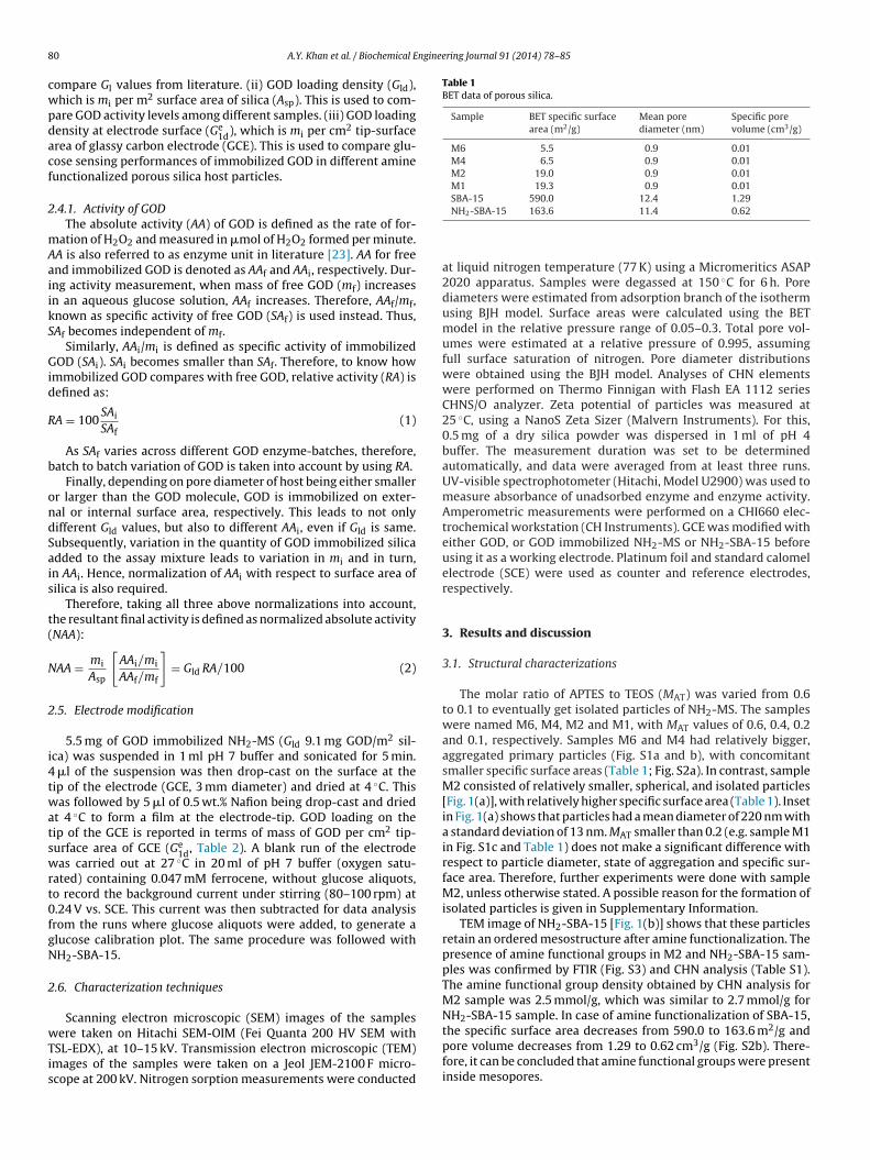
TEM image of NH2-SBA-15 [Fig. 1(b)] shows that these particlesretain an ordered mesostructure after amine functionalization. Thepresence of amine functional groups in M2 and NH2-SBA-15 sam-ples was confirmed by FTIR (Fig. S3) and CHN analysis (Table S1).The amine functional group density obtained by CHN analysis forM2 sample was 2.5 mmol/g, which was similar to 2.7 mmol/g forNH2-SBA-15 sample. In case of amine functionalization of SBA-15,the specific surface area decreases from 590.0 to 163.6 m2/g and
pore volume decreases from 1.29 to 0.62 cm3/g (Fig. S2b). There-fore, it can be concluded that amine functional groups were presentinside mesopores.
A.Y. Khan et al. / Biochemical Engineering Journal 91 (2014) 78–85 81
F 0.2; (d nm. A
3
fmmtdtGrpirG
SiMtSL
F2d
ig. 1. (a) SEM image of amine functionalized microporous silica with MAT value ofistribution with mean particle diameter of 220 nm with standard deviations of 13
.2. GOD immobilization and activity
Water soluble GOD was immobilized on water insoluble, amineunctionalized silica hosts by physical adsorption. Zeta potential
easurements revealed that the surface potential of GOD is notuch sensitive to pH (in the range of 4.0–7.1, Fig. S4), compared
o M2. However, with increase in pH surface potential of M2ecreases. Therefore, immobilization was carried out at pH 4.0 ashe difference between the surface potential in between silica andOD was the largest (Fig. S4). Moreover, immobilization at pH 4.0
esulted in a higher GOD loading (∼200 mg/g), compared to that atH 5.0 (12.5 mg/g) (Fig. S5). Fig. 2 shows that the kinetics of GOD
mmobilization at pH 4.0 was much faster for M2, reaching satu-ation in less than 5 h. Therefore, for all subsequent experiments,OD was immobilized at pH 4.0 with a contact time of 24 h.
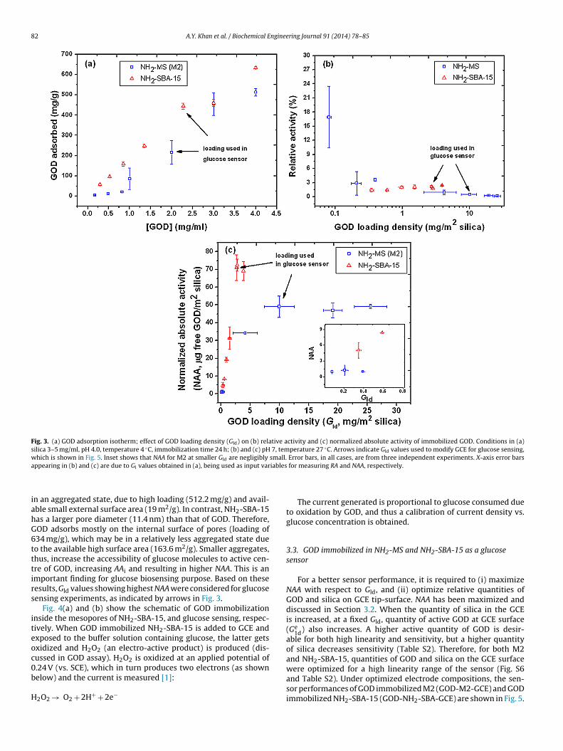
Fig. 3(a) shows GOD adsorption isotherm on M2 and NH2-BA-15 samples. As the initial concentration of GOD increases, Glncreases. In the present experimental condition, Gl achieved for
2 and NH2-SBA-15 samples were 512.5 and 634 mg/g, respec-ively. In this regard, Zhou et al. [15] reported a Gl of 430 mg/g onBA-15 host with circular mesochannels (pore diameter ∼14.8 nm).i et al. [17] obtained a Gl of 487 mg/g with amine functionalized
ig. 2. GOD immobilization kinetics in M2 sample. Experimental condition: GOD mg/ml; silica 4 mg/ml, pH 4, temperature 4 ◦C. Error bars are from three indepen-ent experiments.
b) TEM image of amine functionalized SBA-15. Insets in (a) show particle diameterpproximately 200 particles were used to generate the histogram shown in inset.
mesostructured cellular foam (pore diameter 21.4), which was thehighest reported so far, for GOD in any kind of mesoporous silicahosts. Therefore, the comparison with literature data shows thatboth M2 and NH2-SBA-15 samples have achieved comparably highloadings.
The effect of Gld on relative activity (RA, Eq. (1)) and normalizedabsolute activity (NAA, Eq. (2)) are shown in Fig. 3(b) and (c) respec-tively. Fig. 3(b) shows that as Gld increases, RA of GOD immobilizedin M2 decreases. However, in the NH2-SBA-15 sample, RA is almostindependent of Gld, and is smaller too.1 The reason for decreasedRA is as follows. As a result of interaction of glucose moleculeswith active centre of GOD, H2O2 is produced (Section 2.4), and GODactivity is measured. Since M2 had less number of and also inacces-sible pores, GOD adsorbs on the external surface and possibly getsaggregated. So, with increase in Gld, as aggregation of GOD on theexternal surface of M2 increases, the active centre of GOD withinaggregates become further inaccessible to glucose molecules.2 Incase of NH2-SBA-15, with increase in Gld, similarly aggregation ofGOD inside pores also increases (but to a lower extent than M2).Glucose molecules then encounter two kinds of resistances: (i) dif-fusional resistance offered by GOD aggregates, as before, to GODmolecules present within aggregates, and, in addition (ii) internalpore-diffusion resistance to glucose molecules due to partial poreblockage. Therefore, both SAi and RA were too small in NH2-SBA-15samples. Lei et al. [24] and Ye et al. [25] have also suggested aggre-gation of GOD as a possible reason for lowered activity. However,for biosensing applications, a higher NAA of GOD in the electrode isdesirable, even if RA is low.
Fig. 3(c) shows that, as Gld increases, NAA increases and GODimmobilized NH2-SBA-15 results in higher NAA, compared to M2. Infact, inset shows that NAA for M2 at smaller Gld are negligibly small.Furthermore, NAA does not increase beyond a mean Gld of 10 and2.7 mg/m2 for M2 and NH2-SBA-15, respectively. As the size of GOD
molecule (6 nm × 5.2 nm × 7.7 nm [13]) is larger than pore diameter(0.9 nm) of NH2-MS, GOD cannot adsorb inside the pores, but onlyon the external surface. Furthermore, the adsorbed GOD may be1 For NH2-SBA-15, Gld smaller than 0.35 mg/m2 could not be obtained even in thesmaller GOD concentration range of 0.29–0.85 mg/ml, because of the high adsorp-tion capacity of the NH2-SBA-15 host.
2 Although, the total number of H2O2 molecules formed (i.e. AAi) increases butnumber of H2O2 molecules per molecule of immobilized GOD decreases, whichresults in decreased SAi and RA.
82 A.Y. Khan et al. / Biochemical Engineering Journal 91 (2014) 78–85
Fig. 3. (a) GOD adsorption isotherm; effect of GOD loading density (Gld) on (b) relative activity and (c) normalized absolute activity of immobilized GOD. Conditions in (a)silica 3–5 mg/ml, pH 4.0, temperature 4 ◦C, immobilization time 24 h; (b) and (c) pH 7, temperature 27 ◦C. Arrows indicate Gld values used to modify GCE for glucose sensing,w small.a iables
iahG6tttirs
iteoc0b
H
hich is shown in Fig. 5. Inset shows that NAA for M2 at smaller Gld are negligibly
ppearing in (b) and (c) are due to Gl values obtained in (a), being used as input var
n an aggregated state, due to high loading (512.2 mg/g) and avail-ble small external surface area (19 m2/g). In contrast, NH2-SBA-15as a larger pore diameter (11.4 nm) than that of GOD. Therefore,OD adsorbs mostly on the internal surface of pores (loading of34 mg/g), which may be in a relatively less aggregated state dueo the available high surface area (163.6 m2/g). Smaller aggregates,hus, increase the accessibility of glucose molecules to active cen-re of GOD, increasing AAi and resulting in higher NAA. This is anmportant finding for glucose biosensing purpose. Based on theseesults, Gld values showing highest NAA were considered for glucoseensing experiments, as indicated by arrows in Fig. 3.
Fig. 4(a) and (b) show the schematic of GOD immobilizationnside the mesopores of NH2-SBA-15, and glucose sensing, respec-ively. When GOD immobilized NH2-SBA-15 is added to GCE andxposed to the buffer solution containing glucose, the latter getsxidized and H2O2 (an electro-active product) is produced (dis-ussed in GOD assay). H2O2 is oxidized at an applied potential of
.24 V (vs. SCE), which in turn produces two electrons (as shownelow) and the current is measured [1]:2O2 → O2 + 2H+ + 2e−
Error bars, in all cases, are from three independent experiments. X-axis error bars for measuring RA and NAA, respectively.
The current generated is proportional to glucose consumed dueto oxidation by GOD, and thus a calibration of current density vs.glucose concentration is obtained.
3.3. GOD immobilized in NH2-MS and NH2-SBA-15 as a glucosesensor
For a better sensor performance, it is required to (i) maximizeNAA with respect to Gld, and (ii) optimize relative quantities ofGOD and silica on GCE tip-surface. NAA has been maximized anddiscussed in Section 3.2. When the quantity of silica in the GCEis increased, at a fixed Gld, quantity of active GOD at GCE surface(Ge
1d) also increases. A higher active quantity of GOD is desir-able for both high linearity and sensitivity, but a higher quantityof silica decreases sensitivity (Table S2). Therefore, for both M2and NH2-SBA-15, quantities of GOD and silica on the GCE surface
were optimized for a high linearity range of the sensor (Fig. S6and Table S2). Under optimized electrode compositions, the sen-sor performances of GOD immobilized M2 (GOD-M2-GCE) and GODimmobilized NH2-SBA-15 (GOD-NH2-SBA-GCE) are shown in Fig. 5.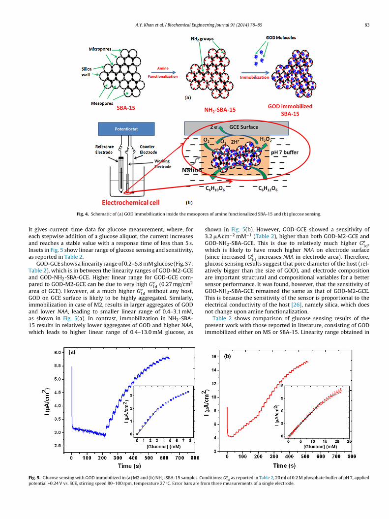
A.Y. Khan et al. / Biochemical Engineering Journal 91 (2014) 78–85 83
sopore
IeaIa
TapaGiaa1w
Fp
Fig. 4. Schematic of (a) GOD immobilization inside the me
t gives current–time data for glucose measurement, where, forach stepwise addition of a glucose aliquot, the current increasesnd reaches a stable value with a response time of less than 5 s.nsets in Fig. 5 show linear range of glucose sensing and sensitivity,s reported in Table 2.
GOD-GCE shows a linearity range of 0.2–5.8 mM glucose (Fig. S7;able 2), which is in between the linearity ranges of GOD-M2-GCEnd GOD-NH2-SBA-GCE. Higher linear range for GOD-GCE com-ared to GOD-M2-GCE can be due to very high Ge
1d (0.27 mg/cm2
rea of GCE). However, at a much higher Ge1d without any host,
OD on GCE surface is likely to be highly aggregated. Similarly,mmobilization in case of M2, results in larger aggregates of GOD
nd lower NAA, leading to smaller linear range of 0.4–3.1 mM,s shown in Fig. 5(a). In contrast, immobilization in NH2-SBA-5 results in relatively lower aggregates of GOD and higher NAA,hich leads to higher linear range of 0.4–13.0 mM glucose, asig. 5. Glucose sensing with GOD immobilized in (a) M2 and (b) NH2-SBA-15 samples. Conotential +0.24 V vs. SCE, stirring speed 80–100 rpm, temperature 27 ◦C. Error bars are fro
s of amine functionalized SBA-15 and (b) glucose sensing.
shown in Fig. 5(b). However, GOD-GCE showed a sensitivity of3.2 �A cm−2 mM−1 (Table 2), higher than both GOD-M2-GCE andGOD-NH2-SBA-GCE. This is due to relatively much higher Ge
1d,which is likely to have much higher NAA on electrode surface(since increased Ge
1d increases NAA in electrode area). Therefore,glucose sensing results suggest that pore diameter of the host (rel-atively bigger than the size of GOD), and electrode compositionare important structural and compositional variables for a bettersensor performance. It was found, however, that the sensitivity ofGOD-NH2-SBA-GCE remained the same as that of GOD-M2-GCE.This is because the sensitivity of the sensor is proportional to theelectrical conductivity of the host [26], namely silica, which does
not change upon amine functionalization.Table 2 shows comparison of glucose sensing results of thepresent work with those reported in literature, consisting of GODimmobilized either on MS or SBA-15. Linearity range obtained in
ditions: Ge1d
as reported in Table 2, 20 ml of 0.2 M phosphate buffer of pH 7, appliedm three measurements of a single electrode.
84 A.Y. Khan et al. / Biochemical Engineering Journal 91 (2014) 78–85
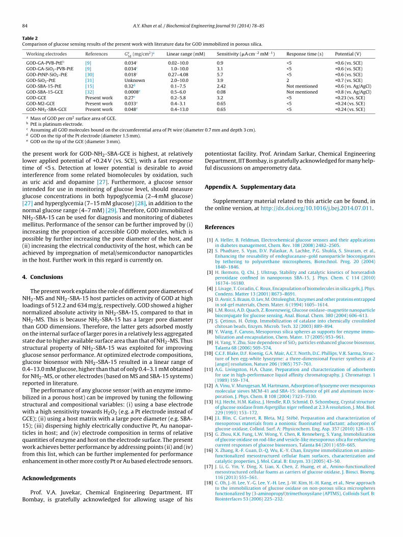
Table 2Comparison of glucose sensing results of the present work with literature data for GOD immobilized in porous silica.
Working electrodes References Ge1d
(mg/cm2)a Linear range (mM) Sensitivity (�A cm−2 mM−1) Response time (s) Potential (V)
GOD-GA-PVB-PtEb [9] 0.034c 0.02–10.0 0.9 <5 +0.6 (vs. SCE)GOD-GA-SiO2-PVB-PtE [9] 0.034c 1.0–10.0 3.1 <5 +0.6 (vs. SCE)GOD-PtNP-SiO2-PtE [30] 0.018c 0.27–4.08 5.7 <5 +0.6 (vs. SCE)GOD-SiO2-PtE [31] Unknown 2.0–10.0 3.9 2 +0.7 (vs. SCE)GOD-SBA-15-PtE [15] 0.32d 0.1–7.5 2.42 Not mentioned +0.6 (vs. Ag/AgCl)GOD-SBA-15-GCE [32] 0.0008e 0.5–6.0 0.08 Not mentioned +0.8 (vs. Ag/AgCl)GOD-GCE Present work 0.27e 0.2–5.8 3.2 <5 +0.23 (vs. SCE)GOD-M2-GCE Present work 0.033e 0.4–3.1 0.65 <5 +0.24 (vs. SCE)GOD-NH2-SBA-GCE Present work 0.048e 0.4–13.0 0.65 <5 +0.24 (vs. SCE)
a Mass of GOD per cm2 surface area of GCE.b
eter
tltiaig[nNmip(ai
4
NlnNtossgg0fr
bswG1tqwfe
A
B
[
[
[
[
[
[
[
[
PtE is platinum electrode.c Assuming all GOD molecules bound on the circumferential area of Pt wire (diamd GOD on the tip of the Pt electrode (diameter 1.5 mm).e GOD on the tip of the GCE (diameter 3 mm).
he present work for GOD-NH2-SBA-GCE is highest, at relativelyower applied potential of +0.24 V (vs. SCE), with a fast responseime of <5 s. Detection at lower potential is desirable to avoidnterference from some related biomolecules by oxidation, suchs uric acid and dopamine [27]. Furthermore, a glucose sensorntended for use in monitoring of glucose level, should measurelucose concentrations in both hypoglycemia (2–4 mM glucose)27] and hyperglycemia (7–15 mM glucose) [28], in addition to theormal glucose range (4–7 mM) [29]. Therefore, GOD immobilizedH2-SBA-15 can be used for diagnosis and monitoring of diabetesellitus. Performance of the sensor can be further improved by (i)
ncreasing the proportion of accessible GOD molecules, which isossible by further increasing the pore diameter of the host, andii) increasing the electrical conductivity of the host, which can bechieved by impregnation of metal/semiconductor nanoparticlesn the host. Further work in this regard is currently on.
. Conclusions
The present work explains the role of different pore diameters ofH2-MS and NH2-SBA-15 host particles on activity of GOD at high
oadings of 512.2 and 634 mg/g, respectively. GOD showed a higherormalized absolute activity in NH2-SBA-15, compared to that inH2-MS. This is because NH2-SBA-15 has a larger pore diameter
han GOD dimensions. Therefore, the latter gets adsorbed mostlyn the internal surface of larger pores in a relatively less aggregatedtate due to higher available surface area than that of NH2-MS. Thustructural property of NH2-SBA-15 was exploited for improvinglucose sensor performance. At optimized electrode compositions,lucose biosensor with NH2-SBA-15 resulted in a linear range of.4–13.0 mM glucose, higher than that of only 0.4–3.1 mM obtainedor NH2-MS, or other electrodes (based on MS and SBA-15 systems)eported in literature.
The performance of any glucose sensor (with an enzyme immo-ilized in a porous host) can be improved by tuning the followingtructural and compositional variables: (i) using a base electrodeith a high sensitivity towards H2O2 (e.g. a Pt electrode instead ofCE); (ii) using a host matrix with a large pore diameter (e.g. SBA-5); (iii) dispersing highly electrically conductive Pt, Au nanopar-icles in host; and (iv) electrode composition in terms of relativeuantities of enzyme and host on the electrode surface. The presentork achieves better performance by addressing points (ii) and (iv)
rom this list, which can be further implemented for performancenhancement in other more costly Pt or Au based electrode sensors.
cknowledgements
Prof. V.A. Juvekar, Chemical Engineering Department, IITombay, is gratefully acknowledged for allowing usage of his
[
0.7 mm and depth 3 cm).
potentiostat facility. Prof. Arindam Sarkar, Chemical EngineeringDepartment, IIT Bombay, is gratefully acknowledged for many help-ful discussions on amperometry data.
Appendix A. Supplementary data
Supplementary material related to this article can be found, inthe online version, at http://dx.doi.org/10.1016/j.bej.2014.07.011.
References
[1] A. Heller, B. Feldman, Electrochemical glucose sensors and their applicationsin diabetes management, Chem. Rev. 108 (2008) 2482–2505.
[2] S. Phadtare, S. Vyas, D.V. Palaskar, A. Lachke, P.G. Shukla, S. Sivaram, et al.,Enhancing the reusability of endoglucanase–gold nanoparticle bioconjugatesby tethering to polyurethane microspheres, Biotechnol. Prog. 20 (2004)1840–1846.
[3] H. Ikemoto, Q. Chi, J. Ulstrup, Stability and catalytic kinetics of horseradishperoxidase confined in nanoporous SBA-15, J. Phys. Chem. C 114 (2010)16174–16180.
[4] J. Livage, T. Coradin, C. Roux, Encapsulation of biomolecules in silica gels, J. Phys.Condens. Matter 13 (2001) R673–R691.
[5] D. Avnir, S. Braun, O. Lev, M. Ottolenghit, Enzymes and other proteins entrappedin sol-gel materials, Chem. Mater. 6 (1994) 1605–1614.
[6] L.M. Rossi, A.D. Quach, Z. Rosenzweig, Glucose oxidase–magnetite nanoparticlebioconjugate for glucose sensing, Anal. Bional. Chem. 380 (2004) 606–613.
[7] S . C etinus, H. Öztop, Immobilization of catalase into chemically crosslinkedchitosan beads, Enzym. Microb. Tech. 32 (2003) 889–894.
[8] Y. Wang, F. Caruso, Mesoporous silica spheres as supports for enzyme immo-bilization and encapsulation, Chem. Mater. 17 (2005) 953–961.
[9] H. Yang, Y. Zhu, Size dependence of SiO2 particles enhanced glucose biosensor,Talanta 68 (2006) 569–574.
10] C.C.F. Blake, D.F. Koenig, G.A. Mair, A.C.T. North, D.C. Phillips, V.R. Sarma, Struc-ture of hen egg-white lysozyme: a three-dimensional Fourier synthesis at 2[angst] resolution, Nature 206 (1965) 757–761.
11] A.G. Livingston, H.A. Chase, Preparation and characterization of adsorbentsfor use in high-performance liquid affinity chromatography, J. Chromatogr. 1(1989) 159–174.
12] A. Vinu, V. Murugesan, M. Hartmann, Adsorption of lysozyme over mesoporousmolecular sieves MCM-41 and SBA-15: influence of pH and aluminum incor-poration, J. Phys. Chem. B 108 (2004) 7323–7330.
13] H.J. Hecht, H.M. Kalisz, J. Hendle, R.D. Schmid, D. Schomburg, Crystal structureof glucose oxidase from Aspergillus niger refined at 2.3 A resolution, J. Mol. Biol.229 (1993) 153–172.
14] J.L. Blin, C. Carteret, R. Bleta, M.J. Stébé, Preparation and characterization ofmesoporous materials from a nonionic fluorinated surfactant: adsorption ofglucose oxidase, Colloid. Surf. A: Physicochem. Eng. Asp. 357 (2010) 128–135.
15] G. Zhou, K.K. Fung, L.W. Wong, Y. Chen, R. Renneberg, S. Yang, Immobilizationof glucose oxidase on rod-like and vesicle-like mesoporous silica for enhancingcurrent responses of glucose biosensors, Talanta 84 (2011) 659–665.
16] X. Zhang, R.-F. Guan, D.-Q. Wu, K.-Y. Chan, Enzyme immobilization on amino-functionalized mesostructured cellular foam surfaces, characterization andcatalytic properties, J. Mol. Catal. B: Enzym. 33 (2005) 43–50.
17] J. Li, G. Yin, Y. Ding, X. Liao, X. Chen, Z. Huang, et al., Amino-functionalizedmesostructured cellular foams as carriers of glucose oxidase, J. Biosci. Bioeng.
116 (2013) 555–561.18] C. Oh, J.-H. Lee, Y.-G. Lee, Y.-H. Lee, J.-W. Kim, H.-H. Kang, et al., New approachto the immobilization of glucose oxidase on non-porous silica microspheresfunctionalized by (3-aminopropyl)trimethoxysilane (APTMS), Colloids Surf. B:Biointerfaces 53 (2006) 225–232.

nginee
[
[
[
[
[
[
[
[
[
[
[[
[
A.Y. Khan et al. / Biochemical E
19] H. Li, J. He, Y. Zhao, D. Wu, Y. Cai, Q. Wei, et al., Immobilization of glucose oxidaseand platinum on mesoporous silica nanoparticles for the fabrication of glucosebiosensor, Electrochim. Acta 56 (2011) 2960–2965.
20] R. Tian, J. Sun, H. Zhang, M. Ye, C. Xie, J. Dong, et al., Large-pore mesoporous SBA-15 silica particles with submicrometer size as stationary phases for high-speedCEC separation, Electrophoresis 27 (2006) 742–748.
21] H.H.P. Yiu, P.A. Wright, N.P. Botting, Enzyme immobilisation using SBA-15mesoporous molecular sieves with functionalised surfaces, J. Mol. Catal. B:Enzym. 15 (2001) 81–92.
22] M.M. Bradford, A rapid and sensitive method for the quantitation of micro-gram quantities of protein utilizing the principle of protein–dye binding, Anal.Biochem. 72 (1976) 248–254.
23] G.K. Kouassi, J. Irudayaraj, G. McCarty, Activity of glucose oxidase functionalized
onto magnetic nanoparticles, Biomagn. Res. Technol. 3 (2005) 1.24] C. Lei, T.A. Soares, Y. Shin, J. Liu, E.J. Ackerman, Enzyme specific activity infunctionalized nanoporous supports, Nanotechnology 19 (2008) 125102.
25] W.N. Ye, D. Combes, The relationship between the glucose oxidase subunitstructure and its thermostability, Biochim. Biophys. Acta 999 (1989) 86–93.
[
ring Journal 91 (2014) 78–85 85
26] Z. Zhu, L. Garcia-Gancedo, A.J. Flewitt, F. Moussy, Y. Li, W.I. Milne, Design ofcarbon nanotube fiber microelectrode for glucose biosensing, J. Chem. Technol.Biotechnol. 87 (2012) 256–262.
27] E. Genter, P.M. Ipp, Accuracy of plasma glucose measurement in the hypo-glycemic range, Diabetes Care 17 (1994) 595–598.
28] K. Zhou, Y. Zhu, X. Yang, J. Luo, C. Li, S. Luan, A novel hydrogen peroxide biosen-sor based on Au–graphene–HRP–chitosan biocomposites, Electrochim. Acta 55(2010) 3055–3060.
29] J. Wang, Electrochemical glucose biosensors, Chem. Rev. 108 (2008) 814–825.30] H. Yang, Y. Zhu, Glucose biosensor based on nano-SiO2 and “unprotected” Pt
nanoclusters, Biosens. Bioelectron. 22 (2007) 2989–2993.31] W.-Z. Jia, K. Wang, Z.-J. Zhu, H.-T. Song, X.-H. Xia, One-step immobilization of
glucose oxidase in a silica matrix on a Pt electrode by an electrochemically
induced sol–gel process, Langmuir 23 (2007) 11896–11900.32] K. Wang, H. Yang, L. Zhu, J. Liao, T. Lu, W. Xing, et al., Direct electrochemistryand electrocatalysis of glucose oxidase immobilized on glassy carbon electrodemodified by Nafion and ordered mesoporous silica-SBA-15, J. Mol. Catal. B:Enzym. 58 (2009) 194–198.