glomerulonephritis- inflammation in the kidneys pt education... · glomerulonephritis-inflammation...
TRANSCRIPT

Glomerulonephritis-
inflammation in the
kidneys A PATIENT EDUCATION TOOL:
RAMY M. HANNA MD FASN FACP
UMUT SELAMET MD
Normal kidney filtering unit-
“glomerulus”
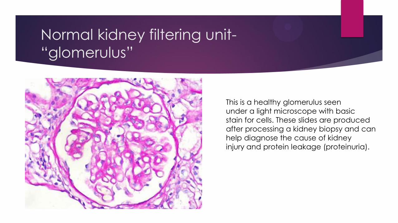
This is a healthy glomerulus seen
under a light microscope with basic
stain for cells. These slides are produced
after processing a kidney biopsy and can
help diagnose the cause of kidney
injury and protein leakage (proteinuria).

Normal electron microscope picture
of kidney filtering unit “glomerulus”
This is a picture of a healthy kidney under an electron microscope. Capillary lumen represents the inner space of small blood vessels( capillaries). Filtration membrane is the tissue barrier that separates blood and
urine. Bowman’s space is the are where urine floats. RBC represents red blood cell in the capillary lumen. Podocyte and pedicels are foot processes that extend from the filtration membrane. Tissues that line inner space of the capillary lumen are
called “endothelial” and the tissues that line outer space of the capillary lumen including podocytes and pedicles are called “epithelial”.

Normal immunofluorescence stain of
kidney filtering unit “glomerulus”

Types of Glomerular Disease 1/2
Proteinuria and blood in urine (hematuria) are the most common manifestations of glomerular diseases.
Proteinuria can be classified by the amount of protein that leaks into the urine:
Nephrotic: > 3.5 grams of protein in 24 hour collection of urine-severe
Sub nephrotic: 0.5-3.5 grams of protein in 24 hour collection of urine-moderate
Mild proteinuria <0.5 g-low grade
Glomerular diseases are called “nephrotic” if proteinuria is severe and called “nephritic” if proteinuria is moderate to low grade but hematuria is also present.
Glomerular diseases can progress slowly or rapidly. When they progress rapidly they are called “ RPGN (rapidly progressive glomerulonephritis)” which can quickly result in need for dialysis.

Types of Glomerular Disease 2/2
Glomerular diseases can also be classified by the parts of the kidney
effect.
Glomerular diseases can be primary or secondary. Primary diseases
originate in the kidney. Secondary diseases are caused because of a
problem somewhere else in the kidney. However both primary and
secondary glomerular diseases share same histological and clinical
characteristics.

1. Nephrotic Disorders
Minimal Change disease
Focal Segmental Glomerulosclerosis
Membranous glomerulonephritis
Membranoproliferative glomerulonephritis
Diabetic nephropathy

Minimal Change Disease (MCD)
High level of protein in urine
Normal light microscopy “Nil disease”
Usually no blood in urine (no hematuria)
Common in children (genetic component)
In some cases easily treated with steroids
like prednisone
Kidney clearance (creatinine) usually normal
or almost normal
No positive antibody tests (serology)for this
found yet
Absent foot
processes
or podocytes
cause
massive leakage
of protein
into urine.

Focal Segmental Glomerulosclerosis
(FSGS)
High level of protein in urine
Can be missed on biopsy if sample obtained
from non scarred area
Many diagnoses fit this pattern
Usually no blood in urine
Some foot process degeneration but
scarring is the most prominent aspect
Many associations: including obesity, HIV,
high blood pressure, diabetes and many
others
Can look like minimal change disease
Can be hard to treat

FSGS associated with HIV is called
“HIVAN”

Membranous Glomerulonephritis
(MGN)
High level of protein in urine
Filtration membrane (basement membrane)
becomes thickened-
That’s why its called “membranous”
Usually no blood in urine
A blood test called anti phospholipase A2
can help diagnose primary types, can stain
for it on biopsy too
Secondary form can be associated with
cancer, autoimmune disease, certain drugs,
and chronic infections
Most common glomerulonephritis in elderly
AntiPLA2R
Membrano-proliferative
Glomerulonephritis (MPGN)
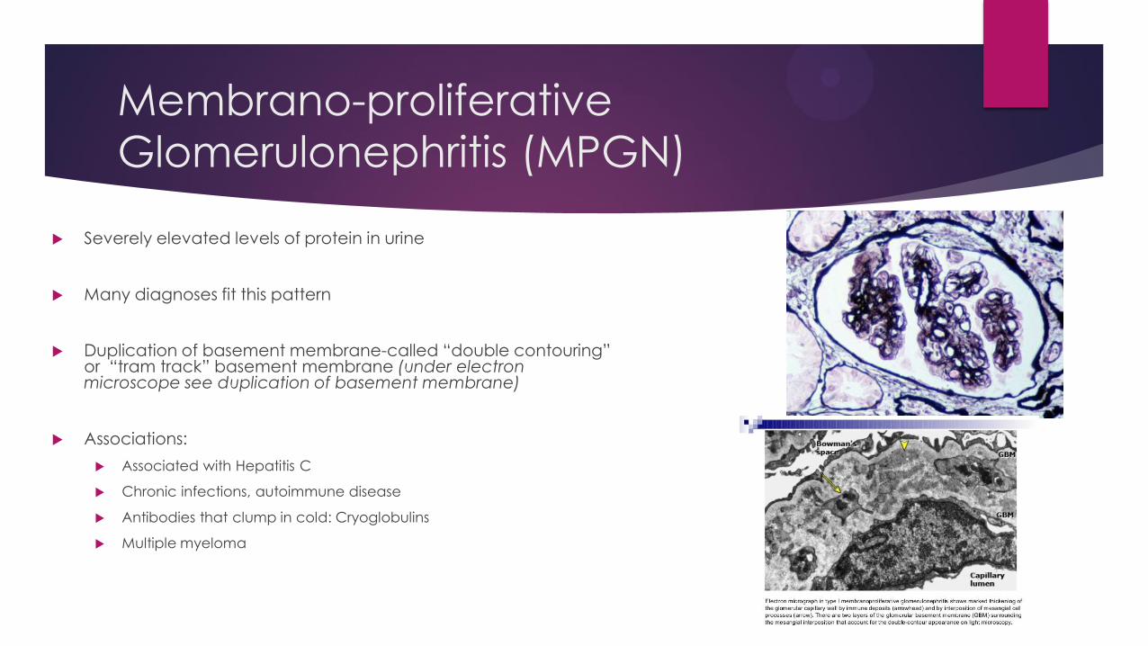
Severely elevated levels of protein in urine
Many diagnoses fit this pattern
Duplication of basement membrane-called “double contouring” or “tram track” basement membrane (under electron microscope see duplication of basement membrane)
Associations:
Associated with Hepatitis C
Chronic infections, autoimmune disease
Antibodies that clump in cold: Cryoglobulins
Multiple myeloma
Diabetic Nephropathy
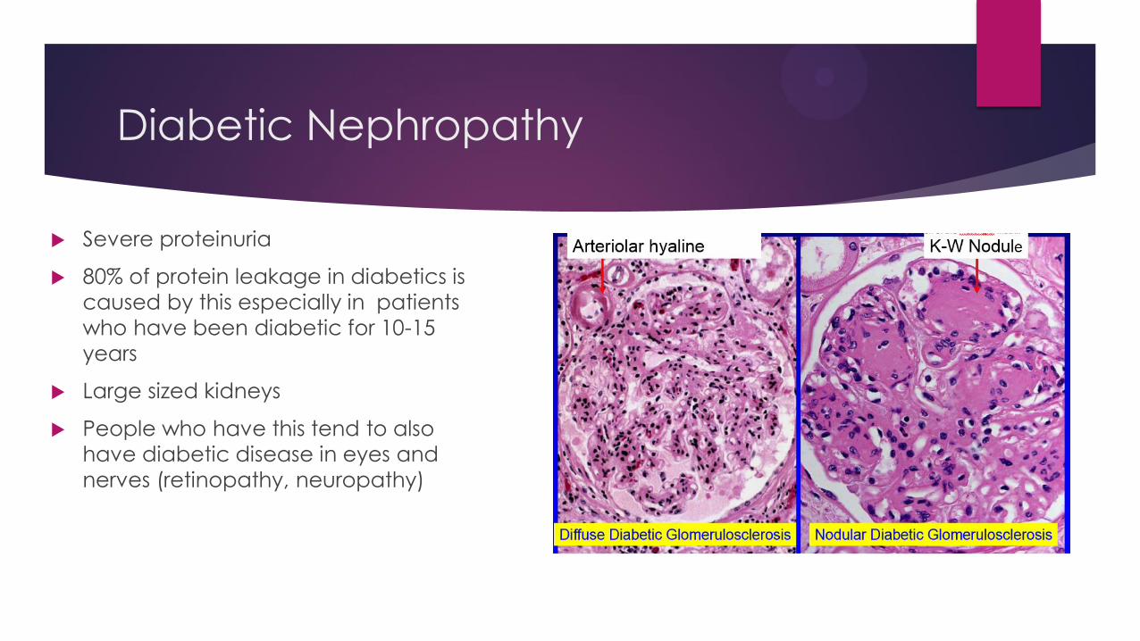
Severe proteinuria
80% of protein leakage in diabetics is
caused by this especially in patients
who have been diabetic for 10-15
years
Large sized kidneys
People who have this tend to also
have diabetic disease in eyes and
nerves (retinopathy, neuropathy)

2. Rapidly Progressing
Glomerulonephritis (RPGN)
Anti-neutrophil cytoplasmic antibody (ANCA) associated
Good Pasture’s Disease
Crescentic IgA nephropathy (Henoch Schonlein Purpura)
Lupus nephritis

ANCA Associated Vasculitis Types
Moderate protein in urine but lots of blood (nephritic)
Crescents are seen: they are a pattern of inflammation and reflect severe damage to kidney filtering units
Group of diseases that cause antibody mediated damage and inflammation at small vessels of the kidney: granulomatosis with polyangiitis (GPA, Wegener's granulomatosis), microscopic polyangiitis (MPA), eosinophilic granulomatosis with polyangitis (EPGA, Churg Strauss)
Antibodies that cause the damage are called C-ANCA (anti-PR3) or P-ANCA (anti-mpo)Old names for ANCA associated vasculitis:
Pauci-immune pattern : nothing shows up when looked for antibodies under immunofluorescence.
Associations:
Cancer
Hepatitis B most often and rarely hepatitis C
Aggressive course can be seen especially in C-ANCA can result in rapid need for dialysis

Good pasture's disease
Usually causes moderate to severe levels of protein and usually causes large amounts of blood in urine
Crescents can be seen due to severe kidney damage
Immunofluorescence shows linear staining of IgG antibodies that target the glomerular basement membrane
Associations:
After transplanting patients with-Alports syndrome who lack type IV collagen-this disease can occur
Can have aggressive course and result in need for dialysis rapidly

Crescentic IgA Nephropathy
Most aggressive subtype of IgA (rare compared to
more common IgA nephropathy-see other slide)
Light microscope shows crescents
Can see IgA antibodies under immune
fluorescence to look for antibodies
Associations:
HIV Can be associated with certain aggressive IgA
nephropathies
Henoch shonlein purpura a skin and blood
autoimmune disease is associated with crescentic IgA
nephropathy
Rapid course can result in dialysis need rapidly
Lupus Nephritis
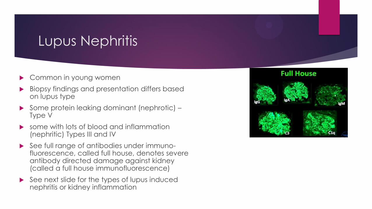
Common in young women
Biopsy findings and presentation differs based on lupus type
Some protein leaking dominant (nephrotic) – Type V
some with lots of blood and inflammation (nephritic) Types III and IV
See full range of antibodies under immuno-fluorescence, called full house, denotes severe antibody directed damage against kidney (called a full house immunofluorescence)
See next slide for the types of lupus induced nephritis or kidney inflammation
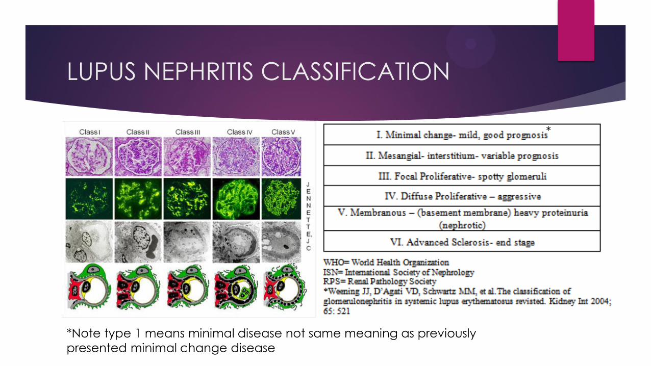
LUPUS NEPHRITIS CLASSIFICATION
*Note type 1 means minimal disease not same meaning as previously
presented minimal change disease
*
3. Other systemic disease that presents
with “tram tracking” pattern
Cryoglobulinemic vasculitis
Myeloma kidney (see MM disease later)
Thrombotic-micro-angiopathy
C3 glomerulonephritis
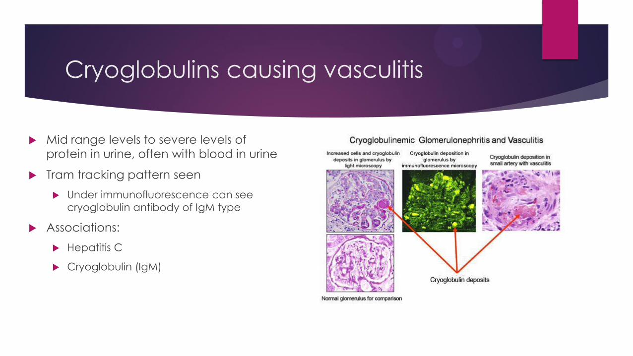
Cryoglobulins causing vasculitis
Mid range levels to severe levels of
protein in urine, often with blood in urine
Tram tracking pattern seen
Under immunofluorescence can see
cryoglobulin antibody of IgM type
Associations:
Hepatitis C
Cryoglobulin (IgM)

Thrombotic microangiopathy
A disorder of complement leading to clots in small vessels of kidney
Tram tracking seen
Low grade to mid range protein in urine, bleeding in urine can be seen as well
Systemic anemia resulting from blood cells being destroyed-hemolysis-can be seen
New specific drugs available to treat some forms of this. (Eculizumab)
Associations:
HIV
Transplant medications
Organ Rejection
Chemotherapy drugs
Vascular endothelial growth factor blocking drugs
In rare cases aspirin or plavix
Shigella infection
Genetic mutations

C3 Glomerulonephritis, Dense deposit
disease
Tram tracking on light microscopy
Deposition of complement protein C3
Can be genetic or acquired see next
Variable protein leakage and bleeding can be seen (nephritic)
No definitive treatment yet but experimental trials ongoing.
Associations:
Genetic
C3 nephritic factor can be an autoimmune cause of this syndrome.

4. Multiple myeloma like diseases
causing inflammation in kidney
Multiple myeloma is a type of bone marrow cancer that can result in
abnormal antibody protein production that can produce some deposition
in organs-especially kidney with different patterns of disease
Light chain disease LCDD
Myeloma cast nephropathy
Heavy chain deposition disease HCDD
Amyloidosis

Light Chain deposition disease
Tram tracking pattern
Evidence of multiple myeloma
Protein leakage usually more than inflammation or blood in urine-but variable amounts of protein in urine and variable presentation
Albumin in urine more than antibody protein in urine
Not amyloid like pattern (negative congo red stain) K or L
Light chain

Cast Nephropathy
Tram tracking pattern
Multiple myeloma diagnosis
Negative congo red stain
Antibody protein in urine > albumin protein in urine
Caused by abnormal myeloma antibody protein in tubules functionally blocking urine flow rather than myeloma protein infiltrating and destroying kidney tissue
K or L
Light chain type
Note cast in tubule not
glomerulus

Heavy chain deposition disease
Tram tracking pattern
Myeloma diagnosis
Albumin levels in urine
more than antibody protein
levels in urine
Same idea as light chain
deposition disease but
caused by the heavy
chains of antibodies
Heavy chain
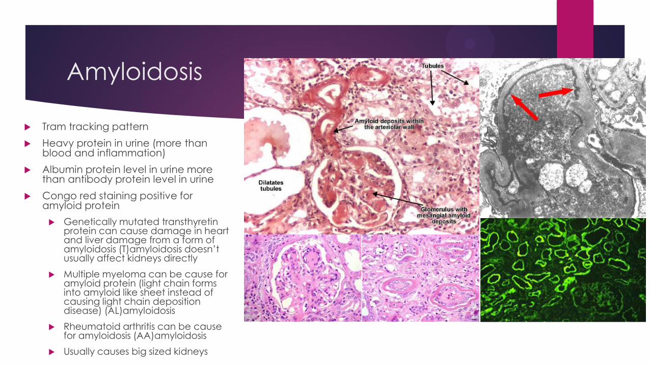
Amyloidosis
Tram tracking pattern
Heavy protein in urine (more than blood and inflammation)
Albumin protein level in urine more than antibody protein level in urine
Congo red staining positive for amyloid protein
Genetically mutated transthyretin protein can cause damage in heart and liver damage from a form of amyloidosis (T)amyloidosis doesn’t usually affect kidneys directly
Multiple myeloma can be cause for amyloid protein (light chain forms into amyloid like sheet instead of causing light chain deposition disease) (AL)amyloidosis
Rheumatoid arthritis can be cause for amyloidosis (AA)amyloidosis
Usually causes big sized kidneys

Can see amyloidosis in fat cells –so
called fat pad biopsy
Congo red stain
5.Other immune glomerulonephritis
types
Non crescentic IgA nephropathy
Post strep GN
Post infectious Immune complex GN
HIVIC
AIN and mimics
Cholesterol emboli
IgG4 GN

IgA nephropathy
MOST COMMON Glomerulonephritis WORLDWIDE
Low grade protein in urine and some blood in urine
Though amount varies
Preserved renal function or only slight worsening
This describes most common cases not “crescentic” IgA
See that slide for more information
Hematuria common and offers a good prognosis
Can flare immediately after infections
Associations
HIV
Asian patients
Patients with cirrhosis
Due to abnormal antibody tagging
with sugar molecules (glycosylation) and
abnormal deposition
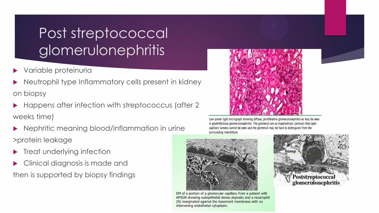
Post streptococcal
glomerulonephritis
Variable proteinuria
Neutrophil type Inflammatory cells present in kidney
on biopsy
Happens after infection with streptococcus (after 2
weeks time)
Nephritic meaning blood/inflammation in urine
>protein leakage
Treat underlying infection
Clinical diagnosis is made and
then is supported by biopsy findings

Typical pattern in post
streptococcus GN on
immunofluorescence-which is
how antibodies are found
when evaluating a biopsy:
shows IgG antibody and C3
Post strep glomerulonephritis
immunofluorescence

Post infectious glomerulonephritis and
immune complex glomerulonephritis
Protein leakage and inflammation is present
Neutrophil type Inflammatory cells present in kidney
Can have complexes of antibodies and bacterial/viral proteins that can be detected as deposits and on immuno-fluorescence
Lag period after infection before it develops-about 2 weeks
Treatment usually involves treating underlying
infection
Associations
Positive signs for systemic Infection
Acute infection
Chronic infection Like endocarditis (heart valve infection)
or osteomyelitis (bone infection)
(with organism other than streptococcus)
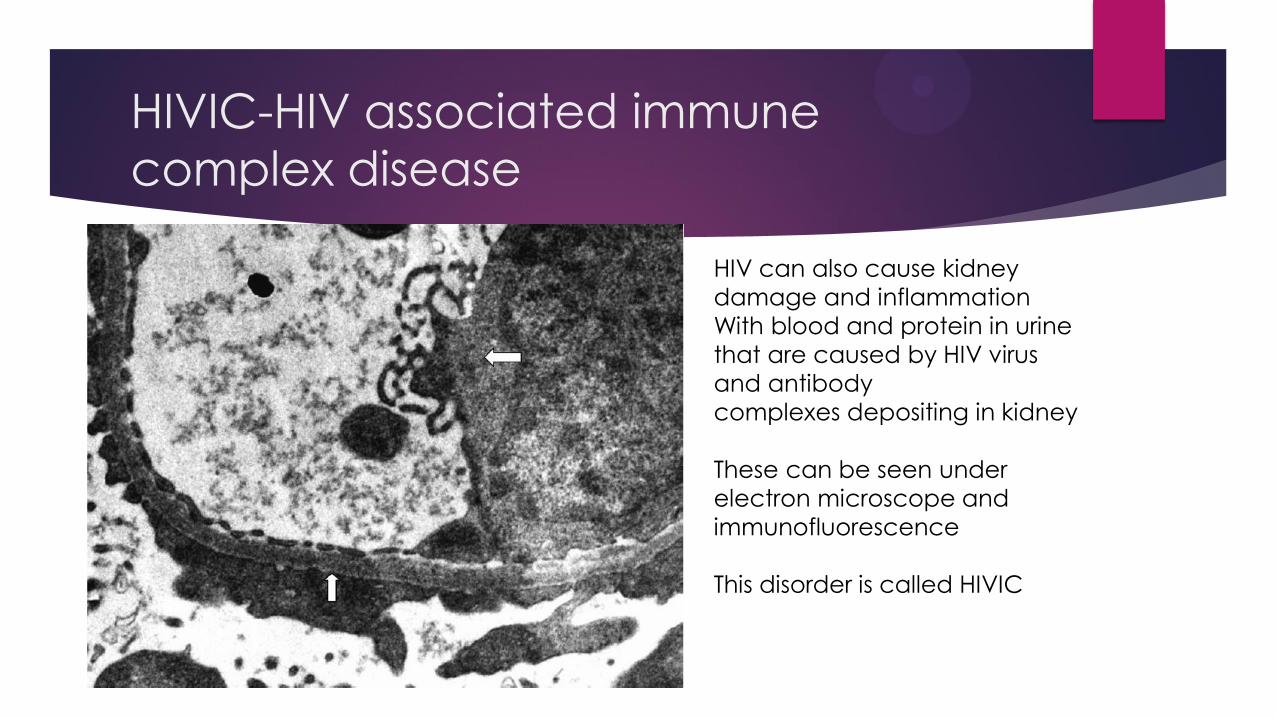
HIVIC-HIV associated immune
complex disease
HIV can also cause kidney
damage and inflammation
With blood and protein in urine
that are caused by HIV virus
and antibody
complexes depositing in kidney
These can be seen under
electron microscope and
immunofluorescence
This disorder is called HIVIC
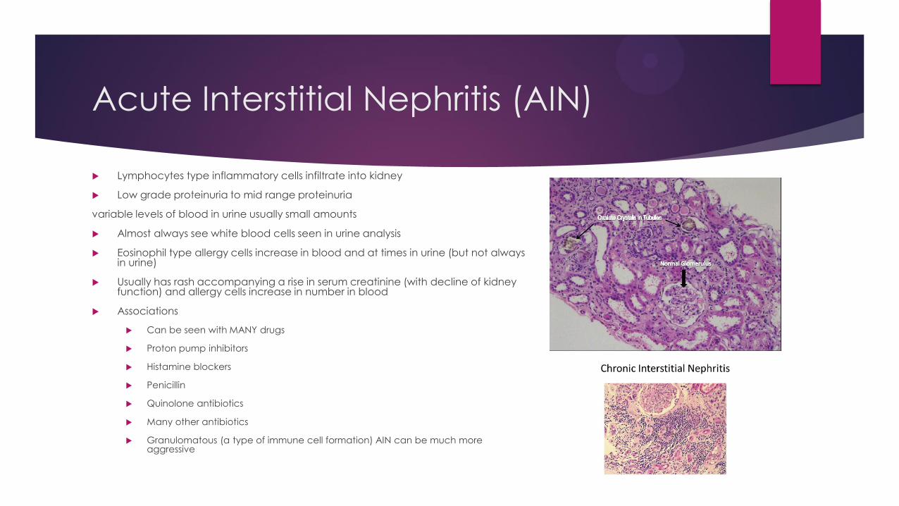
Acute Interstitial Nephritis (AIN)
Lymphocytes type inflammatory cells infiltrate into kidney
Low grade proteinuria to mid range proteinuria
variable levels of blood in urine usually small amounts
Almost always see white blood cells seen in urine analysis
Eosinophil type allergy cells increase in blood and at times in urine (but not always in urine)
Usually has rash accompanying a rise in serum creatinine (with decline of kidney function) and allergy cells increase in number in blood
Associations
Can be seen with MANY drugs
Proton pump inhibitors
Histamine blockers
Penicillin
Quinolone antibiotics
Many other antibiotics
Granulomatous (a type of immune cell formation) AIN can be much more aggressive

Cholesterol Emboli
Can see kidney damage from cholesterol plaque that breaks off and goes to kidney resulting in
Inflammation and damage

6. Genetic diseases of kidney
Alport’s
Thin Basement Membrane subtype
Dense deposit disease and C3 glomerulo-pathy see earlier slide
Muco-polysaccharide deposition disease / mitochondrial disease (beyond
scope but Gaucher’s disease can be dx on renal biopsy)
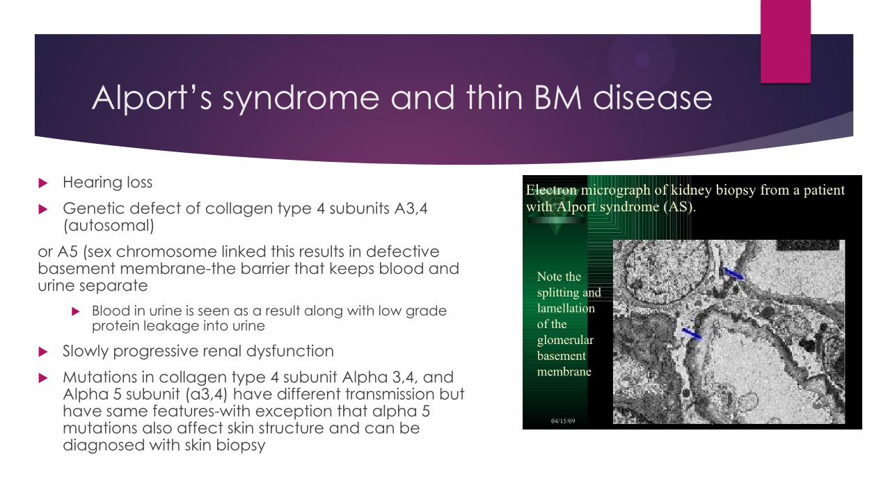
Alport’s syndrome and thin BM disease
Hearing loss
Genetic defect of collagen type 4 subunits A3,4 (autosomal)
or A5 (sex chromosome linked this results in defective basement membrane-the barrier that keeps blood and urine separate
Blood in urine is seen as a result along with low grade protein leakage into urine
Slowly progressive renal dysfunction
Mutations in collagen type 4 subunit Alpha 3,4, and Alpha 5 subunit (a3,4) have different transmission but have same features-with exception that alpha 5 mutations also affect skin structure and can be diagnosed with skin biopsy
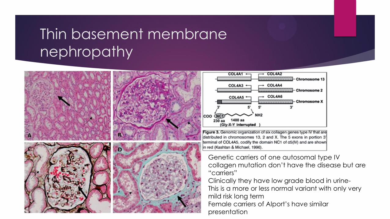
Thin basement membrane
nephropathy
Genetic carriers of one autosomal type IV
collagen mutation don’t have the disease but are
“carriers”
Clinically they have low grade blood in urine-
This is a more or less normal variant with only very
mild risk long term
Female carriers of Alport’s have similar
presentation
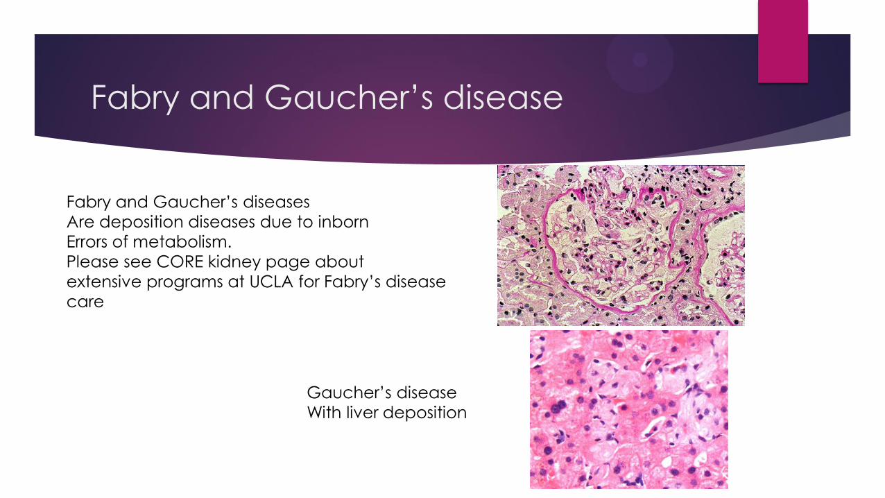
Fabry and Gaucher’s disease
Fabry and Gaucher’s diseases
Are deposition diseases due to inborn
Errors of metabolism.
Please see CORE kidney page about
extensive programs at UCLA for Fabry’s disease
care
Gaucher’s disease
With liver deposition

7. Miscellaneous glomerulopathies
Immunotactoid GN/Fibrillary GN
Malignant HTN/Scleroderma kidney
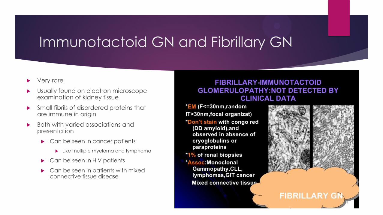
Immunotactoid GN and Fibrillary GN
Very rare
Usually found on electron microscope examination of kidney tissue
Small fibrils of disordered proteins that are immune in origin
Both with varied associations and presentation
Can be seen in cancer patients
Like multiple myeloma and lymphoma
Can be seen in HIV patients
Can be seen in patients with mixed connective tissue disease
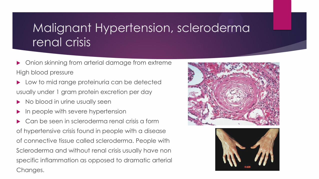
Malignant Hypertension, scleroderma
renal crisis
Onion skinning from arterial damage from extreme
High blood pressure
Low to mid range proteinuria can be detected
usually under 1 gram protein excretion per day
No blood in urine usually seen
In people with severe hypertension
Can be seen in scleroderma renal crisis a form
of hypertensive crisis found in people with a disease
of connective tissue called scleroderma. People with
Scleroderma and without renal crisis usually have non
specific inflammation as opposed to dramatic arterial
Changes.