glomerular diseases: nephrotic and nephritic syndrome prof dr. gülçin kantarcı yeditepe...
TRANSCRIPT
GLOMERULAR DİSEASES:NEPHROTİC AND NEPHRİTİC SYNDROME
Prof Dr. Gülçin KantarcıYeditepe University, Medical Faculty Nephrology Department

Aims & objectives
State the definition of nephritic syndrome. Identify clinical signs of nephritic syndrome. Explain the pathophysiology of nephritic
syndrome. State the definition of nephrotic syndrome. Identify clinical signs of nephrotic syndrome. Explain the pathophysiology of nephrotic
syndrome.

REFERENCE
1. Current Medical Diagnosis and Treatment, Maxine A. Papadakis, Stephen J. McPhee, Eds. Michael W. Rabow, Associate Ed. http://Chapter 22. Glomerular Diseases
2. Bates' Guide to Physical Examination & History Taking 11th edition, Bickleys LS, Szilagyi PG; Lippincott Williams and Wilkins¸
3. Kumar and Clark's Clinical Medicine, 8th edition; Kumar & Clark, Elsevier
4. Andreoli and Carpenter's Cecil Essentials of Medicine 8th edition, Andreoli and Carpenter, Elsevier
PART 11: RENAL AND GENITOURINARY DISEASES 123: Glomerular Disorders and Nephrotic Syndromes
5. http://www.uptodate.com
-Differential diagnosis and evaluation of glomerular disease
- Overview of heavy proteinuria and the nephrotic syndrome

Glomerulus

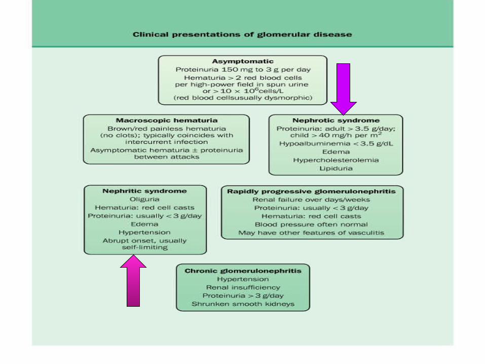
the glomerular capillaries can be injured in a variety of ways, producing many different lesions and several unique changes to urinalysis.
There are many forms of glomerular disease with pathogenesis variably linked to the presence of genetic mutations, infection, toxin exposure, autoimmunity, atherosclerosis, hypertension, emboli, thrombosis, or diabetes mellitus.
Even after careful study, however, the cause often remains unknown, and the lesion is called idiopathic or primary.

NEPHROTİC SYNDROME
The nephrotic syndrome is caused by renal diseases that increase the permeability across the glomerular filtration barrier
Two issues are important in the pathogenesis of nephrotic syndrome: the mechanisms of glomerular injury and proteinuria.

Components of nephrotic syndrome Proteinuria 3.5g/24 hr/1.73m2
Hypoalbum. ( serum alb<3.5 g/d) Hypercholesterolemia (>200mg/dl) Peripheral edema ± anasarca


Tendencies of glomerular diseases to manifest Nephrotic Features
Minimal change glomerulopathy Membranous GN Diabetic GS Amyloidosis Focal segmental GS Fibrilary GN

Common causes of NS
Primary glomerular disorders
Minimal Change Disease FSGS Membranous GN Orthostatik or postural proteinuria Idiopatic MPGN IgA nephropathy Proliferatif GN

Common causes of NS
Secondary glomerular disorders Hereditary-familial: DM, Alport’s Syndrome, Sickle
cell disease Autoimmun: SLE, Goodpasture’s syndrome,
Wegener’s granulomatosis, PAN, RA Infectious: postinfectious glomerulonephritis, HIV
FSGS Drug-induced: NSAIDs, Heroin,gold, mercury, Neoplastic: Hodgkin’s D., Lymphomas, leukemia,
MM. miscellaneous: amyloidosis, preeclampsia-
eclampsia, renovascular HT, intertitial nephritis, fever, exercise

Diabetic nephropathy (1/3 cause in patient on dialysis)

Nodular Sclerosis in DM

Screening for Diabetic nephropathy
BP each visit Urinary alb: Type 2:
annually, beginning at diagnosis
Type 1: annually, 5 years post diagnosis
<130/80 <30 mg/day <20 mic/min <30 mic/mg
creatinine
When Normal Range
ADA 2004
Proteinuria
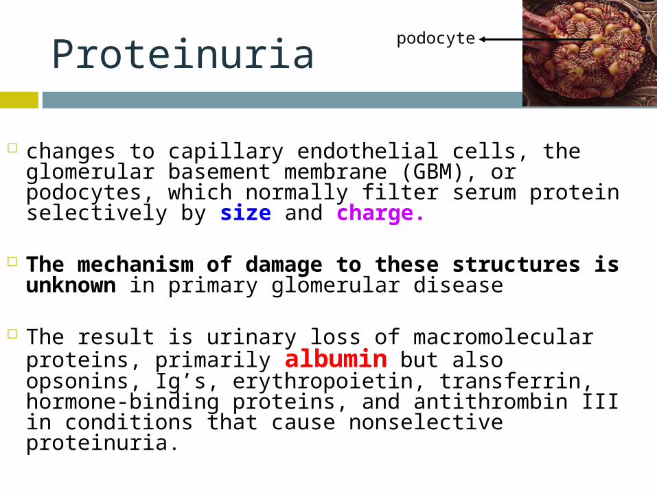
changes to capillary endothelial cells, the glomerular basement membrane (GBM), or podocytes, which normally filter serum protein selectively by size and charge.
The mechanism of damage to these structures is unknown in primary glomerular disease
The result is urinary loss of macromolecular
proteins, primarily albumin but also opsonins, Ig’s, erythropoietin, transferrin, hormone-binding proteins, and antithrombin III in conditions that cause nonselective proteinuria.
podocyte
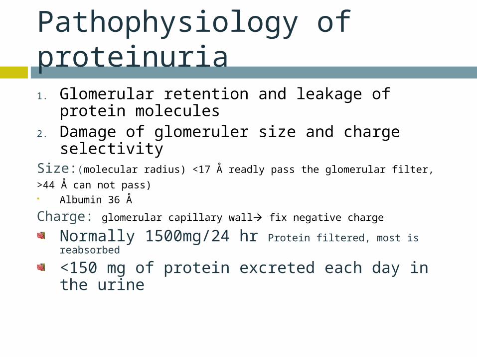
Pathophysiology of proteinuria1. Glomerular retention and leakage of protein
molecules2. Damage of glomeruler size and charge
selectivitySize:(molecular radius) <17 Å readly pass the glomerular filter,
>44 Å can not pass) Albumin 36 Å
Charge: glomerular capillary wall fix negative charge
Normally 1500mg/24 hr Protein filtered, most is reabsorbed
<150 mg of protein excreted each day in the urine

Types of proteinuria
'Glomerular' proteinuria (more than about 1.5 g protein/24 h, mostly albumin)
'Tubular' proteinuria (never excretion of more than 1.5 g/24 h )
'Overflow' proteinuria: immunoglobulin light chains in the urine
Benign proteinuria 'Jogger's nephritis' , orthostatic proteinuria
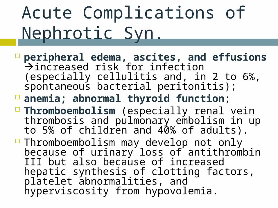
Acute Complications of Nephrotic Syn.
peripheral edema, ascites, and effusions increased risk for infection (especially cellulitis and, in 2 to 6%, spontaneous bacterial peritonitis);
anemia; abnormal thyroid function; Thromboembolism (especially renal vein
thrombosis and pulmonary embolism in up to 5% of children and 40% of adults).
Thromboembolism may develop not only because of urinary loss of antithrombin III but also because of increased hepatic synthesis of clotting factors, platelet abnormalities, and hyperviscosity from hypovolemia.

Hypercholesterolaemia is present in 90 per cent of patients with a urinary protein excretion of over 3 g/24 h
Hyperlipidemia in the NS is the result of both increased synthesis and decreased catabolism of lipoproteins.

Chronic complications of NS
malnutrition in children, coronary artery disease in adults, chronic renal failure, and bone disease.
Malnutrition may mimic kwashiorkor, including brittle hair and nails, alopecia, and stunted growth. Coronary artery disease develops because NS causes hyperlipidemia, hypertension, and hypercoagulability.

Chronic complications of NS
Bone disease develops because of vitamin D deficiency and corticosteroid use.
hypothyroidism from loss of thyroid-binding globulin
proximal tubular dysfunction causing glucosuria, aminoaciduria, K depletion, phosphaturia, and renal tubular acidosis


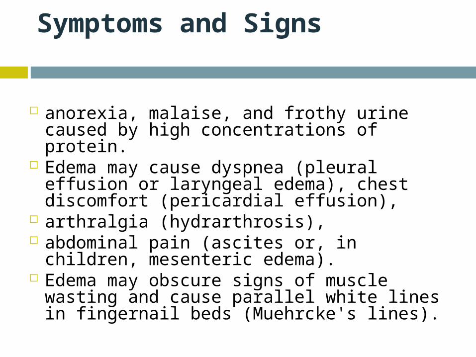
Symptoms and Signs
anorexia, malaise, and frothy urine caused by high concentrations of protein.
Edema may cause dyspnea (pleural effusion or laryngeal edema), chest discomfort (pericardial effusion),
arthralgia (hydrarthrosis), abdominal pain (ascites or, in children, mesenteric
edema). Edema may obscure signs of muscle wasting and
cause parallel white lines in fingernail beds (Muehrcke's lines).

Diagnosis
suspected in patients with edema and proteinuria on urinalysis and confirmed by 24-h measurement of urinary protein. The cause may be suggested by history (eg, cancer); when the cause is unclear, serologic testing and renal biopsy are indicated.
Besides proteinuria, urinalysis may demonstrate RBCs and casts (hyaline, granular, fatty, waxy, RBC, or epithelial cell).
Lipiduria, the presence of free lipid or lipid within tubular cells (oval fat bodies), within casts (fatty casts), or as free globules

Oval Fat Body

Evaluation for secondary causes serum glucose or glycosylated hemoglobin
(HbA1c), antinuclear antibodies, hepatitis B and C serologic tests,

Renal Bx
In adults, a renal biopsy is indicated to diagnose the underlying cause of idiopathic NS.
Idiopathic NS in children is most likely minimal change disease and is usually presumed without biopsy unless the patient fails to improve on a trial of corticosteroids.
NEPHRİTİC SYNDROME
Defined by hematuria and RBC casts on microscopic examination of urinary sediment.

Components of ?Nephritic Syndrome
Often one or more elements of mild to moderate proteinuria, edema, hypertension, elevated serum creatinine, and oliguria are also present.
It has both primary and secondary causes. Diagnosis is based on history, physical examination, and sometimes renal biopsy. Treatment and prognosis vary by cause.

•Asymptomatic Hematuria•Recurrent Gross Hematuria
• Red blood cell/ HP mic >3 cell• Asymptomatic Hematuria
prevalence 5-10 % • Tea or Cola colored urine• Proteinuria< 1 g/24 hr• Serum Cr< 1.5 mg/dl
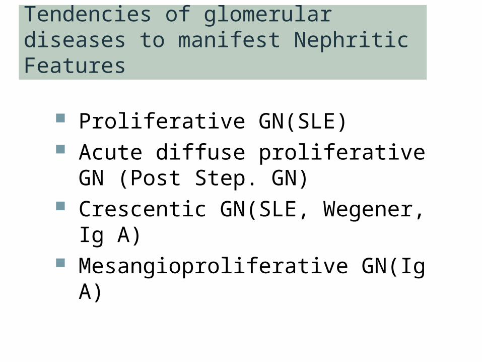
Tendencies of glomerular diseases to manifest Nephritic Features
Proliferative GN(SLE) Acute diffuse proliferative GN (Post
Step. GN) Crescentic GN(SLE, Wegener, Ig A) Mesangioproliferative GN(Ig A)

Asymptomatic glomerular hematuria
• No pathologic abnormality (30%)• Thin basement membrane (26%)• Ig A nephropathy (28%)• Alport syndrome ( rare ? Is it really
rare in Turkey ?)
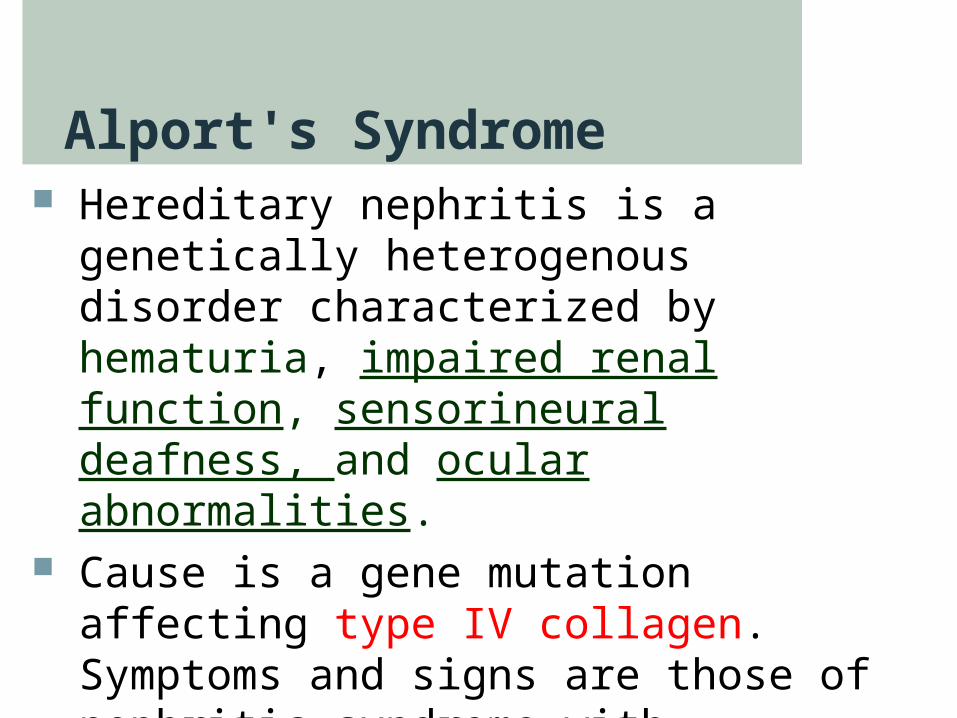
Alport's Syndrome Hereditary nephritis is a genetically
heterogenous disorder characterized by hematuria, impaired renal function, sensorineural deafness, and ocular abnormalities.
Cause is a gene mutation affecting type IV collagen. Symptoms and signs are those of nephritic syndrome with sensorineural deafness and, less commonly, those of ophthalmologic diseases. Diagnosis is by family history and urinalysis.
Alport's Syndrome
Because of X-linked transmission, women usually are asymptomatic and have little functional impairment.
Most men eventually develop renal symptoms and signs similar to those of acute nephritic syndrome and progress to renal insufficiency between ages 20 and 30.

Alport's Syndrome
Diagnosis is suggested by personal and family history and by findings of microscopic hematuria on urinalysis or recurrent episodes of gross hematuria, particularly if abnormalities of hearing or vision are present.

IgA nephropathy
It is the most common form of GN worldwide.
It occurs at all ages, with a peak onset in the teens and 20s; affects men 2 to 6 times more frequently than women; and is more common in whites and Asians than in blacks.
Prevalence estimates are 5% in the US, 10 to 20% in southern Europe and Australia, and 30 to 40% in Asia.
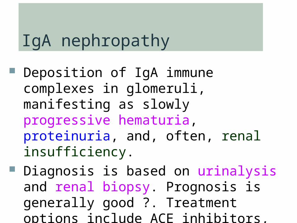
IgA nephropathy
Deposition of IgA immune complexes in glomeruli, manifesting as slowly progressive hematuria, proteinuria, and, often, renal insufficiency.
Diagnosis is based on urinalysis and renal biopsy. Prognosis is generally good ?. Treatment options include ACE inhibitors, corticosteroids, and ω-3 polyunsaturated fatty acids.

PATHOGENESIS
Cause is unknown, pathogenetic mechanisms, increased IgA1 production, defective IgA1 glycosylation causing increased binding to
mesangial cells, decreased IgA1 clearance, a defective mucosal immune system, overproduction of cytokines stimulating mesangial cell
proliferation.
Familial clustering has also been observed, suggesting genetic factors at least in some cases.
Symptoms and Signs
persistent or recurrent macroscopic hematuria (90% of involved children) or
asymptomatic microscopic hematuria with mild proteinuria

Diagnosis
urinalysis and renal biopsy. Urinalysis demonstrates microscopic hematuria,
usually with dysmorphic RBCs and RBC casts. Mild proteinuria (< 1 g/day) is typical and may
occur without hematuria; nephrotic syndrome develops in ≤ 20%.

Ig A mesangial deposition
granular deposition of IgA and C3 on immunofluorescent staining in an expanded mesangium with foci of segmental proliferative or necrotizing lesions.

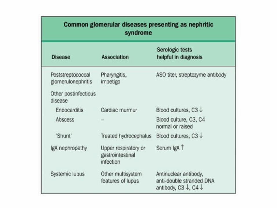
Postinfectious Glomerulonephritis
most common cause of glomerular disease in children between 5 and 15 yr; it is rare in children < 2 yr and in adults > 40 yr.
occurs after infection, usually with a nephritogenic strain of group A β-hemolytic streptococcus.
Diagnosis is suggested by history and urinalysis and confirmed by low complement.

Symptoms and signs
asymptomatic hematuria (in about 50%) and mild proteinuria
full-blown nephritis with microscopic or gross hematuria (cola-colored, brown, smoky, or frankly bloody), proteinuria, oliguria, edema, hypertension, and renal insufficiency.
Severe, late disease is a relatively uncommon cause of nephrotic syndrome. Flank pain may be attributable to stretching of the renal capsule.
Renal failure that causes fluid overload with heart failure and urgent or malignant hypertension and requires dialysis affects 1 to 2% of patients and may present as a pulmonary-renal syndrome with hematuria and hemoptysis
Rapidly Progressive Glomerulonephritis (RPGN)
Crescentic Glomerulonephritis RPGN causes microscopic glomerular
crescent formation with progression to renal failure within weeks to months.
Diagnosis is based on history, urinalysis, serologic tests, and renal biopsy.
Treatment is with corticosteroids, with or without cyclophosphamide, and sometimes plasmapheresis.

Classification of RPGN Based on Immunofluorescence Microscopy
Anti-glomerular basement membrane (GBM) antibody disease (type 1 RPGN)
The combination of GN and alveolar hemorrhage in the presence of anti-GBM antibodies is called Goodpasture's syndrome
autoimmune GN and accounts for 10% of RPGN cases. It may arise when respiratory exposures (eg, cigarette smoke, viral URI) expose alveolar capillary collagen, triggering formation of anticollagen antibodies.
The anticollagen antibodies cross-react with GBM, fixing complement and triggering a cell-mediated inflammatory response in the kidneys and lungs. Immunofluorescent staining of renal biopsy tissue demonstrates linear IgG deposits.

Immune complex RPGN (type 2 RPGN)
complicates numerous infectious and connective tissue disorders and also occurs with other primary glomerulopathies.
Immunofluorescent staining demonstrates nonspecific granular immune deposits.
The condition accounts for 40% of RPGN cases. Pathogenesis is usually unknown.

Pauci-immune RPGN (type 3 RPGN)
distinguished by the absence of immune complex or complement deposition on immunofluorescent staining.
It constitutes 50% of all RPGN cases. Almost all patients have elevated
antineutrophil cytoplasmic antibodies (ANCAs) and systemic vasculitis

crescent formation in a patient with ANCA–associated small vessel vasculitis

Crescentic Glomerulonephritis
Thin Basement Membrane Disease
(Benign Familial Hematuria) Thin basement membrane disease is
diffuse thinning of the glomerular basement membrane from a width of 300 to 400 nm in normal subjects to 150 to 225 nm.
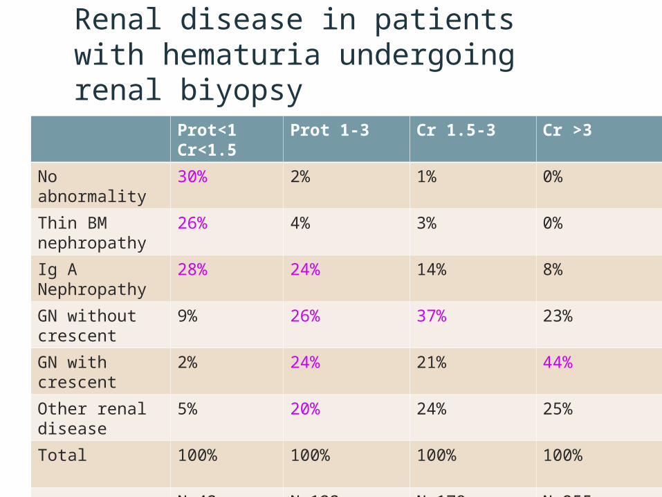
Renal disease in patients with hematuria undergoing renal biyopsy
Prot<1Cr<1.5
Prot 1-3 Cr 1.5-3 Cr >3
No abnormality 30% 2% 1% 0%
Thin BM nephropathy
26% 4% 3% 0%
Ig A Nephropathy
28% 24% 14% 8%
GN without crescent
9% 26% 37% 23%
GN with crescent 2% 24% 21% 44%
Other renal disease
5% 20% 24% 25%
Total 100% 100% 100% 100%
N=43 N=123 N=179 N=255


Renal biopsy: Indications
• The cause cannot be determined or adequatly predicted by less invasive diagnostic procedures
• The sign and symptoms suggest parenchymal disease that can be diagnosis by pathologic evaluation
• The differential diagnosis includes diseases that have different treatment, different prognosis, or both
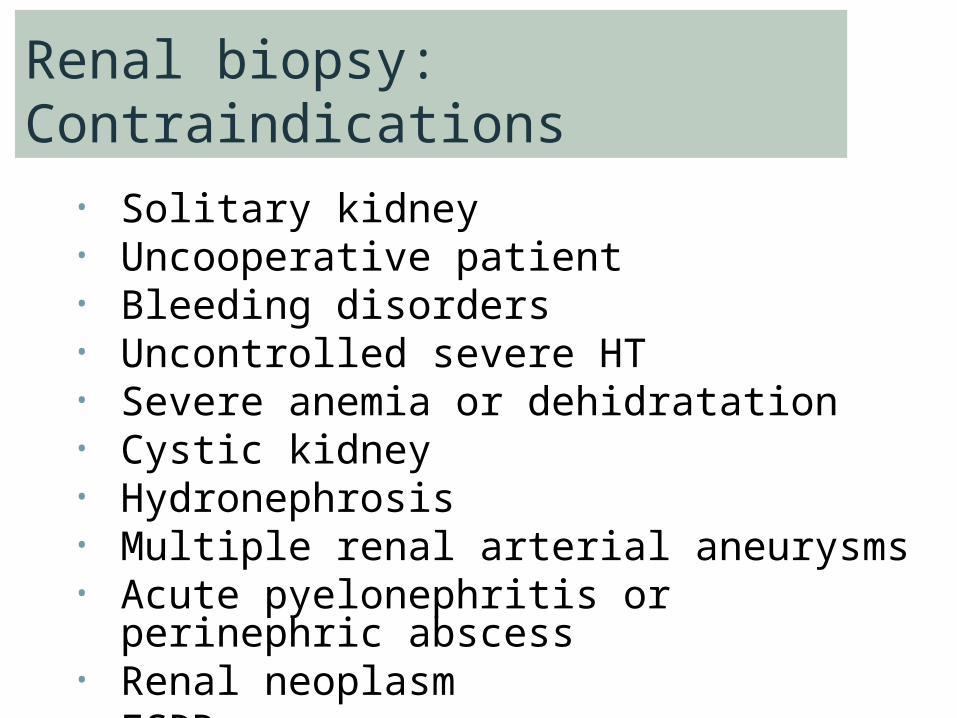
Renal biopsy: Contraindications• Solitary kidney • Uncooperative patient• Bleeding disorders• Uncontrolled severe HT• Severe anemia or dehidratation• Cystic kidney • Hydronephrosis• Multiple renal arterial aneurysms• Acute pyelonephritis or perinephric
abscess• Renal neoplasm• ESRD