glioma stem cells
DESCRIPTION
scienceTRANSCRIPT
6
Glioma Stem Cells: Cell Culture, Markers and Targets for New Combination Therapies
Candace A. Gilbert and Alonzo H. Ross University of Massachusetts Medical School
United States
1. Introduction
Gliomas are brain tumors with glial cell characteristics, and are composed of a heterogeneous mix of cells, which includes glioma stem cells. Gliomas include astrocytomas, oligodendrogliomas, ependymoma, and mixed gliomas. Gliomas account for 32% of all brain and central nervous system tumors (CNS) and 80% of all malignant brain and CNS tumors (CBTRUS, 2010). The WHO grade III anaplastic astrocytomas (AAs) and grade IV glioblastoma multiforme (GBMs) are highly invasive tumors and make up approximately three-quarters of all gliomas (CBTRUS, 2010). GBM is the most common and malignant form of brain tumor. GBMs make up 17% of all primary brain tumors in the United States, with an incidence of 3.17 cases per 100,000 persons per year (CBTRUS, 2010). Although both the knowledge of glioma biology and the available resources for treatment have greatly increased over the past decade, the expected survival of malignant glioma patients remains dismal. For AA patients, the current five-year and ten-year survival rates are 27.4% and 21.3%, respectively (CBTRUS, 2010). GBM patients have a much lower survival. The current five-year and ten-year survival rates for GBM patients are 4.5% and 2.7%, respectively (CBTRUS, 2010). Clinical treatment for gliomas consists of a combination of surgical resection, radiotherapy and chemotherapy. Due to the infiltrative nature of GBMs, complete removal of the tumor by surgery is not possible. Following surgery, the conventional radiation dosage of up to 60 Gy is given daily in 2 Gy fractions (Buatti et al 2008). The
commonly used chemotherapy drug, temozolomide (Temodar®), is an alkylating agent that is taken orally and readily penetrates the blood-brain barrier (Ostermann et al., 2004). 1,3-bis(2-chloroethyl)-1-nitrosourea (BCNU) is an older drug that surgeons deposit in the tumor bed as dissolvable wafers (Grossman et al., 1992). Both of these drugs alkylate DNA at multiple sites, including the O6 position of guanine, which can result in futile cycles of DNA repair and, ultimately, cell death (Sarkaria et al., 2008). These alkylating agents can also induce senescence (Gunther et al., 2003). Temozolomide is administered as both concomitant and adjuvant treatments to radiotherapy. This aggressive treatment increases the two-year survival rate for GBM patients from 10.4%, with radiotherapy alone, to 26.5% (Stupp et al., 2005). Cells that escape radiotherapy- and chemotherapy-induced cell death eventually re-enter the cell cycle and contribute to local tumor recurrence. Despite advances in chemotherapy regimens, the median progression free survival in AA and GBM patients is, 15.2 months (Chamberlain et al., 2008) and 6.9 months (Stupp et al., 2005), respectively. The median overall survival time for GBMs is 14.6 months (Stupp et al., 2005).
www.intechopen.com

Cancer Stem Cells Theories and Practice 80
2. Discovery of neural and glioma stem cells
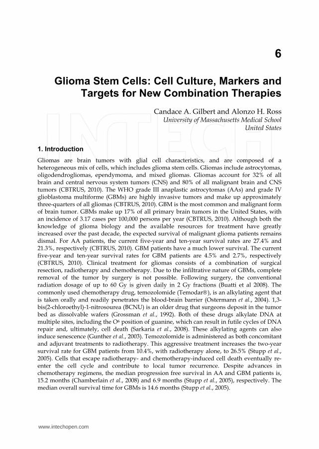
The discovery of adult neural stem cells paved the way for the glioma stem cell field. Until the mid-20th century, the consensus in the neuroscience field was that adult neural stem cells did not exist. The former dogma was that the brain contained mitotic cells only during development. It is now known that neurogenesis persists throughout life. In the adult brain, neural stem cells are located primarily in the subventricular zone (Altman, 1963) and the dentate gyrus (Altman and Das, 1965). In the subventricular zone, adult neural stem cells are termed type B cells and the transit-amplifying cells are type C cells (Kriegstein and Alvarez-Buylla, 2009) (FIG 1a). The type B neural stem cells are mostly quiescent and are derived from embryonic and neonatal radial glial cells. Type B cells structurally resemble astroglial cells (Doetsch et al., 1997). The adult neural stem cells and transit-amplifying cells are closely associated with blood vessels in the subventricular zone (Tavazoie et al., 2008). In the dentate gyrus of the hippocampus, the radial astrocytes are neural stem cells of the subgranular zone of the dentate gyrus (Seri et al., 2004). These cells are also referred to as type I progenitors in the subgranular zone (Fukuda et al., 2003). The subgranular zone is also located next to a vascular network, suggesting a niche for adult neural stem cells (Palmer et al., 2000). Adult neural stem cells from both the subventricular and subgranular zones express the embryonic neural stem cell markers nestin and Sox2, in addition to the astrocytic marker, glial fibrillary acidic protein (GFAP) (Doetsch et al., 1999; Seri et al., 2004; Suh et al., 2007). Unlike their differentiated progeny, these cells possess the ability to form neurospheres in serum-free cultures supplemented with growth factors (Reynolds et al., 1992). Neurospheres are heterogeneous aggregates derived from a single cell. These single cells would be plated at low densities for neurosphere assays, which were originally used to determine the percentage of neural stem cells in a culture or tissue. It is now known that both neural stem cells and transit amplifying cells can form neurospheres; however, neural stem cells are believed to have a greater, long-term proliferation potential than the transit-amplifying cells, and can therefore maintain neurosphere cultures through a large number of serial dissociations (Reynolds and Weiss, 1996). Neural stem cells have been associated with repair after strokes and severe injuries, and have been suggested as means for treatment of neurological disorders, such as Alzheimer’s Disease (Gage, 2000; Zhongling et al., 2009). While neural stem cells are necessary for normal neurological development and activity, cells with aberrant neural stem cell characteristics have been attributed to brain tumors. Glioma stem cells have many characteristics shared with adult neural stem cells, such as self-renewal, neurosphere formation, marker expression, multilineage differentiation, high motility, and localization to stem cell microenvironment niches (Sanai et al., 2005). Normal neural stem cells and glioma stem cells also share similar undifferentiated gene expression profiles, including nestin, EGF receptor, and PTEN. However, the nomenclature ‘stem cell’ in gliomas refers to their function and not their origin. It is currently unknown what is the cell of origin for glioma stem cells. Glioma stem cells may originate from normal neural stem cells that have undergone tumorigenic mutations or from more differentiated transit-amplifying or terminally differentiated neural cells that have undergone multiple mutations that allow the cells to be tumorigenic and revert to stemness properties (FIG 1b). Neural stem cells are probably target cells for malignant transformation. When rodent brains were exposed to avian sarcoma virus or carcinogens, tumors formed in the subventricular zone, where normal neural stem cells are believed to reside (Sanai et al., 2005). In addition, expression of Akt and K-ras in progenitor cells led to tumorigenesis (Holland et al., 2000). Conversely, several laboratories have demonstrated that genetic alterations can
www.intechopen.com
Glioma Stem Cells: Cell Culture, Markers and Targets for New Combination Therapies 81
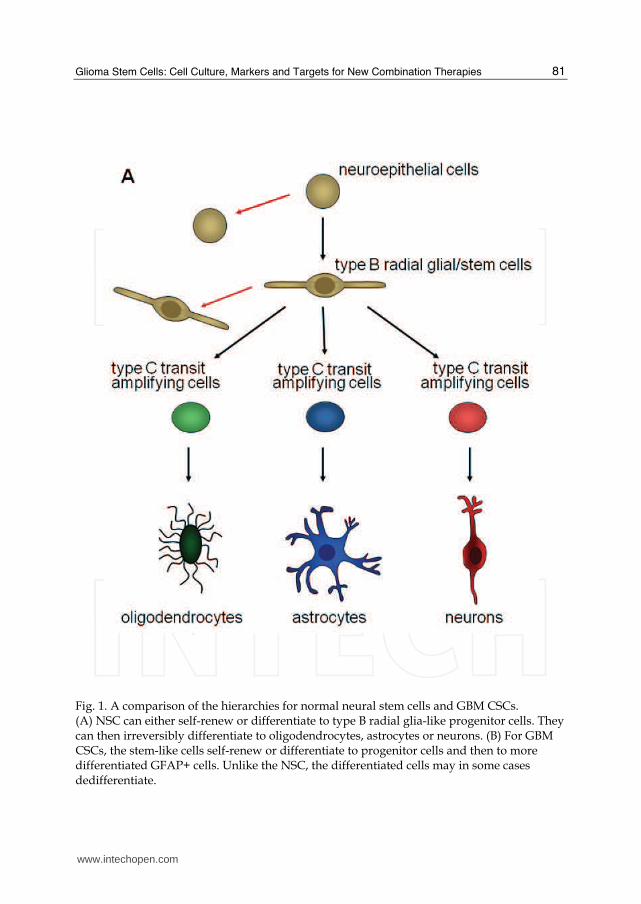
Fig. 1. A comparison of the hierarchies for normal neural stem cells and GBM CSCs. (A) NSC can either self-renew or differentiate to type B radial glia-like progenitor cells. They can then irreversibly differentiate to oligodendrocytes, astrocytes or neurons. (B) For GBM CSCs, the stem-like cells self-renew or differentiate to progenitor cells and then to more differentiated GFAP+ cells. Unlike the NSC, the differentiated cells may in some cases dedifferentiate.
www.intechopen.com
Cancer Stem Cells Theories and Practice 82
Continuation of Fig. 1.
www.intechopen.com

Glioma Stem Cells: Cell Culture, Markers and Targets for New Combination Therapies 83
dedifferentiated terminally differentiated astrocytes and induce tumorigenesis.(Bachoo et al., 2002; Holland et al., 1998) Due to the substantial heterogeneity among gliomas, it is likely that tumors from different patients originate from different stages of the adult neural hierarchy. This is an explanation for the distinct molecular subclasses of gliomas (Phillips et al., 2006). Regardless of the cell of origin, there are three properties that are considered essential for a cell to be universally accepted as a glioma stem cell (Rich, 2008). First, the cell must be capable of self-renewal; second, the cell should possess high proliferative potential; and third, the glioma stem cell must be capable of tumor initiation. There are additional characteristics used to define, but are not required of, glioma stem cells, because they can vary among different glioma grades and individual patients’ tumors. Glioma stem cells may make up a rare population of the tumor or glioma culture; however, recent publications find that the percent of stem cells in different cancers can vary greatly, depending on tumor type and possibly the tumor environment (Eaves, 2008). Many laboratories have used the expression of stem cell markers to identify and isolate glioma stem cells, although there is no single marker that is consistent for all patients, specific to glioma stem cells, and definitely includes all glioma stem cells in a tissue. Finally, similar to neural stem cells, glioma stem cells are capable of multilineage differentiation, albeit aberrant, and the ratio of the differentiated progeny as well as progeny that express markers from multiple lineages can be varied between tumors (FIG 1b and c) (Varghese et al., 2008). However, as it is rare for an individual glioma to exhibit the full hierarchy see in normal brain tissue from neural stem cell differentiation, it is not expected that each glioma stem cell can differentiate into all lineages (Sanai et al., 2005). Therefore, one would expect the differentiation of a glioma stem cell to mimic the lineage composition of the parent tumor.
3. Glioma stem cell cultures
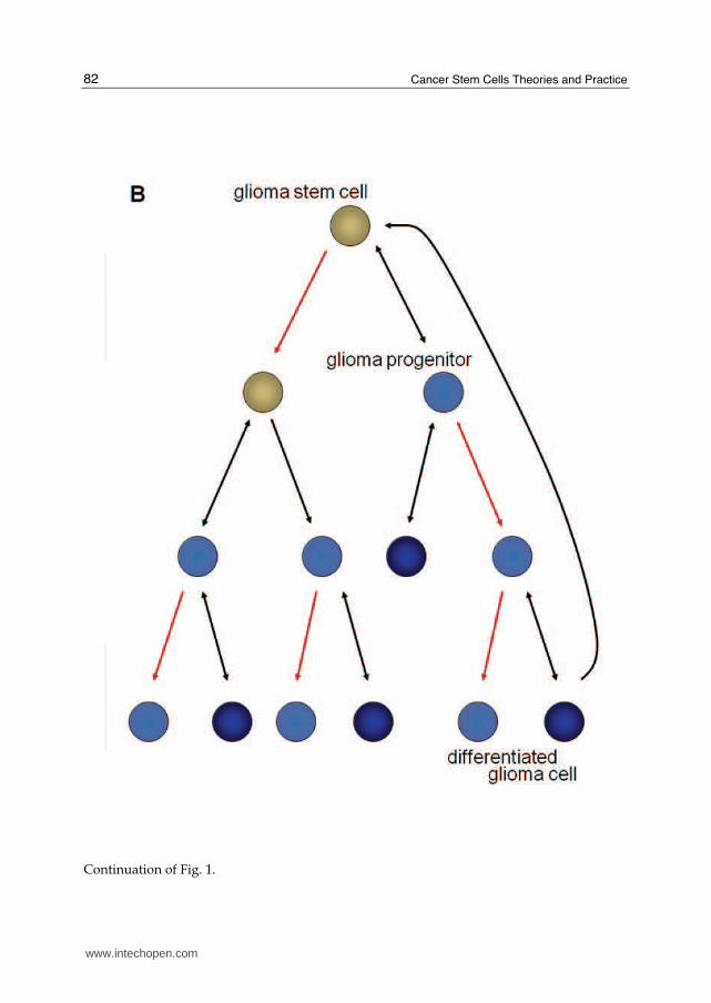
Traditionally, glioma cells were grown in the presence of serum as adherent cultures (FIG 2). The serum-grown cultures are tumorigenic, but unlike the invasive phenotype seen in patient gliomas, serum cultures commonly yield circumscribed tumors in intracranial xenograft models (Radaelli et al., 2009). Gene expression in serum cultures can be drastically different from the original tumor (Lee et al., 2006). Like neural stem cells, glioma stem cells can be grown in serum-free media with the growth factors EGF and FGF (Galli et al., 2004). Neurosphere cultures are currently the most common method used to propagate glioma stem cells, but a new in vitro technique to grow glioma stem cells is emerging, which utilizes laminin-coated plates with serum-free media.
3.1 Neurosphere cultures
The presence of self-renewing glioma stem cells was first demonstrated in 2003. Two laboratories demonstrated that glioma tissue cultured in serum-free media supplemented with growth factors form non-adherent spheroids with an enhanced glioma stem cell population (FIG 2 and 3). The glioma neurosphere cultures maintain genetic profiles similar to the original patients’ tumors and form invasive tumors in intracranial xenografts (Ernst et al., 2009; Lee et al., 2006; Singh et al., 2004). When plated at clonal density, each neurosphere arises from an individual glioma stem cell or transit-amplifying cell. Despite their clonal origin, neurospheres are heterogeneous aggregates that consist of glioma stem cells, transit-amplifying cells, and more differentiated glioma cells. The percentage of neurosphere-initiating can vary greatly among glioma cultures, and neurosphere formation has been
www.intechopen.com

Cancer Stem Cells Theories and Practice 84
demonstrated to increase when neural stem cells are transformed (Li et al., 2009). The majority of cells in a neurosphere are transit-amplifying cells (Ahmed, 2009). When these neurosphere cultures are dissociated to single cells, a small percentage of the cells can form secondary and tertiary neurospheres for many passages (Chen et al., 2010; Reynolds and Weiss, 1996). Glioma stem cells have a high capacity to proliferate and self-renew and robustly form secondary neurospheres. When exposed to fetal bovine serum, neurosphere cells differentiate down the lineage of the parent tumor (Singh et al., 2003). Therefore, gliomas preferentially differentiate to astrocytes, but multilineage differentiation can occasionally be observed with neuronal lineages, and some abnormal cells with mixed phenotypes. It should be noted that these lineages are based on markers but not function. For example, the crucial test for a neuron is an action potential, which is not tested. Also, a significant difference between neural stem cell and glioma stem cell cultures is that serum differentiation of normal neural stem cells is permanent (Lee et al., 2006), while glioma lines established as serum cultures can be converted to neurospheres in serum-free media (Gilbert et al., 2010; Qiang et al., 2009). Neurosphere cultures express known neural stem cell genes, such as Musashi-1, Sox2, and Bmi-1 (Hemmati et al., 2003) (FIG 2). Stem cell membrane markers, such as CD133 and CD15, are also expressed in neurosphere cultures and are discussed in further detail in subsequent sections. Using neurosphere assays to analyze glioma stem cell content can be complicated. As mentioned above, both glioma stem cells and transit amplifying cells are capable of neurosphere formation. In addition, neurospheres aggregate and fuse with one another when the cells are plated at higher densities (Singec et al., 2006). Therefore, the number of neurospheres is a measure of the number of both glioma stem cells and transit amplifying cells and is accurate only when the cells are plated at low densities. Despite these concerns, neurosphere cultures remain a valuable tool in glioma stem cell research.
3.2 Laminin-coated cultures
A key aspect of the neurosphere culture system is that the serum-free, defined media
maintains the glioma stem cell phenotype of the cells. However, in addition to glioma
stem cells, neurospheres contain more differentiated progeny and regions of cell death.
This is thought to be caused by the condensed structure of the neurosphere, which
hinders the diffusion of the growth factors to the innermost cells (Woolard and Fine,
2009). Differentiation and cell death could be limited if glioma cultures were grown in a
monolayer in the presence of serum-free, defined medium. This can be achieved by
culturing glioma samples in the serum-free, defined medium on laminin-coated cell
culture plates (Pollard et al., 2009). When cultured on laminin-coated plates, cells that
would normally form neurospheres grow as an adherent culture, which allows all of the
cells equal access to growth factors. The adherent glioma stem cell lines are less
heterogeneous than neurosphere cultures, and almost all of the cells express glioma stem
cell genes, such as Sox2, Nestin, CD133 and CD44 (FIG 2). There is minimal expression of
differentiation markers. The adherent, laminin cultures are capable of tumor formation
when as few as 100 cells were intracranially injected into immunocompromised mice,
demonstrating the high percentage of tumor-initiating glioma stem cells. An additional
benefit of the laminin glioma stem cell culture system is that all gliomas with good cell
viability formed long-term cell lines.
www.intechopen.com
Glioma Stem Cells: Cell Culture, Markers and Targets for New Combination Therapies 85
Fig. 2. Proposed lineages and culture methods for GBM CSCs. The CSCs (red circles) are cultured in defined medium to enhance stem cell properties. They express stem cell markers, listed below. CSCs are likely heterogeneous and may not express all of these markers, and may show additional tumor-to-tumor variation. CSCs differentiate to transit-amplifying cells (blue circles). The transit-amplifying cells show decreased expression of stem cell markers, and Chen et al (2010) recently suggested that TBR2 and DLX2 are markers for these cells. In mature spheres, a few of the transit-amplifying cells differentiate to astrocytic cells and, to a lesser degree, neuronal and oligodendrocytic cells (astrocytic cells shown as green circles). For cells adhering to laminin-coated substratum, stem cell marker expression is enhances, suggesting that the fraction of CSCs is increased. In addition, there are very few astrocytic cells. Serum treatment rapidly induces astrocytic differentiation.
4. Glioma stem cell markers
Markers are commonly used to identify and isolate different cells types. The most commonly used cell surface markers for glioma stem cells are CD133, CD15, and A2B5. New, less characterized markers are also being tested for glioma stem cells. When cells are isolated from tumors or glioma cultures with these markers, their stem cell characteristics can be analyzed based on stem cell gene expression, multilineage differentiation capabilities and neurosphere formation; however, tumor formation in xenograft models is the most important method to confirm that a marker identifies the glioma stem cell population. Despite many successes using cell surface markers such as CD133, it has become increasingly clear that individual gliomas are very heterogeneous and in addition, tumors vary greatly from patient to patient (Phillips et al., 2006). There is currently no universally accepted collection of markers for isolation of a pure population of glioma stem cells (Gilbert and Ross, 2009). In addition, to complicate the glioma stem cell field, some of the markers used appear to only be relevant when the cells are isolated directly from the tumor tissue. The heterogeneity of malignant gliomas may make it difficult to use a single set of markers to identify and purify glioma stem cells in every glioma.
www.intechopen.com
Cancer Stem Cells Theories and Practice 86
4.1 CD133
CD133 (also known as Prominin-1) was first discovered as a cell surface marker for hematopoietic stem cells (Miraglia et al., 1997). In the human fetal brain, CD133 is a marker for neural stem cells (Uchida et al., 2000). CD133 expression has also been observed in intermediate radial glial cells in the early postnatal brain, and in ependymal cells in the adult brain (Coskun et al., 2008; Pfenninger et al., 2007). Neurogenic astrocytes in the neural stem cell region of the subventricular zone do not express CD133. Despite its inconsistent expression in adult neural stem cells, CD133 has been used to isolate populations of cancer stem cells from multiple types of brain tumors (Singh et al., 2003; Singh et al., 2004). Expression of CD133 in anaplastic astrocytomas and glioblastoma multiforme varies among patients and tumor grade, with reports of 0 – 64% (Ogden et al., 2008; Singh et al., 2003; Singh et al., 2004; Son et al., 2009). CD133+ cells from gliomas are capable of multilineage differentiation and have a high capacity for neurosphere formation. The corresponding CD133- cells did not proliferate in neurosphere cultures. Furthermore, CD133+ glioma cells express significantly higher levels of neural stem cell genes, such as nestin, Msi-1, maternal embryonic leucine zipper kinase (MELK) and CXCR4 (Liu et al., 2006). These data support the stem cell genotype of CD133+ glioma stem cells and suggests that similar signaling pathways may be involved in normal neural stem cells and brain cancers. The gold standard to classify a cell as a glioma stem cell is that it can form a xenograft tumor that is capable of serial transplantations in immunodeficient mice. CD133+ glioma cells have an increased capacity for tumor initiation after intracranial transplantation into mice (Singh et al., 2004). Injection of only 100 CD133+ cells results in tumors capable of serial transplantation, while 100,000 CD133- injected cells do not form tumors. It is important to note that the laboratories that have had the most success studying glioma stem cells based on CD133 expression have isolated the cells from primary patient tissue and fresh xenograft samples (Bao et al., 2006a; Singh et al., 2004; Wang et al., 2010). CD133 knockout mice manifest with a progressive photoreceptor degeneration that leads to total vision loss (Zacchigna et al., 2009). It is surprising that with the wide range of expression of cells expressing CD133 throughout the body and its link to stem cells that there are not more developmental defects. However, the authors suggest that further studies to characterize the mice under stressed conditions may uncover other defects in the CD133 -/- mouse model. An additional explanation is that the family member Prominin-2, which may provide redundant functions, is co-expressed in most tissues, excluding the retina (Fargeas et al., 2003). Other than its involvement in retinal development, little is known about CD133 function. Recent reports demonstrate that its expression may be cell cycle-dependent (Beier et al., 2007; Jaksch et al., 2008) or regulated by hypoxic environments (Griguer et al., 2008). In addition, in the small intestines and the prostate, CD133 marks both the transit-amplifying population and the stem cells (Grey et al., 2009; Snippert et al., 2009). These data imply that CD133 may only identify a subset of glioma stem cells that are actively proliferating, and CD133+ populations may include progenitor cells. A rising concern for CD133 as a glioma stem cell marker is that up to 40% of freshly isolated glioma tumors do not express CD133 (Son et al., 2009). Tumors negative for CD133 expression still included cells with stem cell-like properties of self-renewal, multilineage differentiation, and xenograft tumor formation (Beier et al., 2007). Differences in CD133 expression among gliomas may be result from the origin of the tumor-initiating cell (Lottaz et al., 2010). Cells isolated from CD133+ tumors express a “proneural” gene signature and resemble fetal neural stem cells, while cells from CD133- tumors have “mesenchymal” genes
www.intechopen.com

Gli
ananstiluseCDCoGB
4.2
A2brahuemancel(OobweA2A2
Figneucap
oma Stem Cells: C
d resemble adultd CD133- tumorsll grouped togethe of CD133 as a
D133 that is noollectively, these BMs and highligh
2 A2B5
2B5 is a cell surfaain (Nunes et al.,
uman embryos (mbryonic stem ce
d glioblastoma mlls are capable of
Ogden et al., 2008served in gliom
ere capable of ne2B5+/CD133- po2B5+/CD133+ cell
g. 3. Micrograph ourospheres. Eachpacity of self-rene
ell Culture, Markers
t neural stem cells are not an artifaher when cells aglioma stem cell t recognized bydata demonstra
ht the limits of sel
ace ganglioside e, 2003), and on neTchoghandjian e
ells also express multiforme, 33 - 9f intracranial tum8; Tchoghandjian
ma cells. Howeveeurosphere form
opulation were ls were more cir
of neurosphere. Gh sphere contains ewal, progenitor
s and Targets for N
ls. The gene profact of neurospherre grown as lammarker, many g
y commonly usete that CD133 islecting for glioma
expressed on neueural stem cells iet al., 2010). NeA2B5 (Pruszak e
90% of the cells exmor formation, wn et al., 2010). Cer, A2B5+/CD133ation and tumorhighly infiltrat
rcumscribed (Tch
Glioblastoma cella mix of cells, incand differentiate
New Combination Th
file differences ofre cultures, becau
minin cultures. Fuglioma cells expreed antibodies (Os not a universaa stem cells using
ural precursor cesolated from the
eural stem cells et al., 2007). In axpress A2B5 (Ogd
while A2B5- cells Co-expression of 3+ and the A2B5r initiation. Xenoive, while tumhoghandjian et a
s grown in definecluding stem-liked cells (Fig. 1B).
herapies
f the cells from Cuse the tumor typurther complicatiess a truncated foOsmond et al.,
al stem cell markg CD133.
ells in the adult hsubventricular zderived from h
anaplastic astrocyden et al., 2008). Ado not initiate tA2B5 and CD13
5+/CD133- populograft tumors fro
mors originating al., 2010). Since g
ed medium form e cells with high
87
CD133+
pes are ng the orm of 2010).
ker for
human zone of human ytomas A2B5+ tumors 33 was lations
om the from
glioma
www.intechopen.com

Cancer Stem Cells Theories and Practice 88
cells expressing A2B5 form tumors regardless of their CD133 status, A2B5 appears to identify an additional glioma stem cell population. Ogden et al., state that the A2B5 data do not diminish the utility of CD133 as a glioma stem cell marker, but rather demonstrates a broader population of cells capable of tumor formation. Contrarily, the very high percentage of A2B5+ cells brings up the question of the rarity of the tumor initiating, cancer stem cell in some tissues. It will be interesting to see in the future if additional markers can identify a purer subset of glioma stem cells from the A2B5+ population, or if like observed in melanomas (Quintana et al., 2008), the glioma stem cell population could make up a very large percent of the tumor.
4.3 CD15
CD15 (also known as SSEA-1 and Lewis-X Antigen) is a carbohydrate adhesion molecule associated with glycolipids and glycoproteins. CD15 expression has been shown on neural stem cells derived from human embryonic stem cells and embryonic neural stem cells (Barraud et al., 2007; Pruszak et al., 2007). In freshly isolated GBMs, distinct populations of CD15 varied from 2.4 – 70% (Son et al., 2009). CD15+ cells had increased expression of stem cell genes, such as Sox2 and Bmi1, and were capable of self-renewal and multilineage differentiation. CD15+ cells also form neurospheres in serum-free, defined medium, while CD15- cells had minimal neurosphere formation (Mao et al., 2009). A large percent of CD133+ cells co-expressed CD15, but there was also a unique population of CD15+/CD133- cells. Additionally, tumors negative for CD133 possessed CD15+ cells (Son et al., 2009). CD15+ cells isolated from GBMs were highly tumorigenic, while SSEA-1- cells displayed limited tumor formation in mouse intracranial xenografts. Importantly, 23 out of 24 primary GBMs analyzed contained a subpopulation of CD15+ cells. Cells expressing CD15 that were isolated from CD15+/CD133- neurospheres were capable of forming intracranial tumors in mice (Mao et al., 2009). These results together suggest that CD15 is a useful marker for both normal neural stem cells and glioma stem cells, and may identify new CD133- glioma stem cells.
4.4 New markers: Podoplanin and Integrin Alpha 6
There are two new promising cancer stem cell markers. The first, podoplanin, is a mucin-type transmembrane glycoprotein. It is over expressed in a variety of cancers, including squamous cell carcinomas, colorectal carcinomas and brain tumors (Cortez et al., 2010). For glioblastomas, podoplanin is expressed both in tumors and primary neurospheres in culture (Christensen Neurosurgery 2010). Elevated levels of podoplanin are associated with invasiveness, but the mechanism is not known (Cortez et al., 2010; Shen et al., 2010). The second new marker, integrin alpha 6, plays an important role in normal neural stem cells (Lathia et al., 2010). Integrin alpha 6 binds laminin and plays a role in maintaining the stem cells in the subventricular zone. Lathia et al. provided strong evidence that integrin alpha 6-positive cells have cancer stem cell characteristics. These cells are more proliferative and potent for neurosphere and tumor formation.
5. Glioma stem cell protection mechanisms
5.1 Immunosuppression
The capacity to evade tumor surveillance by the immune system may be a key step in the development of cancer and may involve cancer stem cells (Jaiswal et al., 2010). The immune
www.intechopen.com

Glioma Stem Cells: Cell Culture, Markers and Targets for New Combination Therapies 89
responses to GBMs can be potent (Di Tomaso et al., 2010; Lichtor and Glick, 2003). For example, dendritic cells loaded with glioma cancer stem cells and then injected subcutaneously substantially suppress intracranial tumor growth (Pellegatta et al., 2006). Ironically, antigens associated with glioma cancer stem cells may activate the immune system, and by an independent mechanism, GBM cells may suppress the immune system.
GBMs secrete immunosuppressive factors, including TGF-β, VEGF, PGE2, B7-H1, galectin-3 and CCL-2 (Wei et al., 2010). Conditioned medium from GBMs inhibits T-cell proliferation and induces T regulatory cells (Tregs), which can suppress the functions of T-cells, B-cells, dendritic cells, monocytes, macrophages and natural killer (NK) cells (Humphries et al., 2010; Wei et al., 2010). Di Tomaso and colleagues (2010) found that glioma stem cells were particularly effective for inhibition of T-cell proliferation. In addition, phagocytic cells can play an important role in clearing tumor cells (Jaiswal et al., 2010), and high-grade GBMs may include up to 30% microglia cells (Hanisch and Kettenmann, 2007). Rodrigues et al. (2010) concluded that GBMs suppress activation of microglial cells, and the GBM-suppressed microglial cells, in turn, suppress T-cell activity by secreting immunosuppressive factors, IL-10 and Fas-ligand. The multiple immunosuppressive mechanisms are consistent with our view that interactions with the immune system play a major role in the development of GBMs. It has been suggested that cancer is a result of malignant cells evading the body’s immune
system. Glioma stem cells disrupt tumor immunosurveillance and result in both ineffective
adaptive and innate immune responses. This is another mechanism that glioma stem cells
help protect the tumor, which results in high rates of tumor recurrence and patient death.
Theoretically, targeting the glioma stem cell-induced immunosuppression can enhance the
survival of glioma patients.
5.2 Chemoresistance and radioresistance
By several mechanisms, the stem cell character of glioma stem cells may also contribute to
resistance of tumor cells to therapy (FIG 4). First, normal stem cells can assume a quiescent
state that is regulated by the stem cell niche. Cells that are not proliferating or stop after
DNA damage have an enhanced chance of survival. Several groups have proposed that
cancer stem cells readily assume a quiescent state and later, following DNA repair,
repopulate the tumor (Mellor et al., 2005; Scopelliti et al., 2009). Our laboratory recently
demonstrated that even low doses of temozolomide can induce quiescence followed by a
robust recovery of the culture (Mihaliak et al., 2010). The neurosphere recovery assay
provides a quantitative cell culture assay to test the efficacy of drug combinations at
inhibiting repopulation. We demonstrated that temozolomide drastically diminished initial
neurosphere formation in many glioma cultures; however, these cultures eventually
recovered and formed a robust number of secondary neurospheres (Mihaliak et al., 2010).
The ability of temozolomide treated neurospheres to recover and repopulate the culture
suggests that some cells undergo a transient cell cycle arrest, allowing them to evade cell
death and eventually resume proliferation. CD133+ cells were more resistant to multiple
chemotherapeutic agents, including temozolomide, compared to CD133- cells from the same
primary glioma cultures (Liu et al., 2006). Glioma cells that survived after 1,3-bis(2-
chloroethyl)-1-nitrosourea (BCNU) treatment expressed high levels of CD133+ and retained
their tumorigenic potential in intracranial mouse xenografts (Kang and Kang, 2007). In
addition, ionizing radiation enriched the CD133+ population of human glioma cultures
www.intechopen.com

Cancer Stem Cells Theories and Practice 90
Fig. 4. CSCs and cancer therapy. The CSC model helps us to understand why current cancer therapies fail and aids development of novel, more effective approaches. (A) It has been proposed that CSCs (red circles) are resistant to current cancer therapy, survive even the most rigorous therapies that kill the more differentiated cells (blue circles) and allow tumor repopulation. (B) One of the most appealing aspects of the CSC model is that therapies directed against CSCs might eliminate the cells with long-term self-renewal potential, and the more differentiated cells, which lack self-renewal potential, will eventually cease cell proliferation and die. (C) In a more recent CSC model (Chen et al and Fig. 1), some differentiated cells can revert to CSCs. If this model is correct, then a therapy exclusively directed against the CSCs would allow creation of new CSCs and repopulation of the tumor. (D) A new approach that takes into account dedifferentiation is to combine CSC directed therapy to decrease the number of the most important cells and a nonspecific therapy to clear the differentiated cells and, thereby, reduce the chance of dedifferentiation.
www.intechopen.com
Glioma Stem Cells: Cell Culture, Markers and Targets for New Combination Therapies 91
derived from xenografts and GBM patient samples (Bao et al., 2006a). Based on these data, CD133+ populations were more resistant to ionizing radiation in colony formation assays compared to the corresponding CD133- populations. On the other hand, it has been shown that number of CD133+ glioma cells can decrease or show no significant change after chemotherapy treatment (Beier et al., 2008; Mihaliak et al., 2010). There may be several explanations for these inconsistent results. First, these differences could support the issue that CD133 is not a universal stem cell marker for all gliomas. Second, they could be due to different sources of the glioma stem cells, for example neurosphere cultures, versus xenografts, versus fresh patient tissue. Finally, the disparate results may be on account of the time points that the data are analyzed after treatments and the concentrations of the drug treatments. Mihaliak et al. (2010) demonstrated that the chemotherapy treatments induced a cell cycle arrest in the neurosphere initiating cells at clinically relevant doses, but required higher concentrations to induce cell death in the bulk of the cells. The drug concentrations to achieve this cell cycle arrest varied both by cell line and by the chemotherapy drug used. Therefore, depending on the time point that the culture is analyzed, the ratios of glioma stem cells to the total bulk cells can greatly vary. Another feature that normal stem cells and glioma stem cells share is expression of drug efflux pumps. Adenosine triphosphate-binding cassette (ABC) pumps, ABCG2 and P-glycoprotein are expressed on glioma stem cells and are responsible for efflux of the fluorescent Hoechst 33342 dye, leading to the side population, which is enriched in glioma stem cells (Lu and Shervington, 2008). However, in a model system, ABCG2-positive and -negative cells showed no difference in tumor formation in mice (Patrawala et al., 2005). The ABC transporters are often proposed to enhance survival of glioma stem cells by efflux of chemotherapy drugs, but temozolomide is not a substrate for ABCG2, and expression of ABCG2 did not provide resistance to temozolomide treatment (Bleau et al., 2009). Glioma stem cells express a variety of proteins that promote survival following cancer treatment. The major drug resistance protein, MGMT, and anti-apoptotic genes such as FLIP, BCL-2, BCL-XL, cIAP1 and survivin were upregulated in CD133+ glioma cells (Ghods et al., 2007; Liu et al., 2006). Ionizing radiation resulted in a greater activation of DNA checkpoint responses in CD133+ cells by phosphorylation of Rad15, ATM, Chk1 and Chk2 than in the autologous CD133- cells (Bao et al., 2006a). This indicates that CD133+ glioma stem cells resistance to radiotherapy is partially due to enhanced DNA repair. As a result, pathways related to glioma stem cell functions and resistance to therapy will be promising targets for novel therapies.
6. Therapeutic targets
Current glioma treatments target the bulk of the tumor, but are insufficient (FIG 4). Since tumor recurrence is attributed to glioma stem cell therapy resistance, treatments that directly target glioma stem cells could yield long-term cures. Many have hypothesized that once the glioma stem cells have been eliminated, the bulk tumor would not be able to sustain itself and would disseminate; however, in gliomas, it has been hypothesized that some differentiated tumor cells have the ability to revert to stem cell-like cells (FIG 2 and 4) (Chen et al., 2010; Gupta et al., 2009). The most affective treatments would consist of radiation and chemotherapy against the bulk tumor combined with direct-targeted against the glioma stem cell population. Signaling pathways associated with either mechanisms of resistance or pathways required for the function of glioma stem cells could be targeted to enhance therapy.
www.intechopen.com

Cancer Stem Cells Theories and Practice 92
6.1 Notch pathway The Notch signaling pathway is an important regulator in normal development, adult stem cell maintenance, and tumorigenesis in multiple organs, including the brain (Koch and Radtke, 2007). Notch signaling is a promising pathway to target glioma cells, since the Notch receptors, their ligands, and downstream targets are commonly over-expressed in glioma tissue and cell lines (Fischer and Gessler, 2007; Kanamori et al., 2007; Shih and Holland, 2006). The Notch gene mutation was first discovered in flies with ‘notched’ wings (Radtke and Raj, 2003). There are four mammalian Notch receptors (Notch1 through 4) and five membrane-associated ligands in the Delta and Jagged families. Activation of the Notch pathway through cell-cell interactions initiates a signaling cascade (Pannuti et al., 2010). When a ligand binds to a Notch receptor, the metalloprotease ADAM protein cleaves the extracellular domain of Notch, which initiates intracellular cleavage by the gamma secretase complex (Stockhausen et al., 2010). The gamma-secretase cleavage releases the Notch intracellular domain (NICD), which facilitates its translocation into the nucleus (Miele, 2006). The NICD forms a complex with CSL (CBF1/Suppressor of Hairless/Lag1) and drives transcription of downstream targets, including members of the Hairy enhancer of split (Hes) and Hes-related repressor protein (HERP/Hey) families (Iso et al., 2003), cyclin D (Ronchini and Capobianco, 2001), and c-myc (Sharma et al., 2006), and p21 (Guo et al., 2009). Notch signaling is a promising target for directed therapy since it can be blocked at multiple stages of the pathway (Rizzo et al., 2008). The most common approach to block the Notch pathway in basic research, and in Phase I and Phase II clinical trials, is via small molecule inhibitors of gamma-secretase (Miele, 2006). Administration of gamma-secretase inhibitors blocks the cleavage of the Notch receptor, and the intracellular domain remains bound to the cellular membrane, halting the Notch signaling cascade. Treatment with gamma-secretase inhibitors can lead to gastrointestinal tract cytotoxicity (Barten et al., 2006); however, intermittent treatment schedules can to diminish these side effects (Rizzo et al., 2008). Inhibiting the Notch signaling pathway can target the glioma stem cell population. Notch signaling directly activates transcription of the stem cell marker, nestin (Shih and Holland, 2006). Expression of nestin in a murine K-ras glioma model was demonstrated to correlate
specifically with Notch activation. Likewise, knockdown of Notch by shRNAs or γ-secretase inhibitors decreased the expression of stem cell markers nestin and CD133 and decreased neurosphere formation (Jeon et al., 2008). Inhibiting the Notch pathway through gamma-secretase inhibitors or shRNAs against Notch1, both led to suppression of cell growth and increased differentiation (Kanamori et al., 2007). In glioma cultures, GSI treatment suppressed cell growth and decreased neurosphere formation and tumor growth of CD133+ cells (Fan et al., 2010). Correspondingly, increased Notch signaling enhanced glioma cell survival (Purow et al., 2005). Gamma-secretase inhibitors were also shown to sensitize glioma neurosphere cultures to radiation, thereby, increasing the efficacy of radiotherapy (Wang et al., 2010). The combination of temozolomide chemotherapy with gamma-secretase inhibitor treatment also decreased neurosphere formation and inhibited neurosphere recovery (Gilbert et al., 2010). Ex vivo treatment of glioma xenografts with temozolomide and gamma-secretase inhibitors extended tumor latency and survival, and in vivo temozolomide and gamma-secretase inhibitor treatment blocked tumor progression in 50% of mice with pre-existing tumors. These results suggest that an active Notch pathway maintains the glioma stem cell population and provides protection from chemotherapy and radiation treatments. Therefore, therapies targeting Notch receptors, ligands and downstream targets may enhance current glioma treatments.
www.intechopen.com

Glioma Stem Cells: Cell Culture, Markers and Targets for New Combination Therapies 93
6.2 Hedgehog pathway
The Hedgehog gene was first discovered in flies due to a mutation in the gene that caused Drosophila larvae to possess hedgehog-like spines (Mohler, 1988). The Hedgehog pathway is vital for normal brain development and neural stem cell survival (Ahn and Joyner, 2005; Wechsler-Reya and Scott, 1999). There are three mammalian Hedgehog homologues, the most studied being Sonic Hedgehog (Marti and Bovolenta, 2002). Sonic Hedgehog is the ligand that activates the pathway, and when it is not present, the Patched receptor inhibits the Smoothed membrane-bound protein. When the ligand binds to Patched, Smoothened is activated, which in turn activates the downstream targets of the Gli transcription factors (Gli1 through 3), which were first discovered in human glioma (Kinzler et al., 1987). Glioma cell lines and primary glioma tissues commonly express Patched, Sonic Hedgehog and Gli (Clement et al., 2007; Dahmane et al., 2001). The Hedgehog pathway can be blocked with steroidal alkaloid, Smoothened inhibitor, cyclopamine (Chen et al., 2002). Cyclopamine is a naturally occurring compound that was discovered in a fascinating manner. The compound was named after a group of lambs that were born with one eye in the center of their forehead, imitating a Cyclops (Binns et al., 1964). The parental sheep had grazed on wild corn lilies that produced the cyclopamine, leading to incomplete developmental growth, which was later attributed to the inhibition of the Hedgehog pathway (Cooper et al., 1998). The Hedgehog pathway plays an important role in glioma tumorigenesis. Treatment of neurosphere cultures with cyclopamine, inhibited sphere formation, and enhanced radiation treatment (Bar et al., 2007) and temozolomide chemotherapy (Clement et al., 2007). Cyclopamine treatment also depleted the number of nestin+ cells, CD133+ cells, and the Hoechst 33342 side population, suggesting that inhibition of the Hedgehog pathway decreases the glioma stem cells (Bar et al., 2007). In vivo cyclopamine treatment reduced tumor volume intracranial neurosphere xenografts (Clement et al., 2007). Active Hedgehog signaling in glioma cultures increased survival after chemotherapy and Gli1 expression in patient tissues is associated with glioma recurrence after chemotherapy (Cui et al., 2010). Treatment with cyclopamine to block the Hedgehog pathway increased the cytotoxicity in chemotherapy treated cultures. These data suggest that inhibiting the Hedgehog pathway enhances the sensitivity to current GBM radiation and chemotherapy treatments by targeting the glioma stem cell population.
6.3 VEGF, Angiogenesis, and Bevacizumab
Aberrant, inordinate angiogenesis is a hallmark of malignant gliomas. This abnormal angiogenesis supports tumor growth and has been considered a target for glioma therapy. Glioma stem cells have been associated with a vascular stem cell niche. Nestin+ and CD133+ brain tumor cells were consistently located in the proximity of the tumor’s vascular system (Calabrese et al., 2007). It has been demonstrated that xenografts from CD133+ glioma cells form highly vascular tumors compared to xenografts from CD133- cells (Bao et al., 2006b). In addition, secretion of vascular endothelial growth factor (VEGF) from CD133+ cells was consistently upregulated. The VEGF family and the tyrosine kinase VEGF receptors are important in glioma angiogenesis. When VEGF binds to the receptor, the MAP kinase pathway, the Raf-MEK-Erk pathway, and the PI3K-Akt pathway are activated (Korpanty et al., 2010). The VEGF pathway can be blocked with the FDA-approved bevacizumab
(Avastin™), a neutralizing monoclonal antibody against free VEGF. Bevacizumab in vivo treatment inhibited the growth of subcutaneous and intracranial CD133+ glioma xenografts (Bao et al., 2006b). Anti-angiogenesis treatments have been demonstrated to decrease glioma
www.intechopen.com

Cancer Stem Cells Theories and Practice 94
growth. The vascular niche may regulate glioma stem cell proliferation and provide a protective shield for glioma stem cells against treatment. Therefore, therapies targeting the fundamental angiogenic factors could simultaneously be a treatment against glioma stem cells. Early results from clinical trials with bevacizumab have proved hopeful (Desjardins et al., 2008; Friedman et al., 2009; Kreisl et al., 2009). Although there have been only minimal, well-tolerated side effects (Rahman et al., 2010), the gliomas that recur after bevacizumab treatment are diffuse tumors that are unusually distant from the primary glioma (Zuniga et al., 2009). Current glioma therapies may fail to cure patients because glioma stem cells possess mechanisms to evade treatments and enhance survival. The remaining cells promote tumor regrowth. To circumvent the many protective features of glioma stem cells, such as chemoresistance, radioresistance, and immunosuppression, therapies for glioma stem cells must target the vital pathways for glioma stem cell function and their vascular niche. Combining drugs that target glioma stem cells with surgery, current chemotherapies, and radiation will enhance the overall survival for glioma treatment and decrease tumor recurrence.
7. Summary and future directions of the glioma stem cell field
In our view, the cancer stem cell model has had a positive effect on cancer research, leading to a close examination of tumor cell heterogeneity brought about by differentiation, as well as other causes (Shackleton et al., 2009). However, the model has also led to a series of new questions and controversies. First, is this model applicable to all gliomas? Ogden et al. (2008) found that CD133 only identifies cancer stem cells in a small subset of GBMs (1/6 tumors). In addition, there are CD133- GBMs that are still aggressive tumors (Beier et al., 2007). If CD133 does not select for cancer stem cells in every glioma, is there a better, more universal marker available? As noted in this review, other markers are being tested, but right now there is no agreement in the field. Second, perhaps, the markers are not working because the cancer stem cell model is too simple. Chen et al. (2010) proposed that there is no single marker signature for the cancer stem cell. Instead, they proposed that there are at least three cell types with varying degrees of stemness. An intriguing aspect of this model is the hierarchy. In most models, the cancer stem cells irreversibly differentiate to cells with less potential for self-renewal and tumor formation. In some cases, the more differentiated cells may be able to dedifferentiate back to a stem-like cells (Chen et al., 2010; Gupta et al., 2009). Third, what is the best source of cells for glioma stem cell studies, and what is the best method to culture these cells? Some of the most thorough studies utilize cells shortly after removal of tumors from patients or immunodeficient mice and not cells maintained in culture for many passages (Bao et al., 2006a; Singh et al., 2004; Wang et al., 2010). To enhance stem cell properties, glioma cells are commonly grown as neurospheres in defined medium. A newer method is to grow adherent cells on laminin-coated plastic (Pollard et al., 2009), and there are critics and advocates for this method (Reynolds and Vescovi, 2009; Woolard and Fine, 2009). Fourth, the most important question for the clinic is whether the glioma stem cells are the cells in the tumor that are most resistant to therapy and hence, lead to repopulation of the tumor? The disagreements on this point may relate to the preceding questions and inconsistencies about glioma stem cell markers and methods to culture these cells. Until we agree on what is a glioma stem cell and how to prepare them, disagreements between different groups and studies will continue. Given this complexity, what is the best
www.intechopen.com

Glioma Stem Cells: Cell Culture, Markers and Targets for New Combination Therapies 95
strategy for cancer therapy? Advocates for the cancer stem cell model suggested that therapy be directed against glioma stem cells, and the remainder of the tumor cells will eventually wither away (Cheng et al., 2010). However, since it has been proposed that the differentiated cells can dedifferentiate into stem cell-like cancer cells (Chen et al., 2010; Gupta et al., 2009), only targeting the glioma stem cells could lead to tumor recurrence. This suggests that to have successful long-term cures of gliomas, combined therapy targeting both the bulk of the tumor and the glioma stem cells will be necessary (Cheng et al., 2010). Promising research has recently taken this novel approach combining a directed therapy against the Notch pathway either with chemotherapy (Gilbert et al., 2010) or radiotherapy (Wang et al., 2010). These studies demonstrate the proof-of-principle for the enhancement of current therapies with new cancer stem cell directed drugs. The glioma stem cell field is continuously growing and it will be exciting to see these translational studies tested in the clinical setting.
8. References
Ahmed, S. (2009). The culture of neural stem cells. J Cell Biochem 106, 1-6.
Ahn, S., and Joyner, A.L. (2005). In vivo analysis of quiescent adult neural stem cells
responding to Sonic hedgehog. Nature 437, 894-897.
Altman, J. (1963). Autoradiographic investigation of cell proliferation in the brains of rats
and cats. Anat Rec 145, 573-591.
Altman, J., and Das, G.D. (1965). Autoradiographic and histological evidence of postnatal
hippocampal neurogenesis in rats. J Comp Neurol 124, 319-335.
Bachoo, R.M., Maher, E.A., Ligon, K.L., Sharpless, N.E., Chan, S.S., You, M.J., Tang, Y.,
DeFrances, J., Stover, E., Weissleder, R., et al. (2002). Epidermal growth factor
receptor and Ink4a/Arf: convergent mechanisms governing terminal
differentiation and transformation along the neural stem cell to astrocyte axis.
Cancer Cell 1, 269-277.
Bao, S., Wu, Q., McLendon, R.E., Hao, Y., Shi, Q., Hjelmeland, A.B., Dewhirst,
M.W., Bigner, D.D., and Rich, J.N. (2006a). Glioma stem cells promote
radioresistance by preferential activation of the DNA damage response. Nature
444, 756-760.
Bao, S., Wu, Q., Sathornsumetee, S., Hao, Y., Li, Z., Hjelmeland, A.B., Shi, Q., McLendon,
R.E., Bigner, D.D., and Rich, J.N. (2006b). Stem cell-like glioma cells promote
tumor angiogenesis through vascular endothelial growth factor. Cancer Res 66,
7843-7848.
Bar, E.E., Chaudhry, A., Lin, A., Fan, X., Schreck, K., Matsui, W., Piccirillo, S., Vescovi,
A.L., DiMeco, F., Olivi, A., et al. (2007). Cyclopamine-mediated hedgehog
pathway inhibition depletes stem-like cancer cells in glioblastoma. Stem Cells 25,
2524-2533.
Barraud, P., Stott, S., Mollgard, K., Parmar, M., and Bjorklund, A. (2007). In vitro
characterization of a human neural progenitor cell coexpressing SSEA4 and CD133.
J Neurosci Res 85, 250-259.
www.intechopen.com
Cancer Stem Cells Theories and Practice 96
Barten, D.M., Meredith, J.E., Jr., Zaczek, R., Houston, J.G., and Albright, C.F. (2006). Gamma-
secretase inhibitors for Alzheimer's disease: balancing efficacy and toxicity. Drugs
R D 7, 87-97.
Beier, D., Hau, P., Proescholdt, M., Lohmeier, A., Wischhusen, J., Oefner, P.J., Aigner, L.,
Brawanski, A., Bogdahn, U., and Beier, C.P. (2007). CD133(+) and CD133(-)
glioblastoma-derived cancer stem cells show differential growth characteristics and
molecular profiles. Cancer Res 67, 4010-4015.
Beier, D., Rohrl, S., Pillai, D.R., Schwarz, S., Kunz-Schughart, L.A., Leukel, P., Proescholdt,
M., Brawanski, A., Bogdahn, U., Trampe-Kieslich, A., et al. (2008). Temozolomide
preferentially depletes cancer stem cells in glioblastoma. Cancer Res 68, 5706-
5715.
Binns, W., James, L.F., and Shupe, J.L. (1964). Toxicosis of Veratrum Californicum in Ewes
and Its Relationship to a Congenital Deformity in Lambs. Ann N Y Acad Sci 111,
571-576.
Bleau, A.M., Hambardzumyan, D., Ozawa, T., Fomchenko, E.I., Huse, J.T., Brennan, C.W.,
and Holland, E.C. (2009). PTEN/PI3K/Akt pathway regulates the side population
phenotype and ABCG2 activity in glioma tumor stem-like cells. Cell Stem Cell 4,
226-235.
Calabrese, C., Poppleton, H., Kocak, M., Hogg, T.L., Fuller, C., Hamner, B., Oh, E.Y., Gaber,
M.W., Finklestein, D., Allen, M., et al. (2007). A perivascular niche for brain tumor
stem cells. Cancer Cell 11, 69-82.
CBTRUS (2010). CBTRUS Statistical Report: Primary Brain and Central Nervous
System Tumors Diagnosed in the United States in 2004-2006. (Hinsdale, IL, Central Brain
Tumor Registry of the United States).
Chamberlain, M.C., Wei-Tsao, D.D., Blumenthal, D.T., and Glantz, M.J. (2008). Salvage
chemotherapy with CPT-11 for recurrent temozolomide-refractory anaplastic
astrocytoma. Cancer 112, 2038-2045.
Chen, J.K., Taipale, J., Cooper, M.K., and Beachy, P.A. (2002). Inhibition of Hedgehog
signaling by direct binding of cyclopamine to Smoothened. Genes Dev 16, 2743-
2748.
Chen, R., Nishimura, M.C., Bumbaca, S.M., Kharbanda, S., Forrest, W.F., Kasman, I.M.,
Greve, J.M., Soriano, R.H., Gilmour, L.L., Rivers, C.S., et al. (2010). A hierarchy of
self-renewing tumor-initiating cell types in glioblastoma. Cancer Cell 17, 362-375.
Cheng, L., Bao, S., and Rich, J.N. (2010). Potential therapeutic implications of cancer stem
cells in glioblastoma. Biochem Pharmacol 80, 654-665.
Clement, V., Sanchez, P., de Tribolet, N., Radovanovic, I., and Ruiz i Altaba, A. (2007).
HEDGEHOG-GLI1 signaling regulates human glioma growth, cancer stem cell self-
renewal, and tumorigenicity. Curr Biol 17, 165-172.
Cooper, M.K., Porter, J.A., Young, K.E., and Beachy, P.A. (1998). Teratogen-mediated
inhibition of target tissue response to Shh signaling. Science 280, 1603-1607.
Cortez, M.A., Nicoloso, M.S., Shimizu, M., Rossi, S., Gopisetty, G., Molina, J.R., Carlotti, C.,
Jr., Tirapelli, D., Neder, L., Brassesco, M.S., et al. (2010). miR-29b and miR-125a
regulate podoplanin and suppress invasion in glioblastoma. Genes Chromosomes
Cancer.
www.intechopen.com

Glioma Stem Cells: Cell Culture, Markers and Targets for New Combination Therapies 97
Coskun, V., Wu, H., Blanchi, B., Tsao, S., Kim, K., Zhao, J., Biancotti, J.C., Hutnick, L.,
Krueger, R.C., Jr., Fan, G., et al. (2008). CD133+ neural stem cells in the ependyma
of mammalian postnatal forebrain. Proc Natl Acad Sci U S A 105, 1026-1031.
Cui, D., Xu, Q., Wang, K., and Che, X. (2010). Gli1 is a potential target for alleviating
multidrug resistance of gliomas. J Neurol Sci 288, 156-166.
Dahmane, N., Sanchez, P., Gitton, Y., Palma, V., Sun, T., Beyna, M., Weiner, H., and Ruiz i
Altaba, A. (2001). The Sonic Hedgehog-Gli pathway regulates dorsal brain growth
and tumorigenesis. Development 128, 5201-5212.
Desjardins, A., Reardon, D.A., Herndon, J.E., 2nd, Marcello, J., Quinn, J.A., Rich, J.N.,
Sathornsumetee, S., Gururangan, S., Sampson, J., Bailey, L., et al. (2008).
Bevacizumab plus irinotecan in recurrent WHO grade 3 malignant gliomas. Clin
Cancer Res 14, 7068-7073.
Di Tomaso, T., Mazzoleni, S., Wang, E., Sovena, G., Clavenna, D., Franzin, A., Mortini, P.,
Ferrone, S., Doglioni, C., Marincola, F.M., et al. (2010). Immunobiological
characterization of cancer stem cells isolated from glioblastoma patients. Clin
Cancer Res 16, 800-813.
Doetsch, F., Caille, I., Lim, D.A., Garcia-Verdugo, J.M., and Alvarez-Buylla, A. (1999).
Subventricular zone astrocytes are neural stem cells in the adult mammalian brain.
Cell 97, 703-716.
Doetsch, F., Garcia-Verdugo, J.M., and Alvarez-Buylla, A. (1997). Cellular composition and
three-dimensional organization of the subventricular germinal zone in the adult
mammalian brain. J Neurosci 17, 5046-5061.
Eaves, C.J. (2008). Cancer stem cells: Here, there, everywhere? Nature 456, 581-582.
Ernst, A., Hofmann, S., Ahmadi, R., Becker, N., Korshunov, A., Engel, F., Hartmann, C.,
Felsberg, J., Sabel, M., Peterziel, H., et al. (2009). Genomic and expression profiling
of glioblastoma stem cell-like spheroid cultures identifies novel tumor-relevant
genes associated with survival. Clin Cancer Res 15, 6541-6550.
Fan, X., Khaki, L., Zhu, T.S., Soules, M.E., Talsma, C.E., Gul, N., Koh, C., Zhang, J., Li, Y.M.,
Maciaczyk, J., et al. (2010). NOTCH pathway blockade depletes CD133-positive
glioblastoma cells and inhibits growth of tumor neurospheres and xenografts. Stem
Cells 28, 5-16.
Fargeas, C.A., Florek, M., Huttner, W.B., and Corbeil, D. (2003). Characterization of
prominin-2, a new member of the prominin family of pentaspan membrane
glycoproteins. J Biol Chem 278, 8586-8596.
Fischer, A., and Gessler, M. (2007). Delta-Notch--and then? Protein interactions and
proposed modes of repression by Hes and Hey bHLH factors. Nucleic Acids Res
35, 4583-4596.
Friedman, H.S., Prados, M.D., Wen, P.Y., Mikkelsen, T., Schiff, D., Abrey, L.E., Yung,
W.K., Paleologos, N., Nicholas, M.K., Jensen, R., et al. (2009). Bevacizumab alone
and in combination with irinotecan in recurrent glioblastoma. J Clin Oncol 27,
4733-4740.
Fukuda, S., Kato, F., Tozuka, Y., Yamaguchi, M., Miyamoto, Y., and Hisatsune, T. (2003).
Two distinct subpopulations of nestin-positive cells in adult mouse dentate gyrus. J
Neurosci 23, 9357-9366.
www.intechopen.com
Cancer Stem Cells Theories and Practice 98
Gage, F.H. (2000). Mammalian neural stem cells. Science 287, 1433-1438.
Galli, R., Binda, E., Orfanelli, U., Cipelletti, B., Gritti, A., De Vitis, S., Fiocco, R., Foroni,
C., Dimeco, F., and Vescovi, A. (2004). Isolation and characterization of
tumorigenic, stem-like neural precursors from human glioblastoma. Cancer Res
64, 7011-7021.
Ghods, A.J., Irvin, D., Liu, G., Yuan, X., Abdulkadir, I.R., Tunici, P., Konda, B., Wachsmann-
Hogiu, S., Black, K.L., and Yu, J.S. (2007). Spheres isolated from 9L gliosarcoma rat
cell line possess chemoresistant and aggressive cancer stem-like cells. Stem Cells
25, 1645-1653.
Gilbert, C.A., Daou, M., Moser, R.P., and Ross, A.H. (2010). γ-Secretase Inhibitors Enhance
Temozolomide Treatment of Human Gliomas by Inhibiting Neurosphere
Repopulation and Xenograft Recurrence. Cancer Res 70, 6870-6879.
Gilbert, C.A., and Ross, A.H. (2009). Cancer stem cells: cell culture, markers, and targets for
new therapies. J Cell Biochem 108, 1031-1038.
Grey, B.R., Oates, J.E., Brown, M.D., and Clarke, N.W. (2009). Cd133: a marker of transit
amplification rather than stem cell phenotype in the prostate? BJU Int 103, 856-858.
Griguer, C.E., Oliva, C.R., Gobin, E., Marcorelles, P., Benos, D.J., Lancaster, J.R., Jr., and
Gillespie, G.Y. (2008). CD133 is a marker of bioenergetic stress in human glioma.
PLoS One 3, e3655.
Grossman, S.A., Reinhard, C., Colvin, O.M., Chasin, M., Brundrett, R., Tamargo, R.J., and
Brem, H. (1992). The intracerebral distribution of BCNU delivered by surgically
implanted biodegradable polymers. J Neurosurg 76, 640-647.
Gunther, W., Pawlak, E., Damasceno, R., Arnold, H., and Terzis, A.J. (2003). Temozolomide
induces apoptosis and senescence in glioma cells cultured as multicellular
spheroids. Br J Cancer 88, 463-469.
Guo, D., Ye, J., Dai, J., Li, L., Chen, F., Ma, D., and Ji, C. (2009). Notch-1 regulates Akt
signaling pathway and the expression of cell cycle regulatory proteins cyclin D1,
CDK2 and p21 in T-ALL cell lines. Leuk Res 33, 678-685.
Gupta, P.B., Chaffer, C.L., and Weinberg, R.A. (2009). Cancer stem cells: mirage or reality?
Nat Med 15, 1010-1012.
Hanisch, U.K., and Kettenmann, H. (2007). Microglia: active sensor and versatile effector
cells in the normal and pathologic brain. Nat Neurosci 10, 1387-1394.
Hemmati, H.D., Nakano, I., Lazareff, J.A., Masterman-Smith, M., Geschwind, D.H., Bronner-
Fraser, M., and Kornblum, H.I. (2003). Cancerous stem cells can arise from
pediatric brain tumors. Proc Natl Acad Sci U S A 100, 15178-15183.
Holland, E.C., Celestino, J., Dai, C., Schaefer, L., Sawaya, R.E., and Fuller, G.N. (2000).
Combined activation of Ras and Akt in neural progenitors induces glioblastoma
formation in mice. Nat Genet 25, 55-57.
Holland, E.C., Hively, W.P., DePinho, R.A., and Varmus, H.E. (1998). A constitutively active
epidermal growth factor receptor cooperates with disruption of G1 cell-cycle arrest
pathways to induce glioma-like lesions in mice. Genes Dev 12, 3675-3685.
Humphries, W., Wei, J., Sampson, J.H., and Heimberger, A.B. (2010). The role of tregs in
glioma-mediated immunosuppression: potential target for intervention. Neurosurg
Clin N Am 21, 125-137.
www.intechopen.com

Glioma Stem Cells: Cell Culture, Markers and Targets for New Combination Therapies 99
Iso, T., Kedes, L., and Hamamori, Y. (2003). HES and HERP families: multiple effectors of
the Notch signaling pathway. J Cell Physiol 194, 237-255.
Jaiswal, S., Chao, M.P., Majeti, R., and Weissman, I.L. (2010). Macrophages as mediators of
tumor immunosurveillance. Trends Immunol 31, 212-219.
Jaksch, M., Munera, J., Bajpai, R., Terskikh, A., and Oshima, R.G. (2008). Cell cycle-
dependent variation of a CD133 epitope in human embryonic stem cell, colon
cancer, and melanoma cell lines. Cancer Res 68, 7882-7886.
Jeon, H.M., Jin, X., Lee, J.S., Oh, S.Y., Sohn, Y.W., Park, H.J., Joo, K.M., Park, W.Y., Nam,
D.H., DePinho, R.A., et al. (2008). Inhibitor of differentiation 4 drives brain tumor-
initiating cell genesis through cyclin E and notch signaling. Genes Dev 22, 2028-
2033.
Kanamori, M., Kawaguchi, T., Nigro, J.M., Feuerstein, B.G., Berger, M.S., Miele, L., and
Pieper, R.O. (2007). Contribution of Notch signaling activation to human
glioblastoma multiforme. J Neurosurg 106, 417-427.
Kang, M.K., and Kang, S.K. (2007). Tumorigenesis of chemotherapeutic drug-resistant
cancer stem-like cells in brain glioma. Stem Cells Dev 16, 837-847.
Kinzler, K.W., Bigner, S.H., Bigner, D.D., Trent, J.M., Law, M.L., O'Brien, S.J., Wong, A.J.,
and Vogelstein, B. (1987). Identification of an amplified, highly expressed gene in a
human glioma. Science 236, 70-73.
Koch, U., and Radtke, F. (2007). Notch and cancer: a double-edged sword. Cell Mol Life Sci
64, 2746-2762.
Korpanty, G., Sullivan, L.A., Smyth, E., Carney, D.N., and Brekken, R.A. (2010). Molecular
and clinical aspects of targeting the VEGF pathway in tumors. J Oncol 2010,
652320.
Kreisl, T.N., Kim, L., Moore, K., Duic, P., Royce, C., Stroud, I., Garren, N., Mackey, M.,
Butman, J.A., Camphausen, K., et al. (2009). Phase II trial of single-agent
bevacizumab followed by bevacizumab plus irinotecan at tumor progression in
recurrent glioblastoma. J Clin Oncol 27, 740-745.
Kriegstein, A., and Alvarez-Buylla, A. (2009). The glial nature of embryonic and adult neural
stem cells. Annu Rev Neurosci 32, 149-184.
Lathia, J.D., Gallagher, J., Heddleston, J.M., Wang, J., Eyler, C.E., Macswords, J., Wu, Q.,
Vasanji, A., McLendon, R.E., Hjelmeland, A.B., et al. (2010). Integrin alpha 6
regulates glioblastoma stem cells. Cell Stem Cell 6, 421-432.
Lee, J., Kotliarova, S., Kotliarov, Y., Li, A., Su, Q., Donin, N.M., Pastorino, S., Purow, B.W.,
Christopher, N., Zhang, W., et al. (2006). Tumor stem cells derived from
glioblastomas cultured in bFGF and EGF more closely mirror the phenotype and
genotype of primary tumors than do serum-cultured cell lines. Cancer Cell 9, 391-
403.
Li, L., Dutra, A., Pak, E., Labrie, J.E., 3rd, Gerstein, R.M., Pandolfi, P.P., Recht, L.D., and
Ross, A.H. (2009). EGFRvIII expression and PTEN loss synergistically induce
chromosomal instability and glial tumors. Neuro Oncol 11, 9-21.
Lichtor, T., and Glick, R.P. (2003). Cytokine immuno-gene therapy for treatment of brain
tumors. J Neurooncol 65, 247-259.
www.intechopen.com

Cancer Stem Cells Theories and Practice 100
Liu, G., Yuan, X., Zeng, Z., Tunici, P., Ng, H., Abdulkadir, I.R., Lu, L., Irvin, D., Black, K.L.,
and Yu, J.S. (2006). Analysis of gene expression and chemoresistance of CD133+
cancer stem cells in glioblastoma. Mol Cancer 5, 67.
Lottaz, C., Beier, D., Meyer, K., Kumar, P., Hermann, A., Schwarz, J., Junker, M., Oefner, P.J.,
Bogdahn, U., Wischhusen, J., et al. (2010). Transcriptional profiles of CD133+ and
CD133- glioblastoma-derived cancer stem cell lines suggest different cells of origin.
Cancer Res 70, 2030-2040.
Lu, C., and Shervington, A. (2008). Chemoresistance in gliomas. Mol Cell Biochem 312, 71-
80.
Mao, X.G., Zhang, X., Xue, X.Y., Guo, G., Wang, P., Zhang, W., Fei, Z., Zhen, H.N., You,
S.W., and Yang, H. (2009). Brain Tumor Stem-Like Cells Identified by Neural Stem
Cell Marker CD15. Transl Oncol 2, 247-257.
Marti, E., and Bovolenta, P. (2002). Sonic hedgehog in CNS development: one signal,
multiple outputs. Trends Neurosci 25, 89-96.
Mellor, H.R., Ferguson, D.J., and Callaghan, R. (2005). A model of quiescent tumour
microregions for evaluating multicellular resistance to chemotherapeutic drugs. Br
J Cancer 93, 302-309.
Miele, L. (2006). Notch signaling. Clin Cancer Res 12, 1074-1079.
Mihaliak, A.M., Gilbert, C.A., Li, L., Daou, M., Moser, R.P., Reeves, A., Cochran, B.H., and
Ross, A.H. (2010). Clinically relevant doses of chemotherapy agents reversibly
block formation of glioblastoma neurospheres. Cancer Lett 296, 168-177.
Miraglia, S., Godfrey, W., Yin, A.H., Atkins, K., Warnke, R., Holden, J.T., Bray, R.A., Waller,
E.K., and Buck, D.W. (1997). A novel five-transmembrane hematopoietic stem cell
antigen: isolation, characterization, and molecular cloning. Blood 90, 5013-5021.
Mohler, J. (1988). Requirements for hedgehog, a segmental polarity gene, in patterning
larval and adult cuticle of Drosophila. Genetics 120, 1061-1072.
Nunes, M.C., Roy, N.S., Keyoung, H.M., Goodman, R.R., McKhann, G., 2nd, Jiang, L., Kang,
J., Nedergaard, M., and Goldman, S.A. (2003). Identification and isolation of
multipotential neural progenitor cells from the subcortical white matter of the
adult human brain. Nat Med 9, 439-447.
Ogden, A.T., Waziri, A.E., Lochhead, R.A., Fusco, D., Lopez, K., Ellis, J.A., Kang, J., Assanah,
M., McKhann, G.M., Sisti, M.B., et al. (2008). Identification of A2B5+CD133- tumor-
initiating cells in adult human gliomas. Neurosurgery 62, 505-514; discussion 514-
515.
Osmond, T.L., Broadley, K.W., and McConnell, M.J. (2010). Glioblastoma cells negative for
the anti-CD133 antibody AC133 express a truncated variant of the CD133 protein.
Int J Mol Med 25, 883-888.
Ostermann, S., Csajka, C., Buclin, T., Leyvraz, S., Lejeune, F., Decosterd, L.A., and Stupp, R.
(2004). Plasma and cerebrospinal fluid population pharmacokinetics of
temozolomide in malignant glioma patients. Clin Cancer Res 10, 3728-3736.
Palmer, T.D., Willhoite, A.R., and Gage, F.H. (2000). Vascular niche for adult hippocampal
neurogenesis. J Comp Neurol 425, 479-494.
Pannuti, A., Foreman, K., Rizzo, P., Osipo, C., Golde, T., Osborne, B., and Miele, L. (2010).
Targeting Notch to target cancer stem cells. Clin Cancer Res 16, 3141-3152.
www.intechopen.com
Glioma Stem Cells: Cell Culture, Markers and Targets for New Combination Therapies 101
Patrawala, L., Calhoun, T., Schneider-Broussard, R., Zhou, J., Claypool, K., and Tang, D.G.
(2005). Side population is enriched in tumorigenic, stem-like cancer cells, whereas
ABCG2+ and ABCG2- cancer cells are similarly tumorigenic. Cancer Res 65, 6207-
6219.
Pellegatta, S., Poliani, P.L., Corno, D., Menghi, F., Ghielmetti, F., Suarez-Merino, B.,
Caldera, V., Nava, S., Ravanini, M., Facchetti, F., et al. (2006). Neurospheres
enriched in cancer stem-like cells are highly effective in eliciting a dendritic cell-
mediated immune response against malignant gliomas. Cancer Res 66, 10247-
10252.
Pfenninger, C.V., Roschupkina, T., Hertwig, F., Kottwitz, D., Englund, E., Bengzon, J.,
Jacobsen, S.E., and Nuber, U.A. (2007). CD133 is not present on neurogenic
astrocytes in the adult subventricular zone, but on embryonic neural stem cells,
ependymal cells, and glioblastoma cells. Cancer Res 67, 5727-5736.
Phillips, H.S., Kharbanda, S., Chen, R., Forrest, W.F., Soriano, R.H., Wu, T.D., Misra, A.,
Nigro, J.M., Colman, H., Soroceanu, L., et al. (2006). Molecular subclasses of high-
grade glioma predict prognosis, delineate a pattern of disease progression, and
resemble stages in neurogenesis. Cancer Cell 9, 157-173.
Pollard, S.M., Yoshikawa, K., Clarke, I.D., Danovi, D., Stricker, S., Russell, R., Bayani, J.,
Head, R., Lee, M., Bernstein, M., et al. (2009). Glioma stem cell lines expanded in
adherent culture have tumor-specific phenotypes and are suitable for chemical and
genetic screens. Cell Stem Cell 4, 568-580.
Pruszak, J., Sonntag, K.C., Aung, M.H., Sanchez-Pernaute, R., and Isacson, O. (2007).
Markers and methods for cell sorting of human embryonic stem cell-derived neural
cell populations. Stem Cells 25, 2257-2268.
Purow, B.W., Haque, R.M., Noel, M.W., Su, Q., Burdick, M.J., Lee, J., Sundaresan, T.,
Pastorino, S., Park, J.K., Mikolaenko, I., et al. (2005). Expression of Notch-1 and its
ligands, Delta-like-1 and Jagged-1, is critical for glioma cell survival and
proliferation. Cancer Res 65, 2353-2363.
Qiang, L., Yang, Y., Ma, Y.J., Chen, F.H., Zhang, L.B., Liu, W., Qi, Q., Lu, N., Tao, L., Wang,
X.T., et al. (2009). Isolation and characterization of cancer stem like cells in human
glioblastoma cell lines. Cancer Lett 279, 13-21.
Quintana, E., Shackleton, M., Sabel, M.S., Fullen, D.R., Johnson, T.M., and Morrison, S.J.
(2008). Efficient tumour formation by single human melanoma cells. Nature 456,
593-598.
Radaelli, E., Ceruti, R., Patton, V., Russo, M., Degrassi, A., Croci, V., Caprera, F., Stortini, G.,
Scanziani, E., Pesenti, E., et al. (2009). Immunohistopathological and neuroimaging
characterization of murine orthotopic xenograft models of glioblastoma multiforme
recapitulating the most salient features of human disease. Histol Histopathol 24,
879-891.
Radtke, F., and Raj, K. (2003). The role of Notch in tumorigenesis: oncogene or tumour
suppressor? Nat Rev Cancer 3, 756-767.
Rahman, R., Smith, S., Rahman, C., and Grundy, R. (2010). Antiangiogenic therapy and
mechanisms of tumor resistance in malignant glioma. J Oncol 2010, 251231.
www.intechopen.com

Cancer Stem Cells Theories and Practice 102
Reynolds, B.A., Tetzlaff, W., and Weiss, S. (1992). A multipotent EGF-responsive striatal
embryonic progenitor cell produces neurons and astrocytes. J Neurosci 12, 4565-
4574.
Reynolds, B.A., and Vescovi, A.L. (2009). Brain cancer stem cells: Think twice before going
flat. Cell Stem Cell 5, 466-467; author reply 468-469.
Reynolds, B.A., and Weiss, S. (1996). Clonal and population analyses demonstrate that an
EGF-responsive mammalian embryonic CNS precursor is a stem cell. Dev Biol 175,
1-13.
Rich, J.N. (2008). The Implications of the Cancer Stem Cell Hypothesis for Neuro-Oncology
and Neurology. Future Neurol 3, 265-273.
Rizzo, P., Osipo, C., Foreman, K., Golde, T., Osborne, B., and Miele, L. (2008). Rational
targeting of Notch signaling in cancer. Oncogene 27, 5124-5131.
Rodrigues, J.C., Gonzalez, G.C., Zhang, L., Ibrahim, G., Kelly, J.J., Gustafson, M.P., Lin, Y.,
Dietz, A.B., Forsyth, P.A., Yong, V.W., et al. (2010). Normal human monocytes
exposed to glioma cells acquire myeloid-derived suppressor cell-like properties.
Neuro Oncol 12, 351-365.
Ronchini, C., and Capobianco, A.J. (2001). Induction of cyclin D1 transcription and CDK2
activity by Notch(ic): implication for cell cycle disruption in transformation by
Notch(ic). Mol Cell Biol 21, 5925-5934.
Sanai, N., Alvarez-Buylla, A., and Berger, M.S. (2005). Neural stem cells and the origin of
gliomas. N Engl J Med 353, 811-822.
Sarkaria, J.N., Kitange, G.J., James, C.D., Plummer, R., Calvert, H., Weller, M., and Wick, W.
(2008). Mechanisms of chemoresistance to alkylating agents in malignant glioma.
Clin Cancer Res 14, 2900-2908.
Scopelliti, A., Cammareri, P., Catalano, V., Saladino, V., Todaro, M., and Stassi, G. (2009).
Therapeutic implications of Cancer Initiating Cells. Expert Opin Biol Ther 9, 1005-
1016.
Seri, B., Garcia-Verdugo, J.M., Collado-Morente, L., McEwen, B.S., and Alvarez-Buylla, A.
(2004). Cell types, lineage, and architecture of the germinal zone in the adult
dentate gyrus. J Comp Neurol 478, 359-378.
Shackleton, M., Quintana, E., Fearon, E.R., and Morrison, S.J. (2009). Heterogeneity in
cancer: cancer stem cells versus clonal evolution. Cell 138, 822-829.
Sharma, V.M., Calvo, J.A., Draheim, K.M., Cunningham, L.A., Hermance, N., Beverly, L.,
Krishnamoorthy, V., Bhasin, M., Capobianco, A.J., and Kelliher, M.A. (2006).
Notch1 contributes to mouse T-cell leukemia by directly inducing the expression of
c-myc. Mol Cell Biol 26, 8022-8031.
Shen, Y., Chen, C.S., Ichikawa, H., and Goldberg, G.S. (2010). SRC induces podoplanin
expression to promote cell migration. J Biol Chem 285, 9649-9656.
Shih, A.H., and Holland, E.C. (2006). Notch signaling enhances nestin expression in gliomas.
Neoplasia 8, 1072-1082.
Singec, I., Knoth, R., Meyer, R.P., Maciaczyk, J., Volk, B., Nikkhah, G., Frotscher, M., and
Snyder, E.Y. (2006). Defining the actual sensitivity and specificity of the
neurosphere assay in stem cell biology. Nat Methods 3, 801-806.
www.intechopen.com

Glioma Stem Cells: Cell Culture, Markers and Targets for New Combination Therapies 103
Singh, S.K., Clarke, I.D., Terasaki, M., Bonn, V.E., Hawkins, C., Squire, J., and Dirks, P.B.
(2003). Identification of a cancer stem cell in human brain tumors. Cancer Res 63,
5821-5828.
Singh, S.K., Hawkins, C., Clarke, I.D., Squire, J.A., Bayani, J., Hide, T., Henkelman, R.M.,
Cusimano, M.D., and Dirks, P.B. (2004). Identification of human brain tumour
initiating cells. Nature 432, 396-401.
Snippert, H.J., van Es, J.H., van den Born, M., Begthel, H., Stange, D.E., Barker, N., and
Clevers, H. (2009). Prominin-1/CD133 marks stem cells and early progenitors in
mouse small intestine. Gastroenterology 136, 2187-2194 e2181.
Son, M.J., Woolard, K., Nam, D.H., Lee, J., and Fine, H.A. (2009). SSEA-1 is an enrichment
marker for tumor-initiating cells in human glioblastoma. Cell Stem Cell 4, 440-452.
Stockhausen, M.T., Kristoffersen, K., and Poulsen, H.S. (2010). The functional role of Notch
signaling in human gliomas. Neuro Oncol 12, 199-211.
Stupp, R., Mason, W.P., van den Bent, M.J., Weller, M., Fisher, B., Taphoorn, M.J., Belanger,
K., Brandes, A.A., Marosi, C., Bogdahn, U., et al. (2005). Radiotherapy plus
concomitant and adjuvant temozolomide for glioblastoma. N Engl J Med 352, 987-
996.
Suh, H., Consiglio, A., Ray, J., Sawai, T., D'Amour, K.A., and Gage, F.H. (2007). In vivo fate
analysis reveals the multipotent and self-renewal capacities of Sox2+ neural stem
cells in the adult hippocampus. Cell Stem Cell 1, 515-528.
Tavazoie, M., Van der Veken, L., Silva-Vargas, V., Louissaint, M., Colonna, L., Zaidi, B.,
Garcia-Verdugo, J.M., and Doetsch, F. (2008). A specialized vascular niche for adult
neural stem cells. Cell Stem Cell 3, 279-288.
Tchoghandjian, A., Baeza, N., Colin, C., Cayre, M., Metellus, P., Beclin, C., Ouafik, L., and
Figarella-Branger, D. (2010). A2B5 Cells from Human Glioblastoma have Cancer
Stem Cell Properties. Brain Pathol 20, 211-221.
Uchida, N., Buck, D.W., He, D., Reitsma, M.J., Masek, M., Phan, T.V., Tsukamoto, A.S.,
Gage, F.H., and Weissman, I.L. (2000). Direct isolation of human central nervous
system stem cells. Proc Natl Acad Sci U S A 97, 14720-14725.
Varghese, M., Olstorn, H., Sandberg, C., Vik-Mo, E.O., Noordhuis, P., Nister, M., Berg-
Johnsen, J., Moe, M.C., and Langmoen, I.A. (2008). A comparison between stem
cells from the adult human brain and from brain tumors. Neurosurgery 63, 1022-
1033; discussion 1033-1034.
Wang, J., Wakeman, T.P., Lathia, J.D., Hjelmeland, A.B., Wang, X.F., White, R.R., Rich, J.N.,
and Sullenger, B.A. (2010). Notch promotes radioresistance of glioma stem cells.
Stem Cells 28, 17-28.
Wechsler-Reya, R.J., and Scott, M.P. (1999). Control of neuronal precursor proliferation in
the cerebellum by Sonic Hedgehog. Neuron 22, 103-114.
Wei, J., Barr, J., Kong, L.Y., Wang, Y., Wu, A., Sharma, A.K., Gumin, J., Henry, V., Colman,
H., Sawaya, R., et al. (2010). Glioma-associated cancer-initiating cells induce
immunosuppression. Clin Cancer Res 16, 461-473.
Woolard, K., and Fine, H.A. (2009). Glioma stem cells: better flat than round. Cell Stem Cell
4, 466-467.
www.intechopen.com
Cancer Stem Cells Theories and Practice 104
Zacchigna, S., Oh, H., Wilsch-Brauninger, M., Missol-Kolka, E., Jaszai, J., Jansen, S.,
Tanimoto, N., Tonagel, F., Seeliger, M., Huttner, W.B., et al. (2009). Loss of the
cholesterol-binding protein prominin-1/CD133 causes disk dysmorphogenesis and
photoreceptor degeneration. J Neurosci 29, 2297-2308.
Zhongling, F., Gang, Z., and Lei, Y. (2009). Neural stem cells and Alzheimer's disease:
challenges and hope. Am J Alzheimers Dis Other Demen 24, 52-57.
Zuniga, R.M., Torcuator, R., Jain, R., Anderson, J., Doyle, T., Ellika, S., Schultz, L., and
Mikkelsen, T. (2009). Efficacy, safety and patterns of response and recurrence in
patients with recurrent high-grade gliomas treated with bevacizumab plus
irinotecan. J Neurooncol 91, 329-336.
www.intechopen.com
Cancer Stem Cells Theories and PracticeEdited by Prof. Stanley Shostak
ISBN 978-953-307-225-8Hard cover, 442 pagesPublisher InTechPublished online 22, March, 2011Published in print edition March, 2011
InTech EuropeUniversity Campus STeP Ri Slavka Krautzeka 83/A 51000 Rijeka, Croatia Phone: +385 (51) 770 447 Fax: +385 (51) 686 166www.intechopen.com
InTech ChinaUnit 405, Office Block, Hotel Equatorial Shanghai No.65, Yan An Road (West), Shanghai, 200040, China
Phone: +86-21-62489820 Fax: +86-21-62489821
Cancer Stem Cells Theories and Practice does not 'boldly go where no one has gone before!' Rather, CancerStem Cells Theories and Practice boldly goes where the cutting edge of research theory meets the concretechallenges of clinical practice. Cancer Stem Cells Theories and Practice is firmly grounded in the latest resultson cancer stem cells (CSCs) from world-class cancer research laboratories, but its twenty-two chapters alsotease apart cancer's vulnerabilities and identify opportunities for early detection, targeted therapy, andreducing remission and resistance.
How to referenceIn order to correctly reference this scholarly work, feel free to copy and paste the following:
Candace A. Gilbert and Alonzo H. Ross (2011). Glioma Stem Cells: Cell Culture, Markers and Targets for NewCombination Therapies, Cancer Stem Cells Theories and Practice, Prof. Stanley Shostak (Ed.), ISBN: 978-953-307-225-8, InTech, Available from: http://www.intechopen.com/books/cancer-stem-cells-theories-and-practice/glioma-stem-cells-cell-culture-markers-and-targets-for-new-combination-therapies