gliding motility of cytophaga sp. strain u67
TRANSCRIPT

Gliding Motility of Cytophaga Sp. Strain U67The Harvard community has made this
article openly available. Please share howthis access benefits you. Your story matters
Citation Lapidus, I. Richard, and Howard C. Berg. 1982. Gliding Motilityof Cytophaga Sp. Strain U67. Journal of Bacteriology 151, no. 1:384-398.
Published Version http://jb.asm.org/content/151/1/384
Citable link http://nrs.harvard.edu/urn-3:HUL.InstRepos:12242822
Terms of Use This article was downloaded from Harvard University’s DASHrepository, and is made available under the terms and conditionsapplicable to Other Posted Material, as set forth at http://nrs.harvard.edu/urn-3:HUL.InstRepos:dash.current.terms-of-use#LAA

JOURNAL OF BACTERIOLOGY, July 1982, p. 384-398 Vol. 151, No. 10021-9193/82/070384-15$02.00/0
Gliding Motility of Cytophaga sp. Strain U67I. RICHARD LAPIDUSt AND HOWARD C. BERG*
Division ofBiology, California Institute of Technology, Pasadena, California 91125
Received 12 November 1981/Accepted 18 February 1982
Video techniques were used to analyze the motion of the gliding bacteriumCytophaga sp. strain U67. Cells moved singly on glass along the long axis at aspeed of about 2 ,um/s, advancing, retreating, stopping, pivoting about a pole, orflipping over. They did not flex or roll. Cells of different lengths moved at aboutthe same speed. Cells sometimes spun continuously about a pole at a frequency ofabout 2 Hz, the body moving in a plane parallel to that of the glass or on thesurface of a cone having either a large or a small solid angle. Polystyrene latexspheres moved to and fro on the surfaces of cells, also at a speed of about 2 ,um/s.They moved in the same fashion whether a cell was in suspension, gliding, or atrest on the glass. Two spheres on the same cell often moved in oppositedirections, passing by one another in close proximity. Small and large spheres andaggregates of spheres all moved at about the same speed. An aggregate moveddown the side of a cell with a fixed orientation, even when only one sphere was incontact with the cell. Spheres occasionally left one cell and were picked up byanother. Cells pretreated with small spheres did not adhere to glass. When thecells were deprived of oxygen, they stopped gliding, and the spheres stoppedmoving on their surfaces. The spheres became completely immobilized; they nolonger moved from cell to cell or exhibited Brownian movement. Cytophaga spp.are known to have a typical gram-negative cell envelope: an inner (cytoplasmic)membrane, a thin peptidoglycan layer, and an outer (lipopolysaccharide) mem-brane. Our data are consistent with a model for gliding in which sites to whichglass and polystyrene strongly adsorb move within the fluid outer membranealong tracks fixed to the rigid peptidoglycan framework.
Most motile bacteria swim or glide. Mecha-nisms for swimming are known, but glidingremains "curious, strange, and at present stillunexplained" (54). Common gram-positive andgram-negative organisms swim by rotating fila-ments that project from the surface of the cellinto the surrounding medium (2, 4, 38, 41, 52,53). Spirochetes, another group of gram-nega-tive organisms, swim by rotating filaments thatrun between the protoplasmic cylinder and theouter membrane (3, 5, 12). Gliding bacteria, athird major group of gram-negative organisms,have no flagella and do not swim but move in anonrandom manner when in contact with a solidsurface. They creep steadily back and forthalong their long axes and sometimes roll, bend,or lash about a fixed pole; no locomotor organ-elles associated with this motion have beenidentified (9, 11, 18, 19, 28, 31, 46, 55, 62). Thisgroup of organisms (48) includes the phototro-phic cyanobacteria (57) and some of the photo-trophic green bacteria (45) as well as the chemo-heterotropic filamentous gliding bacteria,
t Present address: Department of Physics and EngineeringPhysics, Stevens Institute of Technology, Hoboken, NJ07030.
fruiting myxobacteria (40), and cytophagae (13).We chose to study gliding of a Cytophaga sp.
because cytophagae are vigorous gliders but arenot very complex morphologically. We usedvideo techniques to record the motion of individ-ual cells on glass in the presence of polystyrenelatex spheres (42). The evidence strongly sup-ports a model in which adsorption sites withinthe outer membrane are driven along tracks thatrun the length of the cell. The data are notconsistent with most other models for glidingmotility. Rotary movements similar to thoseexhibited by other bacteria tethered to glass byflagellar filaments (52), polyhooks (52), or hooks(6) inspired a search for incomplete flagellarstructures (60, 61), but no such structures werefound.
MATERIALS AND METHODSStrains and culture conditions. Cytophaga sp. strain
U67 was the gift of J. Henrichsen (31). This strain wasisolated by H. Lautrop in 1959 from an ulcer on thejaw of a frog, but it was not the causative agent. It is anoxidase-positive, yellow-pigmented, saccharolytic,gram-negative rod that glides but fails to form micro-cysts or fruiting bodies (J. Henrichsen, personal com-munication). Escherichia coli strain MS912, an amber
384
BACTERIAL GLIDING MOTILITY 385
hag derivative of MS1350, was the gift of M. Simon(51).
Strain U67 was grown overnight at 22°C in Henrich-sen medium 62 (0.05% tryptone [Difco Laboratories]-0.05% yeast extract [Difco] adjusted to a final pH of7.0) and harvested in midexponential phase. The cellswere derivatives of a single-colony isolate obtained bystreaking on 1% agar (Difco) in medium 62, pickingfrom the edge of the spreading zone after 2 days at22°C, and restreaking on 1% agar in medium 70 (asmedium 62 but with a tryptone concentration of 0.5%),a medium on which spreading is much reduced (31).
Reagents and particles. Unless otherwise noted,water was glass distilled and chemicals were reagentgrade. Lysozyme (egg white, salt-free, 2x crystal-lized) was from Worthington Diagnostics. DNase (bo-vine pancreas, purified precrystalline), Brij 58, andTriton X-100 were from Sigma Chemical Co. NonidetP-40 (Shell) was from Particle Data Laboratories.
Polystyrene latex spheres 0.13, 0.26, 0.36, 0.56,0.80, and 1.30 ,um in diameter were from Dow Chemi-cal Co. (10% [vol/vol] suspensions). Carboxylate, hy-droxylate, and primary amino derivatives 0.49, 0.52,and 0.52 p.m in diameter, respectively, were fromPolysciences, Inc. (2.5% [vol/vol] suspensions). Silicaspheres 0.4 ,um in diameter were made by hydrolyzing4 ml of tetra-n-pentoxy silicate in 50 ml of n-propanol-methanol (3:1) and 4 ml of ammonium hydroxide(reagent ACS) as described by Stober et al. (58). Thespheres were washed with water before use or mixedwith ca. 100 volumes of medium 62. India ink wastaken directly from the bottle.
Light microscopy. Inverse phase-contrast video re-cordings were made with a Nikon Optiphot micro-scope equipped with a phase-contrast turret condens-er, a CF Plan 40BM objective, a Zeiss Optovar(magnification x2.0), a CF Photo 1Ox eyepiece, and aPanasonic WV-1300A vidicon camera. Recordingswere made on a Sony VO-2800 video cassette recorderon KCA60 cassettes. The recordings were monitoredand played back on a 9-inch (23-cm) Hitachi VM-910U video monitor. Photographs of this monitor weretaken with an oscilloscope camera (Tektronix C-30A).Tracings were made from this monitor or from a 19- or23-inch (48- or 58-cm) Sony monitor. The magni-fication of the system was calibrated by recording theimage of an objective micrometer.
Dark-field video recordings were made with thesame microscope with a different objective (CF Plan40), eyepiece (CFW 15x), and camera (RCA TC-1030H silicon intensifier target). Unless otherwisenoted, however, the data were collected with thestandard vidicon.
Dark-field photographs were taken with the samemicroscope with the CF Plan 40 objective, a CF PL 5 xeyepiece, and a PFX Microflex with an M-100 Polaroidback. Exposures of 30 s were made on Polaroid type667 film (3000 ASA) with the microscope lamp (50-Wtungsten-halogen) set at maximum intensity.Samples were taken directly from the cultures, or
the cells were pelleted by centrifugation and suspend-ed in fresh growth medium, used as such or mixed witha small sample of spheres or India ink, placed within aring of grease (Apiezon L) on a glass slide, andcovered with a cover slip. Care was taken to includeone or more bubbles of air so that cells could beobserved at different levels of oxygenation. The slides
and cover slips were used out of the box or cleaned infuming nitric acid and rinsed with water. In oneexperiment, acid-cleaned glass was coated with Sili-clad. All observations were made at room temperature(23 + 1°C).The displacement of cells and particles could be
measured accurately from single-frame tracings ofvideo images, but their sizes could not, owing tospreading of bright highlights into adjacent areas of thetelevised scene (blooming). This problem was particu-larly serious for the largest polystyrene latex spheresand for large aggregates of spheres, which were muchbrighter than the cells. The spatial resolution washigher when the recordings were viewed in real time,because each frame consisted of only half of the scanlines (2:1 interlace); therefore, the tracings were veri-fied by repeated playback. The temporal resolutionwas high (recording rate, 60 frames per s), but thesingle-frame advance on the recorder skipped overevery sixth or seventh frame, moving the tape ahead116 ± 1 frames per 100 actuations. We corrected forthis by multiplying the number of actuations by 1.16.The system is now equipped with a digital clock TVdisplay, based on an MM58106 integrated circuit (Na-tional Semiconductor). Although many tracings weremade of images of cells moving on the undersurface ofthe cover slip, rotations were scored as clockwise(CW) or counterclockwise (CCW), according to thedirection that would be seen if the cell were gliding onthe top of the slide, i.e., in the frame of reference of anobserver looking down on the cell from within theaqueous medium.
Saturation of sites with spheres. A drop of an expo-nential-phase culture of Cytophaga sp. strain U67 wasmixed with a drop of a suspension of 0.13-,um-diame-ter polystyrene latex spheres (diluted 1:100 in medium62), and the mixture was allowed to stand at roomtemperature for 20 min. In a parallel experiment, adrop of the culture was placed within a ring of greaseon a slide and allowed to stand for 20 min, and then adrop of the suspension of spheres was added. A slidewas prepared of the first mixture, a cover slip wasadded to the second, and the preparations were com-pared by phase-contrast microscopy.
Search for flagellar structures. We followed theprocedures of Suzuki et al. (60) involving spheroplastformation in lysozyme-EDTA, a low-speed spin (5,000x g, 10 min) to pellet spheroplasts, spheroplast lysis inBrij-58, DNA hydrolysis in DNase-MgCl2, one or twomoderate-speed spins (27,000 x g, 15 min) to wash andpellet membrane fragments, membrane lysis in TritonX-100-EDTA, a moderate-speed spin to remove anyremaining membrane fragments, and a high-speed spin(340,000 x g, 30 min) to pellet flagellar structures. Thepellet was dispersed and examined in the electronmicroscope, treated further with Nonidet P-40 (to givefraction BMII), or fractionated on sucrose gradientscontaining Triton X-100 (by the methods of Suzuki etal. [60] or Bryant et al. [8]). Preparations also weremade by the methods of Bryant et al. (8) to purifycyanobacterial phycobilisomes, involving the passageof cells through a French press into a buffer containingTriton X-100, a moderate-speed spin to remove anyremaining membrane-wall fragments, and fraction-ation on a sucrose gradient. All procedures carried outon Cytophaga sp. strain U67 were carried out inparallel on E. coli strain MS912, a strain with hook-
VOL. 151, 1982
386 LAPIDUS AND BERG
basal-body complexes (see Fig. 3 of reference 60 orFig. 3a of reference 61). Samples were negativelystained with 2% uranyl acetate on glow-discharged,carbon-stabilized, collodion-coated grids (8).
RESULTS
Cells of strain U67 adhered strongly to a varietyof solids. Cells of strain U67 glided on glass,acid-cleaned glass, and even Siliclad-coatedglass, but they did not glide at an air interface,e.g., on bubbles of air or at the bottom of ahanging drop. The only motion evident whencells were in suspension was Brownian move-ment. The cells readily adsorbed and propelledalong their surfaces spheres of silica, polysty-rene, and derivatized polystyrene (carboxylate,hydroxylate, and primary amino). The sphereswere adsorbed and propelled as effectively bycells in suspension as they were by cells on a
solid surface. Adherence to glass was so strongthat gliding could be followed for hours either onthe undersurface of the cover slip or on the topof the slide. Cells were not easily displaced byflow of the intervening medium, so it was possi-ble to change the medium, as is done withtethered bacteria (7), without resorting to dialy-sis chambers (20, 21). Most of the experimentsdescribed here were done with glass out of thebox and with polystyrene latex spheres.
Cells did not produce large amounts of slime.Cultures of strain U67 appeared to be no moreviscous than those of E. coli of a similar density.Slime trails were not detectable by either phase-contrast or dark-field microscopy. Particles ofIndia ink moved freely in close proximity to thecell surface. Sometimes such a particle stuck tothe surface and moved along it, but this wasrare, even when the particle density was so highthat the preparations were nearly opaque. Therewas no indication in the motion of these particlesof currents in the vicinity of a gliding cell. Therewas some evidence suggesting the presence ofslime that involved the interaction of polysty-rene latex spheres with glass after contact withthe cell surface. When a large number of 0.13-pLm-diameter spheres were added to cells, thecells became completely covered by them andsoon stopped gliding. Before they did so, how-ever, they shed some of these spheres, whichleft trails on the glass extending for a fewmicrometers behind each cell. Isolated spheresalso stuck to the glass, but the polystyreneappeared to be more adherent after having beenin contact with cells.
Cells moved singly. Cells of strain U67 glidedas soon as they came into contact with the glass,but the motion often was more vigorous after a
few minutes. The cells moved independently ofone another. If a cell happened to collide withanother cell, it might stop or change its direc-
tion, but it would move in front, behind, oralongside of another cell without perturbation.The cells did not follow preestablished paths.
Cells executed a sequence of glides andpivots. Cells moved backward or forward alongtheir long axes in slightly irregular, gentlycurved paths (glided), then abruptly spun clock-wise (CW) or counterclockwise (CCW) abouteither pole, rotating with the cell body nearlyparallel to the surface of the glass through anangle of less than 360 degrees (pivoted). Theyglided at speeds on the order of 2 gtm/s andpivoted at rates of about 0.5 Hz. Instead ofpivoting, cells sometimes lifted one end off theglass and set it down somewhere else (flipped),changing their orientation by a few degrees to asmany as 180 degrees. The cell body might moveon the surface of a cone during this maneuver,but this was difficult to quantify because theevents were brief and the cell moved out offocus. A cell might stop or pivot more than onceabout either pole in the same or in the oppositedirection before continuing to glide. Figure 1shows behavior of this kind for a cell moving onglass for a period of 0.7 min. Figure 2 is aschematic record of the behavior of the samecell for a period of 8.3 min. A summary of thesedata is given in Table 1.
Gliding cells need not flex or roll. Cells ofstrain U67 usually glided without changingshape. No undulations, contractions, or oscilla-tions were visible. Occasionally, a long cellmight bend, but the bending was clearly passive;for example, the bent trailing end of a long cellmight stick to the glass, straighten to someextent, and then snap back into its originalshape. A cell with a planar bend could glide lyingflat on the glass in either of two possible orienta-tions; it might glide for a minute or more in oneorientation and then suddenly roll about its longaxis from one orientation to the other. Similarbehavior was observed with cells carrying oneor more stationary polystyrene latex spheres.Such a cell might glide for several seconds withthe particles on its port side and then abruptlyroll about its long axis so that the particlesappeared to starboard. Later, the cell might rollanother 180 degrees in the same or in the oppo-site direction so that the particles resumed theiroriginal positions.
Cells of different lengths glided at about thesame speed. Figure 3 shows the speed and lengthof 24 cells picked from the same preparation.There was a large variation in the sample of bothspeed and length but no obvious cross-correla-tion. Since the sample was small and was select-ed on the basis of the ability of the cells to glidesteadily in one direction, it is not possible toconclude from this data that cells of any particu-lar length glide most rapidly.
J. BACTERIOL.

BACTERIAL GLIDING MOTILITY 387
2177
2218 21832258 93* 218~~~92443 2490
/ 2455
1633 1830 2473
8>/ ~1447
234 278
774
~185
214
FIG. 1. Cytophaga sp. strain U67 gliding on the undersurface of a glass cover slip, drawn as viewed frombelow. Selected frames of a frame-by-frame analysis spanning 41.5 s (2,490 frames) are shown. The framenumber is shown next to each image. Arrows indicate the beginning of pivots; asterisks indicate the beginning offlips. The cell carried on its trailing end two 0.56-p.m-diameter polystyrene latex spheres which remained nearlystationary relative to the cell during the period shown. The cell appeared at the edge of the screen at frame 0;glided 8 p.m between frames 0 and 174 (2.9 s, 2.8 p.m/s); pivoted 334 degrees CW between frames 174 and 243 (1.1s, 0.8 Hz); glided about 2 p.m between frames 243 and 278 (0.6 s, 3.4 p.m/s); pivoted 27 degrees CW betweenframes 278 and 301 (the latter frame not shown; 0.4 s, 0.2 Hz); continued its glide along a path of length about 60p.m between frames 301 and 2177 (31.3 s, 1.9 ,u/s); pivoted 45 degrees CW between frames 2177 and 2189 (0.2 s,0.6 Hz); lifted one end off of the glass and flipped over between frames 2189 and 2218 (0.5 s), changing itsorientation by 144 degrees; pivoted 15 degrees CCW between frames 2218 and 2258 (0.7 s, 0.1 Hz); glided about3.5 p.m between frames 2258 and 2443 (3.1 s, 1.1 ,um/s); flipped over again between frames 2443 and 2455 (0.2 s),changing its orientation by 162 degrees; pivoted CW 36 degrees between frames 2455 and 2473 (0.3 s, 0.3 Hz);and finally pivoted CCW 64 degrees between frames 2473 and 2490 (0.3 s, 0.6 Hz), at which time the recordingended. These data were collected during the last 0.7 min of the record shown in Fig. 2. Bar, 10 p.m.
0 0.5 1.0 1.5
I
1.5 2.0 2.5 3.0
*}_~~~~~~ [3.0 3.5 4.0 4.5
4.5 5.0 5.5 6.0
6.0 6.5 7.0 7.5
0D a7.5 8.0 8.5
FIG. 2. A schematic record of the behavior of the cell shown in Fig. 1 over a period of 8.3 min. The numbersdenote the elapsed time in minutes. The behavior included forward glides ( ), backward glides (. ), CWpivots (E), CCW pivots (U), and flips (II). All pivots and flips but one (*, ending at 3.3 min) were made about thetrailing end of the cell. The preparation was moved from time to time so that the cell remained in view; theseintervals are shown blank. A statistical summary of the behavior of this cell is given in Table 1.
VOL . 1 51, 1982

388 LAPIDUS AND BERG
TABLE 1. Statistics for the data shown in Fig. 2
Mean ± SD'
Event No.(%Dipaetotal time) Interval (s) Path (,im) Speed ment Rate (Hz)(p.mls) (degreeS)b
Forward glide 38 (79) 10.1 ± 13.9 15.8 ± 18.7 1.7 ± 0.6Backward glide 3 (1) 1.7 ± 0.3 3.8 ± 0.9 2.2 ± 0.2CW pivotc 40(12) 1.4 ± 1.3 152 ± 124 0.6 ± 0.7CCW pivot" 24 (5) 1.0 ± 0.9 124 ± 110 0.5 ± 0.4Flip 7 (3) 2.1 ± 1.7 102 ± 59 0.5 ± 0.8
a Each individual event was weighted equally, including events bordering gaps in the record.b Net angular displacement (-180 degrees in the plane of the glass).CW and CCW in the frame of reference of an observer looking down on the cell from within the aqueous
medium.
Cells could glide or pivot on particles fixed toglass or to other cells. Cells glided on fixedpolystyrene latex spheres at about the samespeed that they glided on glass. The particlescould be fixed to glass or other cells, even cellsin suspension. Pivots also could occur on suchparticles. Sometimes a cell would glide on afixed sphere in a cyclic fashion (Fig. 4). Whenthe cell had gone as far as it could in onedirection, it swung through an arc of 180 degreesand moved in the same direction as before,back-end first. In the frame of reference of thecell, the sphere moved down the length of thecell, around the pole, back up the length of thecell, around the other pole, and down the lengthof the cell once again.
Cells sometimes spun continuously. On rareoccasions, cells of strain U67 pivoted or spuncontinuously in the fashion of tethered E. coli (1,52). The first cell we saw doing this completed90 CW revolutions around one pole in 39 s (meanrotation rate, 2.3 Hz) and then continued toglide, moving both backward and forward andexecuting both CW and CCW pivots. Figure 5shows a more recent example. This cell was
bent. The concave side of the bend always led,the convex side always lagged; the cell wasrotating, not gyrating (bending in a rotary fash-ion, as one might move one's arm). Cells spun ata reasonably uniform rate. This is documentedfor a third cell in Fig. 6, which shows thedistribution of 88 successive rotation periods.One cell was observed to complete more than300 CW revolutions without stopping or chang-ing direction.When most cells spun, the cell body remained
nearly parallel to the surface of the glass, as in apivot, but with some cells, the cell body lifted offof the glass and moved on the surface of a conehaving either a large or a small solid angle. Theaxis of the cone usually was not perpendicular to
0
2.5
1.2C5
2.7 +7
2.3
3.5 Q
2.0h
E
1.5
a)Q-u') 1.0 * 00
10 20Length (Lm)
FIG. 3. Gliding speed versus cell length for 24 cellsfrom the same preparation. Cells of a variety of lengthsthat moved steadily in one direction for at least 10 s
were selected for analysis. The sample was not a
random sample of the population as a whole.
4.6+
FIG. 4. A cell 4.6 ,um long gliding on a polystyrenelatex sphere 0.56 p.m in diameter fixed to the slide. Thefigure shows selected frames of a frame-by-frameanalysis spanning 4.6 s. The cross is fixed in the frameof reference of the microscope. The numbers areelapsed time in seconds. The cell moved lengthwise ata speed of about 1.5 ,um/s until it reached a pole,swung 180 degrees CW in about 0.5 s, and then movedalong the sphere again in the original direction. Thiscycle was repeated more than 75 times. The meanperiod of the cycle was 4.5 s. Bar, 10 p.m.
J. BACTERIOL.
.

BACTERIAL GLIDING MOTILITY 389
34n 40
57%
51
63 69
86cA
84 77
92 97
123 0103
109
115
FIG. 5. A bent cell spinning on the top of a slide. Selected frames of a frame-by-frame analysis spanning 2.0 s(123 frames) are shown. The frame number is shown next to each image. Four revolutions are shown, each tracedseparately. The cell completed 15 CW revolutions in 6.6 s (the last 11 revolutions are not shown; mean rotationrate, 2.3 Hz) and was 6.7 ,um long.
the surface of the glass. This situation wasdifficult to analyze because the cell moved inand out offocus, but in cases involving bent cellsthat we were able to study closely, the cellappeared to rotate, not gyrate; the concave sidealways led or lagged, never both.
Spheres remained at rest or moved on thesurface of a cell in either direction. A polystyrenelatex sphere adsorbed to the surface of a cellcould remain stationary or move at a fairlyconstant speed from one end of the cell to theother. This was true for cells in suspension, cellsstuck to glass, or cells gliding on glass. Often, asphere would move the entire length of a cell,loop around the pole, and then move back againat roughly the same speed, but it might stop orchange direction before reaching the end of thecell. Sometimes it crossed over from one side ofthe cell to the other. If there were severalspheres on the same cell, one could be station-ary, another could be moving along the cell in
20
i. 0.-. < .
0
0)
~~~~-t topeioo(
000)
.5-
0 r-0.3 0.4 0.5 0.6 0.7
Rotation period (s )
FIG. 6. Distribution of rotation periods for a spin-ning cell. The cell completed 88 CW revolutions in37.5 s, with pauses of less than 2 s each after the 22ndand 38th revolutions. The mean period ± standarddeviation was 0.43 ± 0.07 s, corresponding to a
rotation rate of 2.3 Hz.
one direction, and a third could be moving alongthe cell in the same or in the opposite direction.A sphere could move on the surface of the cell inthe direction of the glide or in a direction oppo-site to the glide. Spheres on the port and star-board sides of the same cell sometimes moved inthe same direction and sometimes in oppositedirections (Fig. 7).The behavior of 0.13-p.m-diameter polysty-
rene latex spheres adsorbed to the surface of acell was remarkable. The cell could be coveredwith spheres, many moving at the same time. Asphere might complete several transits down thelength of the cell and around the pole and backagain while the cell continued to glide in onedirection. The sphere often remained on oneside of the cell, e.g., the dorsal side; it did notneed to move around the cell's circumference.Indeed, two spheres on the same side of the cellcould pass close by one another going in oppo-site directions, or two spheres moving side byside a short distance apart could meet a thirdsphere going in the opposite direction, and thethird sphere could pass in between!
Spheres moved on the surface of a cell at aboutthe same speed that the cell glided. Spherestended to remain at rest on the surface of a cellor to move backward or forward at about thespeed that the cell glided, approximately 2 p.m/s.The adsorption of spheres did not appear toaffect the motion of the cell. A sphere thatremained stationary relative to the cell movedacross the field of view at the same velocity asthe cell, a sphere that moved forward relative tothe cell tended to move across the field of viewat twice this velocity, and a sphere that movedbackward relative to the cell tended to remainstationary in the field of view. This observationis documented in Fig. 8, which shows a compari-son of the velocities of two polystyrene latexspheres moving on the surface of the same cellover a period of 23 s.
This motion bore little resemblance to Brown-ian movement; it was not random. A sphere 1.3p.m in diameter could move the length of a cell10 p.m long in about 5 s. The time required forsuch a sphere to diffuse in one dimension
50
28
22
VOL. 151, 1982

390 LAPIDUS AND BERG
+@0 1.2 2.3
4.6
+
5.8 6.9
FIG. 7. Two 0.26-pum-diameter polystyrene latexspheres moving on the surface of a cell, 4.4 p.m long,gliding on the top of a slide. Selected frames of aframe-by-frame analysis spanning 8.1 s are shown.The cross is fixed in the frame of reference of themicroscope. The numbers are the elapsed time inseconds. The first sphere (marked with a dot) wasmoving downward at time 0. It looped around thelower pole, traveled up the right side of the cell,crossed upward from right to left over the dorsalsurface of the cell, and stopped (at 3.5 s); later, itstarted moving again (at 7.9 s) and moved down theleft side of the cell. The second sphere also wasmoving downward at time 0. It crossed downwardfrom left to right over the dorsal surface of the cell,moved down the right side, passed the first sphere (at1.9 s), looped around the lower pole, traveled up theleft side of the cell, backed up (at 4.6 s), looped aroundthe lower pole once again, traveled up the right side ofthe cell, and finally looped around the upper pole. Insubsequent frames (not shown), both spheres moveddown the left side of the cell. Both spheres moved at aspeed of about 1.6 p.m/s. The cell glided upwardbetween 0 and 1.2 s, stopped between 1.2 and 2.3 s,continued to glide upward between 2.3 and 5.2 s,glided downward between 5.2 and 6.9 s, and finallyglided upward again between 6.9 and 8.1 s. In subse-quent frames (not shown), it continued to glide up-ward. The speed of the glide varied from about 1 to 2p,m/s. Note that the times at which the spheres and thecell stopped or changed direction were not the same.This recording was made in dark field with a siliconintensifier vidicon. Bar, 10 p.m.
through water a root-mean-square distance of 10p.m is about 150 s, or 30 times as long.Small and large spheres or aggregates of
spheres moved on the surface of a cell at about thesame speed. The speed at which a sphere movedover the surface of a cell was independent of thesize of the sphere; it was about the same wheth-er the sphere was 0.13, 0.56, or even 1.3 p.m indiameter. This was easily seen when spheres ofdifferent sizes were adsorbed to the same cell or
when monomers and large aggregates of spheresof the same size were adsorbed to the same cell;small particles and large particles moving in thesame direction maintained a fixed separation forconsiderable periods of time.
Linkages between spheres and a cell tended tobe rigid. When an aggregate of two or morespheres moved on the surface of a cell, the anglebetween the major axis of the aggregate and aline perpendicular to the surface of the cell at thepoint of contact tended to remain constant.Thus, an aggregate of particles in the shape of apear with its stem in contact with the surface of acell could move down one side of the cell, swingaround the pole, and then move back againwithout bending the stem (Fig. 9). This aggre-gate completed an entire circuit around the celland returned to its initial position without signifi-cantly changing its orientation relative to thesurface of the cell (Fig. 10). We never sawaggregates tumble end-over-end or rotate in anysystematic way as they moved along the surfaceof a cell. However, an aggregate might remain ata fixed point on the surface of a cell and rotateabout the axis normal to the surface at the pointof contact, but this was rare. Dimers and largeraggregates often moved along the surface of acell making multiple contacts.
Linkages between spheres and a cell could bebroken. Spheres 0.36 pLm in diameter or largerhad a tendency to come off of the surface of acell, move freely in the medium (undergoBrownian movement), and then return to thesame cell or be picked up by another cell. Thisdid not appear to happen with spheres 0.13 p.min diameter, but our experience with this prepa-ration is more limited. A dimer of 0.80-,um-diameter polystyrene latex spheres was followedfor a period of 18 min. It behaved in the follow-ing way: at 0 min, it was moving on cell 1; at 4min, it moved to cell 2; at 7 min, it moved to cell3 and then back and forth between cells 3 and 4;at 9 min, it was carried off by cell 4; at 10 min, itmoved to cell 5; at 14 min, it came off of cell 5and stuck to the glass; at 15 min, it was pickedup by cell 6; at 16 min, it came off of cell 6, stuckto the glass, and then moved to cell 7; at 17 min,it moved to cell 8 and then back to cell 7; at 18min, it came off of cell 7 and stuck to anaggregate of other polystyrene latex spheres.
In the absence of oxygen, all motion ceased.After 1 or 2 h, in regions of a preparation faraway from air bubbles, nearly all motion ceased.Cells stopped gliding, and spheres stopped mov-ing on their surfaces. Spheres on the surfaces ofcells did not exhibit Brownian movement, nordid they leave one cell and move to another.Most cells remained fixed on the glass, and mostspheres remained fixed on the surface of a cellfor minutes or hours without perceptible dis-
J. BACTERIOL.

BACTERIAL GLIDING MOTILITY 391
4 A
E
:12
-2 _
0 '0 20IntervaI
FIG. 8. Mean velocity of a cell in the reference frame of the microscope (A) and mean velocities of twospheres in the reference frame of the cell (B and C) over 20 successive intervals spanning 23 s. The cell was about5 p.m long, and the spheres were 0.56 p.m in diameter. The cell was stationary during the first interval but thenadvanced steadily in one direction along a nearly straight path. The spheres remained at rest relative to the cell ormoved forward (toward the leading end) or backward (toward the trailing end; velocity negative, shaded areas)along its entire length. The mean velocity of the cell over the 23-s period (ignoring the first interval) was 1.7 p.m/s.For the first sphere, the mean forward velocity was 1.4 p.m/s, the mean backward velocity was 1.2 p.m/s, and themean speed (ignoring the fifth interval) was 1.3 p.m/s. For the second sphere, the mean forward velocity was 1.3p.m/s, the mean backward velocity was 1.2 p.m/s, and the mean speed (ignoring the 5th, 12th, 13th, and 19thintervals) was 1.2 p.m/s. These numbers are all of the same order of magnitude. The cell was at rest relative to theglass or a sphere was at rest relative to the cell during the intervals noted.
placement. Some cells or spheres jiggled slight-ly, but for most, the cessation of movement wasabsolute. This phenomenon is shown in Fig. 11for cells coated with 0.13-,um-diameter polysty-rene latex spheres. The photographs were madewith exposures of 30 s, during which time theimages remained completely sharp. A sphere0.13 p.m in diameter, able to diffuse freely in twodimensions in a medium of viscosity equal tothat of water, would be expected to diffuse aroot-mean-square distance of 20 p.m in 30 s. Forit to diffuse a root-mean-square distance lessthan its diameter (0.1 p.m) in 30 s, the viscosityof the medium would have to be 4 x 10⁴ timeslarger than that of water. Evidently, the spheresare adsorbed to the surface of the cell or to siteson the surface of the cell that are linked tightly toits rigid framework. If the cover slip was liftedoff such a preparation for a few seconds and thenreplaced, full activity was restored; cells glidedon glass, spheres moved on cells, and largespheres migrated from cell to cell, as before.
Sites on the cells could be saturated. If 0.13-p.m-diameter latex spheres were added to cellsand the mixture was allowed to stand before aslide was prepared, the cells remained in suspen-
sion. Some appeared to glide on the glass, butcloser examination revealed that they glided onspheres or aggregates of spheres stuck to theglass. In contrast, if the cells were added to theslide first and allowed to stand before thespheres were added, most of the cells continuedto glide on the glass. In both cases, manyspheres moved on the surfaces of the cells; fewspheres remained in suspension. The simplestexplanation for this dramatic difference is thatthe spheres and the glass compete for the samesites on the surface of a cell. A site occupied bya sphere no longer interacts with the glass; thecell glides only when this sphere is fixed to theglass. When these preparations were deprived ofoxygen, the cells that remained in suspensioncontinued to exhibit Brownian movement, butthe spheres on their surfaces stopped moving.No incomplete flagellar structures were found.
No rings, rods, hooks, filaments, or combina-tions thereof that might be components of anincomplete flagellar structure were detected inpellets or in fractions from sucrose gradients infive separate preparations, under conditions inwhich hook-basal-body complexes of E. coliwere readily detected (60, 61). However, the
VOL. 151, 1982
392 LAPIDUS AND BERG
0
1.6
2.9
J. BACTERIOL.
+X0.6
1.8
3.5
+11.2
+22.1
4.1
+1.4
2.3
4.6
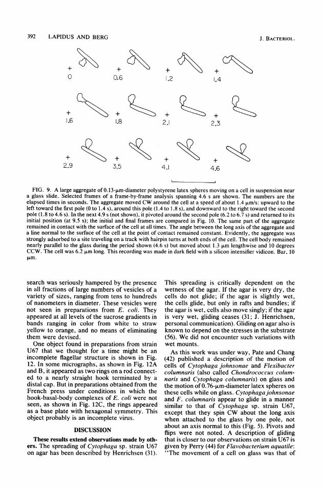
FIG. 9. A large aggregate of 0.13-p.m-diameter polystyrene latex spheres moving on a cell in suspension neara glass slide. Selected frames of a frame-by-frame analysis spanning 4.6 s are shown. The numbers are theelapsed times in seconds. The aggregate moved CW around the cell at a speed of about 1.4 p.m/s: upward to theleft toward the first pole (0 to 1.4 s), around this pole (1.4 to 1.8 s), and downward to the right toward the secondpole (1.8 to 4.6 s). In the next 4.9 s (not shown), it pivoted around the second pole (6.2 to 6.7 s) and returned to itsinitial position (at 9.5 s); the initial and final frames are compared in Fig. 10. The same part of the aggregateremained in contact with the surface of the cell at all times. The angle between the long axis of the aggregate anda line normal to the surface of the cell at the point of contact remained constant. Evidently, the aggregate wasstrongly adsorbed to a site traveling on a track with hairpin turns at both ends of the cell. The cell body remainednearly parallel to the glass during the period shown (4.6 s) but moved about 1.3 p.m lengthwise and 10 degreesCCW. The cell was 6.2 p.m long. This recording was made in dark field with a silicon intensifier vidicon. Bar, 10p.m.
search was seriously hampered by the presencein all fractions of large numbers of vesicles of avariety of sizes, ranging from tens to hundredsof nanometers in diameter. These vesicles werenot seen in preparations from E. coli. Theyappeared at all levels of the sucrose gradients inbands ranging in color from white to strawyellow to orange, and no means of eliminatingthem were devised.One object found in preparations from strain
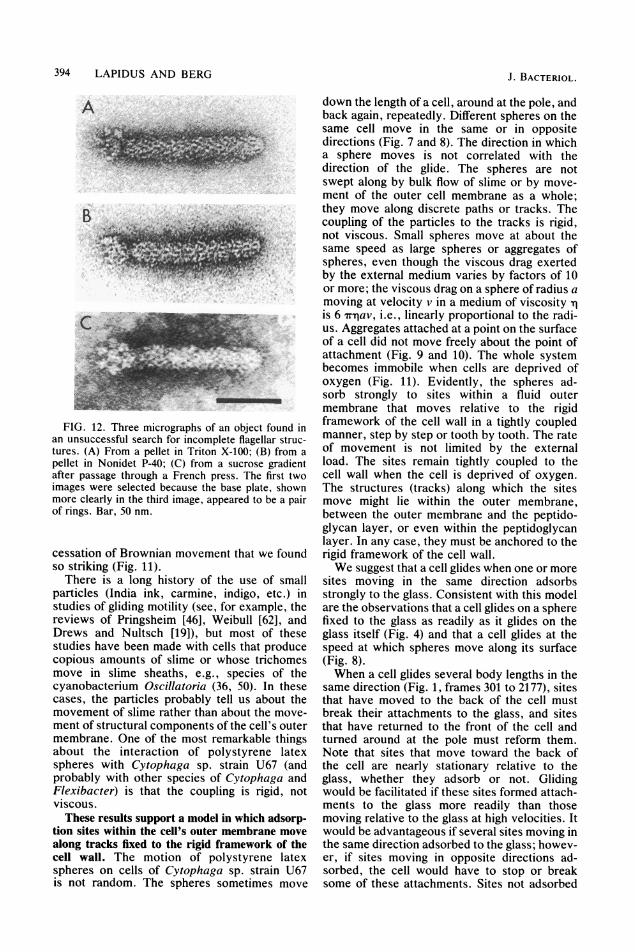
U67 that we thought for a time might be anincomplete flagellar structure is shown in Fig.12. In some micrographs, as shown in Fig. 12Aand B, it appeared as two rings on a rod connect-ed to a nearly straight hook terminated by adistal cap. But in preparations obtained from theFrench press under conditions in which thehook-basal-body complexes of E. coli were notseen, as shown in Fig. 12C, the rings appearedas a base plate with hexagonal symmetry. Thisobject probably is an incomplete virus.
DISCUSSIONThese results extend observations made by oth-
ers. The spreading of Cytophaga sp. strain U67on agar has been described by Henrichsen (31).
This spreading is critically dependent on thewetness of the agar. If the agar is very dry, thecells do not glide; if the agar is slightly wet,the cells glide, but only in rafts and bundles; ifthe agar is wet, cells also move singly; if the agaris very wet, gliding ceases (31; J. Henrichsen,personal communication). Gliding on agar also isknown to depend on the stresses in the substrate(56). We did not encounter such variations withwet mounts.As this work was under way, Pate and Chang
(42) published a description of the motion ofcells of Cytophaga johnsonae and Flexibactercolumnaris (also called Chondrococcus colum-naris and Cytophaga columnaris) on glass andthe motion of 0.76-pum-diameter latex spheres onthese cells while on glass. Cytophagajohnsonaeand F. columnaris appear to glide in a mannersimilar to that of Cytophaga sp. strain U67,except that they spin CW about the long axiswhen attached to the glass by one pole, notabout an axis normal to this (Fig. 5). Pivots andflips were not noted. A description of glidingthat is closer to our observations on strain U67 isgiven by Perry (44) for Flavobacterium aquatile:"The movement of a cell on glass was that of

BACTERIAL GLIDING MOTILITY 393
FIG. 10. Photographs at time 0 (A) and 9.5 s (B) ofthe video images of the aggregate and cell of Fig. 9.The top photograph was printed so that the cellappeared at its final orientation. Note the angle be-tween the two sets of scan lines. The aggregate com-pleted one CW cycle, returning to its initial positionand retaining its initial orientation relative to the cell.The cell rotated CCW about 54 degrees, the directionexpected given the viscous drag on the aggregate. Theimages of the aggregate are not quite identical in thetwo photographs. The cell may have rotated a fewdegrees about its other axes or moved slightly in or outof focus, or the aggregate may have twisted a fewdegrees about its long axis. Note that the wholeassembly was undergoing Brownian movement. Bar, 5j±m.
gliding to and fro, merging with or interrupted byswinging horizontally, or flipping over or possi-bly somersaulting. In other tests, on rare occa-sions, the movement was apparently that of apendulum describing a conical surface and rotat-ing constantly at c. 1 rev./sec." Garnjobst (23)gives a vivid description of this spinning forFlexibacter columnaris: ". . . peculiar rotary orwaving movements, sometimes combined withflexion, were observed in rods which had sud-denly assumed a perpendicular position onglass. The waving was so regular that automati-cally one began to count. The result of countsmade on several different individuals are: 111,218, 193. The horizontal position was suddenlyresumed and sometimes just as suddenly the rodbecame perpendicular again to repeat the proc-ess." The movements in all of these species aremuch more rapid than those observed in manygliders, for example, in species of Myxococcus;the rotary movements are more regular (cf.reference 47).
Cytophaga johnsonae and Flexibacter colum-naris move polystyrene latex spheres in a man-ner similar to that of strain U67, except pairs ofspheres are said to rotate CCW about the longaxis of the cell when localized at one pole andfrequently to flip end-over-end when movingdown the side of a cell (42); we never sawthe latter maneuver. In strain U67, pairs ofspheres or aggregates of spheres are not free torotate or tumble when moving down the side of acell (Fig. 9 and 10). Pate and Chang describeonly the motion of spheres along the port orstarboard side of a cell, with transits around thepole or across the body from one side to theother. In strain U67, particles also move on thedorsal surface of the cells, but this is easier todocument with spheres smaller than 0.76 ,m indiameter. By using spheres as small as 0.13 ,umin diameter, we found that particles canmove in close lateral proximity, even in oppositedirections.
Pate and Chang (42) also note that activemovement ceases when cells are deprived ofoxygen or treated with certain inhibitors (seebelow); however, they do not comment on the
FIG. 11. Dark-field photographs of cells of Cyto-phaga sp. strain U67. (A) Without further treatment;(B) after addition of 0.13-,um-diameter polystyrenelatex spheres. Both preparations were made from thesame culture. The cells were allowed to glide on glass,the particles were added (B), and then the preparationswere depleted of oxygen. Note the distinct spots oflight sprinkled widely over the surfaces of the cells(B). The spots are of different brightness because thespheres tend to form monomers, dimers, trimers, andhigher aggregates; successive exposures of the samecells were identical. Bar, 10 ,um.
VOL. 151, 1982
394 LAPIDUS AND BERG
A
D j .#J, , ; _
FIG. 12. Three micrographs of an object found inan unsuccessful search for incomplete flagellar struc-tures. (A) From a pellet in Triton X-100; (B) from apellet in Nonidet P-40; (C) from a sucrose gradientafter passage through a French press. The first twoimages were selected because the base plate, shownmore clearly in the third image, appeared to be a pairof rings. Bar, 50 nm.
cessation of Brownian movement that we foundso striking (Fig. 11).There is a long history of the use of small
particles (India ink, carmine, indigo, etc.) instudies of gliding motility (see, for example, thereviews of Pringsheim [46], Weibull [62], andDrews and Nultsch [19]), but most of thesestudies have been made with cells that producecopious amounts of slime or whose trichomesmove in slime sheaths, e.g., species of thecyanobacterium Oscillatoria (36, 50). In thesecases, the particles probably tell us about themovement of slime rather than about the move-ment of structural components of the cell's outermembrane. One of the most remarkable thingsabout the interaction of polystyrene latexspheres with Cytophaga sp. strain U67 (andprobably with other species of Cytophaga andFlexibacter) is that the coupling is rigid, notviscous.These results support a model in which adsorp-
tion sites within the cell's outer membrane movealong tracks fixed to the rigid framework of thecell wall. The motion of polystyrene latexspheres on cells of Cytophaga sp. strain U67is not random. The spheres sometimes move
down the length of a cell, around at the pole, andback again, repeatedly. Different spheres on thesame cell move in the same or in oppositedirections (Fig. 7 and 8). The direction in whicha sphere moves is not correlated with thedirection of the glide. The spheres are notswept along by bulk flow of slime or by move-ment of the outer cell membrane as a whole;they move along discrete paths or tracks. Thecoupling of the particles to the tracks is rigid,not viscous. Small spheres move at about thesame speed as large spheres or aggregates ofspheres, even though the viscous drag exertedby the external medium varies by factors of 10or more; the viscous drag on a sphere of radius amoving at velocity v in a medium of viscosity qis 6 zrqav, i.e., linearly proportional to the radi-us. Aggregates attached at a point on the surfaceof a cell did not move freely about the point ofattachment (Fig. 9 and 10). The whole systembecomes immobile when cells are deprived ofoxygen (Fig. 11). Evidently, the spheres ad-sorb strongly to sites within a fluid outermembrane that moves relative to the rigidframework of the cell wall in a tightly coupledmanner, step by step or tooth by tooth. The rateof movement is not limited by the externalload. The sites remain tightly coupled to thecell wall when the cell is deprived of oxygen.The structures (tracks) along which the sitesmove might lie within the outer membrane,between the outer membrane and the peptido-glycan layer, or even within the peptidoglycanlayer. In any case, they must be anchored to therigid framework of the cell wall.We suggest that a cell glides when one or more
sites moving in the same direction adsorbsstrongly to the glass. Consistent with this modelare the observations that a cell glides on a spherefixed to the glass as readily as it glides on theglass itself (Fig. 4) and that a cell glides at thespeed at which spheres move along its surface(Fig. 8).When a cell glides several body lengths in the
same direction (Fig. 1, frames 301 to 2177), sitesthat have moved to the back of the cell mustbreak their attachments to the glass, and sitesthat have returned to the front of the cell andturned around at the pole must reform them.Note that sites that move toward the back ofthe cell are nearly stationary relative to theglass, whether they adsorb or not. Glidingwould be facilitated if these sites formed attach-ments to the glass more readily than thosemoving relative to the glass at high velocities. Itwould be advantageous if several sites moving inthe same direction adsorbed to the glass; howev-er, if sites moving in opposite directions ad-sorbed, the cell would have to stop or breaksome of these attachments. Sites not adsorbed
J. BACTERIOL.
BACTERIAL GLIDING MOTILITY 395
to the glass would not necessarily affect gliding,because relatively little water or slime betweenthese sites and the glass would reduce their dragto a small value.
Competition between sites moving in oppositedirections might explain why spheres 0.36 pLm indiameter or larger tend to come off of glidingcells, whereas particles 0.13 pLm in diameterremain more firmly attached; the latter spheresmight not be large enough to interact with morethan one site. It would also explain why all of thespheres remain fixed to cells deprived ofoxygen; adsorption to more than one site wouldprolong attachment when the sites are station-ary.
If a site adsorbed to the glass moves along atrack through a hairpin turn, the cell must pivotthrough an angle of 180 degrees or flip over,depending on whether the hairpin is on theventral side of the cell in a plane parallel to theglass or loops around the pole in a plane normalto the glass. Pivots or flips through other anglesare harder to understand, unless tracks makeother kinds of turns at the ends of the cell, or asite moving along one track can switch to anoth-er at their points of intersection.
Spins, either in a plane parallel to the glass oron conical surfaces having either a large or asmall solid angle, are readily understood if sometracks exist as closed circles. If a site movesalong a track at a speed of about 2 pLm/s, thespeed at which a cell glides, and the track is bentinto a circle about 0.2 pLm in diameter (0.6 pum incircumference), then the site will move aroundthe circle at a frequency of about 3 Hz, thefrequency at which a cell spins (Fig. 6). The dataat hand are not accurate enough to distinguishrotation about a fixed shaft from motion about acircle as small as this, but note that rotation of acell on a conical surface is easily explained ifcircular tracks are laid out on the hemisphericalcaps at the ends of the cell. If rotation of a cellon a conical surface had to be explained in termsof rotation about a fixed shaft, the shaft wouldhave to flex.Data obtained with spheres 0.13 pLm in diame-
ter indicate that there are many tracks per cell.Some tracks appear to exist as closed loopsrunning the length of the cell in the direction ofits long axis. Some have tight hairpin turns at thepoles. Others go around the pole from one sideof the cell to the other. The tracks probablyoverlap, particularly at the poles of the cell.
These results are not consistent with most othermodels for gliding motility. The ability of cells insuspension to adsorb small particles and topropel them at a uniform speed to and fro alongtheir length, with particles in close proximityoften moving in opposite directions, arguesagainst most mechanisms previously proposed
for gliding motility. These movements cannot beunderstood in terms of differences in interfacialtension generated at the ends of the cell byexcretion of surface-active agents, by the excre-tion of slime, by inchworm contractions or otherundulations of the cell body as a whole, bywaves of contraction running from one end ofthe cell to the other, or by the motion of polarfimbriae. (For discussions of these theories, seethe reviews cited in the introduction.)Mechanisms in which particles are passed
from one site to the next along the cell surface,e.g., from crest to crest in a traveling wave ofadsorption or from ring to ring in an array ofrotating rings (42), seem doubtful but cannot beruled out. Several connections would have to bemaintained at all times between a sphere and acell to explain why spheres of different sizesmove at the same speed, why aggregates ofspheres move so rigidly, or why a cell can glideon a sphere in a cyclic fashion or spin continu-ously. A series of make-then-break connectionsof such high order would be more likely to occurbetween components of a molecular machinedesigned for the purpose, e.g., between interact-ing protein complexes, one in the outer mem-brane (the adsorption site), the other fixed to thepeptidoglycan layer (an element of the track).When we began this work, we considered the
possibility that gliding bacteria might have in-complete flagella, anlage of the endoflagella ofspirochetes, composed of a rotor embedded inthe cytoplasmic membrane, a stator just outsidethe cytoplasmic membrane, a drive shaft thatpenetrated the peptidoglycan layer, and a proxi-mal hook that ran between the peptidoglycanlayer and the outer membrane. If a cell had onlya few such structures, they would be very diffi-cult to find in electron micrographs of negativelystained, sectioned, or freeze-etched cells. If thehook were to roll about its axis in viscouscontact with the outer membrane, the outermembrane would flow, and this flow, in turn,might cause the cell to glide. If the hook were toadsorb strongly to a component of the outermembrane, it could no longer roll about its axis,but it could still revolve rigidly about the axis ofthe drive shaft. This would cause the outermembrane to swirl, and this swirl, in turn, mightcause the cell to spin. This scheme has the meritthat a similar mechanism would underlie motilityin flagellated bacteria, spirochetes, and glidingbacteria. Energy transmission from the cyto-plasmic membrane to the outer membranewould be explained by direct mechanical link-age. But the data at hand do not support a modelin which local regions of the outer membraneundergo viscous flow, nor have we been able tofind such incomplete flagellar structures.Machinery that propels the adsorption sites is
VOL. 151, 1982
396 LAPIDUS AND BERG
not known. Cytophaga spp. have a typical gram-negative cell envelope: an inner (cytoplasmic)membrane, a thin peptidoglycan layer, and anouter (lipopolysaccharide) membrane (13).Strain U67 has neither pili (32) nor flagella (31).When negatively stained with 2% uranyl acetate(G. Guglielmi, unpublished observations), itshows an irregularly undulating surface typicalof other gliding bacteria (14, 17, 22, 37). Wewere not able to find any incomplete flagellarstructures by differential centrifugation, but asnoted above, this search was severely hamperedby large numbers of contaminating vesicles. Wehave no reason to believe that the object that wedid find (Fig. 12) has anything to do with motil-ity.Some time ago, Pate and Ordal (43) described
in Flexibacter columnaris arrays of fibrils in thegap between the peptidoglycan layer and theouter membrane that run parallel to each otheralong the length of the cell and continue inoverlapping bands across the poles. Burchardand Brown (10) failed to find these fibrils infreeze-etched preparations of the same cellsunless the cells were treated with glutaralde-hyde, one of the fixatives used by Pate andOrdal. Without this treatment, Burchard andBrown observed only 10- to 11-nm particles onthe inner face of the outer membrane. They didnot find such particles in another gliding bacteri-um, Myxococcus xanthus. At about the sametime, Glaser and Pate (25) described a mutant ofFlexibacter columnaris, nonmotile under allconditions tested, that lacked the system offibrils. More recently, Strohl (59) described inCytophaga johnsonae double-stranded longitu-dinal fibers, 10 to 12 nm in width, in a fractureplane between the inner and outer membranes,but he considers these fibers to be different fromthose seen in Flexibacter columnaris. Artifactsor not, the fibrils seen in Flexibacter columnarisare oriented in precisely the manner required fora system that would generate or direct themotion that we have observed. A similar argu-ment has been used to implicate a system offibrils in the gliding motility of the cyanobacte-rium Oscillatoria (27-29). It is tempting to spec-ulate that such fibrils (or arrays of 10- to 12-nmparticles) are tracks along which adsorption sitesin the outer membrane move, but we have nodirect evidence for this.More recently, Pate and Chang (42) described
the isolation of ringlike structures from cells ofCytophaga johnsonae and Flexibacter colum-naris that they suggest drive gliding motility.Cell suspensions were homogenized in a buffercontaining 1.2 M KI, EDTA, and dithiothreitol.The soluble fraction was clarified by centrifuga-tion at 100,000 x g for 3 h and dialyzed against abuffer containing 0.1 M KCI, ATP, MgC92, and
dithiothreitol. The rings appeared during thedialysis and were pelleted by centrifugation at100,000 x g for 3 h. The rings also were seen inenvelopes of cells treated with a buffer contain-ing MgCl2 and dithiothreitol. Their resemblanceto the M- and S-rings of the basal-body complexof flagella from E. coli and Bacillus subtilis (16),rings thought to serve as the rotor and stator,respectively, of the flagellar rotary motor (1), isat best superficial; the rings from the glidingbacteria are smaller in diameter and muchthicker than those from the flagellated bacteria.Whether they exist in the cell envelopes ofCytophaga johnsonae and Flexibacter colum-naris as such or form only after treatment withMgCI2 and dithiothreitol is an open question. Wedid not see them in our search for incompleteflagellar structures, using methods known towork in E. coli (61) and Salmonella typhimurium(60).There is a growing body of evidence that the
energy for motility in gliding bacteria, as inflagellated bacteria (see discussion in reference6) and spirochetes (26), is supplied by a proton-motive force in the form of an electrical potentialdifference or a pH difference acting across thecytoplasmic membrane. Evidence for this the-ory has been obtained in Flexibacter poly-morphus by Ridgway (49), in Cytophaga john-sonae and Flexibacter columnaris by Pate andChang (42), in Flexibacter FS-1 by Dayrell-Hartand Burchard (15), in Flexibacter BH3 by Dux-bury et al. (21), and in Phormidium uncinatumand Oscillatoria spp. by Glagoleva et al. (24; seealso 30). If gliding is generated by the motion ofadsorption sites in the outer membrane, or,indeed, by the motion of any component of theouter membrane, how is the power required forthis motion transmitted from the cytoplasmicmembrane through the rigid peptidoglycan lay-er? In flagellated bacteria and spirochetes, thelinkage is mechanical: a drive shaft penetratesthe peptidoglycan layer. The existence of fibrilsbetween the peptidoglycan layer and the outermembrane or of adsorption sites or rings in theouter membrane is not enough; some kind ofcoupling to the cytoplasmic membrane is essen-tial.The genetics of gliding motility has been stud-
ied only in M. xanthus. This fruiting myxobac-terium has two gene systems controlling move-ment (33-35). One system, designated A(adventurous), allows cells to glide when theyare far apart; it has 21 loci. The second system,designated S (social), allows cells to glide whenthey are close together; it has at least 10 loci andis correlated with the presence of pili (39). Thetwo systems have only one locus in common. Ifthe machinery for gliding in Cytophaga sp.strain U67 is similar to that in M. xanthus, then
J . BACTERIOL .

BACTERIAL GLIDING MOTILITY 397
it must be programed by a gene system similar tothe A system; cells of strain U67 glide singly (31;present study), and they have no pili (32).We hope to learn more about the number,
distribution, and nature of adsorption sites in theouter membrane. In summary, our work sup-ports a model for gliding motility in bacteria inwhich adsorption sites within the outer mem-brane of the cell move along tracks fixed to therigid framework of the cell wall. Our observa-tions are inconsistent with most, if not all,alternative models. We would not have obtainedthese results if the adsorption of polystyrenelatex spheres or glass to cells of Cytophaga sp.strain U67 were prevented by a thick interveninglayer of slime. In this respect, species of Cyto-phaga may be particularly well suited to studiesof gliding motility.A number of intriguing questions remain.
How many adsorption sites are there on thesurface of a cell? How are they distributed?What is their chemical nature? When we havethe answers to these questions, we may be in aposition to learn how these sites are propelled.
ACKNOWLEDGMENTS
This work began in the fall of 1978 with a search forincomplete flagellar structures while H.C.B. was on sabbaticalleave in the laboratory of R. Y. Stanier and G. Cohen-Bazire,Unite de Physiologie Microbienne, Institut Pasteur, Paris,whose hospitality is warmly acknowledged. This phase of thework would not have been possible without the help of G.Guglielmi, who did the electron microscopy. The light micros-copy began in earnest the following summer in the laboratoryof J. Henrichsen, Statens Seruminstitut, Copenhagen, whose1972 review on surface translocation inspired much of thiswork. The video measurements were begun in 1980 whileI.R.L. was on sabbatical leave at Caltech and were continuedduring the summer of 1981. We thank Dale Kaiser for com-ments on the manuscript.
This work was supported by grants from the U.S. NationalScience Foundation to H.C.B. (SMI-7717384, PCM-7922601)and I.R.L. (SPI-8165019).
LITERATURE CITED
1. Berg, H. C. 1974. Dynamic properties of bacterial flagellarmotors. Nature (London) 249:77-79.
2. Berg, H. C. 1975. Bacterial behaviour. Nature (London)254:389-392.
3. Berg, H. C. 1976. How spirochetes may swim. J. Theor.Biol. 56:269-273.
4. Berg, H. C., and R. A. Anderson. 1973. Bacteria swim byrotating their flagellar filaments. Nature (London)245:380-382.
5. Berg, H. C., D. B. Bromley, and N. W. Charon. 1978.Leptospiral motility. Symp. Soc. Gen. Microbiol. 28:285-294.
6. Berg, H. C., M. D. Manson, and M. P. Conley. 1982.Dynamics and energetics of flagellar rotation in bacteria.Symp. Soc. Exp. Biol. 35:1-31.
7. Berg, H. C., and P. M. Tedesco. 1975. Transient responseto chemotactic stimuli in Escherichia coli. Proc. Natl.Acad. Sci. U.S.A. 72:3235-3239.
8. Bryant, D. A., G. Guglielmi, N. Tandeau de Marsac, A.-M.Castets, and G. Cohen-Bazire. 1979. The structure ofcyanobacterial phycobilisomes: a model. Arch. Micro-biol. 123:113-127.
9. Burchard, R. P. 1981. Gliding motility of prokaryotes:
ultrastructure, physiology, and genetics. Annu. Rev. Mi-crobiol. 35:497-529.
10. Burchard, R. P., and D. T. Brown. 1973. Surface structureof gliding bacteria after freeze-etching. J. Bacteriol.114:1351-1355.
11. Burkholder, P. R. 1934. Movement in the cyanophyceae.Quart. Rev. Biol. 9:438-459.
12. Canale-Parola, E. 1978. Motility and chemotaxis of spiro-chetes. Annu. Rev. Microbiol. 32:69-99.
13. Christensen, P. J. 1977. The history, biology, and taxono-my of the Cytophaga group. Can. J. Microbiol. 23:1599-1653.
14. Costerton, J. W. F., R. G. E. Murray, and C. F. Robinow.1%1. Observations on the motility and the structure ofVitreoscilla. Can. J. Microbiol. 7:329-340.
15. Dayrell-Hart, B., and R. P. Burchard. 1979. Associationof flexing and gliding in Flexibacter. J. Bacteriol.137:1417-1420.
16. DePamphilis, M. L., and J. Adler. 1971. Fine structureand isolation of the hook-basal body complex of flagellafrom Escherichia coli and Bacillus subtilis. J. Bacteriol.105:384-395.
17. Dickson, M. R., S. Kouprach, B. A. Humphrey, and K. C.Marshall. 1980. Does gliding motility depend on undulat-ing membranes? Micron 11:381-382.
18. Doetsch, R. N., and G. J. Hageage. 1968. Motility inprocaryotic organisms: problems, points of view, andperspectives. Biol. Rev. Biol. Proc. Cambridge Philos.Soc. 43:317-362.
19. Drews, G., and W. Nultsch. 1962. Spezielle Bewegungs-mechanism von Einzellern (Bakterien, Algen), p. 876-919. In E. Bunning (ed.), Handbuch der Pflanzenphysio-logie, vol. 17, part 2, Physiologie der Bewegungen.Springer-Verlag, Berlin.
20. Duxbury, T. 1977. A microperfusion chamber for studyingthe growth of bacterial cells. J. Appl. Bacteriol. 43:247-251.
21. Duxbury, T., B. A. Humphrey, and K. C. Marshall. 1980.Continuous observations of bacterial gliding motility in adialysis microchamber: the effects of inhibitors. Arch.Microbiol. 124:169-175.
22. Follett, E. A. C., and D. M. Webley. 1965. An electronmicroscope study of the cell surface of Cytophaga john-sonii and some observations on related organisms. An-tonie van Leeuwenhoek J. Microbiol. Serol. 31:361-382.
23. Garnjobst, L. 1945. Cytophaga columnaris (Davis) in pureculture. A myxobacterium pathogenic to fish. J. Bacteriol.49:113-128.
24. Glagoleva, T. N., A. N. Glagolev, M. V. Gusev, and K. A.Nikitina. 1980. Protonmotive force supports gliding incyanobacteria. FEBS Lett. 117:49-53.
25. Glaser, J., and J. L. Pate. 1973. Isolation and character-ization of gliding motility mutants of Cytophaga colum-naris. Arch. Mikrobiol. 93:295-309.
26. Goulbourne, E. A., Jr., and E. P. Greenberg. 1980.Relationship between proton motive force and motility inSpirochaeta aurantia. J. Bacteriol. 143:1450-1457.
27. Halfen, L. N. 1973. Gliding motility of Oscillatoria: ultra-structural and chemical characterization of the fibrillarlayer. J. Phycol. 9:248-253.
28. Halfen, L. N. 1979. Gliding movements, p. 250-267. In W.Haupt and M. E. Feinleib (ed.), Encyclopedia of plantphysiology, new series, vol. 7, physiology of movements.Springer-Verlag, Berlin.
29. Halfen, L. N., and R. W. Castenholz. 1970. Gliding in ablue-green alga: a possible mechanism. Nature (London)225:1163-1164.
30. Halfen, L. N., and R. W. Castenholz. 1971. Gliding motil-ity in the blue-green alga Oscillatoria princeps. J. Phycol.7:133-145.
31. Henrichsen, J. 1972. Bacterial surface translocation: asurvey and a classification. Bacteriol. Rev. 36:478-503.
32. Henrichsen, J., and J. Blom. 1975. Examination of fimbri-ation of some gram-negative rods with and without twitch-ing and gliding motility. Acta Pathol. Microbiol. Scand.
VOL. 151, 1982

398 LAPIDUS AND BERG
Sect. B 83:161-170.33. Hodgkin, J., and D. Kaiser. 1977. Cell-to-cell stimulation
of movement in nonmotile mutants of Mvxococcus. Proc.Natl. Acad. Sci. U.S.A. 74:2938-2942.
34. Hodgkin, J., and D. Kaiser. 1979. Genetics of glidingmotility in Myxococcus xanthus (Myxobacteriales): genescontrolling movement of single cells. Mol. Gen. Genet.171:167-176.
35. Hodgkin, J., and D. Kaiser. 1979. Genetics of glidingmotility in Myxococcus xanthus (Myxobacteriales): twogene systems control movement. Mol. Gen. Genet.171:177-191.
36. Hosoi, A. 1951. Secretion of the slime substance in Oscil-latoria in relation to its movement. Bot. Mag. (Tokyo)64:14-17.
37. Humphrey, B. A., M. R. Dickson, and K. C. Marshall.1979. Physicochemical and in situ observations on theadhesion of gliding bacteria to surfaces. Arch. Microbiol.120:231-238.
38. lino, T. 1977. Genetics of structure and function ofbacterial flagella. Annu. Rev. Genet. 11:161-182.
39. Kaiser, D. 1979. Social gliding is correlated with thepresence of pili in Myxococcus xanthu.s. Proc. Natl. Acad.Sci. U.S.A. 76:5952-5956.
40. Kaiser, D., C. Manoil, and M. Dworkin. 1979. Myxobac-teria: cell interactions, genetics, and development. Annu.Rev. Microbiol. 33:595-639.
41. Macnab, R. M. 1979. Bacterial flagella, p. 207-223. In W.Haupt and M. E. Feinleib (ed.), Encyclopedia of plantphysiology, new series, vol. 7, physiology of movements.Springer-Verlag, Berlin.
42. Pate, J. L., and L.-Y. E. Chang. 1979. Evidence thatgliding motility in prokaryotic cells is driven by rotaryassemblies in the cell envelopes. Curr. Microbiol. 2:59-64.
43. Pate, J. L., and E. J. Ordal. 1967. The fine structure ofChondrococcus columnaris III. The surface layers ofChondrococcus columnaris. J. Cell Biol. 35:37-51.
44. Perry, L. B. 1973. Gliding motility in some non-spreadingflexibacteria. J. Appl. Bacteriol. 36:227-232.
45. Pfennig, N. 1977. Phototrophic green and purple bacteria:a comparative, systematic survey. Annu. Rev. Microbiol.31:275-290.
46. Pringsheim, E. G. 1949. The relationship between bacteriaand Myxophyceae. Bacteriol. Rev. 13:47-98.
47. Reichenbach, H. 1964. Myxococcus spp. (Myxobacter-iales) Schwarmentwicklung und Bildung von Protocysten.Film E778. Institut fur den Wissenschaftlichen Film,Gottingen.
48. Reichenbach, H. 1981. Ta,xonomy of the gliding bacteria.Annu. Rev. Microbiol. 35B339-364.
49. Ridgway, H. F. 1977. Source of energy for gliding motilityin Flexibacter polvmorphius: effects of metabolic andrespiratory inhibitors of gliding movement. J. Bacteriol.131:544-556.
50. Schulz, G. 1955. Bewegungsstudien sowie elektronmik-roskopische Membranuntersuchungen an Cyanophyceen.Arch. Mikrobiol. 21:335-370.
51. Silverman, M., and M. Simon. 1973. Genetic analysis offlagellar mutants in Escherichia coli. J. Bacteriol.113:105-113.
52. Silverman, M., and M. Simon. 1974. Flagellar rotation andthe mechanism of bacterial motility. Nature (London)249:73-74.
53. Silverman, M., and M. I. Simon. 1977. Bacterial flagella.Annu. Rev. Microbiol. 31:397-419.
54. Soriano, S. 1973. Flexibacteria. Annu. Rev. Microbiol.27:155-170.
55. Stanier, R. Y. 1942. The Cytophaga group: a contributionto the biology of myxobacteria. Bacteriol. Rev. 6:143-196.
56. Stanier, R. Y. 1942. A note on elasticotaxis in myxobac-teria. J. Bacteriol. 44:405-412.
57. Stanier, R. Y., and G. Cohen-Bazire. 1977. Phototrophicprokaryotes: the cyanobacteria. Annu. Rev. Microbiol.31:225-274.
58. Stober, W., A. Fink, and E. Bohn. 1968. Controlledgrowth of monodisperse silica spheres in the micron sizerange. J. Colloid Interface Sci. 26:62-69.
59. Strohl, W. R. 1979. Ultrastructure of Cvtophaga john-sonae and C. aqaiatilis by freeze-etching. J. Gen. Micro-biol. 112:261-268.
60. Suzuki, T., T. Iino, T. Horiguchi, and S. Yamaguchi. 1978.Incomplete flagellar structures in nonflagellate mutants ofSalmonella tvphimurium. J. Bacteriol. 133:904-915.
61. Suzuki, T., and Y. Komeda. 1981. Incomplete flagellarstructures in Escherichia c oli mutants. J. Bacteriol.145:1036-1041.
62. Weibull, C. 1960. Movement, p. 153-205. In I. C. Gunsa-lus and R. Y. Stanier (ed.), The bacteria, vol. 1. AcademicPress, Inc., New York.
J. BACTERIOL.